Communication

A New Languidulane Diterpenoid from Salvia mexicana var. mexicana

by
Bernardo Antonio Frontana-Uribe
,
Martha Verónica Escárcega-Bobadilla
,
Rosa Estrada-Reyes
,
José Antonio Morales-Serna
,
Manuel Salmón
and
Jorge Cárdenas
*
Instituto de Química, Universidad Nacional Autónoma de México, Circuito Exterior, Ciudad Universitaria, 04510 México D.F., México
*
Author to whom correspondence should be addressed.
Molecules 2011, 16(10), 8866-8873; https://doi.org/10.3390/molecules16108866
Submission received: 27 September 2011 / Revised: 17 October 2011 / Accepted: 18 October 2011 / Published: 21 October 2011

Abstract

:
From the aerial parts of Salvia mexicana var. mexicana, two C-10 epimers (α and β) of salvimexicanolide were isolated. Our interpretation of the data, especially the 13C NMR, led us to conclude that the previously described 13C-NMR spectrum of the α-epimer was not accurately assigned and it actually corresponds to the β-epimer. The structures proposed for the salvimexicanolides were verified by means of NOESY experiments. Dugesin B, arbutin, naringenin and the mixture of oleanolic and ursolic acids were also isolated from this Salvia spp.

1. Introduction

Salvia [1] is one of the largest genera of plants, with over 500 species distributed throughout Mexico, Central and South America. In Mexico, it is represented by over 300 species, many of which are endemic [2]. The diterpenoid constituents of genus Salvia are closely related to the subgenus and section to which they belong. After an intensive study of Mexican Salvia species, a great number of neo-clerodane diterpenoids have been isolated [3,4]. Some examples of the languidulane skeleton, which could be biogenetically originated from the junction of the C-16 and C-1 of a clerodane skeleton [5], have been found in this genus [6].
Salvia Mexicana (Subgenus Calosphace), comprises the varieties major, minor and mexicana [1,2]; which are endemic to the central states of Mexico. In 1999 Esquivel and co-workers reported the results of their study of Salvia mexicana var. major, to our knowledge the only variety that has been investigated. In that study, a new languidulane diterpenoid was described [7]. In this context, this paper describes the isolation and structural elucidation of a new languidulane diterpene, named salvimexicanolide B (2), from the aerial part of Salvia mexicana var. mexicana. In addition to the above mentioned compound, ursolic and oleanolic acids, salvimexicanolide (1), dugesin B (3), the flavanone (-) naringenin (4) and the substituted sugar arbutin (5) were identified.

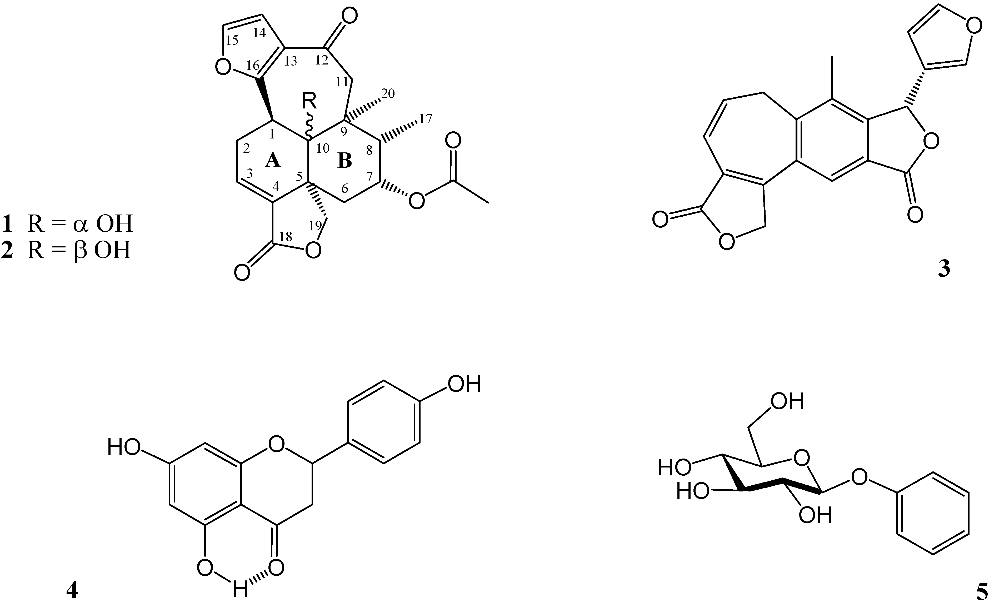
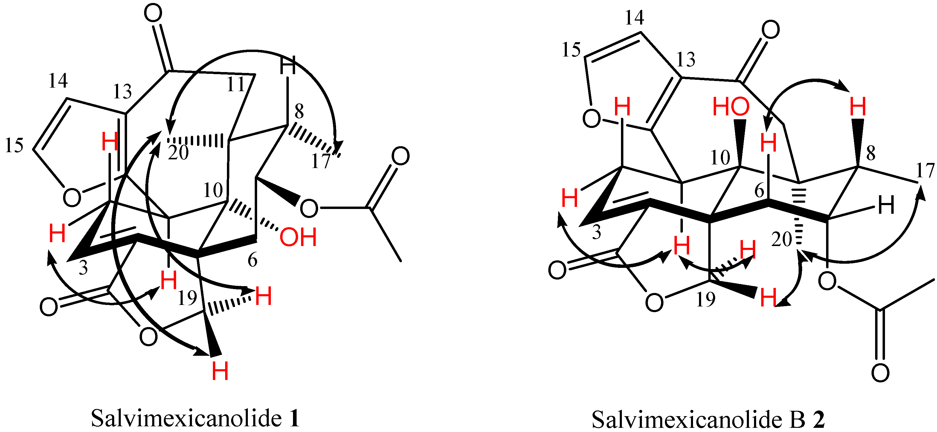
2. Results and Discussion

The aerial part of Salvia mexicana var. mexicana was dried, ground and extracted with acetone at room temperature in a closed container for one week, as described in the Experimental. After extensive column chromatography, six known natural products were isolated and identified. These were ursolic acid, oleanolic acid [8], salvimexicanolide (1) [7], dugesin B (3) [9,10], naringenin (4) [11] and arbutin (5) [12]. Their structures (Figure 1) were confirmed by comparing their spectra with authentic ones. To our delight, were also able to isolate a new compound from the same extract, salvimexicanolide B (2), which is structurally related with salvimexicanolide (1).
Figure 1. Products isolated from S. mexicana var. mexicana.
Molecules 16 08866 g001
Salvimexicanolide B (2) is a yellowish crystalline product (mp. 137–139 °C) whose MS was consistent with a molecular formula of C22H24O7, the molecular formula of 1. The IR spectrum shows the presence of γ-lactone (1777 cm−1), hydroxyl (3604 cm−1), an ester carbonyl (1738 cm−1) and unsaturated ketone carbonyl (1660 cm−1) groups. The hydroxyl group observed in the IR spectrum matches a broad signal exchangeable with D2O in the 1H-NMR spectrum (δ 3.38) and was confirmed by means of a 13C-NMR spectrum (δ 77.8). The 1H-NMR spectrum obtained for this product (Table 1) shows signals similar to those described for the disubstituted furan ring, and the ABCX system, assigned to H-1, H-2eq, H-2ax and H-3 in the α-epimer. However, some other signals are displaced or present a different shape. The most remarkable differences between α and β epimers are the upfield displacement in the chemical shift of H-1 (Δδ = −0.64 ppm) and the methylene protons on C-2 (Δδ = −0.14, 1.04 ppm) to β epimer. Fact that can be explained in terms of a change in the configuration of the C-10 hydroxyl group due to the A/B trans fusion for this compound. This fusion was confirmed by means of the 13C-NMR spectrum (Table 2), in which the chemical shift of the carbons adjacent to the A/B ring fusion (C-1, C-2, C-6 and C-16) are displaced upfield by 3.3, 1.2, 5.3 and 5.2 ppm respectively. On the other hand, C-5 and C-18 are displaced downfield by 2.5 and 5.9 ppm, in comparison to 1, due to the decompressing effect of the trans A/B fusion.
Table 1. 1H-NMR data for compounds 1 and 2. (300 MHz, TMS, J = Hz) a.
H1 (δ ppm) b1 (δ ppm) c1 NOESY2 (δ ppm) c2 NOESY
14.18 (1H) 3.97 (1H)H-2α, H-11α, 3.33 (1H)2α, H-19 pro-S
dd, J = 9.3, 1.8dd, J = 9.9, 2.7H-11βdd, J = 12.3, 5.7
eq2.97 (1H)2.93 (1H)H-1, H-2β, H-32.71 (1H)H-1α, H-2β,
ddd J = 21.0, 9.6, 4.2ddd, J = 21.0, 9.9, 4.8 dt, J = 20.7, 5.7H-3
ax3.28 (1H)3.42 (1H)H-2α, H-32.38 (1H)H-2α, H-3
ddd, J = 21.0, 3.6, 2.1dt, J = 21.0, 3.0 ddd, J = 20.7, 12.3, 3.0
36.93 (1H) dd, J = 4.2,7.09 (1H)H-2α, H-2β7.18 (1H)H-2α, H-2β
3.6t, J = 3.6 dd, J = 5.7, 3.0
eq1.97 (1H)2.08 (1H)H-6β, H-7,1.86 (1H)H-6β, H-7
mdd, J = 15.6, 3.0H-19 pro-Rdd, J = 15.6, 2.4
ax1.06 (1H)1.10 (1H)H-6α, H-7, H-81.79 (1H)H-6α, H-7, H-8
ddd,J = 15.6, 3.6, 2.1dt, J = 15.6, 3.0, 2.1 ddd, J = 15. 6, 4.5, 1.5
74.83 (1H)4.9 (1H)H-6α, H-6β,5.12 (1H)H-6α, H-6β,
q, J = 3.9dt, J = 6.5, 3.3H-8, Me-17td, J = 4.2, 2.4H-8, Me-17
81.97 (1H)m1.95 (1H)qd, J = 7.0, 4.2H-6β, H-7, Me-171.96 (1H)qd, J = 7.2, 4.2H-6β, H-7,Me-17
11α 2.48 (1H)2.62 (1H)H-1α, H-11β,2.48 (1H)H-11β, Me-17,
d, J = 17.1d, J = 17.7Me-17, Me-20d, J = 15.6Me-20
11β3.43 (1H)3.3 (1H)H-1α, H-11α,3.52 (1H)H-11α
d, J = 17.1d, J = 17.7 d, J = 15.6
146.63 (1H)6.72 (1H)H-156.71 (1H)H-15
d, J = 1.4d, J = 1.8 d, J = 2.0
157.57 (1H)7.35 (1H)H-147.29 (1H)H-14
d, J = 1.4d, J = 1.8 d, J = 2.0
170.81 (1H)0.83 (3H)H-7, H-8,0.95 (3H)H-7, H-8,
d, J = 6.9d, J = 7.0H-11α, Μe-20d, J = 7.2H-11α, Μe-20
19 pro-R4.93 (1H)5.0 (1H)H-6α, H-19 pro-S,4.7 (1H)H-19 pro-S,
d, J = 8.1d, J = 8.7Me-20d, J = 8.4Me-20
19 pro-S4.50 (1H)4.47, (1H)H-19 pro-R,4.52, (1H)H-1, H-19 pro-R
dd, J = 8.1, 2.1dd, J = 8.7, 2.1Me-20dd, J = 8.4, 1.5
201.15 (3H)1.15 (3H)H-11α, Me-17, H-191.23 (3H)H-11α, Me-17,
sspro-S and pro-RsH-19 pro-R
CH3COO 2.10 (3H) 2.10 (3H)
s s
-OH 2.2 3.38
a Assignments confirmed by COSY, HETCOR and NOESY NMR experiments; b Acetone-d6 used as solvent; c CDCl3 used as solvent.
Table 2. 13C-NMR data for compounds 1 and 2. (75 MHz, CDCl3, TMS) a.
C1 (δ ppm)2 (δ ppm)
142.7 d39.4 d
223.6 t22.4 t
3133. 6d136.1 d
4130.8 s132.5 s
546.1 s48.6 s
634.7 t29.4 t
770.9 d72.0 d
837.2 d37.3 d
945.3 s46.4 s
1077.6 s77.8 s
1152.0 t52.2 t
12194.6 s194.2 s
13125.7 s122.1 s
14109.1 d110.1 d
15142.4 d142.4 d
16159.8 s154.6 s
1713.0 q11.6 q
18169.7 s175.7 s
1972.5 t73.2 t
2020.0 q21.4 q
CH3COO21.2 t21.2 q
CH3COO169.0 s169.9 s
a Assignements confirmed by HETCOR and FLOCK experiments.
Although long range coupling between H-19 pro S and H-6βax was observed in 1 and 2; H-6βax in 2 was displaced downfield 0.69 ppm in comparison with the corresponding signal observed for the H-6βax proton in 1. The structural disposition of 2 produces a deshielding on H-6β due to a 1–3 diaxial interaction with C-OH, a feature that is not observed in 1. Me-17, H-8, and the AB system assigned to the C-11 methylene protons did not show any significant changes.
The NOESY experiment confirmed the structures proposed for the epimers 1 and 2 (Figure 2). Thus, that experiment show the proton interaction between H-1-and C-11 methylene protons in 1, which is not observed for the epimer 2. The reason is that the epimer 1, as can be observed in the Dreiding model, shows a boat conformation for the C ring due to the cis fusion of the A/B rings. According to the X-ray structure [7], this disposition induces an approach between the H-1 and the Hα-11 proton. Another remarkable difference observed for these products is that the NOESY spectrum for 2 showed the interaction between H-1 and H-19 pro-S, a fact supported by means of the analysis of the Dreiding model of this compound. This NOE interaction is due to the fact that the H-1 proton in the A/B trans languidulane skeleton is located close to H-19 pro-S. From a NOESY experiment performed on 1, it was possible to assign the signals at δ 2.93 and 3.42 which correspond to H-2α and H-2β, whereas the same protons in 2 appear at δ 2.71 and 2.38, respectively (See Table 1).
Figure 2. Key NOESY correlations.
Molecules 16 08866 g002
It is noteworthy that the 13C-NMR data described in this paper shows important differences with the data previously reported for 1 [7], where C-1, C-6, C-13, C-16 were observed at higher chemical shift (3–5 ppm) and C-5, C-18 lower chemical shifts (2–6 ppm). We conclude that these differences are due to the fact that the reported 13C-NMR spectrum for salvimexicanolide (1) actually corresponds to the spectrum obtained for salvimexicanolide B (2) (see Table 2).

3. Experimental

3.1. General

Melting points were obtained with a Fisher-Johns apparatus and are uncorrected. EI-MS spectra were obtained at 70 eV by direct inlet injection on a Jeol JMS-AX505HA instrument. FAB HR-MS spectra were obtained with a Jeol JMS-SX102A. 1H-NMR (300 MHz) and 13C-NMR (75 MHz) spectra were recorded with a Varian Unity 300 spectrometer using TMS as internal standard. Infrared spectra were recorded in solution (CHCl3) using a Nicolet Magna 750 spectrophotometer. UV-spectra were recorded in a Shimadzu U-160 spectrophotometer. Optical rotatory measurements were performed in a Jasco DIP-360 polarimeter.
Plant material was collected in April 1997, at 8 Km N from Izmolitla towards Eloxotitlan (Municipio Molango Hidalgo, México) and voucher specimens (MEXU 864121, 864122, 864123) were deposited in the Herbarium of the Instituto de Biología, UNAM México D.F.

3.1. Extraction and Isolation

Powdered, dried aerial parts of S. mexicana var. mexicana (1.2 kg) were extracted with Me2CO (3 × 10 L) at room temperature for one week. The solvent was removed under reduced pressure. During the evaporation of the solvent a mixture of ursolic and oleanolic acids was isolated by filtration (69 g). The extract (110 g) was partitioned using MeOH-H2O (4:1) and hexane-C6H6 (1:1). The polar fraction (32.2 g) was subjected to vacuum chromatography over silica gel. Mixtures of hexane-EtOAc of increasing polarity were used as eluents. Salvimexicanolide (1) precipitated from the fractions eluted with hexane-EtOAc (4:6). After exhaustive flash chromatography of these fractions, eluted with hexane-EtOAc (7:3), more salvimexicanolide (1) was obtained (0.074 g).
The fractions obtained from the vacuum chromatography eluted with hexane-EtOAc 7:3 to 1:1 (9.32 g) where subjected to a new vacuum chromatography separation over silica gel. Mixtures of hexane-EtOAc of increasing polarity were used as eluents. Dugesin B (3, 0.215 g) was obtained from the fractions eluted with hexane-EtOAc 6:4 to 1:1. Naringenin (4, 0.025 g) was isolated from fractions eluted with hexane-EtOAc 4:6. Both of them obtained after flash chromatography. From the last fractions eluted with hexane-EtOAc 4:6 and the first eluted with hexane-EtOAc 3:7, salvimexicanolide B (2) was obtained (0.045 g). The fractions (2.751 g) from the first vacuum chromatography, eluted with EtOAc and EtOAc-MeOH 9:1, were subjected to a new vacuum chromatography over silica gel. After the column was eluted with a mixture of EtOAc-MeOH 9.9:0.1, arbutin (5, 0.170 g) was isolated.
Salvimexicanolide (1). Rf (hexane-EtOAc 30:70): 0.6; Mp 265–267 °C; UV (MeOH), λmax (ε): 274.5 (5940), 208 (21360); [α]D + 233° (MeOH; c 0.1); IR (CHCl3) νmax cm−1: 3581, 3112, 2941, 1755, 1715, 1661, 1429, 1369, 1126, 1033, 979; 1H-NMR see Table 1; 13C-NMR see Table 2; FAB-MS HR m/z: 401.1628 [M++1]; C22H25O7 requires [M++1] at m/z 401.1600. EI-MS m/z (rel. int.): 400 (100), 382 (17), 358 (55), 340 (26), 322 (10), 295 (27), 278 (12), 267 (26), 249 (24), 235 (23), 223 (22), 199 (17), 189 (19), 161 (23), 135 (41), 121 (38).
Salvimexicanolide B (2). Rf (hexane-EtOAc 30:70): 0.4; Mp 137–139 °C; UV (MeOH), λmax (ε): 304.8 (12580), 218.8 (13620); [α]D + 44° (MeOH; c 0.1); IR (CHCl3) νmax cm−1: 3604, 2973, 2927, 1777, 1738, 1660, 1514, 1434, 1366, 1146, 1015; 1H-NMR see Table 1; 13C-NMR see Table 2; FAB-MS HR m/z: 401.1614 [M++1]; C22H25O7 requires [M++1] at m/z 401.1600. EI-MS m/z (rel. int.): 400 (70), 382 (9), 358 (100), 340 (35), 322 (7), 312 (13), 297 (8), 285 (8), 274 (12), 258 (26), 244 (23), 223 (12), 200 (52), 199 (34), 187 (15), 161 (13), 128 (13).
Dugesin B (3). Rf (hexane-EtOAc 30:70): 0.5; Mp 213–214 °C; UV (MeOH), λmax (ε): 299 (9584), 239 (24500), 216 (33416); [α]D − 120° (MeOH; c 0.1); IR (CHCl3) νmax cm−1: 3110, 1759, 1604, 1296, 1080, 1026; 1H-NMR (CDCl3): 7.82 (s, 1H), 7.54 (d, J = 1.2, 1H), 7.40 (dd, J = 2.1, 1.2, 1H), 6.61 (d, J = 9.6, 1H), 6.46 (s, 1H), 6.09 (d, J = 2.1, 1H), 6.04 (dt, J = 9.6, 6.9, 1H), 5.47 (d, J = 17, 1H), 5.41 (d, J = 17, 1H), 3.22 (dd, J = 13.8, 6.9, 1H), 3.11 (dd, J = 13.8, 6.9, 1H), 2.29 (s, 3H). 13C-NMR (CDCl3): 172.4, 169.3, 155.3, 150.4, 144.3, 142.2, 141.7, 132.2, 130.9, 128.5, 126.7, 124.3, 122.1, 120.7, 119.8, 108.7, 75.1, 69.8, 29.8, 15.6; FAB-MS HR m/z: 335.0943 [M++1]; C20H15O5 requires [M++1] at m/z 335.0919. EI-MS m/z (rel. int.): 334 (100), 319 (8), 305 (32), 277 (7), 239 (71), 210 (6), 202 (4), 189 (5), 181 (5), 152 (6), 95 (10).
(-) Naringenin (4). Rf (hexane-EtOAc 30:70): 0.5; Mp 246–248 °C; IR (CHCl3) νmax cm−1: 3290, 3120, 2918, 2833, 1634, 1601, 1517, 1464, 1311, 1251, 1179, 1156, 1084, 834; 1H-NMR 12.1 (s, 1-H, int-D2O, C5-OH), 9.6 (s, 1-H, int-D2O, C7-OH), 8.77 (s, 1-H, int-D2O, C4-OH), 7.41 (d, 2-H, J = 8.3, H-2’ H-6’), 6.92 (d, 2-H, J = 8.3, H-5’ H-3’), 5.96 (s, 2-H, H-6 H-8), 5.45 (dd, 1-H, J = 12.9, 3.1, H-2), 3.18 (dd, 1-H, J = 17.1, 12.9, H-3α), 2.72 (dd, 1-H, J = 17.1, 3.1, H-3β); 13C-NMR 197.3 (s, C-4), 167.3 (s, C-7), 165.2 (d, C-5), 164.3 (d, C-9), 158.6 (d, C-4’), 130.7 (s, C-1’), 128.9 (d, C-2’ C-6’), 116.1 (d, C3’ C-5’), 103.1 (s, C-10), 96.7 (d, C-6), 95.8 (d, C-8), 79.9 (d, C-2), 43.4 (t, C-3); EIMS m/z (rel. int.): 272 (100) [M+], 255 (7), 244 (5), 229 (5), 179 (22), 166 (23), 153 (71), 120 (41), 107 (12), 91 (14), 69 (13); C15H12O5 requires [M+] at m/z 272.
Arbutin (5). Rf (hexane-EtOAc-MeOH 30:60:10): 0.4; Mp 190–193 °C; IR (CHCl3) νmax cm−1: 3335, 2910, 1649, 1512, 1444, 1402, 1215, 1103, 1066, 1012, 831, 615; 1H-NMR 8.14 (s, 1-H, int-D2O, Ph-OH), 6.92 (d, 2-H, J = 9, H-3’ H-5’), 6.78 (d, 2-H, J = 9, H-2’ H-6’), 4.47 (d, 2-H, J = 7.3, H-1), 4.66 (d, 1-H, J = 3.3, H-5), 4.4 (dd, 2-H, J = 16, 3.3, H-6), 3.95–3.60 (m, 3-H, H-2, H-3, H-4), 3.45 (s, 4-H, int.-D2O 4-OH); 13C-NMR 153.3, 151.9, 118.8, 116.3, 103.1, 77.9, 77.5, 74.6, 71.3, 62.61; EIMS m/z (rel. int.): 272 (1) [M+], 256 (1), 177 (2), 162 (5), 145 (3), 127 (3), 110 (100), 97 (4), 85 (6); C12H16O7 requires [M+] at m/z 272.

4. Conclusions

In summary, we were able to isolate ursolic and oleanolic acids, degustin B (3), the flavanone (-) naringenin (4), the substituted sugar arbutin (5) and two languidulanes, salvimexicanolide (1) and salvimexicanolide B (2) from the aerial part of Salvia mexicana var. Mexicana. Comparison of the spectra of isomers 1 and 2 with those earlier reported for the isomer 1, indicated that the structure of salvimexicanolide (1) was not compatible with the 13C-NMR data described in the literature. The correct structure was assigned by detailed spectroscopic analysis of both isomers.

Supplementary Materials

Supplementary materials can be accessed at: https://www.mdpi.com/1420-3049/16/10/8866/s1.

Supplementary Materials

Supplementary File 1

Acknowledgments

We are very grateful to Eréndira García Ríos, Rubén Gaviño, Alejandrina Acosta and Isabel Chávez, for technical assistance. We thank Biol. Irene Díaz (Botany Department, Instituto de Biología, UNAM) for the botanical classification of the plant material.

Conflict of Interest

The authors declare no conflict of interest.

References and Notes

  1. Rodríguez-Hahn, L.; Cárdenas, J. Comparative chemotaxonomy in labiatae. Curr. Top. Phytochem. 1999, 2, 91–102. [Google Scholar]
  2. Epling, C. A Revision of Salvia Subgenus Salosphace. In Repertorium Specierum Novarum Regni Vegetabilis; Verlag des Repertoriums: Berlin, Germany, 1939. [Google Scholar]
  3. Rodríguez-Hahn, L.; Esquivel, B.; Cárdenas, J. New diterpenoid skeletons of clerodanic origin from Mexican Salvia species. Trends Org. Chem. 1992, 3, 99–111. [Google Scholar]
  4. Rodríguez-Hahn, L.; Esquivel, B.; Cárdenas, J. Clerodane diterpenes in Labiatae. Prog. Chem. Org. Nat. Prod. 1994, 63, 141–157. [Google Scholar]
  5. Cárdenas, J.; Esquivel, B.; Toscano, R.A.; Rodríguez-Hahn, L. Languiduline, a diterpenoid with an unusual structure from Salvia languidula. Heterocycles 1988, 27, 1809–1812. [Google Scholar] [CrossRef]
  6. Maldonado, E.; Ortega, A. Languidulane, clerodane and secoclerodane diterpenes from Salvia tonalensis. Phytochemistry 1997, 45, 1461–1464. [Google Scholar]
  7. Esquivel, B.; Ramírez-Dávalos, N.; Espinosa-Pérez, G. A cis-Languidulane diterpenoid from Salvia mexicana var. major (labiatae). Heterocycles 1999, 51, 1647–1651. [Google Scholar] [CrossRef]
  8. The mixture was identified by comparition with a real sample of the triterpenic acids
  9. Xu, G.; Peng, L.; Niu, X.; Zhao, Q.; Li, R.; Sun, H. Novel diterpenoids from Salvia dugesii. Helv. Chim. Acta 2004, 87, 949–955. [Google Scholar] [CrossRef]
  10. Aoyagi, Y.; Yamazaki, A.; Kato, R.; Tobe, F.; Fukaya, H.; Nishikawa, T.; Nakahashi, A.; Miura, N.; Monde, K.; Takeya, K. Salvileucalin C, a novel rearranged neoclerodane diterpene from Salvia leucantha. Tetrahedron Lett. 2011, 52, 1851–1853. [Google Scholar]
  11. Barros, D.A.D.; de Alvarenga, M.A.; Gottlieb, O.R.; Gottlieb, H.E. The chemistry of Brazilian euphorbiaceae. Part 4. naringenin coumaroylglucosides from Mabea caudata. Phytochemistry 1982, 21, 2107–2109. [Google Scholar]
  12. Pawlowska, A.M.; de Leo, M.; Braca, A. Phenolics of Arbutus unedo L. (Ericaceae) fruits: Identification of anthocyanins and gallic acid derivatives. J. Agric. Food Chem. 2006, 54, 10234–10238. [Google Scholar] [CrossRef]
  • Sample Availability: Samples of the compound 1–4 are available from the authors.

