Article

A Selective Pharmacophore Model for β2-Adrenoceptor Agonists

Key Laboratory of Structure-Based Drugs Design & Discovery of Ministry of Education, School of Pharmaceutical Engineering, Shenyang Pharmaceutical University, Shenyang 110016, China
*
Author to whom correspondence should be addressed.
Molecules 2009, 14(11), 4486-4496; https://doi.org/10.3390/molecules14114486
Submission received: 23 September 2009 / Revised: 22 October 2009 / Accepted: 29 October 2009 / Published: 6 November 2009

Abstract

:
β2-Adrenoceptor selectivity is an important consideration in drug design in order to minimize the possibility of side effects. A selective pharmacophore model was developed based on a series of selective β2-adrenoceptor agonists. The best pharmacophore hypothesis consisted of five chemical features (one hydrogen-bond acceptor, one hydrogen-bond donor, two ring aromatic and one positive ionizable feature). The result was nearly in accordance with the reported interactions between the β2-adrenoceptor and agonists, and it shared enough similar features with the result of field point patterns by FieldTemplater, which mainly validated the pharmacophore model. Moreover, the pharmacophore could predict the selectivity over the β1-adrenoceptor. These results might provide guidance for the rational design of novel potent and selective β2-adrenoceptor agonists.

Graphical Abstract

Introduction

β-Adrenoceptors are among the most thoroughly studied members of the G protein-coupled receptors (GPCRs), the largest signaling family of the human genome. β-Adrenergic receptors have been subdivided into at least three distinct groups: β1, β2 and β3 [1], found predominately in cardiac muscle, airway smooth muscle and adipose tissue, respectively. Stimulation of β2-adrenoreceptor induces relaxation of the bronchiolar, improvement of mucociliary clearance and inhibition of extravasation of plasma proteins [2]. Therefore, β2-agonists are the first-line drugs for treatment of asthma and chronic obstructive pulmonary disease (COPD).
To avoid unwanted β-adrencoptor mediated effects like tachycardia, hypokalemia or muscle tremors, the sub-type specificity of the human β-receptors is considered as one of the main aspects in development of β2-agonists [3]. During the past decades, a lot of selective β2-agonists have been well developed [4]. To simplify the management of asthma and COPD, there has been a renewed interest in the development of β2-adrenoceptor agonists with long duration of action [5]. The key interactions between the β2-adrenoceptor and agonists were identified by a combination of site-directed mutagenesis and molecular modeling. The protonated nitrogen atom formed an ion pair with the carboxylate side chain of Asp113 in transmembrane (TM) 3 [6]. The catechol mimic interacted with Ser203, Ser204 and Ser207 on TM5 [7,8], and the benzylic alcohol bound to the chirally discriminating Asn293 on TM6 [9]. Besides, Tyr308 on TM7 was identified to interact with the amino-substituents of formoterol and salmeterol [10].
Until 2007, no X-ray structures of the human β2-adrenoreceptor had been reported [11,12,13]. Since then, many researchers have devoted themselves to the discovery of novel chemical classes targeting the β2-adrenergic receptor [14,15,16,17,18]. In the case of β2-agonists, biophysical studies revealed that a single small ligand could induce at least two kinetically and functionally distinct conformational states [19]. For an inactive state, the crystal structure of β2-adrenoceptor bound to the inverse agonist carazolol was unsuitable to directly find the pharmacophoric features for the binding of agonists in the absence of crystal packing effects.
The increasing knowledge of the β2-adrenoceptor structure, activity and the mode of interaction between receptor and agonists, is giving momentum to the development of computational models, such as a pharmacophore. Pharmacophore modeling has been one of the important and successful ligand-based approaches for new drug discovery in the last few years [20,21,22]. A pharmacophore hypothesis collects common features distributed in three-dimensional space representing groups in a molecule that participate in important interactions between drug and active site [23].
Since there is no literature concerning pharmacophore modeling for β2-agonists, we collected several selective β2-agonists with long duration of action to construct selective pharmacophore model to shed more light on the chemical features which might contribute to the β2-adrenogic receptor agonistic activity.

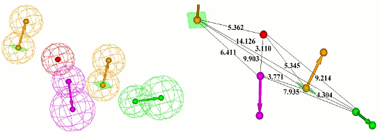
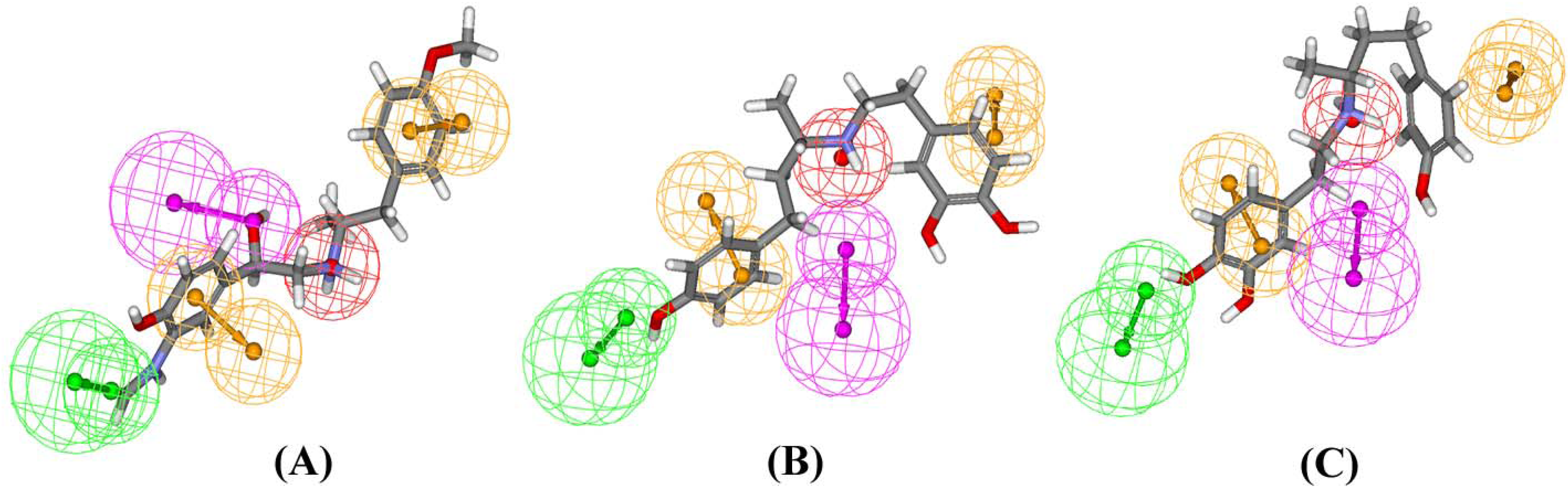
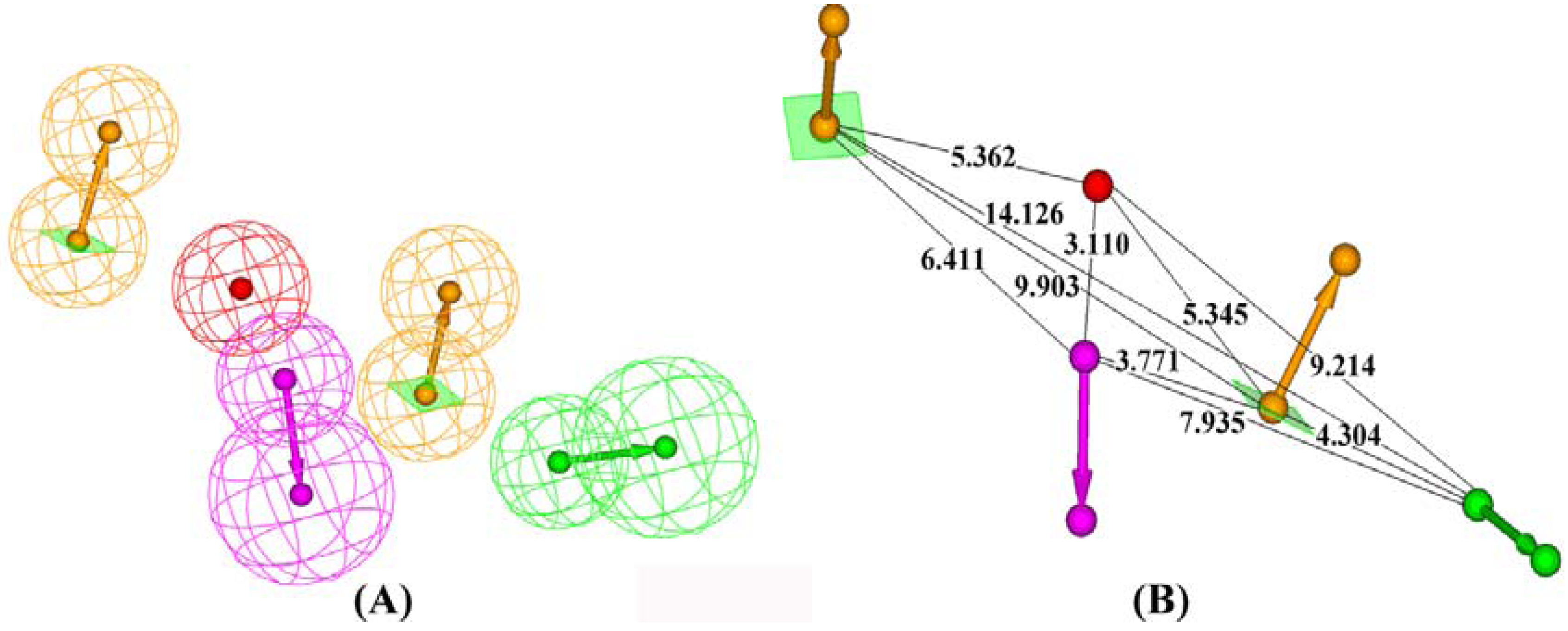
Results and Discussion
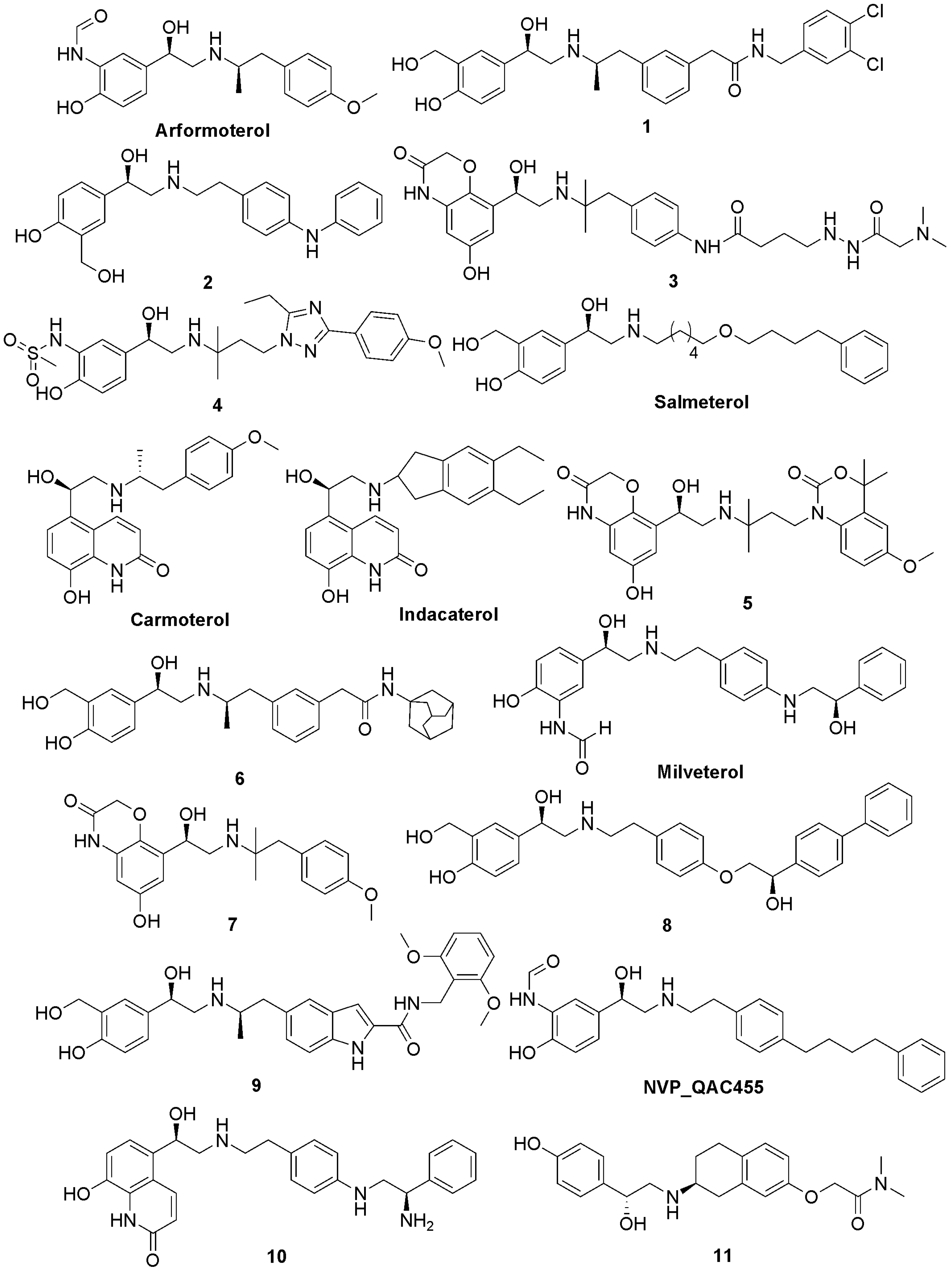
The HipHop module within Catalyst is the common features hypotheses generation program which is widely used in ligand-based approaches to rational drug design [24]. HipHop attempts to derive a pharmacophore based on features that are common to active molecules. The constructed 3D pharmacophore model can be used for identification of original lead compounds from a database [25,26].
Based on the published literature, we selected a series of potent and selective β2-agonists (Figure 1) to generate a pharmacophore model. These compounds were taken from different literature sources [10,27,28,29,30,31,32,33,34,35,36,37,38,39,40], and the activity values were measured in various systems. In addition, due to limited structural diversity, qualitative HipHop pharmacophore modeling was performed based on the collected β2-agonists.
Figure 1. Chemical structures of the selective β2-agonists used to construct the HipHop pharmacophore hypotheses.
Molecules 14 04486 g001
Finally ten pharmacophore hypotheses were automatically generated with alignment scores ranging from 232.507 to 240.129. (Table 1) The best hypothesis (referred to as Hypo1_I) was found to be associated with a five point pharmacophore containing one hydrogen-bond acceptor (A), one hydrogen-bond donor (D), two ring aromatic (R) and one positive ionizable feature (P). It was denoted as RRPDA and is depicted in Figure 2 showing the 3D space and distance between pharmacophoric sites.
Table 1. The results of the top 10 hypotheses for β2-adrenoreceptor agonists generated by the HipHop program.
HypothesesFeaturesRankDirect HitPartial HitMax Fit
1RRPDA240.12911111111111111111000000000000000005
2RRPDA240.12911111111111111111000000000000000005
3RRPDA240.12911111111111111111000000000000000005
4RRPAA236.72911111111111111111000000000000000005
5RRPAA236.72911111111111111111000000000000000005
6RRPAA236.72911111111111111111000000000000000005
7RRPDA232.57911111111111111111000000000000000005
8RRPDA232.57911111111111111111000000000000000005
9RRPAA232.50711111111111111111000000000000000005
10RRPAA232.50711111111111111111000000000000000005
Figure 2. Pharmacophore model for β2-adrenoreceptor agonists generated by HipHop. (A) The best HipHop model Hypol_I. (B) 3D spatial relationship and geometric parameters of Hypol_I. These features are portrayed as mashed spheres, color-coded as follows: green, hydrogen-bond acceptor, magenta, hydrogen-bond donor, orange, aromatic ring, red, positive ionizable feature.
Molecules 14 04486 g002
The alignment of the most potent compound arformoterol onto Hypo1_I is shown in 3A. It was revealed that one hydrogen-bond acceptor and one hydrogen-bond donor matched the 3-formamido group on the benzene ring and the benzylic alcohol, respectively. One positive ionizable feature was at the protonated nitrogen atom and the two ring aromatic features are located in the mother ring and amino-substituents. All the pharmacophores were located in the proximity, which was in good agreement with the key interactions between the β2-adrenoceptor and agonists.
Table 2. The information about the alignment of β2-adrenoreceptor agonists onto Hypo1_I.
IndexNamePrincipalMaxOmitFeatFitValue
1Arformoterol205
2NVP_QAC455104.669
34104.371
46104.358
5Carmoterol104.322
62104.172
78104.092
810103.886
95103.886
101103.881
11Indacaterol103.87
129103.847
13Milveterol103.735
147103.672
153103.63
1611103.402
17Salmeterol102.66
Figure 3. Hypol_I aligned with arformoterol (A), (S)-dobutamine (B) and (R)-dobutamine (C).
Molecules 14 04486 g003
Molecular field-based similarity analysis with the FieldTemplater software provided the necessary three-dimensional molecular field properties of the β2-adrenoceptor agonists. FieldTemplater took three β2-adrenoceptor agonists (arformoterol, salmeterol and indacaterol), optimally aligned their conformer fields and yielded a series of templates which were ranked according to an incorporated score. The top-ranking multi structural template (T1) from FieldTemplater is shown in Figure 4. Four types of 3D molecular field descriptors were produced for describing electrostatic (negative and positive), van der Waals and hydrophobic properties of the β2-adrenoceptor agonists. As shown in the field point patterns (large points indicating strong interactions), it was abundant in hydrophobic fields along the amino-substituents, positive ionic fields surrounding protonated nitrogen atoms (strong interactions) and para-positions of aromatic ring and negative fields near the meta-positions of aromatic rings (strong interactions) and surrounding the benzylic hydroxyl group. This pharmacophore shared enough similar features with Hypol_I and they were almost consistent. The results reflected the validity of the pharmacophore model Hypol_I.
Figure 4. Field point patterns of the three conformers of arformoterol, salmeterol and indacaterol in Template T1. Field points are color coded as follows: negative charge, blue; positive charge, red; van der Waal’s surface, yellow; hydrophobes, orange.
Molecules 14 04486 g004
Figure 5. Chemical structures of selective β1-agonists used to map with Hypol_I.
Molecules 14 04486 g005
To test the selectivity of pharmacophore over β1-adrenoceptor, several selective β1-adrenoceptor agonists (Figure 5) were collected and aligned with the Hypol_I model. As a result, no β1-adrenoceptor agonists could map the features using the Hypol_I model, except for dobutamine. The fit values for (R)-dobutamine and (S)-dobutamine were only 0.803 and 1.528, respectively. The alignment of (R)-dobutamine and (S)-dobutamine onto the pharmacophore is shown in Figure 3B, Figure 3C. It was found that the mapping could not match the hydrogen-bond donor and one ring aromatic feature encoded in Hypol_I. According to this partial mapping, it suggested this hydrogen-bond donor and one ring aromatic feature on the N-substituent might be the key areas for the differentiation of β2-agonists from β1-agonists. It has been reported that the substituent on the amino portion was important for subtype selective agonist binding [41], which further validated our phamacophore. In summary, it may be concluded that our pharmacophore model for β2-agonists could predict the sub-type specificity of the human β-receptors.
Molecular docking has established itself as a valuable in silico technique alongside traditional high-throughput screening (HTS) for discovering new active compounds in the pharmaceutical industry. However, virtual screening of large databases via docking is expensive in terms of CPU-time, especially for GPCRs where the protein conformation changes upon binding with different ligands (ligand-induced fit). Therefore, pharmacophore models can be used as a first-screen before docking studies. Our pharmacophore model for β2-adrenergic receptor agonists can be used for virtual screening of new potent and selective candidates targeting the β2-adrenergic receptor.

Experimental

Pharmacophore modeling with Discovery Studio

A common-features pharmacophore model was derived with the HipHop module of Catalyst. All molecules were built in 2D Visualizer using the Discovery Studio software package and the nitrogen atom of aliphatic amine in each structure was protonated. Arformoterol was considered as ‘reference compound’ specifying a ‘Principal’ value of 2 and a ‘MaxOmitFeat’ value of 0, meaning its structure and conformation would have the strongest influence in the model building phase. The ‘Principal’ value and ‘MaxOmitFeat’ value for the remaining compounds were set to 1 and 0, respectively. Diverse conformational models for each compound were generated using the ‘best conformational analysis’ method and an energy threshold of 20 kcal/mol above the global energy minimum for conformation searching. The maximum number of conformers for each molecule was specified as 250 to ensure maximum coverage of the conformational space. Due to the basic structures of the compounds, five kinds of features including hydrogen-bond acceptor (A), hydrogen-bond donor (D), hydrophobic group (H), positive ionizable (P) and ring aromatic (R) features were selected to initiate the pharmacophore hypotheses generation process.

Molecular field-based similarity analysis with FieldTemplater

Using the XEDEX software, a conformational search was performed with the XED force field. Three β2-adrenoceptor agonists (arformoterol, salmeterol and indacaterol) were submitted to the FieldTemplater software for generation of putative bioactive field template [42,43]. FieldTemplater overlaid the field patterns of all conformers of these compounds to find a single common field pattern assumed to reflect the binding requirements for the β2-adrenoreceptor agonists.

Ligand pharmacophore mapping with Discovery Studio

The β1-agonists were built in 2D Visualizer using Discovery Studio software package and the nitrogen atom of aliphatic amine in each structure was protonated. Diverse conformational models for each compound were generated using the ‘best conformational analysis’ method and an energy threshold of 20 kcal/mol above the global energy minimum for conformation searching. A flexible fitting method was adopted to map these β1-agonists with Hypol_I.

Conclusions

In summary, we have presented the first study using a ligand-based computational approach to generate specific 3D pharmacophore hypotheses for the β2 aderonergic receptor from its selective agonists. The best hypothesis with a five point pharmacophore was consistent with the interactions between the β2-adrenoceptor and agonists, and it was further validated by molecular field-based similarity analysis with FieldTemplater. Moreover, the best pharmacophore hypothesis could perfectly differentiate β2-agonists from β1-agonists, so this pharmacophore model may be considered a valuable tool to predict agonist activity and identify diverse structures with desired biological activity and selectivity by 3D virtual screening.

Acknowledgements

This work was supported by the National High Technology Research and Development Program of China (863 Program, 2005AA233030). The authors would like to thank Cresset BioMolecular Discovery Ltd. for kindly allowing us a free evaluation of their software packages.

References and Notes

  1. Johnson, M. The β-adrenoceptor. Am. J. Respir. Crit. Care. Med. 1998, 158, S146–S153. [Google Scholar]
  2. Broadley, K.J. Adrenoceptor responses of the airways: For better or worse? Eur. J. Pharmacol. 2006, 533, 15–27. [Google Scholar] [CrossRef]
  3. Sears, M.R.; Lötvall, J. Past, present and future—β2-adrenoceptor agonists in asthma management. Respir. Med. 2005, 99, 152–170. [Google Scholar] [CrossRef]
  4. Tatterfield, A.E. Current issues with β2-adrenoceptor agonists. Clin. Rev. Allergy. Immunol. 2006, 31, 107–117. [Google Scholar] [CrossRef]
  5. Matera, M.G.; Cazzola, M. Ultra-long-acting β2-adrenoceptor agonists: An emerging therapeutic option for asthma and COPD? Drugs 2007, 67, 503–515. [Google Scholar] [CrossRef]
  6. Strader, C.D.; Sigal, S.I.; Dixon, R.A.F. Structural basis of β-adrenergic receptor function. FASEB J. 1989, 3, 1825–1832. [Google Scholar]
  7. Strader, C.D.; Candelore, M.R.; Hill, W.S.; Sigal, I.S.; Dixon, R.A.F. Identification of two serine residues involved in agonist activation of the β2-adrenergic receptor. J. Biol. Chem. 1989, 264, 13572–13578. [Google Scholar]
  8. Kikkawa, H.; Isogaya, M.; Nagao, T.; Kurose, H. The role of the seventh transmembrane region in high affinity binding of a β2-selective agonist TA-2005. Mol. Pharm. 1998, 53, 128–134. [Google Scholar]
  9. Zuurmond, H.M.; Hessling, J.; Blüml, K.; Lohse, M.; Ijzerman, A.P. Study of interaction between agonists and Asn293 in Helix VI of human β2-adrenergic receptor. Mol. Pharm. 1999, 56, 909–916. [Google Scholar]
  10. Alikhani, V.; Beer, D.; Bentley, D.; Bruce, I.; Cuenoud, B.M.; Fairhurst, R.A.; Gedeck, P.; Haberthuer, S.; Hayden, C.; Janus, D.; Jordan, L.; Lewis, C.; Smithies, K.; Wissler, E. Long-chain formoterol analogues: an investigation into the effect of increasing amino-substituent chain length on the β2-adrenoceptor activity. Bioorg. Med. Chem. Lett. 2004, 14, 4705–4710. [Google Scholar]
  11. Rosenbaum, D.M.; Cherezov, V.; Hanson, M.A.; Rasmussen, S.G.F.; Thian, F.S.; Kobilka, T.S.; Choi, H.J.; Yao, X.J.; Weis, W.I.; Stevens, R.C.; Kobilka, B.K. GPCR engineering yields high-resolution structural insights into β2-adrenergic receptor fuction. Science 2007, 318, 1266–1273. [Google Scholar]
  12. Rasmussen, S.G.F.; Choi, H.J.; Rosenbaum, D.M.; Kobilka, T.S.; Thian, F.S.; Edwards, P.C.; Burghammer, M.; Ratnala, V.R.P.; Sanishvili, R.; Fischetti, R.F.; Schertler, G.F.X.; Weis, W.I.; Kobilka, B.K. Crystal structure of the human β2-adrenergic G-protein-coupled receptor. Nature 2007, 450, 383–388. [Google Scholar]
  13. Cherezov, V.; Rosenbaum, D.M.; Hanson, M.A.; Rasmussen, S.G.F.; Thian, F.S.; Kobilka, T.S.; Choi, H.J.; Kuhn, P.; Weis, W.I.; Kobilka, B.K.; Stevens, R.C. High-resolution crystal structure of an engineered human β2-adrenergic G-protein-coupled receptor. Science 2007, 318, 1258–1265. [Google Scholar]
  14. Kobilka, B.; Schertler, G.F.X. New G-protein-coupled receptor crystal structures: insights and limitations. Trends Pharmacol. Sci. 2008, 29, 79–83. [Google Scholar] [CrossRef]
  15. Topiol, S.; Sabio, M. Use of the X-ray structure of the beta2-adrenergic receptor for drug discovery. Bioorg. Med. Chem. Lett. 2008, 18, 1598–1602. [Google Scholar] [CrossRef]
  16. Sabio, M.; Jones, K.; Topiol, S. Use of the X-ray structure of the beta2-adrenergic receptor for drug discovery 2. Identification of active compounds. Bioorg. Med. Chem. Lett. 2008, 18, 5391–5395. [Google Scholar] [CrossRef]
  17. Costanzi, S. On the applicability of GPCR homology models to computer-aided drug discovery: a comparison between in silico and crystal structures of the β2-adrenergic receptor. J. Med. Chem. 2008, 51, 2907–2914. [Google Scholar] [CrossRef]
  18. Graaf, C.; Rognan, D. Selective structure-based virtual screening for full and partial agonists for the β2 adrenergic receptor. J. Med. Chem. 2008, 51, 4978–4985. [Google Scholar] [CrossRef]
  19. Gayathri, S.; Yang, X.; Tae, W.L.; Jacqueline, S.; Charles, P.; Brian, K.K. Sequential binding of agonists to the β2-adrenoceptor. J. Biol. Chem. 2004, 279, 686–691. [Google Scholar]
  20. Karki, R.G.; Kulkarni, V.M. A feature based pharmacophore for Candida albicans MyristoylCoA: protein N-myristoyltransferase inhibitors. Eur. J. Med. Chem. 2001, 36, 147–163. [Google Scholar] [CrossRef]
  21. Singh, N.; Nolan, T.L.; McCurdy, C.R. Chemical function-based pharmacophore development for novel, selective kappa opioid receptor agonists. J. Mol. Graph. Model. 2008, 27, 131–139. [Google Scholar] [CrossRef]
  22. Burnett, J.C.; Wang, C.; Nuss, J.E.; Nguyen, T.L.; Hermone, A.R.; Schmidt, J.J.; Gussio, R.; Wipf, P.; Bavari, S. Pharmacophore-guided lead optimization: The rational design of a non-zinc coordinating, sub-micromolar inhibitor of the botulinum neurotoxin serotype a metalloprotease. Bioorg. Med. Chem. Lett. 2009, 19, 5811–5813. [Google Scholar]
  23. Shah, U.A.; Deokar, H.S.; Kadam, S.S.; Kulkarni, V.M. Pharmacophore generation and atom-based 3D-QSAR of novel 2-(4-methylsulfonylphenyl)pyrimidines as COX-2 inhibitors. Mol. Divers. 2009. [Google Scholar] [CrossRef]
  24. Accelrys Inc. 10188 Telesis Court. Suite 100 San Diego. CA 92121, USA, 2008.
  25. Michaux, C.; Leval, X.; Julémont, F.; Dogné, J.M.; Pirott, B.; Durant, F. Structure-based pharmacophore of COX-2 selective inhibitors and identification of original lead compounds from 3D database searching method. Eur. J. Med. Chem. 2006, 41, 1446–1455. [Google Scholar] [CrossRef]
  26. Ren, J.; Li, L.; Zou, J.; Yang, L.; Yang, J.; Yang, S. Pharmacophore modeling and virtual screening for the discovery of new transforming growth factor-β type I receptor (ALK5) inhibitor. Eur. J. Med. Chem. 2009, 44, 4259–4265. [Google Scholar] [CrossRef]
  27. Revill, P.; Serradell, N.; Bolós, J.; Bayés, M. Aformoterol tartrate. Drugs of the Future 2006, 31, 944–952. [Google Scholar] [CrossRef]
  28. Linsell, M.S.; Jacobasen, J.R.; Khossravi, D.; Paborji, M.; Zhang, W. Crystalline β2-adrenergic receptor agonist. WO Pat. 2004011416, 2004. [Google Scholar]
  29. Voss, H.P.; Donnell, D.; Bast, A. Atypical molecular pharmacology of a new long-acting β2-adrenoceptor agonist, TA 2005. Eur. J. Pharmacol. 1992, 227, 403–409. [Google Scholar] [CrossRef]
  30. Battram, C.; Charlton, S.J.; Cuenoud, B.; Dowling, M.R.; Fairhurst, R.A.; Farr, D.; Fozard, J.R.; Leighton-Davies, J.R.; Lewis, C.A.; McEvoy, L.; Turner, R. J.; Trifilieff, A. In vitro and in vivo pharmacological characterization of 5-[(R)-2-(5,6-diethyl-indan-2-ylamino)-1-hydroxy-ethyl]-8-hydroxy-1H-quinolin-2-one (Indacaterol), a novel inhaled b2 adrenoceptor agonist with a 24-h duration of action. J. Pharmacol. Exp. Ther. 2006, 317, 762–770. [Google Scholar] [CrossRef]
  31. Ball, D.I.; Brittain, R.T.; Coleman, R.A.; Denyer, L.H.; Jack, D.; Johnson, M.; Lunts, L.H.C.; Nials, A.T.; Sheldrick, K.E.; Skidmore, I.F. Salmeterol, a novel, long-acting β2-adrenoceptor agonist: characterization of pharmacological activity in vitro and in vivo. Br. J. Pharmacol. 1991, 104, 665–671. [Google Scholar] [CrossRef]
  32. Brown, A.D.; Bunnage, M.E.; Glossop, P.A.; James, K.; Jones, R.; Lane, C.A.L.; Lewthwaite, R.A.; Mantell, S.; Perros-Huguet, C.; Price, D.A.; Trevethick, M.; Webster, R. The discovery of adamantyl-derived, inhaled, long acting β2-adrenoreceptor agonists. Bioorg. Med. Chem. Lett. 2008, 18, 1280–1283. [Google Scholar]
  33. Brown, A.D.; Bunnage, M.E.; Glossop, P.A.; James, K.; Jones, R.; Lane, C.A.L.; Lewthwaite, R.A.; Mantell, S.; Perros-Huguet, C.; Price, D.A.; Trevethick, M.; Webster, R. The discovery of long acting β2-adrenoreceptor agonists. Bioorg. Med. Chem. Lett. 2007, 17, 4012–4015. [Google Scholar]
  34. Norman, P. Combinations of a long-acting β2 agonist; is it BI-1744-CL? Expert Opin. Ther. Patents 2007, 17, 1401–1404. [Google Scholar] [CrossRef]
  35. Brown, A.D.; Bunnage, M.E.; Glossop, P.A.; Holbrook, M.; Jones, R.D.; Lane, C.A.L.; Lewthwaite, R.A.; Mantell, S.; Perros-Huguet, C.; Price, D.A.; Webster, R. The discovery of indole-derived long acting β2-adrenoceptor agonists for the treatment of asthma and COPD. Bioorg. Med. Chem. Lett. 2007, 17, 6188–6191. [Google Scholar]
  36. Bouyssou, T.; Rudolf, K.; Hoenke, C.; Lustenberger, P.; Schnapp, A.; Konetzki, I. Studies towards topical selective β2-adrenoceptor agonists with a long duration of action. Bioorg. Med. Chem. Lett. 2009, 19, 5237–5240. [Google Scholar] [CrossRef]
  37. Linsell, M.; Jacobsen, J.R.; Saito, D.R. Amino-substituted ethylamino b2 adrenergic receptor agonists. WO Pat. 2005030678, 2005. [Google Scholar]
  38. Kitazawa, M.; Okazaki, K.; Tamai, T.; Saito, M.; Muranaka, H.; Tanaka, N.; Kobayashi, H.; Kikuchi, K. Phenylethanolaminotetralincarboxamide derivatives. US Pat. 6747043, 1997. [Google Scholar]
  39. Moran, E.J.; Fournier, E. Alkoxy aryl β2 adrenergic receptor agonists. US Pat. 6747043, 2004. [Google Scholar]
  40. Moran, E.J.; Jacobsen, J.R.; Leadbetter, M.R.; Nodwell, M.B.; Trapp, S.G.; Aggen, J.; Church, T.J. Aryl aniline beta-2 adrenergic receptor agonists. WO Pat. 03042164 2003. [Google Scholar]
  41. Kurose, H.; Isogaya, M.; Kikkawa, H.; Nagao, T. Domains of β1 and β2 adrenergic receptors to bind subtype selective agonists. Life Sci. 1998, 62, 1513–1517. [Google Scholar] [CrossRef]
  42. Cheeseright, T.; Mackey, M.; Rose, S.; Vinter, A. Molecular field extrema as descriptors of biological activity: definition and validation. J. Chem. Info. Mod. 2006, 46, 665–676. [Google Scholar] [CrossRef]
  43. Cheeseright, T.; Mackey, M.; Rose, S.; Vinter, A. Molecular field technology applied to virtual screening and finding the bioactive conformation. Expert Opin. Drug Discov. 2007, 2, 131–144. [Google Scholar] [CrossRef]
  • Sample availability: Not available.

