Communication

A New Cytotoxic Friedelane Acid – Pluricostatic Acid – and Other Compounds from the Leaves of Marila pluricostata

by
Dionisio A. Olmedo
1,
José L. López-Pérez
2,
Esther Del Olmo
2,
Yelkaira Vásquez
1,
Arturo San Feliciano
2 and
Mahabir P. Gupta
1,*
1
Centro de Investigaciones Farmacognósticas de la Flora Panameña (CIFLORPAN), Facultad de Farmacia, Universidad de Panamá, Apartado 0824-00172,Panama
2
Departamento de Química Farmacéutica, Facultad de Farmacia, CIETUS, Universidad de Salamanca, Campus Miguel de Unamuno, E-37007, Salamanca, Spain
*
Author to whom correspondence should be addressed.
Molecules 2008, 13(11), 2915-2924; https://doi.org/10.3390/molecules13112915
Submission received: 16 October 2008 / Revised: 14 November 2008 / Accepted: 18 November 2008 / Published: 25 November 2008

Abstract

:
Bioassay-guided fractionation of the dichloromethane extract of the leaves of Marila pluricostata led to the isolation of 2α,3β-dihydroxy-D:A-friedoolean-28-oic acid (pluricostatic acid), a new friedelane triterpenoid, (1), ten known triterpenoids and three sterols. Their chemical structures were elucidated through spectroscopic analysis. The less polar fractions, on GC/MS analysis and comparison with a MS library, resulted in the identification of twenty four sesquiterpenoids. The new triterpenoid acid 1 showed cytotoxicity against the MCF-7, H-460, and SF-268 human cancer cell lines with GI50 values from 1.2 to 3.3 μg/mL.

Introduction

Marila pluricostata Standl. & L.O. Williams (Clusiaceae) is a tree which has a restricted distribution in Panama [1,2,3], Costa Rica [1,2], and Colombia [4]. There is no common name nor any ethnomedical use reported for this species, while Marila tomentosa is used in Colombia for the treatment of dysentery [5]. Previous phytochemical studies on Marila pluricostata by our group reported several 4-phenylcoumarins [6], some of them showed some degree of antineoplastic cytotoxicity [6], while others displayed transcription inhibitory activity of the HIV Tat receptor [7]. We now report the isolation and structure assignment of one new triterpenoid acid – 2α,3β-dihydroxy-D:A-friedoolean-28-oic acid (1) – and the identification of 10 known triterpenoids 2-11, three sterols 12-14 and 24 sesquiterpenoids as components of its dichloromethane extract. The cytotoxic activity of compound 1 (Figure 1), against three human cancer cell lines (H-460, MCF-7, and SF-268) has also been assessed.
Figure 1. Structures of compound 1 and 1a.
Molecules 13 02915 g001

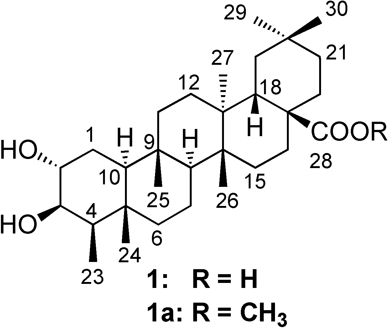
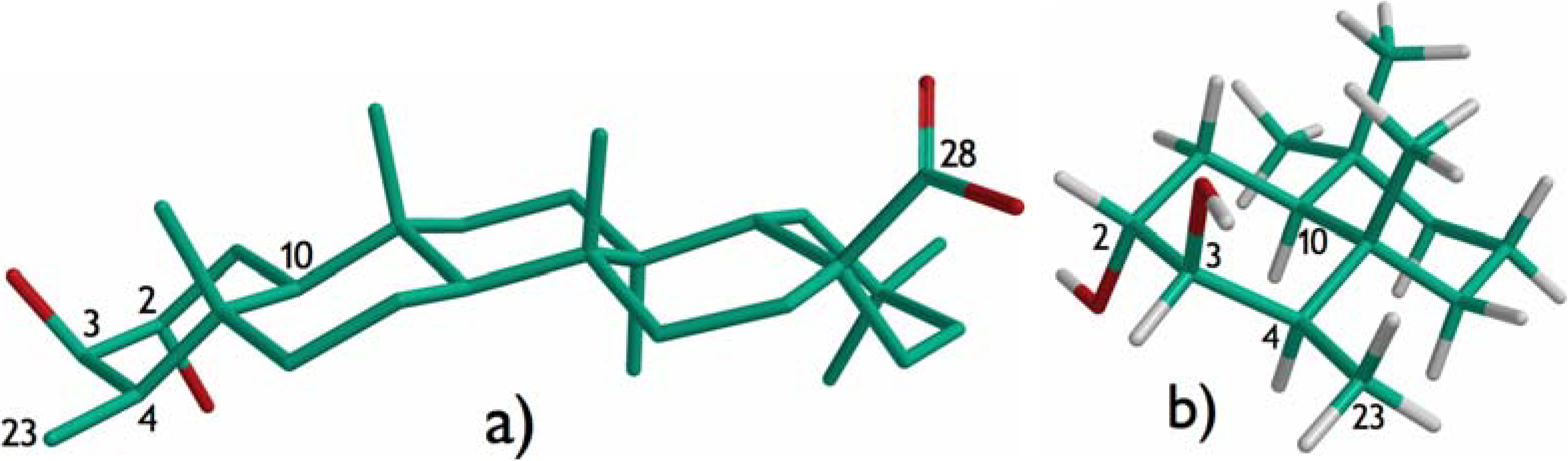
Results and Discussion

Compound 1 was isolated as a white amorphous solid. HRFABMS showed [M]+ at m/z 474.7162, corresponding to the molecular formula of C30H50O4, typical for a triterpenoid. The IR spectrum exhibited one broad absorption band due to a hydroxyl function (3,410 cm−1) and another intense band at 1,688 cm−1 corresponding to a carboxylic acid group. The skeletal basis of a friedelane triterpenoid skeleton in compound 1 was deduced from an iterative search carried out within our NAPROC-13 database [8]. This finding was also corroborated by the presence in the 1H-NMR spectrum of a doublet (J = 7.3 Hz) centered at δ 0.77 ppm, characteristic of the methyl group at position C-23 of friedelane compounds. The search in NAPROC-13 also revealed the presence of a carboxyl group (δ 180.0 ppm) at position C-28 of the friedelane skeleton. Nevertheless, the overlapping of a number of signals in the 1H-NMR spectrum did not allow us to perform the complete analysis of many hindered signals, important for the structure assignment. Consequently, some 2D-NMR experiments (HMQC, COSY, HMBC and ROESY) were run in order to obtain more information about the compound. The 1H-NMR spectrum showed a double doublet at δ 3.64 (J1 = 7.8 Hz; J2 = 2.4 Hz) and a triplet at δ 3.23 (J = 2.4 Hz) corresponding to two oxygenated methines, that simultaneously were correlated in the COSY spectrum, cross-linked in the HMQC spectrum with their corresponding carbon signals at δ 70.1 and 74.7 ppm, respectively, and long-range interconnected in the HMBC spectrum. Therefore, both hydroxylated methines must be vicinal. Additionally, in the HMBC spectrum a long-range correlation was observed between the C-23 methyl signal [δ 11.3 ppm] and that of the oxygenated methine proton at 3.64 ppm. These data support the localization of the hydroxyl groups at C-2 and C-3, which were associated with the carbon signals at δ 70.01 [3.64 (J1 = 7.8 Hz; J2 = 2.4 Hz)] and 74.7 [3.23 (J = 2.4 Hz)], respectively. Correlations were also observed for the signals of H-1 (1.36 ppm) and those of the methine carbon C-2 and the quaternary carbon at 51.3 ppm, thus assigned to C-10. The signal of the carboxylic carbon (δ 180.0) was correlated with those of the protons of two methylene carbons at 29.4 and 35.4 ppm, thus assigned to C-16 and C-22, respectively. The free carboxylic group signal also correlated with that of the methine carbon at 37.2 ppm, assigned to C-18. These facts confirmed that the carboxyl group was located at position C-28. The remaining connectivities observed in the HMBC spectrum allowed the unambiguous assignment of most 1H- and 13C-NMR signals. The configuration of the hydroxylated methines C-2 and C-3 were established by ROESY experiments. In the 2D-spectrum a correlation was observed between H-10 and H-4, thus indicating the axial disposition for the latter and, hence, the methyl group at position C-23 must display an equatorial disposition. The coupling pattern of the signal assigned to the proton geminal to the hydroxyl group at position C-3, a triplet with a coupling constant of 2.4 Hz, allowed us to discard the antiperiplanar disposition for H-2 and H-3 because of their observed small coupling constants. The correlations observed in the ROESY spectrum between H-3 and H-4 and H-23 indicated that the proton corresponding to the signal at δ 3.23 ppm was in the alpha disposition, consequently, the hydroxyl group at C-3 must have a beta disposition. Consequently the alpha disposition for the hydroxyl group at C-2 was established. All these statements are in agreement with the structure obtained by molecular modeling studies after a conformational analysis carried out for compound 1 (Figure 2). These data permitted us to establish the absolute configuration of this new compound, which is corroborated by biogenetic pathway for the friedelane type triterpenoids obtained from the squalene oxide, yielding the stereochemistry indicated in this paper. In the literature reviewed there in no mention of any friedelanes of the opposite series.
Figure 2. a) Low-energy conformer and full geometry optimization of compound 1; b) Partial representation of rings A and B for compound 1.
Molecules 13 02915 g002
Accordingly, attempts to obtain the acetonide derivative of 1 by treatment with 2,2-dimethoxypropane in acetone in the presence of acid, such as p-toluenesulfonic acid (p-TsOH) or trimethylsilyl chloride, left the starting compound 1 unchanged, in agreement with the trans-diaxial disposition of the hydroxyl groups. Thus, the compound was identified as the new friedelane 2α,3β-dihydroxy-D:A-friedoolean-28-oic acid (1), which we named pluricostatic acid. This compound is being reported here for the first time, however, its epimer 2α,3α-dihydroxy-D:A-friedoolean-28-oic acid has been isolated previously from Euonymus revolutus [9].
The ten known triterpenoids were identified as: squalene (2), friedelin (3) [10,11], 4-epi-friedelin (4) [12], canophyllal (5) [13], friedelinol (6) [14], canophyllol (7) [15,16], 3-oxo-friedelan-28 oic acid (8) [9], D:A-friedo-3,4-seco-olean-3-oic acid (9) [17], epi-betulinic acid (10) [18], betulinic acid (11) [19] and three sterols as β-sitosterol (12) [20] stigmasterol (13) [21] and 3-O-β-glucopyranosyl-sitosterol (14) [20] through the complete analysis of their NMR and MS data and comparison with those included in our database NAPROC-13 [8]. NMR data for pluricostatic acid methyl ester (1a) are also reported here.

Biological activity

The dichloromethane extract, fractions and pure compounds were evaluated against three human cancer cell lines, MCF-7 (human breast adenocarcinoma), H-460 (human lung carcinoma) and SF-268 (human CNS carcinona), using the sulphorhodamine B assay [22]. The compounds 1, 8-11 showed GI50 values as indicated in the Table 1. The other compounds were considered inactive as their GI50 values were higher than 10 μg/mL.
Table 1. Antineoplastic cytotoxicity of extracts and compounds 1, 8-11.
GI50 (μg/mL)
MCF-7H-460SF-268
dichloromethane extract5.96.24.6
methanolic fraction defatted8.37.85.5
neutral fraction1.61.50.7
acid fraction2.42.72.2
13.01.23.3
86.35.14.6
97.87.2> 10
107.25.36.5
115.03.65.1
Adriamycin0.370.420.50

Experimental

General

Melting points were determined on a Büchi 510-K melting point apparatus and are uncorrected. Optical rotation was determined on an AUTOPOL III automatic polarimeter. IR spectra were obtained on a Nicolet Impact 410 spectrophotometer with KBr pellets. 1H-NMR (200 and 400 MHz), and 13C- NMR (100 and 50 MHz) spectra were recorded on Brüker AC 200 (200 MHz) and Brüker DRX 400 (400 MHz) spectrometers, with TMS as an internal reference. 2D-NMR spectra were measured with Brüker DRX 400 spectrometer. HRFABMS analysis, a VG-TS250 mass spectrometer (70 eV) was used. Gas chromatography/mass spectrometry (GC/MS) analysis was performed on a Hewlett Packard 5890 serie II, chromatograph apparatus equiped with Hewlett-Packard 5971 mass spectrometer operating in the EIMS-mode at 70 eV and Supelco SPB-1 column (12 m x 0.20 mm, with 0.33 μm film thickness) with helium (10 Psi), as carrier gas at a flow rate of 1 mL/min. The GC oven temperature was kept at 90 ºC for 5 minutes and programmed to 250 oC with a gradient of 2 oC/min and maintained for 10 min at 250 ºC. Injector and detector temperatures were 250 oC and 260 oC, respectively. The injection volume: 1.0 µL of the 10% solution of the fraction A-D. Silica gel (230–400 mesh) for column chromatography and GF254 for TLC were obtained from Merck KGaA, 64271 Darmstadt, Germany. Sephadex LH-20 was obtained from Fluka, BioChemika Switzerland. The relative percentages of the identified components were computed from GC peak area on SPB-1 without any correction factor. The minimum source temperature was 90ºC; acquisition mass range, m/z 40-600.

Plant Material

Leaves of M. pluricostata Standl. & L.O. Williams were collected from Llano-Cartí in the province of Panama, Republic of Panama, in November of 2000. Its taxonomic identity was established by Mireya Correa, Director the Herbarium of the University of Panama. Voucher specimens (F-4740 and F-4937) were deposited in the Herbarium of the University of Panama (PMA).

Extraction, Isolation and Identification

The dried and powdered plant material of Marila pluricostata (3.4 kg) was extracted with dichloromethane (10 L) at room temperature for five days. Evaporation of the solvent yielded a crude extract (186.5 g), which was further solubilized in hot n-hexane (4 L twice) and cooled (-20 oC) overnight, yielding a soluble fraction (136.1 g), which was defatted successively with MeOH (3L twice) and a saturated solution of urea in MeOH (3L twice). The final soluble part (74.5 g) was partitioned with basic aqueous solution (NaOH 4% x 3 times), yielding an acid fraction (3.0 g) and neutral fraction (71.1 g). The neutral fraction was successively fractionated on a silica gel column using a gradient solvent system n-hexane, n-hexane/EtOAc (95:5 → 50:50), EtOAc, MeOH to give subfractions (A-K). The subfraction A-D (0.81, 1.03, 0.30 and 1.62 g) were analyzed by GC/MS and 24 known sesquiterpenes were identified by comparison of their retention times and mass spectra (Table 2) with those stored in the spectrometric electronic library (Wiley 275.l) and those reported in the literature [23].
Workup of subfraction E (6.92 g) by repeated CC over silica gel, using n-hexane, n-hexane/ dichloromethane (95:5 → 60:40), CH2Cl2, MeOH) led to the isolation of triterpenoids 3 (46.4 mg, 0.024 %), 4 (28.9 mg, 0.015 %), 5 (333.2 mg, 0.780 %) and 6 (23.2 mg, 0.012 %). Subfraction F (29.96 g) was further fractionated on silica gel using n-hexane/EtOAc (95:5 → 50:50), EtOAc, MeOH) to yield the triterpenoids 7 (139.1 mg, 0.010 %), and 8 (8 mg, 0.004 %). Subfraction G (5.87 g) on chromatography over silica gel using CH2Cl2-Et2O (80:20) as eluent afforded triterpenoid 9 (118 mg, 0.100 %). Subfraction H (11.32 g) on CC over Sephadex LH 20 using n-hexane-CH2Cl2-MeOH (2:1:1), as eluent gave triterpenoids 10 (115 mg, 0.061 %) and 11 (118 mg, 0.100 %). Subfraction I (11.32 g) by repeated CC on Sephadex LH 20, using n-hexane-CH2Cl2-MeOH (2:1:1) furnished sterols 12 (120 mg, 0.064 %) and 13 (154 mg, 0.082 %). Subfraction J (7.14 g) on CC over silica gel using CH2Cl2-MeOH (1:1), afforded the new friedelane acid 1 (78 mg, 0.042 %). Finally, subfraction K (6.11 g) on silica gel CC using CH2Cl2-MeOH (1:1) afforded sterol 14 (39 mg, 0.021 %).
Table 2. Chemical composition of fractions A-D obtained by GC/MS a.
ComponentsRetention
time (Rt)
(min.)
Relative
(%)
M+
m/z
fraction A
α-copaene17.332.6220
β-caryophyllene20.0129.9204
zingiberene20.831.1204
germacrene-d21.840.6204
α-amorphene23.0211.7204
β-selinene23.193.8204
calarene23.714.1204
α-muurolene24.173.6204
χ-cadinene24.877.7204
calamenene25.011.6204
δ-cadinene25.423.6204
α-cadinene26.093.0204
calacorene26.851.3200
α-caryophyllene oxide27.9010.9220
Total compounds
identified
85.5
fraction B
α-copaene17.3736.0220
β-elemene18.410.9204
β-caryophyllene20.0313.4204
β-cubenene20.253.0204
α-humulene21.180.8204
α-amorphene23.0511.7204
α-aromadendrene23.113.8204
Total compounds
identified
69.6
fraction C
α-cubebene16.065.1204
α-copaene17.3538.2220
β-bourbonene17.767.6204
β-cubenene20.2511.3204
α-aromadendrene23.130.7204
β-sesquiphellandrene25.340.9204
Total compounds
identified
36.4
fraction D
β-farnesene16.482.8204
α-curcumene16.921.8202
β-bisabolene17.721.9204
Squalene42.4089.9410
Total compounds
identified
96.4
aQuality 95 to 99 %.
2α,3β-Dihydroxy-D:A-friedoolean-28-oic acid (1): White amorphous solid (CHCl3/MeOH): mp 310-313 ºC; [α]D25 +2.0o (c.1.0, MeOH); IR (CHCl3) υmax 3410, 2933, 2871, 1688, 1431, 1388, 1247 cm–1; 1H-NMR (DMSO-d6, 400 MHz) δ 3.64 (1H, dd, J1 = 7.8 Hz; J2 = 2.4 Hz, H-2), 3.23 (1H, t, J = 2.4 Hz, H-3), 2.36 (1H, dd, J1 = 9.8 Hz, J2 = 2.4 Hz, H-18), 2.22 (1H, dd, J1 = 7.8 Hz, J2 = 4.2 Hz, H-16α), 1.59 (1H, m, H-6β), 1.50 (1H, m, H-16β), 1.40 (1H, m, H-4), 1.37 (1H, m, H-10), 1.36 (2H, m, H-22; 1H, m, H-1β), 1.35 (1H, t, H-21β), 1.34 (1H, t, H-12β), 1.29 (1H, d, H-19β), 1.28 (1H, d, H-1α), 1.26 (2H, m, H-7; 1H, m, H-12α), 1.15 (1H, m, H-21α), 1.12 (1H, m, H-8), 1.10 (1H, m, H-19α), 1.08 (2H, m, H-15; 2H, m, H-11), 0.99 (3H, s, H-30), 0.94 (3H, s, H-27), 0.89 (3H, s, H-29), 0.88 (1H, m, H-6α), 0.82 (3H, bs, H-24), 0.77 (3H, d, J = 7.3 Hz, H-23), 0.76 (3H, s, H-25), 0.74 (3H, m, H-26); 13C-NMR (DMSO-d6, 100 MHz) δ 180.0 (CO, COOH, C-28), 74.7 (CHOH, C-3), 70.1 (CHOH, C-2), 52.8 (CH, C-8), 51.3 (CH, C-10), 43.9 (C, C-17), 43.0 (CH, C-4), 41.3 (CH2, C-6), 38.4 (C, C-13), 37.4 (C, C-14), 37.3 (C, C-5), 37.2 (C, C-18), 36.3 (C, C-9), 35.5 (CH3, C-30), 35.4 (CH2, C-22), 34.7 (CH2, C-11), 34.6 (CH2, C-19), 32.5 (CH2, C-21), 32.4 (CH2, C-15), 30.8 (CH2, C-12), 29.8 (CH3, C-29), 29.4 (CH2, C-16), 28.2 (C, C-20), 23.3 (CH2, C-1), 20.0 (CH3, C-26), 18.3 (CH3, C-27), 17.7 (CH3, C-25), 17.1 (CH2, C-7), 15.9 (CH3, C-24), 11.3 (CH3, C-23); HRFABMS m/z 474.7162 (calcd. for C30H50O4, 474.7201).
Methyl 2α,3β-dihydroxy-D:A-friedoolean-28-oate (1a): Cooled ethereal diazomethane solution (5 mL) was added dropwise to a cooled solution of D:A 2α,3β-dihydroxy-friedoolean-28oic acid (30 mg) in 4 mL of diethyl ether and the reaction mixture was maintained at 0 ºC for 5 h, then concentrated in vacuo to give 1a (32 mg). White amorphous solid (CHCl3/MeOH); [α]D25 +0.0o (c.1.0, MeOH); 1H- NMR (CDCl3/CD3OD 9:1, 200 MHz) δ 3.84 (1H, dd, J1 = 7.8 Hz, J2 = 2.4 Hz, H-2), 3.66 (3H, s, COOCH3), 3.44 (1H, t, J = 2.4 Hz, H-3), 2.42 (1H, dd, J1 = 9.8 Hz, J2 = 2.4 Hz, H-18), 2.32 (1H, dd, J1 = 7.8 Hz, J2 = 4.2 Hz, H-16α), 1.71 (1H, m, H-1β), 1.69 (1H, m, H-6β), 1.64 (1H, m, H-16β), 1.50 (H, m, H-4), 1.49 (1H, d, J1 = 2.4 Hz, H-1α), 1.46 (1H, m, H-22β), 1.44 (1H, t, H-12β; ), 1.43 (1H, m, H-10), 1.42 (2H, m, H-15), 1.41 (1H, m, H-22α), 1.38 (3H, s, H-25), 1.36 (1H, m, H-8; 1H, m, H-19β), 1.31 (1H, m, H-12α),1.22 (1H, m, H-11β), 1.17 (2H, t, J = 2.4 Hz, H-21), 1.16 (1H, t, J = 2.4 Hz, H-19α), 1.14 (1H, m, H-11α), 1.02 (3H, bs, H-26; 3H, bs, H-30), 0.97 (1H, m, H-6α), 0.91 (3H, s, H-29), 0.90 (3H, s, H-24), 0.88 (3H, d, J = 7.3 Hz, H-23), 0.84 (3H, s, H-27; 1H, s, H-7β), 0.81 (1H, s, H-7α), 13C-NMR (CDCl3/CD3OD 9:1, 50 MHz) δ 182.0 (CO, COOCH3, C-28), 74.1 (CHOH, C-3), 69.5 (CHOH, C-2), 51.6 (CH3, COOCH3), 51.7 (CH, C-8), 50.1 (CH, C-10), 43.1 (C, C-17), 42.1 (CH, C-4), 39.9 (CH2, C-6), 37.2 (C, C-13), 36.3 (C, C-18), 36.2 (CH, C-14), 36.0 (C, C-5), 35.1 (C, C-9), 34.3 (CH2, C-22), 33.4 (CH2, C-11), 33.3 (CH2, C-19), 32.3 (CH3, C-30), 31.0 (CH2, C-15), 30.9 (CH2, C-21), 29.6 (CH2, C-12), 28.1 (CH2, C-16), 27.7 (CH3, C-29), 26.6 (C, C-20), 21.8 (CH2, C-1), 18.4 (CH3, C-27), 16.4 (CH3, C-26), 15.8 (CH2, C-7), 15.7 (CH3, C-25), 13.8 (CH3, C-24), 8.9 (CH3, C-23).
Since 13C-NMR data of canophyllal (5) have not been reported before, these are included here: 13C- NMR (CDCl3, 50 MHz) δ 213.0 (CO, C-3), 209.0 (CHO, C-28), 59.2 (CH, C-10), 58.2 (CH, C-4), 52.8 (CH, C-8), 47.7 (C, C-17), 42.0 (C, C-5), 41.5 (CH2, C-2), 41.0 (CH2, C-6), 38.7 (C, C-13), 37.7 (C, C-14), 37.1 (C, C-9), 36.4 (CH, C-18), 35.4 (CH2, C-11), 34.9 (CH2, C-19), 34.5 (CH3, C-29), 33.4 (CH2, C-21), 32.4 (CH2, C-15, C-16), 30.6 (CH2, C-12), 29.4 (CH3, C-30), 28.3 (C, C-20), 28.0 (CH2, C-22), 22.3 (CH2, C-1), 20.0 (C CH3, C-26), 18.8 (CH3, C-27), 18.0 (CH2, C-7), 17.2 (CH3, C-25), 14.3 (CH3, C-24), 6.8 (CH3, C-23).

Test for in vitro Antineoplastic Cytotoxicity

The antineoplastic cytotoxicity bioassay was performed against breast (MCF-7), lung (H-460) and CNS (SF-268) human cancer cell lines using the sulforhodamine B assay [22].

Molecular Modeling

Calculations were performed on a Silicon Graphics Indigo computer. Compounds were built using Macromodel v.4. [24]. Conformational analysis was performed by a Monte Carlo random search. All freely rotating bonds were searched with MM2 [24], minimization to a gradient of less than 0.001 kcal/mol.

Acknowledgment

Financial support from the Organization of American States for Project SEDI/AICD036/06, Junta de Castilla y León SAO30A06 and SENACYT grant Col-06-204 are gratefully acknowledged. Thanks are due to Alex Espinosa and Carlos Guerra for their help in plant collection and Mireya Correa, Director of the Herbarium of the University of Panama for identification of the plant.

References

  1. Woodson, R.E.; Shery, R.W. Flora of Panama. Ann. Mo. Bot. Gard. 1980, 67, 969–1043. [Google Scholar] [CrossRef]
  2. D’Arcy, W.G. Flora of Panama, Checklist and Index. Part II. Ann. Mo. Bot. Gard. 1987, 1–670. [Google Scholar]
  3. Correa, M.D.; Foster, R.; Galdames, C.; Stapf, M.S. Catálogo de Plantas Vasculares de Panamá, 1st ed.; Novo Art: Bogotá, Colombia, 2004; pp. 1–600. [Google Scholar]
  4. Jorgensen, P.; León, Y. Catalogue of the Vascular Plants of Ecuador. Monographs in Systematic Botany. Ann. Mo. Bot. Gard 1999, 75, 1–1182. [Google Scholar]
  5. Schultes, R. De Plantis toxicariis e mundo novo tropicale comentationes. XXX. Biodynamic Gutiferous plants of Northwest Amazon. Bot. Mus. Leafl. Harv. Univ. 1983, 29, 49–57. [Google Scholar]
  6. López-Pérez, J.L.; Olmedo, D.A.; Del Olmo, E.; Vásquez, Y.; Solís, P.N.; Gupta, M.P.; San Feliciano, A. Cytotoxic 4-phenylcoumarins from the leaves of Marila pluricostata. J. Nat. Prod. 2005, 68, 369–373. [Google Scholar] [CrossRef]
  7. Bedoya, L.M.; Beltrán, M.; Sancho, R.; Olmedo, D.A.; Sánchez-Palomino, S.; Del Olmo, E.; López-Pérez, J.L.; Muñoz, E.; San Feliciano, A. Alcamí. 4-Phenylcoumarins as HIV transcription inhibitors. Bioorg. Med. Chem. Lett. 2005, 15, 4447–4450. [Google Scholar] [CrossRef]
  8. López-Pérez, J.L.; Therón, R.; Del Olmo, E.; Díaz, D. NAPROC-13: a database for the dereplication of natural product mixtures in bioassay-guided protocols. Bioinformatics 2007, 23, 3256–3257. [Google Scholar] [CrossRef]
  9. Kumar, N.S.; Muthukuda, P.M.; MacLeod, J.K. New D: A-friedooleananes from Euonymus revolutus (Celastraceae). J. Chem. Soc. Perkin Trans. I. 1985, 685–689. [Google Scholar]
  10. Patra, A.; Chaudhuri, S.K.; Acharya, A.K. Applications of two-dimensional NMR in spectral assignments of some friedelanes and secofriedelanes. Magn. Reson. Chem. 1990, 28, 85–89. [Google Scholar] [CrossRef]
  11. Akihisa, T.; Yamamoto, K.; Tamura, T.; Kumura, Y.; Iida, T.; Nambara, T.; Chang, F.C. Triterpenoid ketones from Lingnania chungii McClure: arborinone, friedelin and glutinone. Chem. Pharm. Bull. 1992, 40, 789–791. [Google Scholar] [CrossRef]
  12. Chang, W-C.; Wu, T-S.; Hsieh, Y-S.; Kuo, S-C.; Cha, P-D. Terpenoids of Syzygium formosanum. J. Nat. Prod. 1999, 62, 327–328. [Google Scholar] [CrossRef]
  13. Nozaki, H.; Suzuki, H.; Hirayama, T.; Kasai, R.; Wu, R.-Y.; Lee, K.H. Antitumor triterpenes of Maytenus diversifolia. Phytochemistry 1986, 25, 479–485. [Google Scholar] [CrossRef]
  14. Salazar, G.C.M.; Silva, G.D.F.; Duarte, L.P.; Filho, S.A.V.; Lula, I.S. Two epimeric friedelane triterpenes isolated from Maytenus truncata Reiss: 1H and 13C chemical shift assignments. Magn. Reson. Chem. 2000, 38, 977–980. [Google Scholar] [CrossRef]
  15. Ngouamegne, E.T.; Fongang, R.S.; Ngouela, S.; Boyom, F.F.; Rohmer, M.; Tsamo, E.; Gut, J.; Rosenthal, P.J. Endodesmiadiol, a friedelane triterpenoid, and other antiplasmodial compounds from Endodesmia calophylloides. Chem. Pharm. Bull. 2008, 56, 374–377. [Google Scholar] [CrossRef]
  16. Dharmaratne, H.R.W.; Sotheeswaran, S.; Balasubramaniam, S. Triterpene and neoflavonoids of Calophyllum lankaensis and Calophyllum thwaitesii. Phytochemisty 1984, 23, 2601–2603. [Google Scholar] [CrossRef]
  17. Katakawa, J.; Tetsumi, T.; Hasegawa, H.; Iida, T.; Katai, M. A new triterpene from leaves and stems of Euonymus fortunei. Nat. Med. 2000, 54, 18–21. [Google Scholar]
  18. Kitajima, J.; Shindo, M.; Tanaka, Y. Two new triterpenoid sulfates from the leaves of Schefflera octophylla. Chem. Pharm. Bull. 1990, 38, 714–716. [Google Scholar] [CrossRef]
  19. Siddiqui, S.; Hafeez, F.; Begum, S.; Siddiqui, B.S. Oleanderol, a new pentacyclic triterpene from the leaves of Nerium oleander. J. Nat. Prod. 1988, 51, 229–233. [Google Scholar] [CrossRef]
  20. Queiroz de Assis Galotta, A.L.; Boaventura, M.D.A. Constituintes químicos da raiz e do tallo da folha do acaí (Euterpe precatoria Mart., Arecaceae). Quím. Nova 2005, 28, 610–613. [Google Scholar]
  21. Amaral, M.D.C.E.; Faria, A.D.; Magalhaes, A.F.; Magalhaes, E.G.; Ruiz, A.L.T.G. Steroids and triterpenes from Eleocharis acutangula and E. sellowiana (Cyperaceae). Phytochem. Anal. 2004, 15, 125–129. [Google Scholar] [CrossRef]
  22. Monks, A.; Scudiero, D.A.; Johnson, G.S.; Paull, K.D.; Sausville, E.A. The NCI anti-cancer drug screen: A smart screen to identify effectors of novel drug targets. Anticancer Drug Des. 1997, 12, 533–541. [Google Scholar]
  23. Adams, R.P. Identification of Essential Oil Components by Gas Chromatography/Mass Spectroscopy; Allured Publishing Corporation: Illinois, USA, 1995; pp. 1–469. [Google Scholar]
  24. Mohamadi, F.; Richards, N.G.J.; Guida, W.C.; Liskamp, R.; Lipton, M.; Caufield, C.; Chang, G.; Hendrickson, T.; Still, W.C. Macromodel - an integrated software system for modeling organic and bioorganic molecules using molecular mechanics. J. Comput. Chem. 1990, 11, 440–467. [Google Scholar] [CrossRef]
  • Sample Availability: Milligram quantities of compounds 3-7 and 9-14 are available from the authors.

