Neuroprotective Activity of Triterpenoid Saponins from Platycodi radix Against Glutamate-induced Toxicity in Primary Cultured Rat Cortical Cells

Abstract
:Introduction
Results and Discussion

| Compounds | Cell viability b (%) | ||
| 0.1 | 1 | 10 | |
| Control c | 100 | ||
| Glutamate-treated c, e | 0 | ||
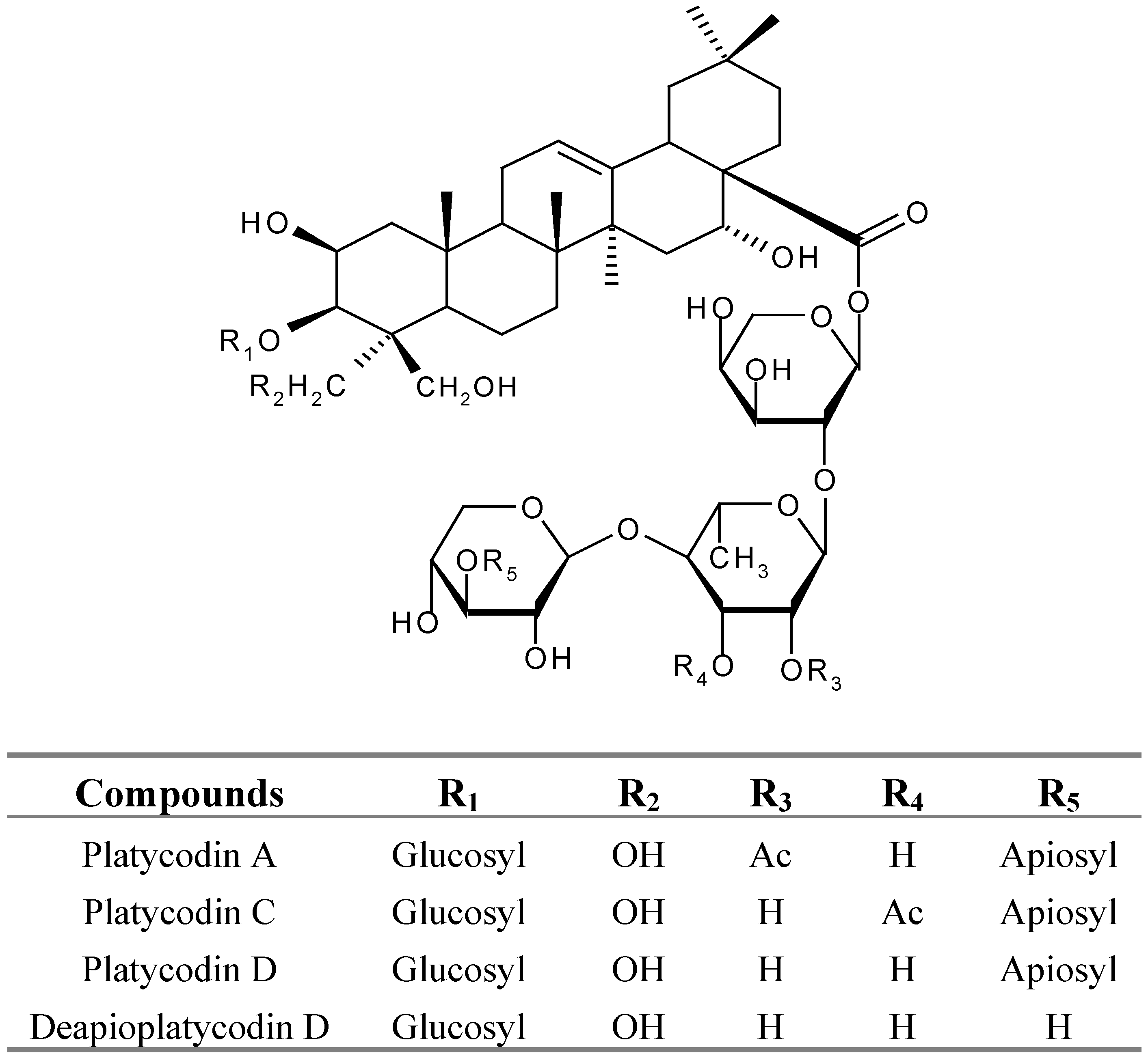
| Platycodin A | 13.6 ± 0.4* | 32.5 ± 4.8* | 52.4 ± 3.8*** |
| Platycodin C | 11.6 ± 2.8 | 20.3± 2.5 | 16.3 ± 2.1 |
| Platycodin D | 16.5 ± 2.7 | 21.6 ± 2.1 | 26.5 ± 3.7 |
| Deapioplatycodin D | 22.3 ± 3.8 | 21.2 ± 1.3 | 26.5 ± 2.0 |
| APV f | 10.9 ± 2.2 | 23.9 ± 2.8 | 40.0 ± 3.8* |
| MK-801 g | 51.8 ± 4.8** | 63.8 ± 5.4*** | 72.8 ± 4.9*** |
| CNQX h | 28.3 ± 3.4* | 41.5 ± 3.6* | 51.5 ± 4.8*** |
- a
- Rat cortical cell cultures were incubated with test compounds for 1h. The cultures were then exposed to 100 μM glutamate for 24 hrs. After the incubation, the cultures were assessed for the extent of neuronal damage;
- b
- Cell viability was measured by the LDH assay;
- c
- LDH released from control and glutamate-treated cultures were 11.9 ± 1.2 and 48.3 ± 4.1 units/mL, respectively;
- d
- Cell viability was calculated as 100 x (LDH released from glutamate-treated-LDH released from glutamate+test compound-treated)/(LDH released from glutamate-treated-LDH released from control). The values shown are the mean ± STD of three experiments (3-4 cultures per experiment). Results differ significantly from the glutamate-treated: * p < 0.05, ** p < 0.01, *** p < 0.001;
- e
- Glutamate-treated value differed significantly from the untreated control at the level of p < 0.001;
- f
- APV: DL-2-amino-5-phosphonovaleric acid, a competitive NMDA receptor antagonist;
- g
- MK-801: dizocilpine maleate, a noncompetitive NMDA receptor;
- h
- CNQX: 6-cyano-7-nitroquinoxaline-2,3-dione, non-NMDA receptor antagonist.
Experimental
General
Plant material
Plant material extraction, the total saponin from P. radix.
Isolation of platycodins from total saponins
Cortical cell culture and Cell viability assessment
Statistical analysis
Acknowledgments
References
- Wang, R.; Zhou, J.; Tang, X.C. Tacrine attenuates hydrogen peroxide-induced apoptosis by regulating expression of apoptosisrelated genes in rat PC12 cells. Brain Res. Mol. Brain Res. 2002, 107, 1–8. [Google Scholar] [CrossRef] [PubMed]
- Sucher, N. J.; Awobuluyi, M.; Choi, Y. B.; Lipton, S. A. NMDA receptors: from genes to channels. Trends Pharmacol. Sci. 1996, 17, 348–355. [Google Scholar]
- Cacabelos, R.; Takeda, M.; Winblad, B. The glutamatergic system and neurodegeneration in dementia: preventive strategies in Alzheimer’s disease. Int. J. Geriatr. Psychiatry 1999, 14, 3–47. [Google Scholar]
- Trist, D. G. Excitatory aminoacid agonists and antagonists: pharmacology and therapeutic applications. Pharm. Acta Helv. 2000, 74, 221–229. [Google Scholar]
- Ahn, K.S.; Hahn, B.S.; Kwack, K.; Lee, E.B.; Kim, Y.S. Platycodin D-induced apoptosis through nuclear factor-kappaB activation in immortalized keratinocytes. Eur. J. Pharmacol. 2006, 537, 1–11. [Google Scholar] [CrossRef] [PubMed]
- Tada, A.; Kaneiwa, Y.; Shoji, J.; Shibata, S. Studies on the saponins from roots of Platycodon grandiflorum A. De Candolle. I. Isolation and the structure of platycodin-D. Chem. Pharm. Bull. 1975, 23, 2965–2972. [Google Scholar]
- Ishii, H.; Tori, K.; Tozyo, T.; Yoshimura, Y. Saponins from roots of Platycodon grandiflorum. Part 2. Isolation and structure of new triterpene glycosides. J. Chem. Soc. Perkin Trans. I. 1984, 661–668. [Google Scholar]
- Kang, S.Y.; Lee, K.Y.; Sung, S.H.; Kim, Y.C. Four new neuroprotective dihydropyranocoumarins from Angelica gigas. J. Nat. Prod. 2005, 68, 56–69. [Google Scholar]
- Sample Availability: Samples of the compounds are available from authors.
© 2007 by MDPI (http://www.mdpi.org). Reproduction is permitted for noncommercial purposes.
Share and Cite
Son, I.H.; Park, Y.H.; Lee, S.I.; Yang, H.D.; Moon, H.-I. Neuroprotective Activity of Triterpenoid Saponins from Platycodi radix Against Glutamate-induced Toxicity in Primary Cultured Rat Cortical Cells. Molecules 2007, 12, 1147-1152. https://doi.org/10.3390/12051147
Son IH, Park YH, Lee SI, Yang HD, Moon H-I. Neuroprotective Activity of Triterpenoid Saponins from Platycodi radix Against Glutamate-induced Toxicity in Primary Cultured Rat Cortical Cells. Molecules. 2007; 12(5):1147-1152. https://doi.org/10.3390/12051147
Chicago/Turabian StyleSon, Il Hong, Yong Hoon Park, Sung Ik Lee, Hyun Duk Yang, and Hyung-In Moon. 2007. "Neuroprotective Activity of Triterpenoid Saponins from Platycodi radix Against Glutamate-induced Toxicity in Primary Cultured Rat Cortical Cells" Molecules 12, no. 5: 1147-1152. https://doi.org/10.3390/12051147
APA StyleSon, I. H., Park, Y. H., Lee, S. I., Yang, H. D., & Moon, H.-I. (2007). Neuroprotective Activity of Triterpenoid Saponins from Platycodi radix Against Glutamate-induced Toxicity in Primary Cultured Rat Cortical Cells. Molecules, 12(5), 1147-1152. https://doi.org/10.3390/12051147