Article

Synthesis and X-ray Structure of the Inclusion Complex of Dodecamethylcucurbit[6]uril with 1,4-Dihydroxybenzene

Institute of Applied Chemistry, Guizhou University, Guiyang, 550025, People’s Republic of China
*
Author to whom correspondence should be addressed.
Molecules 2007, 12(4), 716-722; https://doi.org/10.3390/12040716
Submission received: 25 February 2007 / Revised: 19 March 2007 / Accepted: 19 March 2007 / Published: 5 April 2007

Abstract

:
The synthesis, and X-ray crystal structure of the inclusion host-guest complex of dodecamethylcucurbit[6]uril (DDMeQ[6]) with 1,4-dihydroxybenzene (DHOBEN) are reported. The complex crystallizes in the space group P21/c (No.14) with a =12.2847(4), b = 12.6895(4), c = 15.1310(4) Å, α = 74.6960(10), β = 71.4090(10), γ = 86.5090(10)° and Z = 1. A novel approach to dodecamethylcucurbit[6]uril synthesis is also described. To separate dodecamethylcucurbit[6]uril, 1,4-dihydroxybenzene is used as a guest molecule for crystallization of the fully methyl-substituted cucurbituril. The driving force for the self-assembled inclusion host-guest complex can be attributed to not only the cavity interaction of dodecamethylcucurbit[6]uril (host), but also to the hydrogen bonding between the carbonyl oxygen at the portals of the host and the hydroxy groups of the guest.

Introduction

The chemistry of cucurbit[n]uril (Q[n]s) has expanded dramatically with the discovery of cucurbituril(Q[6]) and its homologues (Q[5], Q[7], Q[8], and Q[10]) [1,2,3]. Recently, the direct functionalization of CB[n][4,5,6] and introduction of building blocks for the preparation of Q[n] derivatives[7,8,9] and analogues[10,11] providing CB[n]s with solubility in both organic and aqueous solution has further expanded the range of the research and applications, which have been summarized in related reviews [12,13,14,15,16,17,18,19]. More recently, we have found that the solubilities of substituted cucurbit[n]urils (SQ[n]s) are dependent upon the kind, position and number of substituent groups on the substituted cucurbit[n]urils Q[n]s [20].
Since the discovery in 1992 of the first substituted Q[n], decamethylcucurbit[5]uril, Me10Q[5] [21], a number of fully and partially substituted Q[n]s have been reported since 2001. An approach to partial methyl substituted cucurbit[n]urils was demonstrated by Day in 2003 [8]. In this work, a similar approach to substituted cucurbituril synthesis is reported (Scheme 1), whereby one mole equivalent of monomethylglycoluril (1) was added to four mole equivalent of dimethylglycoluril (2) in 12 M HCl with the addition of formaldehyde to produce a mixture of methyl-substituted cucurbit[n=5,6]urils (Me-SQ[5, 6]).
Scheme 1. Synthesis of methyl-substituted cucurbit[n]urils.
Molecules 12 00716 g003
From this mixture a very rare dodecamethycucurbit[6]uril (DDMeQ [6] shown in Scheme 2) can be obtained in moderate yield (compared to the yield of 0.2% in the literature method [22]). The crystal structure of DDMeQ[6] has been characterized for the first time as the inclusion host-guest complex of dodecamethycucurbit[6]uril (DDMeQ[6], host) and 1,4-dihydroxybenzene (DHOBEN, guest).
Scheme 1. Structures of DDMeQ[6].
Molecules 12 00716 g004

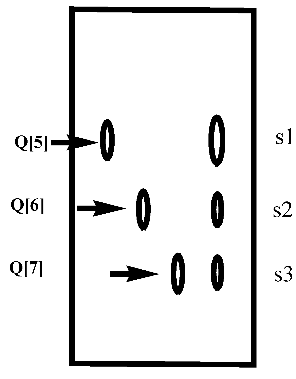
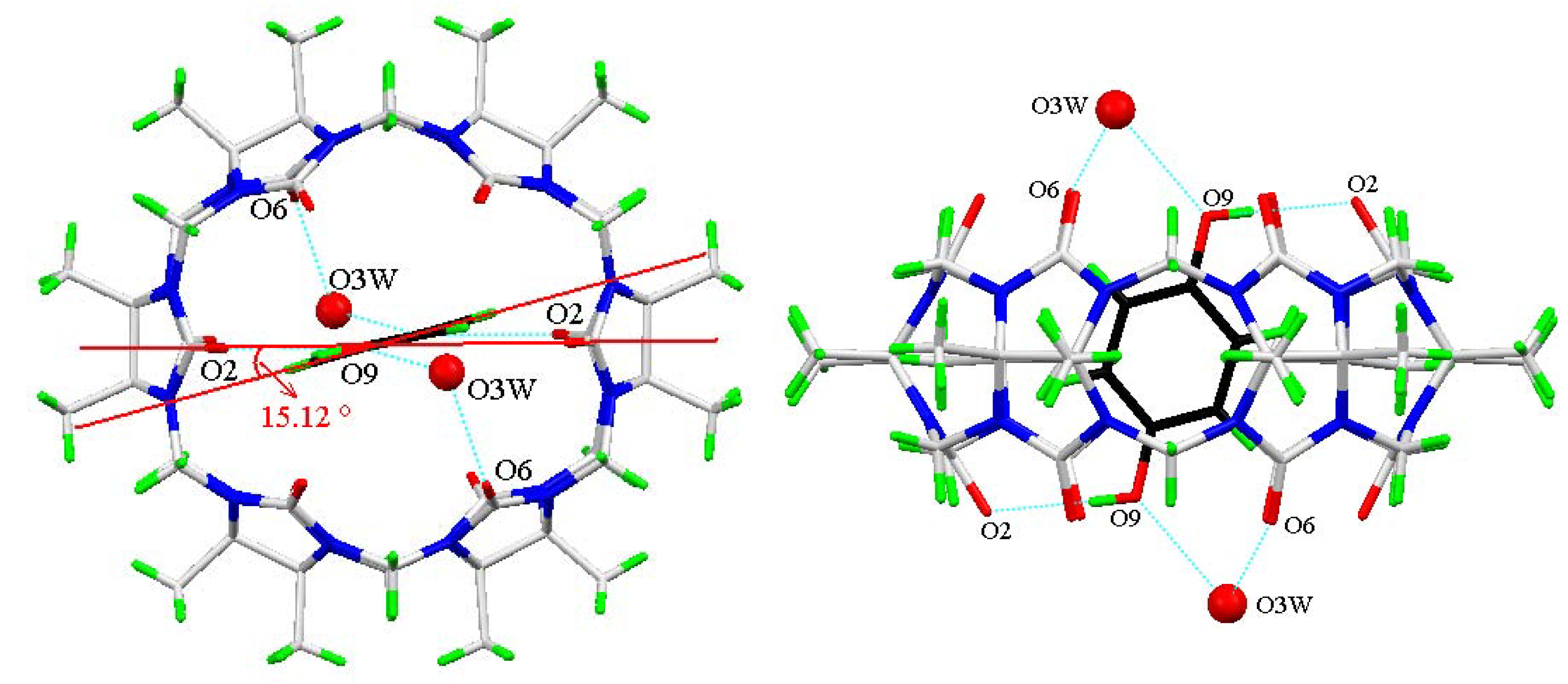
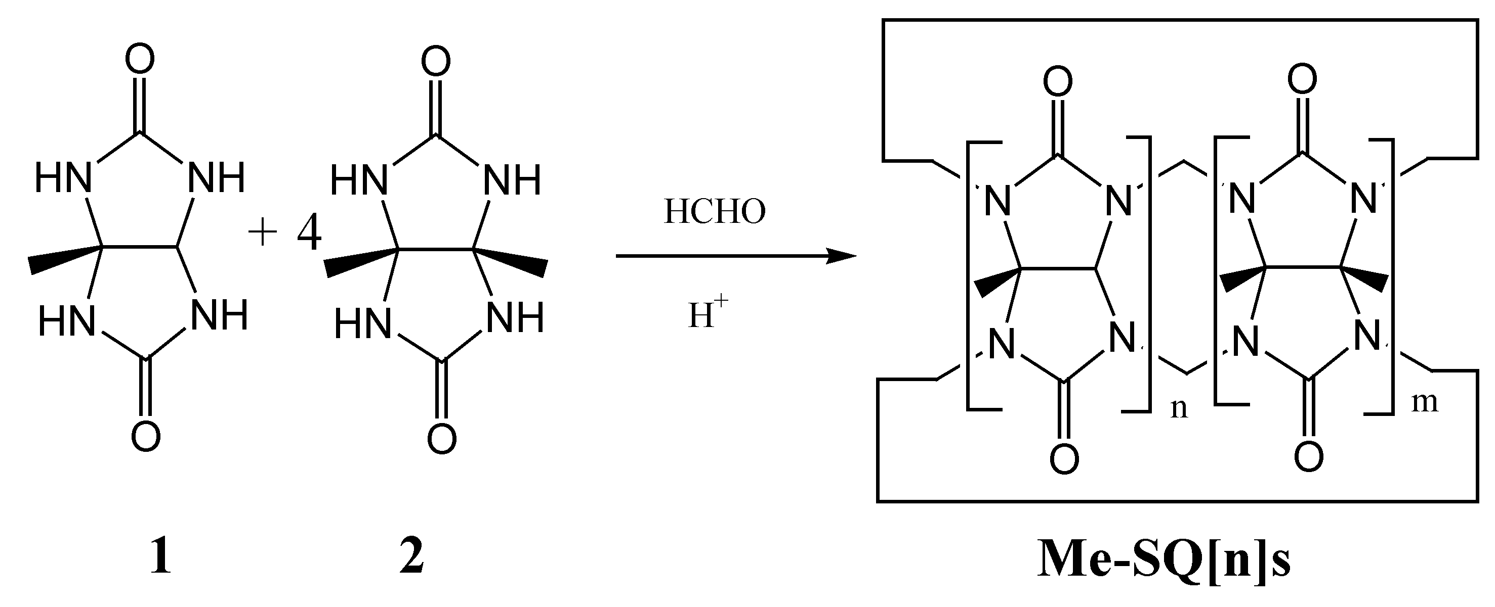
Results and Discussion

The TLC analysis showed three well separated spots with a ratio of 3(s1):1(s2):1(s3), which corresponded to the normal cucurbit[5, 6, 7]urils (Q[5], Q[6], Q[7], Figure 1). The typical guest probe tests showed that the substituted s3 cucurbit[n]uril(s) were not SQ[7], but rather SQ[6], because no included 1-adamantaneamine (ad) resonance signals were observed in the 1H-NMR spectrum of the s3-ad system [23], while the 1H-NMR spectra exhibited two sets of signals for the bound (indicated by wedges) and unbound protons of the HCl salt of 2,2’-bispyridine (bpy·1HCl) which suggested that the s3 was definitely SQ[6]s [24].
To isolate the DDMeQ[6] from s3, various guests were used for crystallizing DDMeQ[6] and the DDMeQ[6]-DHOBEN system was the first one in which the single crystals of DDMeQ[6] adduct with DHOBEN were obtained by dissolving DDMeQ[6] in a solution of DHOBEN in water. The final solution was mixed thoroughly and allowed to stand at room temperature; crystals formed after several days, and were collected.
Figure 1. The TLC profile of the mixture of the substituted Qs (I2 as a visualization agent).
Molecules 12 00716 g001
The crystal structure of inclusion host-guest complex of DDMeQ[6] with DHOBEN has been determined by single crystal X-ray diffraction at 223K. The complex crystallizes in the triclinic space group P21/c (No.14) with cell dimensions: a = 12.2847(4) Å, b = 12.6895(4) Å, c = 15.1310(4) Å, α = 74.6959(10)°, β = 71.4090(10)°, γ = 86.5090(10)°. Figure 2 shows two views of the DDMeQ[6]- DHOBEN inclusion complex. In the solid state, the whole DHOBEN molecule is clearly included in the cavity center of the DDMeQ[6] host. It is notable that a preferential orientation of two OH groups of the guest protruding towards the portal of DDMeQ[6] will cause a obvious distortion of the host, so the macrocycle is not circular but an ellipsoid. The distance between the portal carbonyl oxygens O1 and O3 is about 5.681 Å, and between O4 and O6 it is about 5.853 Å, while the distance between the portal carbonyl oxygens O2 and O5 is up to 7.091 Å, which is approximately about 20% longer than the closer sides. In addition, the guest is inserted to the extent that the aromatic ring sits essentially in line with opposite sets of oxygen donors O2 and O5, but is twisted away from linearity by ~15.0° (referring to Figure 2, top view). Moreover, the formation of hydrogen bonds between the OH groups of DHOBEN with the portal rimmed carbonyls of DDMeQ[6] increases the stability of the title inclusion complex. The main hydrogen bonds are O9-H9···O2, O3W-H3W···O9, and O3W-H3W···O6, the distances are 2.828, 2.703 and 2.929 Å. Thus, in the self-assembled structure of the inclusion complex of DDMeQ[6]- DHOBEN, the main driving forces can be attributed to the hydrophobic interaction between the cavity of DDMeQ[6] and aromatic moiety of the DHOBEN, and the hydrogen bonds between the hydroxyl group of the guest and the carbonyl oxygen atom(s).
Figure 2. Crystal structure of the inclusion host-guest complex of DDMeQ[6]-DHOBEN, showing partial atomic numbering and drawn at the 50% probability level in top view(left) and side view(right).
Molecules 12 00716 g002

Experimental

General

All reagents and chemicals were obtained from Beijing Chemical Company (P.R. China) and used without further purification. Column chromatography elution was carried out with formic acid (HCOOH, ≥88%) and acetic acid (CH3COOH, ≥99.5%) at different ratios. 1H- and 13C-NMR spectra were recorded on an INOVA-400 (Varian, 400 MHz) spectrometer. The solvent used was D2O. ES-MS measurements were performed on an HP 1100 LC-MSD at room temperature.

Syntheses and separation

12M HCl (350 mL) was added to a mixture of monomethylglycoluril (6.44 g, 0.04 mol), dimethylglycoluril (27.2 g, 0.16 mol) and paraformaldehyde (15.0 g), and the mixture was stirred at room temperature for approx ~30 min (at which point all solids dissolved) and then heated at 90°C for 8 hr to give a brown coloured solution, which was allowed to cool to room temperature, and then the acidic solvent was evaporated in vacuo to give a crude brown solid (38~41 g). The solid was dissolved in water (85 mL) and this solution was added dropwise to acetone (1000 mL) with vigorous stirring. The yellow precipitate formed was collected by filtration and air dried to give a yellow-red mixture of methyl substituted SQ[n]s 34~373g, yield: 69.9~76.7% (based on starting materials). TLC analysis on a silica gel plate showed that the mixture of methyl substituted SQ[n]s obtained can be readily separated into three bands (s1, s2 and s3) by elution with HCOOH/CH3COOH (1:2, v/v), compared to the normal Q[5], Q[6] and Q[7]; the ratio of s1:s2:s3 was ~60:20:20. A mixture of the substituted Qs (20 g) was loaded onto a silica gel column (200-300 mesh, 90 cm x 8.0 cm) and eluted with HCOOH/CH3COOH (1:1 ~ 1:2 v:v), to give a total of 10.1 g of s1, 3.4 of s2 and 3.5 g of (overall yield: ~ 85%). To isolate the DDMeQ[6] from s3, a series of guest molecules such as dioxane, 2,2’-bipyridine, 5,5’-dimethyl-2,2’-bipyridine, 1,ω-alkylenediamines etc. were used for attempted crystallization of DDMeQ[6]. Single crystals of DDMeQ[6]@DHOBEN were obtained by adding 1,4-dihydroxybenzene (DHOBEN, 0.058 g, 0.53 mmol) to a solution of DDMeQ[6] (0.50 g, 0.43 mmol) in water (12 mL). The final solution was mixed thoroughly and allowed to stand at room temperature; crystals formed after several days and were collected by filtration.

X-ray analysis of inclusion complex

The data were collected on a Bruker Apex-2000 CCD diffractometer at 223 K, using graphite monochromated Mo Kα radiation (λ= 0.71073 Å) with ω scan mode. Lorentz polarization and absorption corrections were applied. Structural solution and full matrix least-squares refinement based on F2 were performed with the SHELXS-97 and SHELXL-97 program package, respectively. All the non-hydrogen atoms were refined anisotropically. H atoms were placed in calculated positions and refined using a riding model, with isotropic thermal parameters equal to 1.2-1.5 times those of their parent atoms. In the final cycles of refinement, least squares weights of the form w = 1/[σ2(Fo)2 + (aP)2 +bP], P = [max(Fo2, 0) + 2Fc 2]/3 were employed (a = 0.1328, b = 2.2163). The ORTEP-3 [12] and WebLab programs were used for illustrations. Crystallographic data for DDMeQ[6] are listed in Table 1. CCDC 633953 contains the supplementary crystallographic data for this paper. These data can be obtained free of charge via www.ccdc.cam.ac.uk/conts/retrieving.html.
Table 1. Crystallographic parameters for DDMeQ[6].
Chemical formulaC78H90N24O36
Formula weight139.74
Crystal Color, Habitcolorless, diamond like
Crystal Dimensions0.1° 0.1 ° 0.15
Crystal Systemtriclinic
Lattice Typeprimitive
Space GroupP-1
Lattice Parametersa = 12.2847(4) Å
b = 12.6895(4) Å
c = 15.1310(4) Å
α =74.6960(10)°
β=71.4090(10)°
γ =86.5090(10)°
Volume2155.66(11) Å3
Z value1
Dcalc1.494 g/cm3
F(000)1014
μ (MoKα)0.120 mm-1
Reflections/restraints/parameters7477 / 0 / 640
Residuals: R1, wR2 [I > 2 σ (I)]0.0767, 0.2216
Goodness of Fit, S1.079
Max. shift/error0.000
Max. peak in final Δρ synthesis0.825
Min. peak in final Δρ synthesis-0.812

Acknowledgments

This work was supported by the National Natural Science Foundation of China (NSFC; No. 20662003), the International Collaborative Project of the Ministry of Science and Technology (ICPMST; No. 2003DF000030), and the Foundations of the Governor of Guizhou Province.

References and Notes

  1. Kim, J.; Jung, I. S.; Kim, S.-Y.; Lee, E.; Kang, J.-K.; Sakamoto, S.; Yamaguchi, K.; Kim, K. New cucurbituril homologues: syntheses, isolation, characterization, and X-ray crystal structures of cucurbit[n]uril (n = 5, 7, and 8). J. Am. Chem. Soc. 2000, 122, 540–541. [Google Scholar]
  2. Day, A. I.; Arnold, A. P.; Blanch, R. J.; Snushall, B. Controlling factors in the synthesis of cucurbituril and its homologues. J. Org. Chem. 2001, 66, 8094–8100. [Google Scholar] [CrossRef]
  3. Day, A. I.; Blanch, R. J.; Arnold, A. P.; Lorenzo, S.; Lewis, G. R.; Dance, I. A cucurbituril-based gyroscane: a new supramolecular form. Angew. Chem., Int. Ed. 2002, 41, 275–277. [Google Scholar] [CrossRef]
  4. Jon, S. Y.; Selvapalam, N.; Oh, D. H.; Kang, J. -K.; Kim, S. -Y.; Jeon, Y. J.; Lee, J. W.; Kim, K. Facile synthesis of cucurbit[n]uril derivatives via direct functionalization: expanding utilization of cucurbit[n]uril. J. Am. Chem. Soc. 2003, 125, 10186–10187. [Google Scholar]
  5. Jeon, Y. J.; Kim, H.; Jon, S.; Selvapalam, N.; Oh, D. H.; Seo, I.; Park, C. S.; Jung, S. R.; Koh, D.-S.; Kim, K. Artificial ion channel formed by cucurbit[n]uril derivatives with a carbonyl group fringed portal reminiscent of the selectivity filter of K+ channels. J. Am. Chem. Soc. 2004, 126, 15944–15945. [Google Scholar] [CrossRef]
  6. Lee, H.-K.; Park, K. M.; Jeon, Y. J.; Kim, D.; Oh, D. H.; Kim, H. S.; Park, C. K.; Kim, K. Vesicle formed by amphiphilc cucurbit[6]uril: Versatile, noncovalent modification of the vesicle surface, and multivalent binding of sugar-decorated vesicles to lectin. J. Am. Chem. Soc. 2005, 127, 5006–5007. [Google Scholar]
  7. Isobe, H.; Sato, S.; Nakamura, E. Synthesis of disubstituted cucurbit[6]uril and its rotaxane derivative. Org. Lett. 2002, 4, 1287–1289. [Google Scholar] [CrossRef]
  8. Day, A. I.; Arnold, A. P.; Blanch, R. J. A method for synthesizing partially substituted cucurbit[n]uril. Molecules 2003, 8, 74–84. [Google Scholar] [CrossRef]
  9. Zhao, Y.-J.; Xue, S. -F.; Zhu, Q. -J.; Tao, Z.; Zhang, J. -X.; Wei, Z. -B.; Long, L. -S.; Hu, M.-L.; Xiao, H. -P.; Day, A. I. Synthesis of a symmetrical tetrasubstituted cucurbit[6]uril and its host-guest compound with 2,2'-bipyridine. Chin. Sci. Bull. 2004, 49, 1111–1116. [Google Scholar] [CrossRef]
  10. Lagona, J.; Fettinger, J. C.; Isaacs, L. Cucurbit[n]uril analogues. Org. Lett. 2003, 5, 3745–3747. [Google Scholar] [CrossRef]
  11. Wagner, B. D.; Boland, P. G.; Lagona, J.; Isaacs, L. A cucurbit[6]uril analogue: host properties monitored by fluorescence spectroscopy. J. Phys. Chem. B 2005, 109, 7686–7691. [Google Scholar] [CrossRef]
  12. Lagona, J.; Mukhopadhyay, P.; Chakrabarti, S.; Isaacs, L. The cucurbit[n]uril family. Angew. Chem. Int. Edit. 2005, 44, 4844–4870. [Google Scholar] [CrossRef]
  13. Huang, F. H.; Gibson, H. W. Polypseudorotaxanes and polyrotaxanes. Prog. Polym. Sci. 2005, 30, 982–1018. [Google Scholar] [CrossRef]
  14. Sliwa, W.; Zujewska, T. Interlocked molecules containing quaternary azaaromatic moieties. Heterocycles 2005, 65, 1713–1739. [Google Scholar] [CrossRef]
  15. Gerasko, O. A.; Sokolov, M. N.; Fedin, V. P. Mono- and polynuclear aqua complexes and cucurbit[6]uril: versatile building blocks for supramolecular chemistry. Pure Appl. Chem. 2004, 76, 1633–1646. [Google Scholar]
  16. Sokolov, M. N.; Dybtsev, D. N.; Fedin, V. P. Supramolecular compounds of cucurbituril with molybdenum and tungsten chalcogenide cluster aqua complexes. Russ. Chem. Bull. 2003, 52, 1041–1060. [Google Scholar] [CrossRef]
  17. Kim, K. Mechanically interlocked molecules incorporating cucurbituril and their supramolecular assemblies. Chem. Soc. Rev. 2002, 31, 96–107. [Google Scholar] [CrossRef]
  18. Buschmann, H. J.; Mutihac, L.; Jansen, K. Complexation of some amine compounds by macrocyclic receptors. J. Incl. Phenom. Macro. 2001, 39, 1–11. [Google Scholar] [CrossRef]
  19. Mock, W. L. Cucurbituril. Top. Curr. Chem. 1995, 175, 1–24. [Google Scholar] [CrossRef]
  20. We have found that the symmetry of fully methyl-substituted Q[5] (DEMeQ[5]) is higher than that of the partially methyl-substituted Q[5]; the corresponding dipole (0.0393 Debye) is quite smaller than that of dimethyl-substituted Q[5], (0.7444 Debye), or hexamethyl-substituted Q[5] (1.2115 Debye). The normal Q[5] has the smallest dipole, 0.0216 Debye, due to its high symmetry. On the other hand, among the four Q[5]s, HMeQ[5] has the best water solubility (up to 17 g/100 g water); while the Q[5] has the least water solubility (only 0.03 g/100 g water). The order of dipoles of the four Q[5]s is qualitatively consistent with that of the solubility of these Q[5]s, that is HMeQ[5] > DMeQ[5] > DEMeQ[5] > Q[5].
  21. Flinn, A.; Hough, G. C.; Stoddart, J. F.; Williams, D. J. Decamethylcucurbit[5]uril. Angew. Chem. Int. Ed. 1992, 31, 1475–7. [Google Scholar] [CrossRef]
  22. Sanjita, S.; Mantosh, K.; Sinha, E. K. Facile purification of rare cucurbiturils by affinity chromatography. Org. Lett. 2004, 6, 1225–1228. [Google Scholar] [CrossRef]
  23. Ma, P.-H.; Dong, J.; Xiang, S.-C.; Xue, S.i-F.; Zhu, Q.-J.; Tao, Z.; Zhang, J.-X.; Zhou, X. Interaction of host-guest complexes of cucurbit[n]urilswith double probe guests. Sci. China Ser. B. 2004, 47, 301–310. [Google Scholar] [CrossRef]
  24. Fu, H.-Y.; Xue, S.-F.; Zhu, Q.-J.; Tao, Z.; Zhang, J.-X.; Day, A. I. Investigation of host-guest compounds of cucurbit[n = 5-8]uril with some ortho pyridiniumammonium Ions. J. Incl. Phenom. Macro. 2005, 52, 101–107. [Google Scholar] [CrossRef]
  • Sample availability: Contact the authors.

