Article

Isolation and Structure Elucidation of a Novel Yellow Pigment from the Marine Bacterium Pseudoalteromonas tunicata

1
School of Chemistry, The University of New South Wales, Australia
2
School of Biotechnology and Biomolecular Sciences, The University of New South Wales, Australia
*
Author to whom correspondence should be addressed.
Molecules 2005, 10(10), 1286-1291; https://doi.org/10.3390/10101286
Submission received: 12 December 2004 / Revised: 21 April 2005 / Accepted: 4 May 2005 / Published: 31 October 2005
(This article belongs to the Special Issue 2004 RACI One-day Natural Products Group Symposium)

Abstract

:
The marine environment is a major source for many novel natural compounds. A new yellow pigment has been isolated from the marine bacterium P. tunicata and identified as a new member of the tambjamine class of compounds. The structural identification was achieved by a combination of 1D and 2D-NMR spectroscopy and high resolution mass spectrometry data.

Introduction

Members of the bacterial genus Pseudoalteromonas are commonly found associated with living and inert surfaces in the marine environment [1]. Pseudoalteromonas species produce a wide range of biologically active compounds that have activity against common fouling organisms. These include compounds that target bacteria, invertebrate larvae, algal spores, protozoan grazing and fungi, and are believed to provide protection for host marine organisms colonised by Pseudoalteromonas species [1,2,3,4,5,6,7,8]. In a preliminary screen of ten Pseudoalteromonas species, Holmström et al. [6] found that P. tunicata has the highest and broadest range of biological activities. These activities are linked to the production of unidentified yellow and purple pigments [8]. As part of our ongoing research on biologically active compounds produced by P. tunicata, we report the isolation and identification of a novel yellow pigment produced by P. tunicata. This pigment is a new member of the tambjamine class of compounds.
The tambjamine alkaloids have been isolated previously from bacteria and marine invertebrates including bryozoans, nudibranchs and ascidians [9,10]. They comprise a 2,2’-bipyrrole ring system containing an enamine moiety at the C2 position of the pyrrole ring, and an adjacent methoxy group at C3. The enamine nitrogen is normally substituted with a two to four carbon saturated alkyl chain. Furthermore, a tambjamine analogue, BE-18591, containing a saturated twelve carbon alkyl chain has been isolated from the cultures of Streptomyces sp [11]. Other members of this class, which include tri- and tetra-pyrrole compounds, possess a range of biological activities including antimicrobial [12], antitumour [13] and immunosuppressive activities [14]. BE-18591 has also been shown to inhibit immunoproliferation and gastritis in rabbits [15].

Experimental

General

High resolution mass spectra were acquired in methanol using a Bruker BioApex IIe 7 T Fourier Transform Ion Cyclotron Resonance mass spectrometer with an Analytica electrospray ionisation source. Samples were run in positive-ion mode under high resolution conditions (<2 ppm). 1H- and 13C-NMR spectra were run in neutralized CDCl3 (prepared by passing CDCl3 through a pad of anhydrous potassium carbonate) on a Bruker AMX300 spectrometer, while two-dimensional NMR experiments were conducted on a Bruker DMX600 spectrometer.

Bacterial culture conditions and isolation of the yellow pigment

P. tunicata [16] (100 litres) was cultured in VNSS medium [17] to stationary phase (12 hours) and harvested by centrifugation. The cells were freeze dried and extracted with methanol. The methanol extract was reduced under vacuum and partitioned using a dichloromethane-water (1:1 v/v) mixture. The DCM fraction was separated, evaporated and loaded onto an Altec SPE column using diethyl ether. The column was eluted using steps of 100% hexane, 10% ethyl acetate/hexane, 100% chloroform, and 4% isopropanol/chloroform. The pigmented yellow fractions (90 mg) (in the 100% chloroform fraction) were further fractionated on another Altec SPE column. The sample was loaded using DCM and eluted with 2% isopropyl alcohol (IPA)/chloroform. The resultant yellow fraction was chromatographed on preparative thin layer glass plates coated with silica gel to remove the large amounts of oleic acid that co-eluted with the yellow pigment. In some cases preparative plates containing silica gel mixed with 12% sodium acetate were employed.The yellow pigmented band was scraped from the silica plate, and eluted with dichloromethane. Evaporation of the solvent in vacuo yielded a compound as a yellow oil (11 mg). A purple band was also isolated from the crude mixture. The purple compound was identified as violacein by comparison, and the yellow pigment as a novel tambjamine alkaloid (1). The structure of the yellow compound was elucidated as follows.

Results and Discussion

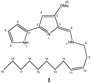
An ESI-HRMS of compound 1 gave a molecular ion at m/z 356.270484 [MH+] corresponding to a molecular formula of C22H33N3O (calc: 356.269605 [MH+]). The structure 1 was deduced through the extensive use of high resolution nuclear magnetic spectroscopy experiments including COSY, TOCSY, NOESY, HSQC, 1H-13C and 1H-15N HMBC experiment.
Molecules 10 01286 i001
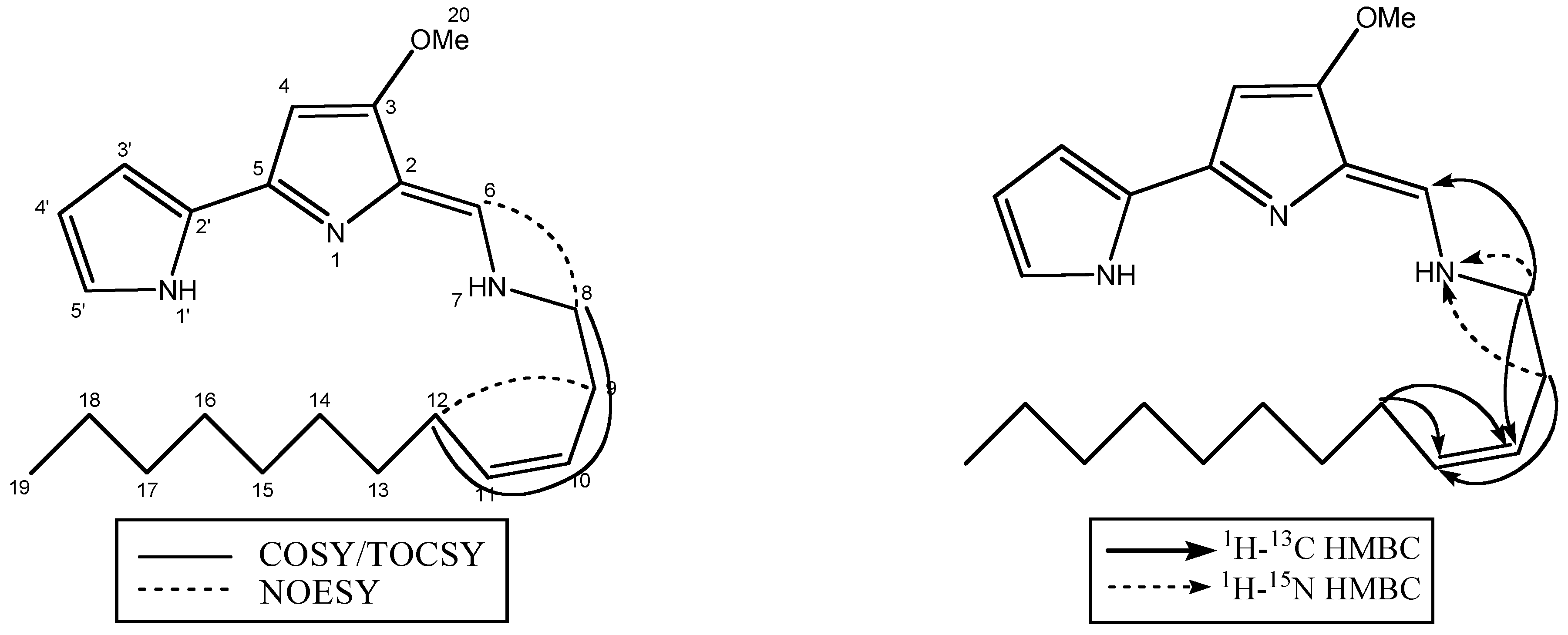
The presence of a 2,2’-bipyrrole ring system was established from the COSY, TOCSY and NOESY experiments (Figure 1, Table 1) and comparison with the reported 1H and 13C chemical shifts for tambjamines [9,10]. The 1H-NMR spectrum of 1 showed signals for the bipyrrole protons at δ 7.06, 6.25, 6.70 and 5.95 ppm corresponding to protons H5’, H4’, H3’ and H4 respectively (Table 1). The presence of a methoxy group at C3 was confirmed by a proton singlet at δ 3.90 ppm and the C3 carbon at 163.9 ppm. The enamine CH appeared at δ 7.29 ppm in the proton spectrum with the corresponding carbon signal at 141.1 ppm. The enamine nitrogen was found to be substituted with an unsaturated twelve carbon alkyl chain. Comparison of the spectral data of the new tambjamine with reported tambjamine structures indicated the presence of a longer chain and a double bond between C10 and C11 carbon atoms.
Figure 1. Selected 2D NMR Data
Molecules 10 01286 g001
The C8 CH2 group next to an enamine nitrogen appeared at δ 3.46 ppm and showed through space coupling to H6 and direct through bond correlation to C9 CH2. The large coupling constant between C6 proton and the enamine NH (J= 14 Hz) further confirmed the trans-antiplanar arrangement of the enamine [18].
Table 1. 1H and 13C chemical shifts for 1
Yellow Pigment
Position13C1H15N
1 142
2111.3
3163.9
491.65.95, s
5143.4
6141.17.29, d
7 132
851.23.46, m
928.82.46, m
10124.15.35, dt
11134.55.55, dt
1227.82.01, m
13 – 1826.5-27.71.21-1.70
1914.50.86, t
2058.83.90, s
1’ 155
2’123.5
3’113.06.70, m
4’110.56.25, m
5’124.17.06, m
Furthermore, protons at C8 and C9 showed long range 1H-15N couplings to N7 confirming the linkage of the alkyl chain (Figure 1). The double bond between C10 and C11 in the alkyl chain was located through COSY/TOCSY experiments and assigned a cis configuration with a coupling constant J10,11 = 10.8 Hz and a NOESY correlation between H9 and H12. Both H8 and H9 showed 3-bond heteronuclear correlations to the double bond, with H8 showing coupling to C10 and H9 to C11. The CH2 at C12 showed long range couplings to both double bond carbons. A large broad multiplet at δ 1.21-1.70 ppm accounted for the C13-C18 methylene protons with the terminal methyl group appearing at δ 0.86 ppm as a triplet.

Conclusions

The isolation and characterisation of a new yellow pigment from the marine bacterium P. tunicata is described. This is the first reported isolation of a tambjamine with an unsaturated alkyl chain from nature. Furthermore, the isolation and structural similarity of 1 to tambjamines isolated from sponges and bryozoans suggests that these compounds may be of bacterial origin and related to the presence of Pseudoaltermonas either within tissue or on the surface of the organism as is the case with P. tunicata.

Acknowledgements

We thank Anthony Carroll from Natural Product Discovery, Griffith University and Adrian Blackman from University of Tasmania for providing spectra of tambjamines. The authors also wish to thank Hilda Stender for her help in performing NMR experiments.

References

  1. Holmström, C.; Kjelleberg, S. Marine Pseudoalteromonas species are associated with higher organisms and produce active extracellular agents. FEMS Microbiol. Ecol. 1999, 30, 285–293. [Google Scholar] [CrossRef]
  2. Burkholder, P. R.; Pfister, R. M.; Leitz, F. H. Production of a pyrrole antibiotic by a marine bacterium. Appl. Microbiol. Biotechnol. 1966, 14, 649–653. [Google Scholar]
  3. McCarthy, S. A.; Johnson, R. M.; Kakimoto, D. Characterization of an antibiotic produced by Alteromonas luteoviolacea Gauthier 1982, 85 isolated from Kinko Bay, Japan. J. Appl. Bacteriol. 1994, 77, 426–32. [Google Scholar] [CrossRef] [PubMed]
  4. Gauthier, M. J. Alteromonas citrea, a new gram-negative yellow-pigmented species from seawater. Int. J. Syst. Bacteriol. 1977, 27, 349–354. [Google Scholar]
  5. Eaton, R. W.; Chapman, P. J. Formation of indigo and related compounds from indolecarboxylic acids by aromatic acid-degrading bacteria: chromogenic reactions for cloning genes encoding dioxygenases that act on aromatic acids. J. Bacteriol. 1995, 177, 6983–6988. [Google Scholar] [PubMed]
  6. Holmström, C.; Egan, S.; Franks, A.; McCloy, S.; Kjelleberg, S. Antifouling activities expressed by marine surface associated Pseudoalteromonas species. FEMS Microbiol. Ecol. 2002, 41, 47–58. [Google Scholar] [CrossRef]
  7. Egan, S.; Holmström, C.; Kjelleberg, S. Pseudoalteromonas ulvae sp. nov., a bacterium with antifouling activities isolated from the surface of a marine alga. Int. J. Syst. Evol. Microbiol. 2001, 51, 1499–1504. [Google Scholar] [CrossRef] [PubMed]
  8. Egan, S.; James, S.; Holmström, C.; Kjelleberg, S. Correlation between pigmentation and antifouling compounds produced by Pseudoalteromonas tunicata. Environ. Microbiol. 2002, 4, 433–442. [Google Scholar] [CrossRef] [PubMed]
  9. Blackman, A. J.; Li, C. New tambjamine alkaloids from the marine bryozoan Bulgula dentata. Aust. J. Chem. 1994, 47, 1625–1629. [Google Scholar] [CrossRef]
  10. Davis, R. A.; Carroll, A. R.; Quinn, R. J. The synthesis of a combinatorial library using a tambjamine natural product template. Aust. J. Chem. 2001, 54, 355–359. [Google Scholar] [CrossRef]
  11. Kojiri, K.; Nakajima, S.; Suzuki, H. A new antitumour substance, BE-18591, produced by Streptomycete II. Structure determination. J. Antibiotics. 1993, 46, 1894–1896. [Google Scholar]
  12. Boger, D. L.; Patel, M. Total synthesis of prodigiosin, prodigiosene, and desmethoxyprodigiosin: Diels-Alder reactions of heterocyclic azadienes and development of an effective palladium(II)- promoted 2,2’-bipyrrole coupling procedure. J. Org. Chem. 1988, 53, 1405–1415. [Google Scholar] [CrossRef]
  13. Nakajima, S.; Kojiri, K.; Suda, H. A new antitumour substance, BE-18591, produced by Streptomycete I. Fermentation, isolation, physico-chemical and biological properties. J. Antibiotics. 1993, 46, 1799–1803. [Google Scholar] [CrossRef] [Green Version]
  14. Han, S. B.; Kim, H. M.; Kim, Y. H.; Lee, C. W.; Jang, E. S.; Son, K. H.; Kim, S. U.; Kim, Y. K. T- cell specific immunosuppression by prodigiosin isolated from Serratia marcescens. Int. J. Immunopharmacol. 1998, 20, 1–13. [Google Scholar] [CrossRef]
  15. Tanigaki, K.; Sato, T.; Tanaka, Y.; Ochi, T.; Nishikawa, A.; Nagai, K.; Kawashima, H.; Ohkuma, S. BE-18591 as a new H+/Cl- symport ionophore that inhibits immunoproliferation and gastritis. FEBS Lett. 2002, 524, 37–42. [Google Scholar] [CrossRef]
  16. Holmström, C.; James, S.; Neilan, B.; White, D.; Kjelleberg, S. Pseudoalteromonas tunicata sp. nov., a bacterium that produces antifouling agents. Int. J. Syst. Bacteriol. 1998, 48, 1205–1212. [Google Scholar] [CrossRef] [PubMed]
  17. Marden, P.; Tunlid, A.; Malmcronafriberg, K.; Odham, G.; Kjelleberg, S. Physiological and morphological changes during short-term starvation of marine bacterial isolates. Arch. Microbiol. 1985, 142, 326–332. [Google Scholar] [CrossRef]
  18. Carté, B.; Faulkner, D. J. Defensive metabolites from three nembrothid nudibranchs. J. Org. Chem. 1983, 48, 2314–2318. [Google Scholar]
  • Sample availability: Contact the authors.