Article

Electron Mediation and Photocurrent Enhancement in Dunalliela salina Driven Bio-Photo Electrochemical Cells

1
Grand Technion Energy Program, Technion, Haifa 32000, Israel
2
Schulich Faculty of Chemistry, Technion, Haifa 320000, Israel
3
Faculty of Biology, Technion, Haifa 32000, Israel
*
Author to whom correspondence should be addressed.
Catalysts 2021, 11(10), 1220; https://doi.org/10.3390/catal11101220
Submission received: 10 September 2021 / Revised: 6 October 2021 / Accepted: 7 October 2021 / Published: 10 October 2021
(This article belongs to the Special Issue NanoBio Hybrids and Photocatalysis)

Abstract

:
In recent years, finding alternatives for fossil fuels has become a major concern. One promising solution is microorganism-based bio-photo electrochemical cells (BPECs) that utilize photosynthetic solar energy conversion as an energy source while absorbing CO2 from the atmosphere. It was previously reported that in cyanobacterial-based BPECs, the major endogenous electron mediator that can transfer electrons from the thylakoid membrane photosynthetic complexes and external anodes is NADPH. However, the question of whether the same electron transfer mechanism is also valid for live eukaryotic microalgae, in which NADPH must cross both the chloroplast outer membrane and the cell wall to be secreted from the cell has remained elusive. In this work, we show that NADPH is also the major endogenous electron mediator in the microalgae Dunalliela salina (Ds). We show that the ability of Ds to tolerate high salinity enables the production of a photocurrent that is 5–6 times greater than previously reported for freshwater cyanobacterial-based BPECs in the presence or absence of exogenous electron mediators. Additionally, we show that the electron mediator Vitamin B1 can also function as an electron mediator enhancing photocurrent production. Finally, we show that the addition of both FeCN and NADP+ to Ds has a synergistic effect enhancing the photocurrent beyond the effect of adding each mediator separately.

Graphical Abstract

1. Introduction

In recent years, the increase in global pollution and the concern from climate change has led to extensive efforts to develop green energy technologies that have the potential to replace the use of fossil fuels. One of promising method for electrical energy generation is the utilization of microbial fuel cells (MFCs) [1,2,3,4,5,6,7,8]. This method is based on the ability coupling of exo-electrogenic bacteria with an electrochemical setup. Upon association with the anode, the bacteria can conduct external electron transport and function as electron donors [9]. Alternatively, several bacterial species can also function as terminal electron acceptors while attached to the cathode [9,10].
A small number of bacterial species have been reported to perform external electron transport (ExET) by a mechanism of direct electron transport (DET) [1,11,12]. For this mechanism to be efficient, the cell walls must contain either conductive protein complexes such as c-types cytochromes [13,14,15,16], or conductive pili [13,14,15,16], and MTR complexes [17,18]. A different ExET mechanism is based on mediated electron transport (MET) [3,4,19], which is performed by secretion of small metabolites that function as electron mediators between bacteria and the anode of an electrochemical cell. The electron mediation in MFCs can be enhanced by exogenous artificial electron mediators such as different quinones and phenazines derivatives, sulfides, humic acids, neutral red, thionine, and ferric chelated complexes [2,20,21,22,23,24]. It should be noted that in bacteria exhibiting DET type EET, there are still diffusion limited steps within the cells that link energetic reduced compounds such as NADPH to the transmembrane molecular wires that transport the electrons to external substrates [25,26]. In the presence of an excess small molecule reductants in liposome reconstituted MTR complexes, maximal electron transfer rates of 8500 e/sec/complex have been reported [25].
Bacteria have the potential to become a cheap resource for electricity production, however, a carbon source must be provided for the cell metabolism [27]. Some of the bacterial species used in MFCs may be pathogenic so safety considerations must be considered if one wishes to cultivate them in large scales. Another method, that has similar working principles to MFCs are bio-photo electrochemical cells (BPEC) [28,29,30,31]. These cells operate based on the electric coupling of photosynthetic microorganisms (PM) such as cyanobacteria or eukaryotic microalgae to the anode of an electrochemical cell. The utilization of PM may afford several benefits that enhance their potential to be used for future applicative technologies. Unlike non-photosynthetic bacteria, PM fix CO2, thereby synthesizing (and storing) their own source of carbon [32]. In addition, PM species such as the green algae Dunaliella salina (Ds) or the cyanobacterium Spirulina (mostly A. platensis) are already being cultivated in agroindustry around the world for the production of food additives, cosmetic products, and biofuels [33,34]. Therefore, they have the potential to be integrated with BPEC-based innovative energy technologies.
The current density production of intact cyanobacterial-based BPECs was reported to be about ~4 µA/cm [2,35,36]. Enhancement of photocurrent production was reported by the utilization of a microfluidizer device to apply gentle external pressure on the cells [35]. Such a gentle pressure can induce minor changes to the cell membrane, thereby increasing its permeability to allow enhanced trafficking of electron mediators between the cells and the anode. This gentle treatment did not damage the cells and they could be recovered and continued to multiply. In this system, addition of the herbicide 3-(3,4-chlorophenyl)-1,1-dimethylurea (DCMU) that typically inhibits electron transport between Photosystem II (PSII) and the plastoquinone pool in the photosynthetic pathway actually increased the harvested current, indicating that the source of the electrons was from the respiratory system, via Photosystem I (PSI) [35,36]. Additional experiments confirmed this observation, however in reports of other cyanobacterial BPECs, DCMU inhibited current harvesting [37]. Thus, multiple sources of electrons are potentially possible, depending on the method of cell preparation and the BPEC composition. Another way for enhancement of the current that does not involve internalization of components by the cells can be done simply by utilization of an electrolyte with high salinity, which increases the conductivity of the system [28]. However, such a high salinity requires the utilization of biocompatible microorganisms whose natural habitat is in saltwater. Another possibility to enhance the photocurrent is the utilization of exogenous electron mediators such as potassium ferricyanide [38]. However, it is a toxic chemical and therefore may not be feasible for usage in large-scale applicative technologies [39]. Recently, the major endogenous electron mediator in cyanobacteria was identified to be NADPH [36]. Molecules such as NADPH are active in many biological metabolic pathways and are essential for the cells [40,41,42,43]. Therefore, intact cyanobacterial cells spend energy to internalize NADPH and do not tend to secrete it. Nevertheless, the anode of the BPEC functions as a driving force that enables their release [36]. Although this EET mechanism was reported for several cyanobacterial species, it was not clear whether NADPH is also the major electron mediator in microalgae, especially concerning the fact that to exit the cells, NADPH must first cross the chloroplast membrane that does not exist in cyanobacteria.
In this work, we show that intact cells of Ds can produce photocurrents in the presence of high ionic strength buffer serving as the electrolyte, which is significantly higher than reported for cyanobacteria. This current is further enhanced by the addition of FeCN. We show that NADPH is also the major electron mediator in Ds-driven BPECs and that the addition of its oxidized form (NADP+) significantly enhances the photocurrent. Finally, we show that vitamin B1, can also perform MET while its performance is like NADPH.

2. Results

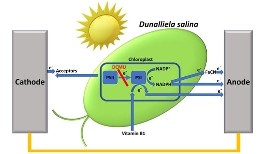
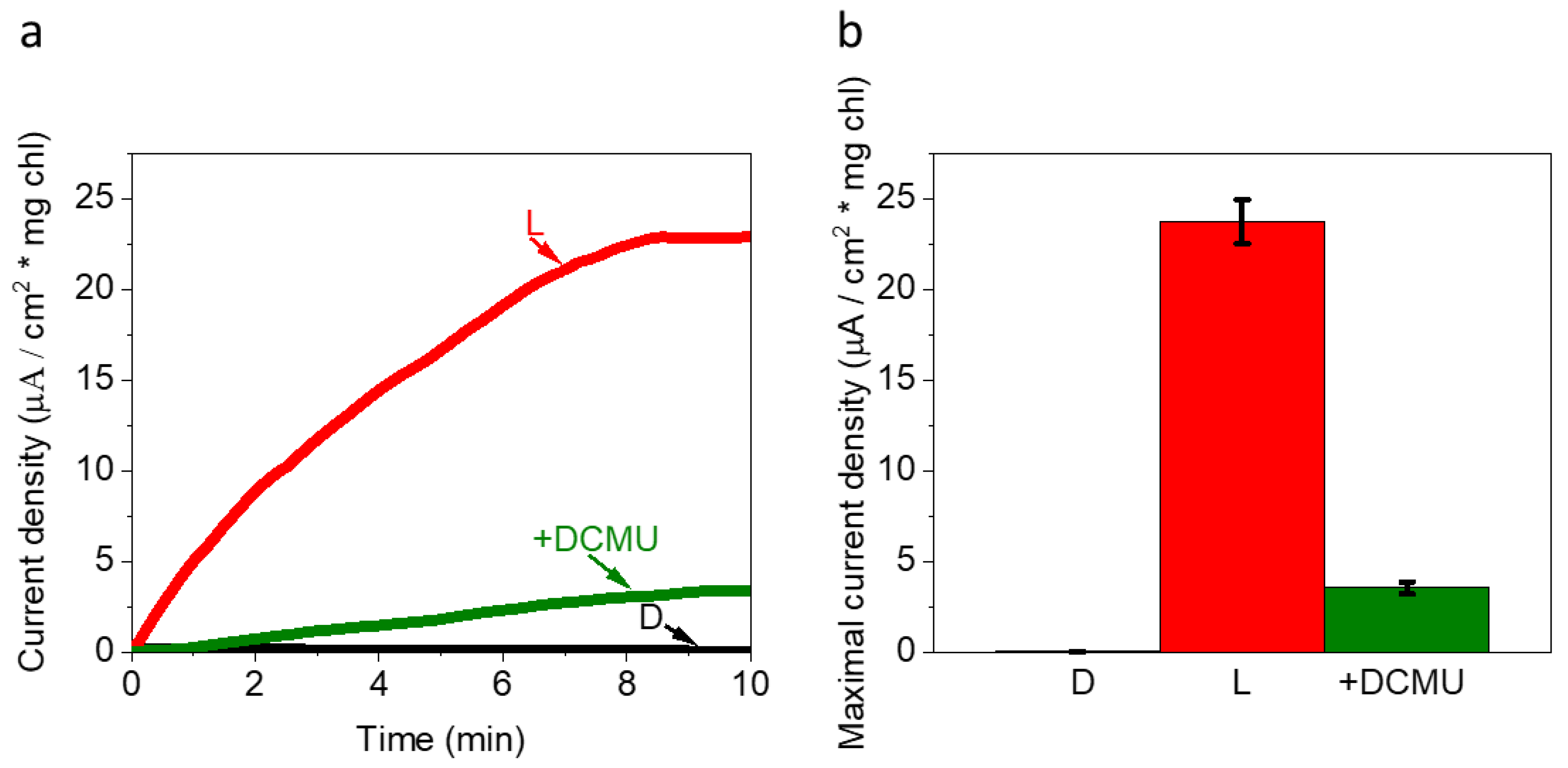
2.1. Live Ds Cells in High Ionic Strength Solution Produce Photocurrents That Originate from PSII

We have previously shown that cyanobacterial cells can produce electricity by applying them to the anode of an electrochemical cell, without the need for electrochemical linkage. In this system, upon illumination, intracellular NADP+ is reduced by ferrodoxin NADP+ reductase (FNR) within the cytoplasm to form NADPH which is secreted by the cells and is then oxidized by the anode [36]. NADPH secretion is enhanced by the application of bias between the electrodes in the BPEC.
Unlike the prokaryotic cyanobacteria, microalgae are eukaryotes, and their photosynthetic apparatus is contained within the chloroplast, whose membrane serves as an additional barrier for the secretion of electron mediators on their way to the BPEC anode. We first wished to explore whether live microalgal cells can produce significant photocurrent without the addition of an exogenous mediator. Chronoamperometry (CA) measurements were conducted for concentrated live Ds cells in dark and under solar illumination of 1.5 SUN (1500 W/m2) using screen-printed electrodes with a graphite anode (WE), platinum cathode (CE), and Ag/AgCl reference electrode (RE) as described in our previous work [36]. A solution of 1.5 M NaCl was used as an electrolyte. This concentration was chosen because it was also used in the cultivation medium of the cells. Following system stabilization, no significant current was obtained in the dark which was unlike the minor dark current that was obtained with cyanobacteria. A photocurrent of ~23 µA/cm2 ·mg chl was obtained in the light. (Figure 1). This current was about 5–6 times greater than previously reported for untreated freshwater cyanobacteria in our previous work [36]. This result indicated that the chloroplast secreted electron mediators to the cell and the cell secreted the mediators further, to produce photocurrent. Addition of the PSII inhibitor 100 µM 3-(3,4-dichlorophenyl)-1,1-dimethylurea (DCMU) reduced the photocurrent production to ~2.5 µA/cm2 mg chl (Figure 1). Based on this result, we propose that most of the photocurrent originated from PSII.

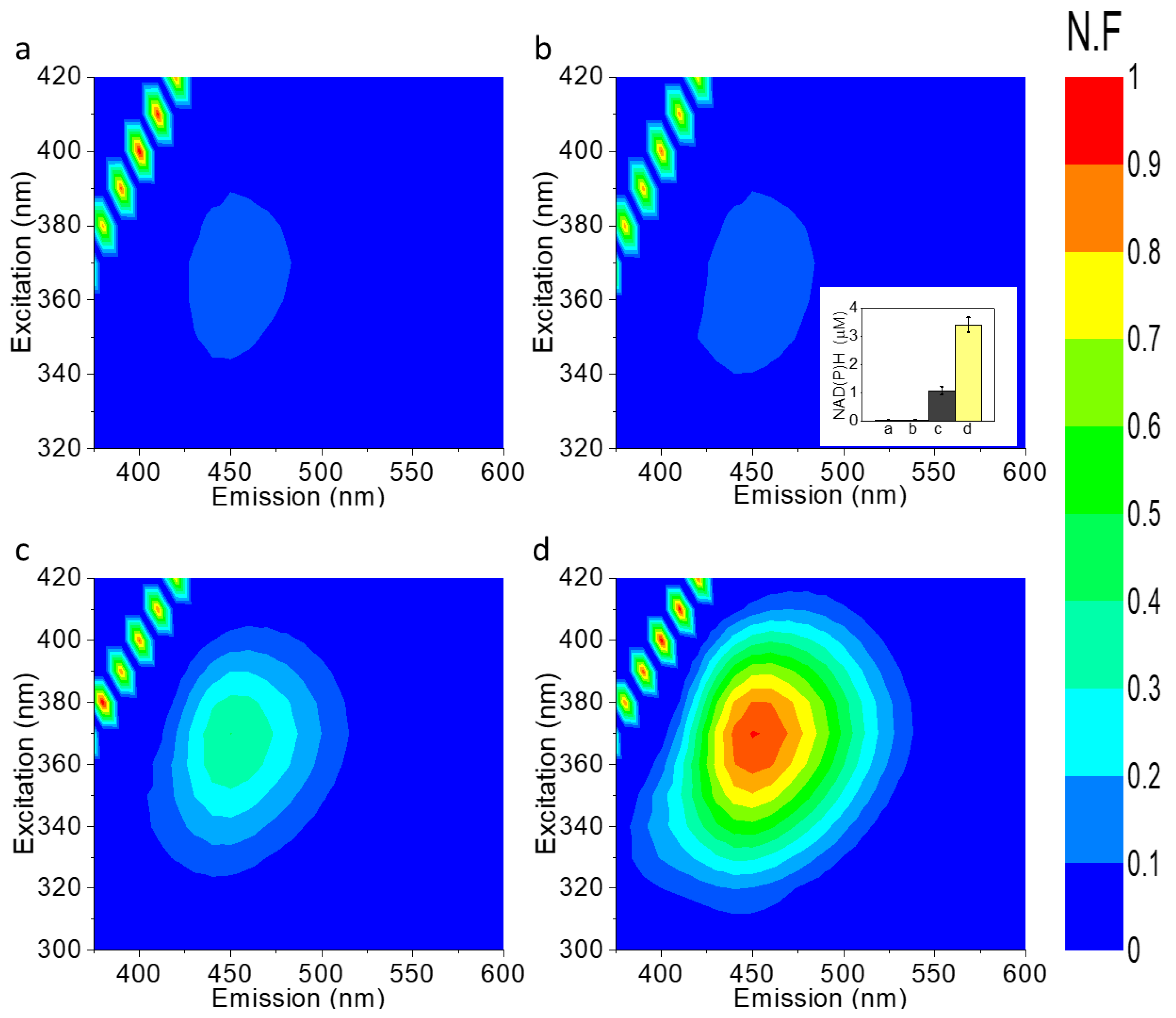
2.2. NAD(P)H Accumulates in the External Cellular Media of Ds

We previously showed that cyanobacteria secrete NADH and NADPH (NAD(P)H) molecules to the ECM by 2D fluorescence maps (2D-FM). The secretion was significantly enhanced by the association of the cyanobacteria with the electrochemical cell and upon illumination [36]. These results led us to explore whether the same phenomena occurred in Ds. To assess this, 2D-FM of the ECM of Ds was measured after 10 min in dark and light on non-conductive plastic panels without association to an electrochemical setup or on the screen-printed electrodes immediately following CA measurement. The ECM of Ds that was obtained from cells not in association with the electrochemical setup showed a low intensity of the spectral fingerprint of NAD(P)H (λ(ex)max = ~350 nm, λ(em)max = ~450 nm), in either dark or light. A higher intensity signal was obtained for the ECM of Ds in the BPEC, after CA was performed in the dark, that was significantly enhanced by illumination of the system (Figure 2). The concentration of NAD(P)H in the ECM was calculated based on the calibration curve of fluorescence intensity vs. NADH concentration that was reported in our previous work [36]. The calculated concentrations of NAD(P)H for the ECM of Ds on plastic (dark, light), or in the BPEC (dark, light) were ~0.035, 0.04, 1, and 3.4 µM, respectively, similar to the values obtained for the cyanobacterial based BPECs [36]. As is seen in Figure 1, the dark current was quite low, indicating that while there was some release of NAD(P)H (or the oxidized molecules), cycling back into the cells may be light dependent as well.

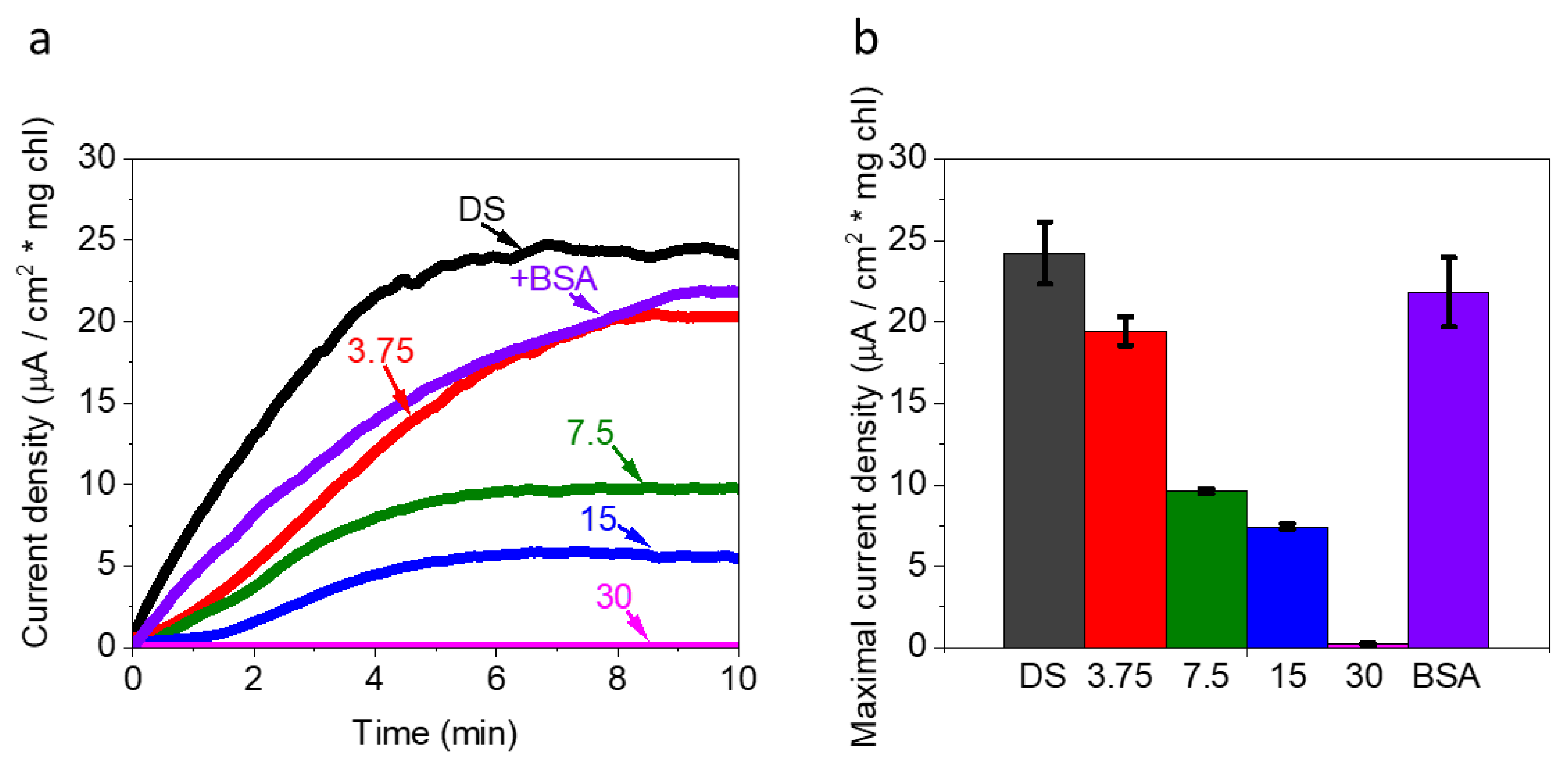
2.3. FNR Eliminates the Photocurrent Production in Ds-Based BPEC

The ECM of microalgae may contain hundreds of molecules, many which are redox active, that originate from the cultivation media, secreted molecules, degraded cells, and chemical reactions. In our previous results with cyanobacteria [36] we showed that NAD(P)H was the major exogenous electron mediator and that addition of exogenously added FNR almost totally eliminated photocurrent production. We wished to assess whether NAD(P)H was also the major electron mediator in Ds-based BPECs. CA of Ds was measured with the addition of increasing concentrations of FNR (0, 3.75, 7.5, 15, and 30 µM). As a control experiment, CA of Ds was measured with addition of 1 mg/mL of bovine serum albumin (BSA). A concentration-dependent reduction in the photocurrent was obtained in the presence of FNR, down to total elimination of the current at 30 µM FNR, while addition of BSA did not influence the photocurrent (Figure 3). This result indicated that in similar to cyanobacteria [36], NAD(P)H was also the main electron mediator in Ds.

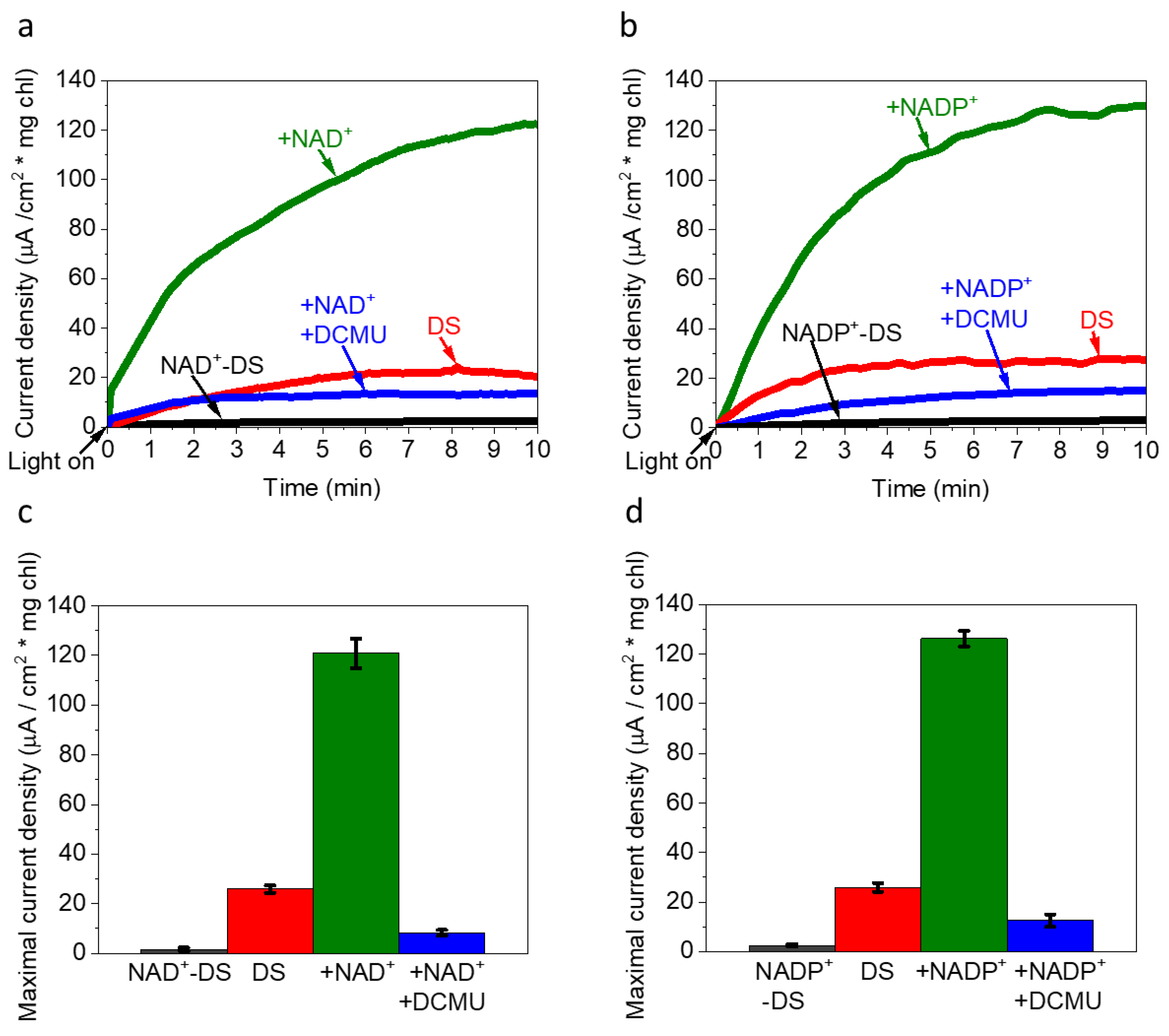
2.4. The Addition of Exogenous NADP+, NAD+ Enhance Photocurrent Production

In our study of current harvesting from cyanobacteria, we showed that the electron mediation by NADP+ or NAD+ (NAD(P+)) was cyclic, and that addition of exogenous NAD(P+) significantly increased photocurrent production. This indicates that after oxidation by the anode, NAD(P+) is taken up by cells for further cycles of reduction [36]. This led us to explore if Ds could also uptake NAD(P+) and re-reduce it to increase photocurrent production. In addition, we wished to assess if the re-reduction of the internalized NAD(P+) derived from the photosynthesis pathway or by other components in the cell. 10 mM NAD+ or NADP+ were incubated for 2 h with Ds in the presence or absence of 100 µM DCMU, followed by CA in the light (Figure 4). The addition of either NAD+ or NADP+ increased the photocurrent from ~25 to ~120 µA/cm2 mg chl. Addition of DCMU inhibited the photocurrent by more than 80% (~20 µA/cm2 mg chl). Based on the obtained results we postulate that exogenous NAD(P+) could enter the cells and be re-reduced by the photosynthesis pathway. Nevertheless, we could not exclude the participation of additional small redox active molecules that existed in the Ds ECM.

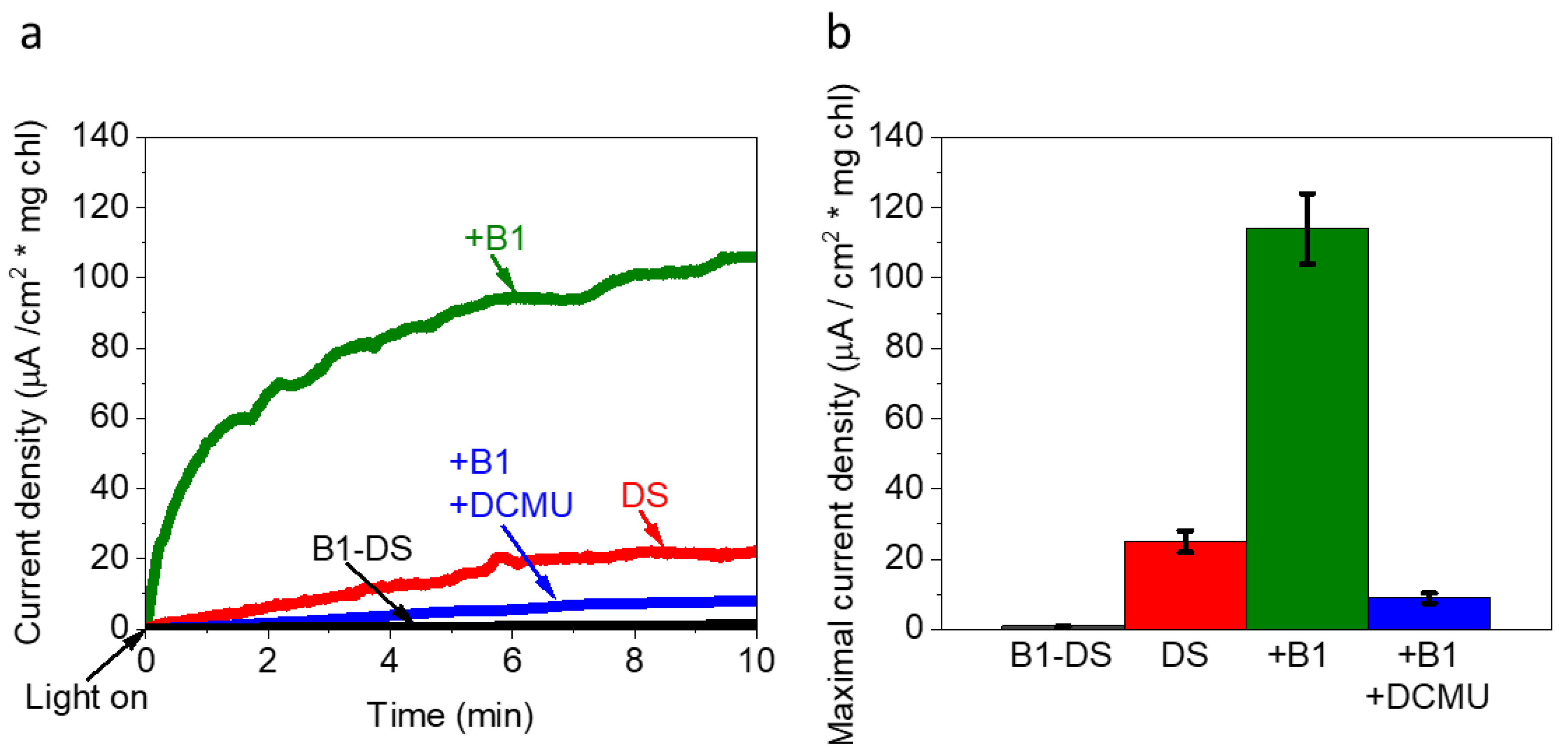
2.5. Vitamin B1 Can Mediate Electrons from the Photosynthesis Pathway to the Anode

Thiamine (vitamin B1) is a water-soluble molecule that functions as an electron mediator in biological reactions [44]. We wished to explore whether it could enhance the photocurrent in the Ds-based BPEC. Ds cells were incubated for 2 h with 5 mM commercial thiamine and their CA was measured for 10 min in dark and light. As for both NAD+ or NADP+ (Figure 4) the addition of vitamin B1 enhanced the photocurrent to ~110 µA/cm2 ·mg chl in light and did not produce any significant current in dark. (Figure 5). To identify that the source of the electrons was from photosynthesis, we measured DS + vitamin B1 with the addition of DCMU. Indeed, the addition of DCMU almost totally eliminated the photocurrent, as was observed for NAD+ and NADP+ (Figure 4).

2.6. The Addition of Both FeCN and NADP+ Produces a Greater Photocurrent

One of the known bio-compatible exogenous electron mediators that can significantly enhance the current in bio-electrochemical cells is FeCN. We wished to estimate the ability of this mediator to produce photocurrent and to explore whether the addition of both NADP+ and FeCN could produce more current synergistically. CA of Ds was measured with addition of either FeCN or a mixture of FeCN and NADP+ (Figure 6). A maximal photocurrent of ~700 and ~950 µA/cm2 mg chl was obtained, respectively. These values were 6–8 fold greater that the currents harvested with Ds cells without addition of mediators, which was dependent on the rate of internal NADPH concentrations. Addition of FeCN without Ds produced a current of 2.5 µA/cm2 (equivalent to 25 µA/cm2 mg chl, when normalized by the same chl normalization value used in the presence of Ds). Addition of the mixture of FeCN and NADP+ produced a photocurrent than was about 16% greater than the sum of the currents obtained separately. We postulate that upon irradiation, the addition of NADP+ increased the concentration of NADPH in the cells, which could then reduce external FeCN molecules in the ECM, without the need to diffuse out of the cells, thereby enhancing the photocurrent. A similar transfer of electrons to external FeCN has been reported for cyanobacteria [45].

3. Materials and Methods

All chemicals were purchased from Merck unless mentioned otherwise.
Millipore hydrophilic PVDF filter Membrane 0.22 µm was purchased from Millex-HV.
Chlamydomonas reinhardtii Ferredoxin NADP+ Reductase (crFNR) was produced in E. coli using an expression plasmid harbouring the FNR cDNA, kindly provided by Prof. Iftach Yacoby at Tel Aviv University and purified as described by Marco et al. [36,46].

3.1. Ds Cultivation

Ds was cultivated in BG11 medium with 1.5 M NaCl in the growth chamber at a light intensity of about 100 µE/m2 s, shaking at 100 rpm and 29 °C.

3.2. Samples Preparation

OD measurements of the cells were done at 750 nm (Nanodrop 2000 UV-Vis spectrophotometer, Thermo Fisher Scientific). For the measurements in the BEPC, log phase grown cells (O.D.750 nm = 0.6–1.0) were centrifuged (Multifuge X1R, Thermo Scientific) for 10 min at 5000 rpm. The supernatant was removed, and the pellet was resuspended in a solution of MES buffer (MES 50 mM (pH = 6) with 1.5 M NaCl. Chlorophyll concentration was determined by 90% methanol extraction followed by absorption measurements [47,48]. For use in the BPEC, live Ds were diluted in MES buffer solution to have a final concentration of 0.1 mg/mL total chl. The cells were allowed to precipitate for 0.5 min before initiating CA measurements.

3.3. Chronoamperometry Measurements

Chronoamperometry measurements were performed as previously described [36] using a Plamsens3 potentiostat (Palmsens, Houten, The Netherlands) connected to screen printed electrodes (AC1.W4.R1, BVT Technologies, Strážek, Czech Republic). The working electrode (WE) had a surface area of 0.79 mm2 and the distances between electrodes were: 1.95 mm (WE-counter electrode (CE)), 0.68 mm (WE-reference electrode (RE)), and 0.25 mm (CE-RE). Illumination of 1.5 SUN (1000 w/m2, calibrated at the electrode surface height without samples) was performed using a solar simulator (Abet Technologies, Milford, CN, USA). In all measurements, a bias potential of 0.5 V was applied on the graphite anode (this bias value was chosen because it produced the maximal signal/noise in the CA measurements). Total current calculations were done by summing all current values of the measurement which were acquired in time steps of 0.5 s. The volume of all measured samples was 50 µL, pipetted directly onto the printed electrode panel. The round boundaries of the measuring area of the panel induced a similar drop shape in all measurements that did not leak to the sides by the surface tension of the liquid. The drop was not stirred, and the cells typically did not sediment spontaneously over the short measurement duration of 10 min. The addition of the electron mediators NADP+, NADP+, or vitamin B1 to Ds cells was done 2 h prior to initiation of the measurements. This time was determined as the minimal time that produced maximal photocurrent production in our previous work [36]. FeCN was added immediately prior to measurement initiation. As preparation for fluorescence measurements, 50 µL of bacterial cells were filtrated through a 0.22 µm filter membrane to produce the external cell media (ECM) solution. Each ECM sample was diluted with fresh MES buffer (MES 50 mM (pH = 6) with 1.5 M NaCl to a final volume of 2 mL. CA values obtained in the absence of Ds cells were multiplied by the chlorophyll concentration value used to normalize the samples that contained Ds cells.

3.4. Fluorescence Measurements

All fluorescence measurements, unless mentioned otherwise, were performed using a Fluorolog 3 fluorimeter (Horiba) with excitation and emission slits bands of 4 nm. Quantification of NAD(P)H concentrations was calculated based on the NADH calibration curve which was based on increasing concentrations measured at (λ(ex) = 350 nm, λ(em) = 450 nm), that was previously used [36]. The lines of diagonal spots that appear in all of the maps presented here and in the following figures result from the light scattering of the Xenon lamp [49].

3.5. Statistical Analysis

In all experiments, the average and the standard deviation were calculated based on three independent repetitions.

4. Conclusions

In this work, we show that NADPH which is the major endogenous mediator in cyanobacterial cells based BPECs is also the major endogenous mediator in the microalgae DS. We show that is the unique ability of Ds to habitat in a high salinity environment enables the production of photocurrent that is about 5–6 times greater than freshwater cyanobacteria. This current can be further increased by the addition of exogenous NAD+ or NADP+ as previously reported for cyanobacterial-based BPECs. We show for the first time that vitamin B1 can also mediate electrons from the photosynthetic pathway and increase the photocurrent in the same magnitude as NAD(P)+. Finally, we show that there is synergism in the electron mediation of NADPH and FeCN while adding both mediators together enhances the photocurrent more the sum of each of them separately. All in all, the understanding of the electron mediation mechanism in DS and the utilization of the electron mediators described in this work may contribute to future green applicative technologies that are based on microalgal BPECs.

Author Contributions

Y.S., G.S. and N.A. conceived the idea. Y.S. and N.A. designed the experiments. Y.S. performed the main experiments. B.E., T.N.T. and L.K. assisted in performing the experiments. Y.S., G.S. and N.A. wrote the paper. N.A. supervised the entire research. All authors have read and agreed to the published version of the manuscript.

Funding

Funding was provided by a “Nevet” grant from the Grand Technion Energy Program (GTEP) and a Technion VPR Berman Grant for Energy Research. Yaniv Shlosberg and Tünde N. Tóth are supported by fellowships of the Nancy and Stephen Grand Technion Energy Program (GTEP) and by a Schulich Graduate fellowship.

Acknowledgments

Some of the results reported in this work were obtained using central facilities at the Technion’s Hydrogen Technologies Research Laboratory (HTRL) supported by the Nancy and Stephen Grand Technion Energy Program (GTEP), the ADELIS Foundation, and the Solar Fuels I-CORE. We thank Rachel Edreii for her technical support. We thank Iftach Yacoby for supplying biological materials. We thank Sonya Copperstein for her assistance.

Conflicts of Interest

The authors declare no competing interests.

References

  1. Wei, X.; Lee, H.; Choi, S. Biopower Generation in a Microfluidic Bio-Solar Panel. Sens. Actuators B Chem. 2016, 228, 151–155. [Google Scholar] [CrossRef]
  2. Rabaey, K.; Boon, N.; Höfte, M.; Verstraete, W. Microbial Phenazine Production Enhances Electron Transfer in Biofuel Cells. Environ. Sci. Technol. 2005, 39, 3401–3408. [Google Scholar] [CrossRef] [PubMed]
  3. Chaudhuri, S.K.; Lovley, D.R. Electricity Generation by Direct Oxidation of Glucose in Mediatorless Microbial Fuel Cells. Nat. Biotechnol. 2003, 21, 1229–1232. [Google Scholar] [CrossRef] [PubMed]
  4. Liu, Z.-D.; Lian, J.; Du, Z.-W.; Li, H.-R. Construction of Sugar-Based Microbial Fuel Cells by Dissimilatory Metal Reduction Bacteria. Chin. J. Biotechnol. 2006, 22, 131–137. [Google Scholar] [CrossRef]
  5. Ringeisen, B.R.; Henderson, E.; Wu, P.K.; Pietron, J.; Ray, R.; Little, B.; Biffinger, J.C.; Jones-Meehan, J.M. High Power Density from a Miniature Microbial Fuel Cell Using Shewanella Oneidensis DSP10. Environ. Sci. Technol. 2006, 40, 2629–2634. [Google Scholar] [CrossRef] [Green Version]
  6. Park, D.; Zeikus, J. Impact of Electrode Composition on Electricity Generation in a Single-Compartment Fuel Cell Using Shewanella Putrefaciens. Appl. Microbiol. Biotechnol. 2002, 59, 58–61. [Google Scholar] [CrossRef]
  7. Pham, C.A.; Jung, S.J.; Phung, N.T.; Lee, J.; Chang, I.S.; Kim, B.H.; Yi, H.; Chun, J. A Novel Electrochemically Active and Fe(III)-Reducing Bacterium Phylogenetically Related to Aeromonas Hydrophila, Isolated from a Microbial Fuel Cell. FEMS Microbiol. Lett. 2003, 223, 129–134. [Google Scholar] [CrossRef] [Green Version]
  8. Min, B.; Cheng, S.; Logan, B.E. Electricity Generation Using Membrane and Salt Bridge Microbial Fuel Cells. Water Res. 2005, 39, 1675–1686. [Google Scholar] [CrossRef]
  9. Fang, X.; Kalathil, S.; Reisner, E. Semi-Biological Approaches to Solar-to-Chemical Conversion. Chem. Soc. Rev. 2020, 49, 4926–4952. [Google Scholar] [CrossRef]
  10. Bergel, A.; Féron, D.; Mollica, A. Catalysis of Oxygen Reduction in PEM Fuel Cell by Seawater Biofilm. Electrochem. Commun. 2005, 7, 900–904. [Google Scholar] [CrossRef] [Green Version]
  11. Sekar, N.; Jain, R.; Yan, Y.; Ramasamy, R.P. Enhanced Photo-Bioelectrochemical Energy Conversion by Genetically Engineered Cyanobacteria. Biotechnol. Bioeng. 2016, 113, 675–679. [Google Scholar] [CrossRef] [PubMed]
  12. Thirumurthy, M.A.; Hitchcock, A.; Cereda, A.; Liu, J.; Chavez, M.S.; Doss, B.L.; Ros, R.; El-Naggar, M.Y.; Heap, J.T.; Bibby, T.S.; et al. Type IV Pili-Independent Photocurrent Production by the Cyanobacterium Synechocystis Sp. PCC 6803. Front. Microbiol. 2020, 11, 1–11. [Google Scholar] [CrossRef] [PubMed]
  13. Heidary, N.; Kornienko, N.; Kalathil, S.; Fang, X.; Ly, K.H.; Greer, H.F.; Reisner, E. Disparity of Cytochrome Utilization in Anodic and Cathodic Extracellular Electron Transfer Pathways of Geobacter Sulfurreducens Biofilms. J. Am. Chem. Soc. 2020, 142, 5194–5203. [Google Scholar] [CrossRef] [Green Version]
  14. Lovley, D.R. Electromicrobiology. Annu. Rev. Microbiol. 2012, 66, 391–409. [Google Scholar] [CrossRef] [PubMed]
  15. Nevin, K.P.; Richter, H.; Covalla, S.F.; Johnson, J.P.; Woodard, T.L.; Orloff, A.L.; Jia, H.; Zhang, M.; Lovley, D.R. Power Output and Columbic Efficiencies from Biofilms of Geobacter Sulfurreducens Comparable to Mixed Community Microbial Fuel Cells. Environ. Microbiol. 2008, 10, 2505–2514. [Google Scholar] [CrossRef]
  16. Yi, H.; Nevin, K.P.; Kim, B.-C.; Franks, A.E.; Klimes, A.; Tender, L.M.; Lovley, D.R. Selection of a Variant of Geobacter Sulfurreducens with Enhanced Capacity for Current Production in Microbial Fuel Cells. Biosens. Bioelectron. 2009, 24, 3498–3503. [Google Scholar] [CrossRef]
  17. Hartshorne, R.S.; Jepson, B.N.; Clarke, T.A.; Field, S.J.; Fredrickson, J.; Zachara, J.; Shi, L.; Butt, J.N.; Richardson, D.J. Characterization of Shewanella Oneidensis MtrC: A Cell-Surface Decaheme Cytochrome Involved in Respiratory Electron Transport to Extracellular Electron Acceptors. JBIC J. Biol. Inorg. Chem. 2007, 12, 1083–1094. [Google Scholar] [CrossRef] [Green Version]
  18. Shi, L.; Rosso, K.M.; Zachara, J.M.; Fredrickson, J.K. Mtr Extracellular Electron-Transfer Pathways in Fe(III)-Reducing or Fe(II)-Oxidizing Bacteria: A Genomic Perspective. Biochem. Soc. Trans. 2012, 40, 1261–1267. [Google Scholar] [CrossRef]
  19. Bond, D.R.; Lovley, D.R. Electricity Production by Geobacter Sulfurreducens Attached to Electrodes. Appl. Environ. Microbiol. 2003, 69, 1548–1555. [Google Scholar] [CrossRef] [Green Version]
  20. Rabaey, K.; Boon, N.; Siciliano, S.D.; Verhaege, M.; Verstraete, W. Biofuel Cells Select for Microbial Consortia That Self-Mediate Electron Transfer. Appl. Environ. Microbiol. 2004, 70, 5373–5382. [Google Scholar] [CrossRef] [Green Version]
  21. Simoska, O.; Sans, M.; Eberlin, L.S.; Shear, J.B.; Stevenson, K.J. Electrochemical Monitoring of the Impact of Polymicrobial Infections on Pseudomonas Aeruginosa and Growth Dependent Medium. Biosens. Bioelectron. 2019, 142, 111538. [Google Scholar] [CrossRef]
  22. Ieropoulos, I.; Greenman, J.; Melhuish, C.; Hart, J. Comparative Study of Three Types of Microbial Fuel Cell. Enzyme Microb. Technol. 2005, 37, 238–245. [Google Scholar] [CrossRef]
  23. Lovley, D.R.; Coates, J.D.; Blunt-Harris, E.L.; Phillips, E.J.P.; Woodward, J.C. Humic Substances as Electron Acceptors for Microbial Respiration. Nature 1996, 382, 445–448. [Google Scholar] [CrossRef]
  24. Lovley, D.R.; Holmes, D.E.; Nevin, K.P. Dissimilatory Fe(III) and Mn(IV) Reduction; Academic Press: Cambridge, MA, USA, 2004; Volume 49, pp. 219–286. [Google Scholar] [CrossRef] [Green Version]
  25. White, G.F.; Shi, Z.; Shi, L.; Wang, Z.; Dohnalkova, A.C.; Marshall, M.J.; Fredrickson, J.K.; Zachara, J.M.; Butt, J.N.; Richardson, D.J.; et al. Rapid Electron Exchange between Surface-Exposed Bacterial Cytochromes and Fe(III) Minerals. Proc. Natl. Acad. Sci. USA 2013, 110, 6346–6351. [Google Scholar] [CrossRef] [Green Version]
  26. Edwards, M.J.; White, G.F.; Butt, J.; Richardson, D.J.; Clarke, T.A. The Crystal Structure of a Biological Insulated Transmembrane Molecular Wire. Cell 2020, 181, 665–673. [Google Scholar] [CrossRef] [PubMed]
  27. Xavier, J.C.; Preiner, M.; Martin, W.F. Something Special about CO-Dependent CO2 Fixation. FEBS J. 2018, 285, 4181–4195. [Google Scholar] [CrossRef] [PubMed] [Green Version]
  28. Tschörtner, J.; Lai, B.; Krömer, J.O. Biophotovoltaics: Green Power Generation From Sunlight and Water. Front. Microbiol. 2019, 10, 866. [Google Scholar] [CrossRef] [Green Version]
  29. Grattieri, M.; Beaver, K.; Gaffney, E.M.; Minteer, S.D. Tuning Purple Bacteria Salt-Tolerance for Photobioelectrochemical Systems in Saline Environments. Faraday Discuss. 2019, 215, 15–25. [Google Scholar] [CrossRef]
  30. Grattieri, M. Purple Bacteria Photo-Bioelectrochemistry: Enthralling Challenges and Opportunities. Photochem. Photobiol. Sci. 2020, 19, 424–435. [Google Scholar] [CrossRef]
  31. Grattieri, M.; Rhodes, Z.; Hickey, D.P.; Beaver, K.; Minteer, S.D. Understanding Biophotocurrent Generation in Photosynthetic Purple Bacteria. ACS Catal. 2019, 9, 867–873. [Google Scholar] [CrossRef]
  32. Singh, S.K.; Sundaram, S.; Sinha, S.; Rahman, M.A.; Kapur, S. Recent Advances in CO2 Uptake and Fixation Mechanism of Cyanobacteria and Microalgae. Crit. Rev. Environ. Sci. Technol. 2016, 46, 1297–1323. [Google Scholar] [CrossRef]
  33. Thangam, K.R.; Santhiya, A.; Sri, S.R.A.; MubarakAli, D.; Karthikumar, S.; Kumar, R.S.; Thajuddin, N.; Soosai, M.R.; Varalakshmi, P.; Moorthy, I.G.; et al. Bio-Refinery Approaches Based Concomitant Microalgal Biofuel Production and Wastewater Treatment. Sci. Total Environ. 2021, 785, 147267. [Google Scholar] [CrossRef]
  34. Mehariya, S.; Goswami, R.K.; Karthikeysan, O.P.; Verma, P. Microalgae for High-Value Products: A Way towards Green Nutraceutical and Pharmaceutical Compounds. Chemosphere 2021, 280, 130553. [Google Scholar] [CrossRef]
  35. Saper, G.; Kallmann, D.; Conzuelo, F.; Zhao, F.; Tóth, T.N.; Liveanu, V.; Meir, S.; Szymanski, J.; Aharoni, A.; Schuhmann, W.; et al. Live Cyanobacteria Produce Photocurrent and Hydrogen Using Both the Respiratory and Photosynthetic Systems. Nat. Commun. 2018, 9, 1–9. [Google Scholar] [CrossRef] [Green Version]
  36. Shlosberg, Y.; Eichenbaum, B.; Tóth, T.N.; Levin, G.; Liveanu, V.; Schuster, G.; Adir, N. NADPH Performs Mediated Electron Transfer in Cyanobacterial-Driven Bio-Photoelectrochemical Cells. iScience 2020, 24, 101892. [Google Scholar] [CrossRef]
  37. Cereda, A.; Hitchcock, A.; Symes, M.D.; Cronin, L.; Bibby, T.S.; Jones, A.K. A Bioelectrochemical Approach to Characterize Extracellular Electron Transfer by Synechocystis Sp. PCC6803. PLoS ONE 2014, 9, 91484. [Google Scholar] [CrossRef] [PubMed]
  38. Lee, J.; Cho, H.; Kim, S. Enhanced Photocurrent Generation From a Single-Mediated Photo-Bioelectrochemical Cell Using Wild-Type Anabaena Variabilis Dispersed in Solution. ChemElectroChem 2020, 7, 4075–4083. [Google Scholar] [CrossRef]
  39. Pablo, F.; Buckeny, R.T.; Lim, R.P. Toxicity of Cyanide, Iron-Cyanide Complexes, and a Blast Furnace Effluent to Larvae of the Doughboy Scallop, Chlamys Asperrimus. Bull. Environ. Contam. Toxicol. 1997, 58, 93–100. [Google Scholar] [CrossRef]
  40. Murakawa, S.; Takahashi, T. Stimulation of Glutamate Uptake by NADH and NADPH in Intact Cells of Escherichia Coli Mutant Defective in Mg2+, Ca2+-ATPase. Agric. Biol. Chem. 1978, 42, 1803–1804. [Google Scholar] [CrossRef]
  41. Zhou, Y.; Wang, L.; Yang, F.; Lin, X.; Zhang, S.; Zhao, Z.K. Determining the Extremes of the Cellular NAD(H) Level by Using an Escherichia Coli NAD +-Auxotrophic Mutant. Appl. Environ. Microbiol. 2011, 77, 6133–6140. [Google Scholar] [CrossRef] [PubMed] [Green Version]
  42. Major, P.; Embley, T.M.; Williams, T.A. Phylogenetic Diversity of NTT Nucleotide Transport Proteins in Free-Living and Parasitic Bacteria and Eukaryotes. Genome Biol. Evol. 2017, 9, 480–487. [Google Scholar] [CrossRef]
  43. Gruber, A.; Haferkamp, I. Nucleotide Transport and Metabolism in Diatoms. Biomolecules 2019, 9, 761. [Google Scholar] [CrossRef] [Green Version]
  44. Lovander, M.D.; Lyon, J.D.; Parr, D.L.; Wang, J.; Parke, B.; Leddy, J. Critical Review—Electrochemical Properties of 13 Vitamins: A Critical Review and Assessment. J. Electrochem. Soc. 2018, 165, G18–G49. [Google Scholar] [CrossRef]
  45. Gonzalez-Aravena, A.C.; Yunus, K.; Zhang, L.; Norling, B.; Fisher, A.C. Tapping into Cyanobacteria Electron Transfer for Higher Exoelectrogenic Activity by Imposing Iron Limited Growth. RSC Adv. 2018, 8, 20263–20274. [Google Scholar] [CrossRef] [Green Version]
  46. Marco, P.; Elman, T.; Yacoby, I. Binding of Ferredoxin NADP+ Oxidoreductase (FNR) to Plant Photosystem I. Biochim. Biophys. Acta Bioenerg. 2019, 1860, 689–698. [Google Scholar] [CrossRef] [PubMed]
  47. Ritchie, R.J. Consistent Sets of Spectrophotometric Chlorophyll Equations for Acetone, Methanol and Ethanol Solvents. Photosynth. Res. 2006, 89, 27–41. [Google Scholar] [CrossRef] [PubMed]
  48. Ritchie, R.J. Universal Chlorophyll Equations for Estimating Chlorophylls a, b, c, and d and Total Chlorophylls in Natural Assemblages of Photosynthetic Organisms Using Acetone, Methanol, or Ethanol Solvents. Photosynthetica 2008, 46, 115–126. [Google Scholar] [CrossRef]
  49. Lawaetz, A.J.; Stedmon, C.A. Fluorescence Intensity Calibration Using the Raman Scatter Peak of Water. Appl. Spectrosc. 2009, 63, 936–940. [Google Scholar] [CrossRef]
Figure 1. Live Ds cells in high salt produce photocurrent that originates from PSII. CA measurements of live Ds were conducted in 1.5 M NaCl with an applied bias of 0.5 V on the anode in dark, light, and in light in the presence of 100 µM DCMU. (a) CA in dark (D), light (L), and in light with DCMU (+DCMU). (b) Maximal current densities after 10 min of CA in dark (D), light (L), and in light with DCMU (+DCMU). The error bars represent the standard deviation over three independent measurements.
Catalysts 11 01220 g001
Figure 2. NAD(P)H accumulates in the external cellular media of Ds. 2D-FM spectra were measured for the ECM of Ds cells after 10 min in dark and light, with or without association to the anode. (a). ECM of DS in dark without association to the electrochemical cell. (b). ECM of Ds in light without association to the electrochemical cell. (c). ECM of Ds in dark with association to the electrochemical cell. (d). ECM of Ds in light with association to the electrochemical cell. The insert in panel b shows the NAD(P)H concentrations that were calculated based on the maximal fluorescence intensities at λmax = 340 nm [36]. The X axis labels of the insert are: a, dark without association to the anode; b, light without association to the anode; c, dark with association with the anode; and d, light with association with the anode, respectively. The error bars represent the standard deviation over three independent experimental repetitions.
Catalysts 11 01220 g002
Figure 3. FNR eliminates the photocurrent production in DS-based BPEC. CA of DS was measured without or with increasing concentrations of FNR or with addition of BSA. The photocurrent production was decreased in proportion with the increasing FNR concentrations down to total elimination. (a) CA of Ds without the addition of FNR (black) and with the addition of 3.75 µM FNR (red), 7.5 µM FNR (green), 15 µM FNR (blue), 30 µM FNR (magenta) and 1 mg/mL BSA (violet). (b) Maximal current density over 10 min of CA. The error bars represent the standard deviation over three independent measurements.
Catalysts 11 01220 g003
Figure 4. Addition of exogenous NADP+ or NAD+ enhances the photocurrent production. CA of Ds with addition of NAD+ or NADP+ and with or without DCMU were measured. (a) CA of NAD+ (without cells), intact Ds, Ds with addition of NAD+, and with addition of NAD+ + DCMU. (b) CA of NADP+ (without cells), intact Ds, Ds with Addition of NADP+, and with addition of NADP+ + DCMU. (c) Maximal current densities of the CA measurement that are described in panel a. (d) Maximal current densities of the CA measurement that are described in panel b. The error bars represent the standard deviation over three independent measurements. Values obtained in the absence of Ds cells were multiplied by the chlorophyll concentration value used to normalize the samples that contain Ds.
Catalysts 11 01220 g004
Figure 5. Vitamin B1 can mediate electrons from the photosynthesis pathway to the anode. CA of DS with the addition of vitamin B1 with or without DCMU was measured. (a) CA of vitamin B1 without Ds (black), Ds (red), Ds + vitamin B1 (green), and Ds + vitamin B1 + DCMU (blue). (b) Maximal current densities over 10 min of CA measurements. The error bars represent the standard deviation over three independent measurements. Values obtained in the absence of Ds were multiplied by the chlorophyll concentration value used to normalize the samples that contain Ds.
Catalysts 11 01220 g005
Figure 6. The addition of both FeCN and NADP+ produces a greater photocurrent. CA of FeCN, Ds, Ds with the addition of FeCN, and Ds with the addition of both FeCN and NADP+ were measured. (a) CA of FeCN without Ds (black), Ds (red), Ds + FeCN (green), and Ds + FeCN + NADP+ (blue). (b) Maximal current densities over 10 min of CA measurements. The error bars represent the standard deviation over three independent measurements.
Catalysts 11 01220 g006
Publisher’s Note: MDPI stays neutral with regard to jurisdictional claims in published maps and institutional affiliations.

