Article

Pharmacokinetic Behavior and Pharmacokinetic/Pharmacodynamic Integration of Danofloxacin Following Single or Co-Administration with Meloxicam in Healthy Lambs and Lambs with Respiratory Infections

1
Department of Pharmacology and Toxicology, Faculty of Veterinary Medicine, University of Selcuk, 42031 Konya, Turkey
2
Pendik Veterinary Control Institute, Bati Mah. Yunus Cad. 2/1, Pendik, 34890 Istanbul, Turkey
*
Author to whom correspondence should be addressed.
Antibiotics 2021, 10(10), 1190; https://doi.org/10.3390/antibiotics10101190
Submission received: 28 June 2021 / Revised: 18 August 2021 / Accepted: 27 September 2021 / Published: 30 September 2021
(This article belongs to the Special Issue Antibiotic Use in Veterinary)

Abstract

:
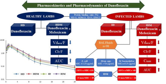
The aim of this study was to determine the pharmacokinetics and pharmacodynamics of danofloxacin (DAN; 6 mg/kg) following subcutaneous administration alone or co-administration with meloxicam (MLX; 1 mg/kg) in healthy lambs and lambs with respiratory infections. The study was carried out using a total of four groups: HD (healthy; n = 6) and ID (infected; n = 7) groups who were administered DAN only, and HDM (healthy; n = 6) and IDM (infected; n = 7) groups who were administered DAN and MLX simultaneously. The plasma concentrations of DAN were determined using high-performance liquid chromatography–UV and analyzed by the non-compartmental method. DAN exhibited a similar elimination half-life in all groups, including both the healthy and infected lambs. The total clearance in the HDM, ID and IDM groups and volume of distribution in the HDM and IDM groups were significantly reduced. MLX in the IDM group significantly increased the area under the curve (AUC) and peak concentration (Cmax) of DAN compared to the HD group. The Mannheimia haemolytica, Escherichia coli, and Streptococcus spp. strains were isolated from bronchoalveolar lavage fluid samples of the infected lambs. When co-administration with meloxicam, DAN at a 6 mg/kg dose can provide optimum values of ƒAUC0–24/MIC (>56 h) and ƒCmax/MIC (>8) for susceptible M. haemolytica isolates with an MIC90 value of 0.25 µg/mL and susceptible E. coli isolates with an MIC value of ≤0.125 µg/mL.

Graphical Abstract

1. Introduction

Respiratory diseases, which are common in all countries where lamb breeding is carried out, are considered to be complex diseases that include interactions between multiple agents such as the host’s immune system and physiological state, viruses, bacteria, Mycoplasma, and environmental factors. They cause serious financial losses due to low body weight gain, treatment costs, and death [1,2,3]. Bacterial agents frequently isolated from lambs with pneumonia are Mannheimia haemolytica and Pasteurella multocida [4,5]. Antibiotics are used to treat acute pneumonia, to reduce the effects of chronic nonprogressive pneumonia, and to treat the remainder of the flock to prevent more deaths from acute pneumonia. The quick detection of infected lambs is very important for the success of antibiotic treatment [3,6]. The antibiotic selection for the treatment of infection depends on many factors, including the infective microorganism, the susceptibility of the microorganism to antibiotics, the site of infection, local factors (pH, inoculum size, etc.), host factors, the potential side effects of antibiotics, and antibiotic resistance. Following the selection of the ideal antibiotic, the pharmacokinetic and pharmacodynamic data of the drug should be determined to establish the most appropriate dosage regimen [7,8].
Danofloxacin, a third-generation synthetic fluoroquinolone developed for use in veterinary medicine, is approved for the treatment of respiratory infections in cattle, chickens, and pigs [9,10]. Danofloxacin reaches a 4–5 times higher concentration in the lung tissue than in the plasma and has activity against most Gram-negative bacteria, such as P. multocida, M. haemolytica, Histophilus somni, and Enterobacterales (Escherichia coli, Klebsiella, and Salmonella), and some Gram-positive bacteria, such as Staphylococcus aureus and Streptococcus pneumoniae [11,12,13,14]. Danofloxacin has a very wide distribution volume in sheep and goats due to having high lipid solubility, low binding to plasma proteins, and being a substrate for ATP-dependent efflux transporters [15,16]. The clinical efficacy of danofloxacin has been established in sheep and cattle with respiratory infections [17,18].
Meloxicam, a non-steroidal anti-inflammatory drug (NSAID) of the oxicam class, is used to alleviate the symptoms of acute respiratory infection with appropriate antibiotic therapy in cattle [19]. In Canada, Australia, and New Zealand, it is approved for use in the treatment of pain and inflammation in sheep [20]. It has been determined that meloxicam has a stronger anti-inflammatory effect by reducing the synthesis of tumor necrosis factor (TNF)-alpha, a cytokine with proinflammatory effects, as well as inhibiting the cyclooxygenase (COX)-2 enzyme in respiratory infections [21,22].
In respiratory infections, the host’s excessive inflammatory response plays an important role in the pathogenesis of the disease [23]. Therefore, the use of NSAIDs is recommended against the harmful effects of inflammation in the treatment of pneumonia [24,25]. It has been reported that when NSAIDs are used in combination with antibiotics compared to antibacterial therapy alone, clinical findings improve faster in respiratory infection [25,26,27]. However, pathophysiological changes in respiratory infections and antibiotics–NSAID interactions resulting from concurrent drug administration during treatment may cause unpredictable changes in the pharmacokinetics of antibiotics. These changes may lead to the development of resistance to antibiotics, the emergence of undesirable effects, and a suboptimal level of drug exposure, leading to failure in the treatment of infection [28,29]. Although there are many studies investigating the effects of NSAIDs on the pharmacokinetics of antibiotics in healthy animals [30,31,32,33,34,35,36], studies conducted in the case of disease are limited [37,38,39]. We hypothesized that the effect of meloxicam on the pharmacokinetics of danofloxacin will change with the systemic inflammatory response caused by respiratory infection in lambs, and that the results obtained will contribute to the rational use of danofloxacin.
Therefore, the current study was conducted to provide the following information: (1) the subcutaneous (SC) pharmacokinetics of danofloxacin (6 mg/kg) administered alone or simultaneously with meloxicam (1 mg/kg; SC) in healthy lambs and lambs with respiratory infections, (2) isolation of bacterial agents from bronchoalveolar lavage (BAL) fluid samples of lambs with respiratory infections, (3) determination of the minimum inhibitory concentration (MIC) of danofloxacin for isolated bacterial agents, and (4) estimation of the surrogate markers (ƒCmax/MIC and ƒAUC/MIC) of antibacterial activity by integration of the pharmacokinetic parameters obtained in vivo with in vitro MIC data.

2. Materials and Methods

2.1. Animals

This study was carried out on lambs with healthy and respiratory infections, who had not received any medication in the month prior to administration. During the study, lambs were fed with commercial feed (CP-5621, Ankara) twice a day, and alfalfa hay, grass hay, and water were given ad libitum. All procedures on animals were approved by the local ethics committee (Faculty of Veterinary Medicine, University of Selcuk, Konya, Turkey).

2.2. Clinical Examination and Creating Groups

Before the study, lamb farms were screened for the presence of respiratory infections. Lambs (Kivircik breed, 3–4 months old, n = 174) on a farm (TR4223131, Kadınhanı/Konya) in whom respiratory infections were detected were examined for appetite, coughing, nasal and eye discharge, increased respiratory rate, rectal temperature, and lung sounds. The clinical scoring parameters (scores of 0–3) reported by Christodoulopoulos et al. were used to determine the disease and its severity [40]. Lambs (n = 76) determined to be healthy upon clinical examination were taken into clean pens and were observed clinically for 7 days. To determine the pharmacokinetics of danofloxacin, 12 lambs were randomly selected from the healthy lambs following clinical examination and placed into two pens of 20 m2 with 6 lambs each, who were then numbered using oil paint.
The lambs (n = 98) with respiratory infections were placed into different pens according to their clinical scores (scores of 1–3) and numbered using oil paint. Bronchoalveolar lavage (BAL) fluid samples from these lambs were taken for the isolation and identification of bacterial agents caused respiratory infections. In infected lambs, an increased respiratory rate (46% with >48 breaths/min); abnormal findings, including abnormal sounds (audible, predominantly antero-ventrally, and throughout the entire lung field) under lung auscultation (92%); coughing (48%); nasal discharge (42%), including scanty serous, abundant serous, and mucopurulent discharge; eye discharge (27%), including scanty serous, abundant serous, and purulent discharge; and increased rectal temperature (46% with >40 °C) were revealed with a detailed clinical examination. To determine the pharmacokinetics of danofloxacin, 14 lambs were randomly selected from the infected lambs (n = 23) with a score of 3, coughing, an increased respiratory rate (>55 breaths/min), and an increased rectal temperature (>40.5 °C) and were placed into two pens of 20 m2 with 7 lambs each.

2.3. Pharmacokinetic Study

The age, body weight, rectal temperature, and respiration rate of the healthy lambs and those with respiratory infections selected to determine the pharmacokinetics of danofloxacin are presented in Table 1. The pharmacokinetic study was carried out using a total of 4 groups: HD (healthy; n = 6) and ID (infected; n = 7) groups, who were administered danofloxacin only, and HDM (healthy; n = 6) and IDM (infected; n = 7) groups, who were administered danofloxacin and meloxicam simultaneously. Danofloxacin (6 mg/kg, Advocin 180, 180 mg/mL, injection solution, Zoetis, France) and meloxicam (1 mg/kg, Maxicam X4, 20 mg/mL, injection solution, Sanovel, Turkey) were administered subcutaneously to the right and left axillary regions of the lambs, respectively. Blood samples (2 mL) were collected in lithium heparin-containing anticoagulant tubes via a catheter from the right jugular vein before (0 min) drug administration and at 0.25, 0.3, 0.45, 1, 2, 4, 6, 8, 10, 12, 18, 24, and 48 h after drug administration. All blood samples were centrifuged at 3000× g for 10 min, and the harvested plasma were stored at −70 °C until analysis.

2.3.1. Danofloxacin Analysis

The plasma concentration of danofloxacin was analyzed using the high-performance liquid chromatography (HPLC)–UV system (Shimadzu, Tokyo, Japan) as per a previously reported method [41,42,43,44]. First, 200 μL of acetonitrile was added to 200 μL of the plasma sample. The mixture was vortexed for 30 s and centrifuged at 9167× g for 10 min at 15 °C. Then, 20 μL of supernatant was injected into the HPLC–UV system, comprising a pump (LC-20AT controlled CBM-20A), degasser (DGU-14A), autosampler (SIL-20A), column oven (CTO-10A), and ultraviolet detector (SPD-10A), which was set to 278 nm. Chromatographic separation was performed using a Gemini ™ C18 column (250 × 4.6 mm; internal diameter, 5 µm; Phenomenex, Torrance, CA, USA). The column oven and autosampler temperatures were kept at 30 and 22 °C, respectively. The mobile phase comprised orthophosphoric acid of 0.4% in water, including 0.4% triethylamine and acetonitrile (87:13, v/v), pumped at a flow rate of 1 mL/min. HPLC system control and data analysis were performed with LC solution software (Shimadzu, Japan). To prepare the calibration standards (0.04–10 μg/mL) and the quality control samples (0.05, 0.5, and 5 μg/mL), control plasma from animals that had received no treatment was spiked with working standards of danofloxacin (98%, analytical purity; Sigma-Aldrich, St. Louis, MO, USA) prepared in water. The calibration standards and quality control samples were run with each assay. The linearity of the standard curve created with the calibration standards was r2 > 0.999. The lower limit of quantitation (LLOQ) was 0.04 μg/mL with a coefficient of variation of less than 20% and a bias of ±15%. To determine the recovery, precision, and accuracy of the assay, five replicates for quality control samples were tested for five consecutive days. The recovery from the plasma was >91%. The percent coefficient of variation for the intra- and interday precision was <7.9%, while the percent bias for the intra- and interday accuracy of the assay was ±5.3%.

2.3.2. Pharmacokinetic Analysis

WinNonlin 6.1.0.173 software (Pharsight Corporation, Scientific Consulting Inc., Sunnyvale, CA, USA) was used to plot the plasma concentration–time curve of danofloxacin for each lamb and to calculate the pharmacokinetic parameters by the non-compartmental method using the formulae described by Gibaldi and Perrier [45]. After SC administration, the elimination rate constant (ʎz), terminal elimination half-life (t1/2ʎz), area under the curve (AUC), total body clearance (ClT/F), volume of distribution (Vdarea/F), and mean residence time (MRT) were calculated. The volume of distribution at a steady state (Vdss/F) after SC administration was calculated as MRT * ClT/F. The AUC was determined by the linear up/log down method. The ʎz was determined from the linear portion of the terminal phase by linear regression analysis. The peak concentration (Cmax) and the time to reach the Cmax (Tmax) were determined by direct observation from the individual plasma concentration–time curves plotted for each lamb that received danofloxacin.

2.3.3. In Vitro Protein Binding Assay of Danofloxacin

In plasma samples obtained from healthy lambs and lambs with respiratory infections, the plasma protein binding ratio of danofloxacin alone or in combination with meloxicam was evaluated using ultrafiltration [46]. Studies were carried out at physiologic temperature and pH (7.4). Pooled plasma was separately collected from healthy lambs (n = 12, HD and HDM groups) and lambs (n = 14, ID and IDM groups) with respiratory infections that had received no drug treatment. Then, the obtained plasma samples were divided into 4 groups: PBHD and PBID groups who were spiked with danofloxacin only, and PBHDM and PBIDM groups who were spiked with danofloxacin and meloxicam simultaneously. Plasma samples were spiked with danofloxacin and meloxicam to make a 0.05, 0.5, and 5 μg/mL concentration. Three replicates of each concentration of danofloxacin were made. To achieve equilibrium between drugs and plasma proteins, the spiked plasma samples were incubated at 37 °C for 30 min prior to ultrafiltration. Samples of 1 mL were transferred to an Amicon Ultra Centrifugal Filter (Ultracel 10 kD; Millipore Corporation, Bedford, MA, USA) and centrifuged at 4000× g for 15 min. The ultrafiltrate was analyzed directly by HPLC for determination of free danofloxacin concentrations. The protein binding of danofloxacin was determined the following formula: Protein binding (%) = [(Total drug−Free drug)/Total drug] × 100. In addition, a preliminary study was performed to identify danofloxacin nonspecific binding to the ultrafiltration device.

2.3.4. Statistical Analysis

All values for the pharmacokinetic parameters and pharmacokinetic/pharmacodynamic (PK/PD) data are expressed as the geometric mean (min–max). Other values are presented as the mean ± SD. The normality of the data distribution was assessed with the Shapiro–Wilk test and the homogeneity of variance with the Levene’s test. A two-way between groups ANOVA was used to evaluate the interaction of health/infection condition and meloxicam administration for the pharmacokinetic parameters. The interaction of health/infection condition, meloxicam and concentrations of danofloxacin for the protein binding of danofloxacin was evaluated by a three-way ANOVA. The intergroup differences of the pharmacokinetic parameters and protein binding were analyzed using the post hoc Tukey’s test following the ANOVA (SPSS 22.0, SPSS for Windows, SPSS Inc., Chicago, IL, USA). A p-value of <0.05 was considered statistically significant.

2.4. Microbiological Analysis

2.4.1. Collection of Bronchoalveolar Lavage Fluid Samples

BAL fluid samples were taken from 98 lambs whose clinical scoring results were 1, 2 or 3 in order to isolate and identify the bacterial agents caused infection in lambs with respiratory infections. The trans-nasal method was used for the collection of BAL fluid. Before the collection of BAL fluid, the nostrils of the lambs were cleaned with an alcohol swab. Following extension of the head and neck of the lamb, a sterile nasogastric probe (3.33 × 1210 mm; Bıcakcilar, Istanbul, Turkey) was advanced trans-nasally until a weak resistance was encountered in the trachea. With the recurrent cough reflex, it was understood that the hull area was reached. Then, the catheter was withdrawn 1–2 cm, and 10 mL of sterile saline (0.9% NaCl) was injected into the trachea with the help of an injector placed at the tip of the catheter, followed by immediate aspiration. The BAL fluid (1–2 mL) was kept at 4 °C until microbiological analysis. The microbiological isolation process was started within 12 h following the sampling.

2.4.2. Bacterial Isolation and Identification

Direct streaking was performed on 5% sheep blood agar and MacConkey agar (Merck, Darmstadt, Germany) plates from the BAL fluid samples. The plates were incubated aerobically at 37 °C for 24–48 h. For the Mycoplasma culture, the samples were inoculated on Mycoplasma-selective agar (Mycoplasma agar base, Oxoid, CM0401 + Mycoplasma-selective supplement G, Oxoid, SR0059C). The Mycoplasma agar plates were incubated at 37 °C for 2–7 days in an environment with 5% CO2. The Mycoplasma-selective agar plates were examined daily under a microscope after 48 h incubation and were evaluated for growth. Isolated bacteria were identified by colony morphology, growth characteristics, and biochemical tests [47]. The isolated strains were stored at −80 °C for antimicrobial susceptibility tests.

2.4.3. Minimum Inhibitory Concentration

In this study, the broth microdilution method described by the Clinical and Laboratory Standards Institute [48] was used to determine the MIC values of danofloxacin on the bacteria isolated from lambs with respiratory infections. E. coli ATCC 25922 and Staphylococcus aureus ATCC 29213 were used as quality control isolates. Danofloxacin was tested in a range from 0.016 to 16 µg/mL in doubling dilutions. The final test concentration of bacteria was 5 × 10⁵ colony forming units/mL in Mueller–Hinton broth (Merck, Germany). The MICs were read after 24 h incubation at 37 °C. The lowest concentration of drug that visibly inhibited bacterial growth was accepted as the MIC. The concentrations that inhibited 50% (MIC50) and 90% (MIC90) of the isolates were calculated for danofloxacin. The MIC50 and MIC90 were calculated using “n × 0.5” and “n × 0.9”, respectively, in which n was the number of test strains. Then, if the resulting number was not an integer, the next integer following the respective value was taken as the MIC50 or MIC90 value [49].

2.4.4. Pharmacokinetic/Pharmacodynamic Integration

The PK/PD surrogate indices used to determine the inhibitory activity or effect of danofloxacin against susceptible pathogens were AUC0–24/MIC and Cmax/MIC ratios [50,51,52,53]. In this study, the ƒAUC0–24/MIC and ƒCmax/MIC ratios were calculated using the ƒAUC and ƒCmax obtained from the free concentrations of danofloxacin in the plasma of the ID and IDM groups after danofloxacin administration and the MIC values determined for the bacterial strains isolated from the lambs with respiratory infections.

3. Results

3.1. Pharmacokinetics

Semi-logarithmic plasma concentration–time curves of danoflaxacin alone or co-administered with meloxicam in healthy lambs and lambs with respiratory infections are presented in Figure 1. In the HD, HDM, ID and IDM groups, the plasma concentration of danofloxacin following administration of a 6 mg/kg dose of SC was 1.72 ± 0.64, 1.90 ± 0.77, 2.01 ± 0.84, and 2.56 ± 0.85 µg/mL, respectively, at the first observational point (15 min). Then, it dropped to 0.057 ± 0.015, 0.086 ± 0.026, 0.075 ± 0.027, and 0.108 ± 0.026 µg/mL, respectively, at 24 h.
The pharmacokinetic parameters of danofloxacin alone or co-administered with meloxicam in healthy lambs and lambs with respiratory infections are presented in Table 2. A two-way ANOVA revealed no significant interaction between the healthy/infected conditions and meloxicam administration of any of the pharmacokinetic parameters (p > 0.05), but significant main effects were found for the healthy/infected conditions (p < 0.05) on ClT/F and AUC and for meloxicam administration (p < 0.05) on the ClT/F, Vdarea/F, Vdss/F, Cmax, and AUC, indicating that these factors acted independently. Danofloxacin exhibited similar t1/2ʎz and MRT in all groups (p > 0.05). However, the ClT/F and Vdarea/F of danofloxacin showed significant differences between the groups (p < 0.05). The ClT/F decreased significantly (p < 0.05) upon meloxicam administration (HDM and IDM groups) and the effect of infection (ID and IDM groups). The administration of meloxicam to healthy (HDM) and infected lambs (IDM) significantly increased the Vdarea/F and Vdss/F of danofloxacin compared to the HD group (p < 0.05). In the HD, HDM, ID, and IDM groups, danofloxacin reached different Cmax values of 3.36, 3.73, 3.61, and 4.59 µg/mL (p < 0.05), respectively, with Tmax values of 1.0 h (p > 0.05). In particular, the administration of meloxicam (IDM) to infected lambs significantly increased the Cmax of danofloxacin compared to the HD group (p < 0.05). The AUC increased significantly in the HDM and IDM groups compared to the HD and ID groups (p < 0.05). In the ID and IDM groups, the ƒAUC was 10.36 (7.43–14.99) h·µg/mL and 14.80 (11.25–20.33) h·µg/mL, respectively, and ƒCmax was 2.42 (2.00–3.00) µg/mL and 3.60 (2.58–5.05) µg/mL, respectively.

3.2. In Vitro Protein Binding of Danofloxacin

The protein binding ratio of danofloxacin alone or in combination with meloxicam in plasma samples of healthy lambs and lambs with respiratory infections is presented in Table 3. The protein binding ratio (mean ± SD) of danofloxacin for PBHD, PBHDM, PBID and PBIDM groups were 32.0 ± 4.8%, 22.7 ± 3.1%, 32.8 ± 3.9% and 21.6 ± 4.0%, respectively. No interaction of the health/infection condition, meloxicam and concentration of danofloxacin for the protein binding of danofloxacin was determined by the three-way ANOVA, but the meloxicam and concentration of danofloxacin acted independently. No significant effects on the plasma protein binding of danofloxacin were found for the healthy/infected conditions (p > 0.05). When danofloxacin was in combination with meloxicam (PBHDM and PBIDM groups), the plasma protein binding of danofloxacin decreased significantly (p < 0.05). In PBHD group, the protein binding ratio at the concentration of 5 µg/mL is statistically lower than that at the concentration of 0.05 µg/mL (p < 0.05). Preliminary studies found negligible binding (<1%) of danofloxacin to the ultrafiltration device.

3.3. Pharmacodynamics

In the BAL fluid samples taken from the infected lambs, the E. coli (n = 6), Streptococcus spp. (n = 3), and M. haemolytica (n = 83) strains were isolated and identified. No growth was detected on the Mycoplasma cultures. The MIC values of danofloxacin were determined as 0.063–0.125 μg/mL for the E. coli strain and >16 μg/mL for the Streptococcus spp. strain. The breakpoints of danofloxacin for E. coli and Streptococcus spp. have not been defined. However, all isolates of E. coli isolated from the lambs could be susceptible to danofloxacin according to the susceptibility breakpoint (8 µg/mL) of danofloxacin reported for E. coli isolated from pigs [54]. The MIC90 value could not be determined for the E. coli or Streptococcus spp. strains due to the insufficient number of isolates.
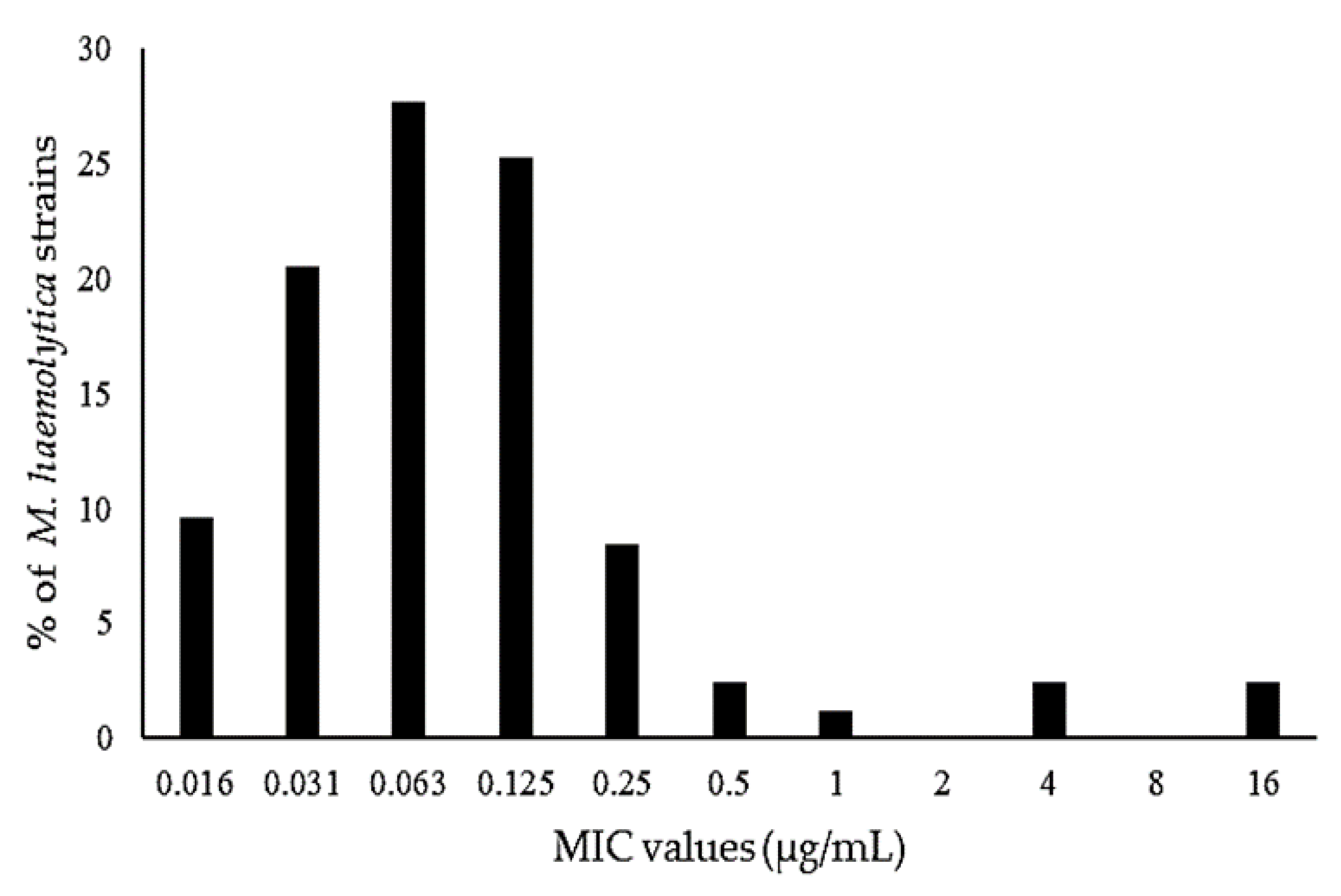
In this study, the MIC values of danofloxacin for the 83 M. haemolytica isolates isolated from the lambs ranged from 0.016 to >16 µg/mL. The percent MIC distribution of danofloxacin for the 83 isolates of M. haemolytica is presented in Figure 2. The percent MIC distribution for the concentrations of 0.016, 0.031, 0.063, 0.125, 0.25, 0.5, 1, 4, and 16 μg/mL was 9.6%, 20.5%, 27.7%, 25.3%, 8.4%, 2.4%, 1.2%, 2.4%, and 2.4%, respectively. There are no CLSI-approved breakpoint MIC values of danofloxacin for M. haemolytica isolated from sheep. The susceptible (0.25 µg/mL), intermediate (0.5 µg/mL), and resistant (1 µg/mL) MIC breakpoint values for M. haemolytica isolated from cattle have been determined [55,56]. For M. haemolytica isolated from the lambs, the MIC50 value was 0.063 µg/mL and the MIC90 value was 0.25 µg/mL, in which 91.6% of the population was below the susceptible breakpoint of 0.25 µg/mL.

3.4. Pharmacokinetic/Pharmacodynamic Integration

The PK/PD surrogate indices determined using the pharmacokinetic parameters obtained from the free concentrations of danofloxacin and the MIC values determined for the bacterial strains isolated from the lambs with respiratory infections are presented in Table 4. In the ID and IDM groups, the AUC0–24/MIC90 estimated using the in vitro MIC90 data (0.25 µg/mL) for M. haemolytica and the in vivo pharmacokinetic parameters of danofloxacin was 41.44 h and 59.21 h, while the ƒCmax/MIC90 was 9.69 and 14.40, respectively. In the ID and IDM groups, the ƒAUC0–24/MIC for the E. coli isolates ≤0.125 µg/mL was 82.89 h and 118.43 h, while the ƒCmax/MIC was 19.39 and 28.80, respectively. The PK/PD surrogate markers for the Streptococcus spp. isolates with an MIC value >16 µg/mL were not determined.

4. Discussion

4.1. Pharmacokinetics of Danofloxacin

Danofloxacin has been recommended for use at a dose of 6 mg/kg against M. haemolytica isolates with an MIC value of 0.25 μg/mL by PK/PD modeling in sheep [51,57]. In addition, the clinical efficacy of a 6 mg/kg dose of danofloxacin has been demonstrated in an experimental infection caused by M. haemolytica in lambs [17]. In this study, the injection route (SC) and dose (6 mg/kg) of danofloxacin administered to lambs were determined based on previous studies [17,50,56]. Meloxicam was administered at the approved dose (1 mg/kg) in sheep [20].
In this study, danofloxacin exhibited a similar t1/2ʎz in all groups, including the healthy and infected lambs. The t1/2ʎz is a hybrid parameter affected by changes in the Vd and Cl of the drug [58]. In this study, the Vdarea/F and ClT/F values of danofloxacin significantly decreased with the administration (HDM and IDM groups) of meloxicam and the effect (ID and IDM groups) of infection. However, the decrease in both the Vdarea/F and ClT/F values caused by the infection and the administration of meloxicam may be the reason for similar t1/2ʎz parameters in all groups. These results are also consistent with the results of studies in which the ClT and Vd of enrofloxacin decreased, but its t1/2 did not change [30,59].
In this study, the ClT/F of danofloxacin significantly decreased with the administration (HDM and IDM groups) of meloxicam and the effect (ID and IDM groups) of infection. The lower ClT/F of danofloxacin in the HDM (0.32 L/h·kg) and IDM (0.28 L/h·kg) groups compared to the HD (0.45 L/h·kg) and ID (0.38 L/h·kg) groups indicates that danofloxacin interacts with meloxicam. The elimination of danofloxacin and meloxicam has not been observed in sheep. The excretion of danofloxacin occurs approximately equally in urine and feces; while approximately 80% of the drug is removed unchanged, 20% of the drug is removed as a β-glucuronide conjugate of danofloxacin, desmethyldanofloxacin and danofloxacin-N-oxide, which are formed as a result of phase I and extensive phase II reactions [60]. Meloxicam is metabolized extensively in the liver through phase I reactions (conjugate derivative has not been determined) and less than <10% as unchanged meloxicam is excreted in the urine [61,62]. These data may indicate that danofloxacin and meloxicam interact primarily at the kidney level due to differences in their metabolism pathways. It is known that NSAIDs reduce the renal blood flow and glomerular filtration rate by inhibiting the synthesis of vasodilator prostaglandin (PG)E2 and PGI2, which play a role in maintaining normal kidney function [63,64]. PGs are synthesized in the kidneys by both COX-1 and COX-2 enzymes. Selective COX-2 inhibitors have been shown to have similar effects on renal hemodynamics as non-selective NSAIDs [65,66]. The effect of meloxicam, a selective COX-2 inhibitor, on the ClT/F of danofloxacin may be related to its decrease in renal blood flow and glomerular filtration rate.
Meloxicam is a substrate of BCRP, a member of the ATP-dependent efflux transporters [67,68]. Danofloxacin is the substrate of P-gp and MRP2, in addition to BCRP [16,43]. In sheep, ivermectin reduces the passage of danofloxacin entering the milk by a BCRP-mediated interaction (40%) [43]. Ciprofloxacin, a substrate of BCRP but not of P-gp or MRP2, reduces the secretion and absorption of danofloxacin in a Caco-2 cell model [16]. In this study, the decrease in the ClT/F of danofloxacin in the IDM and HDM groups may have resulted from the limitation of the excretion of danofloxacin due to a BCRP-mediated danofloxacin–meloxicam interaction in the excretion routes, such as the intestines, bile duct, or proximal tubule cells.
In this study, the lower ClT/F of danofloxacin in the ID (0.38 L/h·kg) and IDM (0.28 L/h·kg) groups than that in the HD (0.45 L/h·kg) and HDM (0.32 L/h·kg) groups indicates the effect of infection. These findings are consistent with other studies that reported a reduced ClT/F of danofloxacin, marbofloxacin, and tilmicosin in respiratory infections [69,70,71,72]. However, the ClT/F of marbofloxacin increased in lambs naturally infected with M. haemolytica [73]. The ClT of drugs is influenced by a variety of factors, particularly the blood flow to the excretion organs, the binding ratio of the drug to plasma proteins, and the activity of drug-metabolizing enzymes. These factors can be affected by sepsis and septic shock and may alter drug excretion and metabolism, leading to a reduced ClT [74,75]. It is reported that inflammation and infection could lead to downregulation of drug-metabolizing enzymes and transporters (primarily of the ABC superfamily), resulting in higher plasma concentrations and altering some distribution processes. Many inflammatory mediators (interleukines, cytokines, transforming growth factor, tumor necrosis factor or interferons) reduced the gene expression of cytochrome P450 complex in liver. On the other hand, inflammation and infection also downregulate drug efflux transporters ATP-binding cassette (ABC) superfamily. These transporters are ubiquitous in the organism and play a major role in drug transport of antimicrobials, among other drugs. A specific ABC transporter, the BCRP, and ABCG2 family transporter, is present in the human mammary gland and is responsible to excrete fluoroquinolones to milk. BCRP was present in mammary glands of animals including sheep and goats, and it is reported that danofloxacin is a substrate of BCRP, and inflammation and infection could lead to a downregulation of this transporter, affecting the disposition of the drug in the organism [76,77,78,79,80]. In lambs infected with M. haemolytica, the release of pro-inflammatory cytokines such as interleukin-1β, interleukin-8, and tumor necrosis factor-alpha has been shown to increase [81]. Pro-inflammatory cytokines reduce the activities of various cytochrome P450 and phase II enzymes, as well as the expression and function of transporter proteins such as P-gp, BCRP, and MRP2, which play an important role in the pharmacokinetics of drugs [82,83,84,85]. These effects have been reported as the cause for the decreased ClT of danofloxacin in infected animals [16,86]. In this study, the decreased ClT/F of danofloxacin in the ID group may be related to changes in blood flow into the excretion organs caused by infection and the decrease in the functions of phase II enzymes and the P-gp, BCRP, and MRP2 transporters. The partial decrease in the ClT/F of danofloxacin in the IDM group compared to the HDM group may be due to the therapeutic effect of meloxicam on the inflammation and fever caused by infection in the infected lambs.
In this study, the Vdarea/F and Vdss/F of danofloxacin were significantly decreased in the HDM and IDM groups compared to the HD group. These data show that the Vdarea/F and Vdss/F decreased with the administration of meloxicam (HDM and IDM). Similarly, the Vd of enrofloxacin and moxifloxacin has been shown to decrease with upon administration of some NSAIDs in healthy animals [30,31]. The protein binding ratio of danofloxacin in HD, HDM, ID and IDM groups were 32.0 ± 4.8%, 22.7 ± 3.1%, 32.8 ± 3.9% and 21.6 ± 4.0%, respectively. The binding ratio of meloxicam to plasma proteins is high (96% in dogs and 98% in calves) [60]. In this study, meloxicam decreased significantly the plasma protein binding of danofloxacin in HDM and IDM groups. A decrease in the binding ratio of danofloxacin to plasma proteins can be expected to cause an increased Vd. However, an increased concentration of danofloxacin in the plasma due to its reduced elimination by meloxicam can lead to a low calculation of Vd/F. Another reason for the decreased Vd/F in the HDM and IDM groups may be the limitation of the body disposition of danofloxacin, which is a substrate of P-gp, MRP2, and BCRP, by meloxicam [16,43,87]. Danofloxacin reaches 4 times higher concentrations in the lungs, bronchial mucosa, and bronchial secretions and 10 times higher in the milk than that in the plasma [12,57,88]. ABC transporters such as P-gp, MRP2, and especially BCRP are responsible for the high concentrations of danofloxacin in the lung tissues and milk [16,43]. The limitation of the excretion and passage of danofloxacin to highly accumulated tissues such as the lungs due to a BCRP-mediated danofloxacin–meloxicam interaction may be the reason for the decreased Vd/F in the HDM and IDM groups. In particular, given that the interest lies with the influence of meloxicam in the pharmacokinetics of danofloxacin, a more robust approach would be to also analyze the concentration–time profile of meloxicam to examine whether there is a potential concentration-dependent effect.
The rate of drug absorption from the site of administration is affected by conditions such as septicemia and septic shock, which affect the regional blood flow [74,89]. A systemic inflammatory response has been reported in pneumonia caused by M. haemolytica [81]. In this study, an increase (>40.5 °C) in body temperature, indicating the systemic effects of the infection, was determined in infected lambs. Therefore, the absorption rate of danofloxacin can be expected to change following SC administration in infected lambs. However, in this study, the Cmax/AUC and Tmax parameters used to evaluate the rate of drug absorption from the site of administration [90] were similar between the groups.
In this study, the Cmax of danofloxacin in the HD, HDM, ID, and IDM groups was 3.42, 3.76, 3.64, and 4.72 µg/mL, respectively. The administration of meloxicam (IDM) to infected lambs significantly increased the Cmax of danofloxacin compared to the HD group. The AUC of danofloxacin increased significantly in the HDM and IDM groups compared to in the HD and ID groups. The absorption extent, Cl, and Vd of a drug contribute to the formation of Cmax and AUC [91,92]. Danofloxacin, following SC administration, in sheep has been shown to achieve a high bioavailability of 94% [51,93]. In this study, the decreased Vdarea/F and ClT/F in the HDM and IDM groups may lead to the formation of a high Cmax and AUC. A high Cmax and AUC has been reported for enrofloxacin co-administered with flunixin meglumine and for danofloxacin in calves with respiratory tract infections [35,70]. Danofloxacin has been shown to demonstrate linear pharmacokinetics at doses of 1.25–10 mg/kg following SC administration and no toxic symptoms at doses of 10 and 20 mg/kg for six days or doses of 18 and 24 mg/kg for three days in calves [94]. In this study, a 6 mg/kg dose of danofloxacin in the IDM group created a high Cmax within the therapeutic range. This may have resulted in an increased therapeutic effect of danofloxacin in the IDM group.

4.2. Pharmacokinetic/Pharmacodynamic Integration

The PK/PD surrogate parameters showing the efficacy of fluoroquinolones are the ƒAUC0–24/MIC and ƒCmax/MIC ratios [95,96]. In the treatment of infections caused by Gram-negative bacteria, an ƒAUC0–24/MIC ratio should be 100–125 and a ƒCmax/MIC ratio should be 8–10 in order to achieve an ideal bactericidal effect and reduce the risk of resistance [97,98]. In in vitro studies, it has been shown that while fluoroquinolone dosages with a ƒCmax/MIC ratio of >3 cause a 99% reduction in the number of bacteria, dosages with a ƒCmax/MIC ratio of ≥8 are sufficient to prevent the emergence of resistant bacteria [99]. In the present study, the ƒAUC0–24/MIC and ƒCmax/MIC ratios were calculated only for the ID and IDM groups. In the ID and IDM groups, the ƒAUC0–24/MIC ratio of danofloxacin for susceptible E. coli isolates with an MIC value of 0.125 µg/mL was 82.89 h and 118.43 h, respectively, while the ƒCmax/MIC ratio was 19.39 and 28.80, respectively. Danofloxacin in the ID and IDM groups provided the ideal ƒCmax/MIC ratios following SC administration at a dose of 6 mg/kg. However, the ideal ƒAUC0–24/MIC ratio was obtained for the IDM group only.
A susceptible MIC breakpoint of ≤0.25 µg/mL for danofloxacin against M. haemolytica has been reported [56,100]. In this study, the MIC90 value of danofloxacin for M. haemolytica isolates isolated from lambs with respiratory infections was 0.25 µg/mL. In the ID and IDM groups, the ƒAUC0–24/MIC90 ratio was 41.44 h and 59.21 h, respectively, while the ƒCmax/MIC90 ratio was 9.69 and 14.40. Danofloxacin at a dose of 6 mg/kg achieved the ideal ƒCmax/MIC90 value of 8–10 but did not reach the ƒAUC0–24/MIC90 ratio of ≥125 h previously reported for fluoroquinolones [95,96]. However, studies have reported that the optimal ƒAUC0–24/MIC90 value is not an absolute value, and the AUC0–24/MIC90 value of danofloxacin, which can provide bacterial elimination of M haemolytica, is 2–4 times lower than 125 h [50,51,52,101]. The ideal ex vivo and in vivo AUC0–24/MIC values of danofloxacin for the elimination of M. haemolytica isolates from sheep have been reported to be 28.7 and 55.9 h, respectively [51]. When evaluated on the basis of these data, danofloxacin provided the ideal in vivo ƒAUC0–24/MIC90 ratio (55.9 h) for M. haemolytica isolates with MIC values of ≤0.25 µg/mL in the ID and IDM groups, but not in the ID group. Furthermore, previous studies have shown that danofloxacin in the lung tissues and bronchial mucosa reaches approximately 5- and 3-times higher concentrations, respectively, than those obtained in the plasma [11]. Therefore, in the present study, the concentration of danofloxacin obtained in the lung tissues and bronchial mucosa of the lambs in the ID group may have significantly exceeded the MIC values required for the elimination of M. haemolytica, which is an extracellular localization [70,102].

5. Conclusions

The administration of meloxicam and the presence of infection in lambs reduced the Vd/F and ClT/F of danofloxacin but did not affect the t1/2λz. Meloxicam increased the plasma concentration of danofloxacin in both healthy lambs and lambs with respiratory infections. In lambs with respiratory infections, an increased plasma concentration of danofloxacin within the therapeutic range may result in a better therapeutic effect. However, the clear definition of an increase in the therapeutic effect requires the determination of the concentration of danofloxacin in the target tissues and the effects of the resulting concentration on bacterial eradication. In addition, repeated doses of danofloxacin are required for the treatment of infection. A danofloxacin–meloxicam interaction may not be predicted following administration at repeated doses. Therefore, it is necessary to determine the pharmacokinetics of danofloxacin and meloxicam following the administration of repeated doses before simultaneous use in lambs with respiratory infections.
When co-administration with meloxicam, danofloxacin following SC administration at a dose of 6 mg/kg in lambs with respiratory infections provided optimum AUC0–24/MIC (>56 h) and Cmax/MIC (>8) values for susceptible M. haemolytica isolates with an MIC90 value of 0.25 µg/mL and for susceptible E. coli isolates with an MIC value of ≤0.125 µg/mL. However, due to bacterial isolation in a small number of samples in this study, the determination of pharmacodynamic data on bacterial agents isolated from more lambs with respiratory infections will be more valuable for the rational use of danofloxacin.

Author Contributions

M.N.U. and K.U. contributed to conception, experimental design, and analysis, drafted and critically revised the manuscript, provided final approval, and agreed to be accountable for all aspects of the work, ensuring integrity and accuracy. All authors have read and agreed to the published version of the manuscript.

Funding

This research was funded by the Coordination of Scientific Research Projects, University of Selcuk, Turkey, grant number 17202070.

Institutional Review Board Statement

The study protocol (2018/02) was approved by the Ethical Committee of the Faculty of Veterinary Medicine of Selcuk University, Turkey, and carried out in accordance with the European Directive (2010/63/UE).

Informed Consent Statement

Not applicable.

Data Availability Statement

The data presented in this study are available on request from the corresponding author.

Acknowledgments

This study was summarized from a PhD thesis.

Conflicts of Interest

The authors declare no conflict of interest.

References

  1. Goodwin, K.; Jackson, R.; Brown, C.; Davies, P.; Morris, R.; Perkins, N. Pneumonic lesions in lambs in New Zealand: Patterns of prevalence and effects on production. N. Z. Vet. J. 2004, 52, 175–179. [Google Scholar] [CrossRef]
  2. Lacasta, D.; Ferrer, L.; Ramos, J.; González, J.; De las Heras, M. Influence of climatic factors on the development of pneumonia in lambs. Small Rumin. Res. 2008, 80, 28–32. [Google Scholar] [CrossRef]
  3. Scott, P.R. Treatment and control of respiratory disease in sheep. Vet. Clin. N. Am. Small Anim. Pract. 2011, 27, 175–186. [Google Scholar] [CrossRef]
  4. Odugbo, M.; Odama, L.; Umoh, J.; Lamorde, A. Pasteurella multocida pneumonic infection in sheep: Prevalence, clinical and pathological studies. Small Rumin. Res. 2006, 66, 273–277. [Google Scholar] [CrossRef]
  5. Rawat, N.; Gilhare, V.R.; Kushwaha, K.K.; Hattimare, D.D.; Khan, F.F.; Shende, R.K.; Jolhe, D.K. Isolation and molecular characterization of Mannheimia haemolytica and Pasteurella multocida associated with pneumonia of goats in Chhattisgarh. Vet. World 2019, 12, 331. [Google Scholar] [CrossRef]
  6. Goodwin-Ray, K.A. Pneumonia and Pleurisy in Sheep: Studies of Prevalence, Risk Factors, Vaccine Efficacy and Economic Impact. Ph.D. Thesis, Massey University, Palmerston North, New Zealand, 2006. [Google Scholar]
  7. Estes, L. Review of pharmacokinetics and pharmacodynamics of antimicrobial agents. Mayo Clin. Proc. 1998, 73, 1114–1122. [Google Scholar] [CrossRef]
  8. Ulldemolins, M.; Roberts, J.A.; Lipman, J. Optimizing Antibiotic Use in the Intensive Care Unit. Clin. Pulm. Med. 2010, 17, 162–169. [Google Scholar] [CrossRef]
  9. EMA. Committee for Veterinary Medicinal Products. Danofloxacin Summary Report (2). 1997. Available online: https://www.ema.europa.eu/en/documents/mrl-report/danofloxacin-summary-report-2-committee-veterinary-medicinal-products_en.pdf (accessed on 27 April 2021).
  10. EMA. Committee for Veterinary Medicinal Products. Danofloxacin (Extension to All Food Producing Species) Summary Report (6). 2002. Available online: https://www.ema.europa.eu/en/documents/mrl-report/danofloxacin-extension-all-food-producing-species-summary-report-6-committee-veterinary-medicinal_en.pdf (accessed on 27 April 2021).
  11. Friis, C. Penetration of danofloxacin into the respiratory tract tissues and secretions in calves. Am. J. Vet. Res. 1993, 54, 1122–1127. [Google Scholar]
  12. Giles, C.J.; Magonigle, R.A.; Grimshaw, W.T.; Tanner, A.C.; Risk, J.E.; Lynch, M.J.; Rice, J.R. Clinical pharmacokinetics of parenterally administered danofloxacin in cattle. J. Vet. Pharmacol. Ther. 1991, 14, 400–410. [Google Scholar] [CrossRef]
  13. Riviere, J.E.; Papich, M.G. Fluoroquinolone Antimicrobial Drugs. In Veterinary Pharmacology and Therapeutics, 10th ed.; Papich, M.G., Ed.; JohnWiley & Sons, Inc.: Hoboken, NJ, USA, 2018; pp. 953–987. [Google Scholar]
  14. Stetsko, T.; Kocjumbas, I.; Muzyka, V.; Lisova, N.; Pyatnichko, O.; Ostrovska, L. Antimicrobial activity of danofloxacin, effectiveness and safeness of a new antimicrobial preparation based on its basis for treatment of calves with respiratory bacterial infections. Sci. Tech. Bull. State Sci. Res. Control Inst. Vet. Med. Prod. Fodd. Addit. Inst. Animal Biol. 2019, 20, 126–137. [Google Scholar] [CrossRef]
  15. Atef, M.; El-Gendi, A.Y.; Amer, M.M.; Abd El-Aty, A.M. Some pharmacokinetic data for danofloxacin in healthy goats. Vet. Res. Commun. 2001, 25, 367–377. [Google Scholar] [CrossRef]
  16. Schrickx, J.; Fink-Gremmels, J. Danofloxacin-mesylate is a substrate for ATP-dependent efflux transporters. Br. J. Pharmacol. 2007, 150, 463–469. [Google Scholar] [CrossRef] [Green Version]
  17. Godinho, K.; Benchaoui, H.; Tilt, N.; Ramage, C.; Quirie, M.; Donachie, W.; De La Puente-Redondo, V.; Rowan, T. Efficacy of danofloxacin in the treatment of pneumonic pasteurellosis in specific pathogen-free lambs. Vet. Rec. 2007, 160, 770–771. [Google Scholar] [CrossRef]
  18. Rowan, T.; Sunderland, S.; Smith, D.; Sarasola, P.; Giles, C. Efficacy of danofloxacin in the treatment of respiratory disease in European cattle. Vet. Rec. 2004, 154, 585–589. [Google Scholar] [CrossRef]
  19. EMA. Committee for Veterinary Medicinal Products. Meloxicam Summary Report (1). 1997. Available online: https://www.ema.europa.eu/en/documents/mrl-report/meloxicam-summary-report-1-committee-veterinary-medicinal-products_en.pdf (accessed on 27 April 2021).
  20. Sim, T. Meloxicam Product Approved for Sheep in Australia after World-First Tri-Lateral Review. sheepcentral.com. Available online: https://www.sheepcentral.com/meloxicam-product-approved-for-sheep-in-australia-after-world-first-tri-lateral-review/ (accessed on 29 July 2020).
  21. Bednarek, D.; Zdzisinska, B.; Kondracki, M.; Kandefer-Szerszen, M. Effect of steroidal and non-steroidal anti-inflammatory drugs in combination with long-acting oxytetracycline on non-specific immunity of calves suffering from enzootic bronchopneumonia. Vet. Microbiol. 2003, 96, 53–67. [Google Scholar] [CrossRef]
  22. Curry, S.L.; Cogar, S.M.; Cook, J.L. Nonsteroidal antiinflammatory drugs: A review. J. Am. Anim. Hosp. Assoc. 2005, 41, 298–309. [Google Scholar] [CrossRef]
  23. Ramírez-Romero, R.; Brogden, K.A.; Gallup, J.M.; Sonea, I.M.; Ackermann, M.R. Mast cell density and substance P-like immunoreactivity during the initiation and progression of lung lesions in ovineMannheimia (Pasteurella) haemolytica pneumonia. Microb. Pathog. 2001, 30, 325–335. [Google Scholar] [CrossRef] [Green Version]
  24. Lekeux, P. A therapeutic strategy for treatment of the bovine respiratory disease complex: The rationale for the combination of a nonsteroidal anti-inflammatory drug with an antibiotic. Cattle Pract. 2007, 15, 115–119. [Google Scholar]
  25. Lockwood, P.; Johnson, J.; Katz, T. Clinical efficacy of flunixin, carprofen and ketoprofen as adjuncts to the antibacterial treatment of bovine respiratory disease. Vet. Rec. 2003, 152, 392–394. [Google Scholar] [CrossRef]
  26. Bednarek, D.; Kondracki, M.; Friton, G.; Trela, T.; Niemczuk, K. Effect of steroidal and non-steroidal anti-inflammatory drugs on inflammatory markers in calves with experimentally-induced bronchopneumonia. Berl. Munch. Tierarztl. Wochenschr. 2005, 118, 305–308. [Google Scholar]
  27. Friton, G.; Cajal, C.; Ramirez-Romero, R. Long-term effects of meloxicam in the treatment of respiratory disease in fattening cattle. Vet. Rec. 2005, 156, 809–811. [Google Scholar] [CrossRef] [PubMed]
  28. Jager, N.G.; van Hest, R.M.; Lipman, J.; Taccone, F.S.; Roberts, J.A. Therapeutic drug monitoring of anti-infective agents in critically ill patients. Expert Rev. Clin. Pharmacol. 2016, 1–19. [Google Scholar] [CrossRef] [PubMed]
  29. Roberts, J.A.; Norris, R.; Paterson, D.L.; Martin, J.H. Therapeutic drug monitoring of antimicrobials. Br. J. Clin. Pharmacol. 2012, 73, 27–36. [Google Scholar] [CrossRef] [PubMed] [Green Version]
  30. Abo-EL-Sooud, K.; Al-Anati, L. Pharmacokinetic study of flunixin and its interaction with enrofloxacin after intramuscular administration in calves. Vet. World 2011, 4. [Google Scholar] [CrossRef]
  31. Altan, F.; Corum, O.; Yildiz, R.; Eser Faki, H.; Ider, M.; Ok, M.; Uney, K. Intravenous pharmacokinetics of moxifloxacin following simultaneous administration with flunixin meglumine or diclofenac in sheep. J. Vet. Pharmacol. Ther. 2020, 43, 108–114. [Google Scholar] [CrossRef] [PubMed]
  32. Baroni, E.; Rubio, S.; Rodríguez, C.; De Lucas, J.; Fernández, H.; Andrés, M. Pharmacokinetic interactions of marbofloxacin with anti-inflammatory drugs in buffalo calves. Vet. Rec. 2011. [Google Scholar] [CrossRef]
  33. Durna Corum, D.; Corum, O.; Yildiz, R.; Eser Faki, H.; Ider, M.; Cetin, G.; Uney, K. Influences of tolfenamic acid and flunixin meglumine on the disposition kinetics of levofloxacin in sheep. Acta Vet. Hung. 2020, 68, 65–70. [Google Scholar] [CrossRef]
  34. Ogino, T.; Arai, T. Pharmacokinetic interactions of flunixin meglumine and enrofloxacin in ICR mice. Exp. Anim. 2007, 56, 79–84. [Google Scholar] [CrossRef] [Green Version]
  35. Ogino, T.; Mizuno, Y.; Ogata, T.; Takahashi, Y. Pharmacokinetic interactions of flunixin meglumine and enrofloxacin in dogs. Am. J. Vet. Res. 2005, 66, 1209–1213. [Google Scholar] [CrossRef]
  36. Tekeli, I.O.; Turk, E.; Durna Corum, D.; Corum, O.; Kirgiz, F.C.; Sakin, F.; Uney, K. Effect of ketoprofen co-administration on pharmacokinetics of cefquinome following repeated administration in goats. J. Vet. Pharmacol. Ther. 2020, 43, 440–447. [Google Scholar] [CrossRef]
  37. Elmas, M.; Yazar, E.; Uney, K.; Er, A.; Traş, B. Pharmacokinetics of enrofloxacin and flunixin meglumine and interactions between both drugs after intravenous co-administration in healthy and endotoxaemic rabbits. Vet. J. 2008, 177, 418–424. [Google Scholar] [CrossRef]
  38. Koc, F.; Atila, A.; Karakuş, E.; Üney, K. Influences of flunixin and tenoxicam on the pharmacokinetics of florfenicol in lipopolysaccharide-induced endotoxemia. Turk. J. Vet. Anim. Sci. 2015, 39, 168–173. [Google Scholar] [CrossRef]
  39. Sadariya, K.; Patel, J.; Bhavsar, S.; Thaker, A. Effect of Febrile Condition and Ketoprofen Co-administration on Pharmacokinetics of Moxifloxacin Following Intravenous Administration in Sheep. Isr. J. Vet. Med. 2014, 69, 68–73. [Google Scholar]
  40. Christodoulopoulos, G.; Warnick, L.; Papaioannou, N.; Fthenakis, G. Tilmicosin administration to young lambs with respiratory infection: Safety and efficacy considerations. J. Vet. Pharmacol. Ther. 2002, 25, 393–397. [Google Scholar] [CrossRef] [PubMed]
  41. Corum, O.; Altan, F.; Yildiz, R.; Ider, M.; Ok, M.; Uney, K. Pharmacokinetics of enrofloxacin and danofloxacin in premature calves. J. Vet. Pharmacol. Ther. 2019, 42, 624–631. [Google Scholar] [CrossRef]
  42. Corum, O.; Durna Corum, D.; Atik, O.; Eser Faki, H.; Altan, F.; Uney, K. Pharmacokinetics and bioavailability of danofloxacin in chukar partridge (Alectoris chukar) following intravenous, intramuscular, subcutaneous, and oral administrations. J. Vet. Pharmacol. Ther. 2019, 42, 207–213. [Google Scholar] [CrossRef] [PubMed]
  43. Real, R.; Egido, E.; Perez, M.; Gonzalez-Lobato, L.; Barrera, B.; Prieto, J.; Alvarez, A.; Merino, G. Involvement of breast cancer resistance protein (BCRP/ABCG2) in the secretion of danofloxacin into milk: Interaction with ivermectin. J. Vet. Pharmacol. Ther. 2011, 34, 313–321. [Google Scholar] [CrossRef] [PubMed]
  44. Ruennarong, N.; Wongpanit, K.; Sakulthaew, C.; Giorgi, M.; Kumagai, S.; Poapolathep, A.; Poapolathep, S. Dispositions of enrofloxacin and its major metabolite ciprofloxacin in Thai swamp buffaloes. J. Vet. Med. Sci. 2015, 15-0464. [Google Scholar] [CrossRef] [PubMed]
  45. Gibaldi, M.; Perrier, D. Noncompartmental analysis based on statistical moment theory. Pharmacokinetics 1982, 2, 409–417. [Google Scholar]
  46. Greko, C.; Finn, M.; Franklin, A.; Bengtsson, B. Pharmacokinetic/pharmacodynamic relationship of danofloxacin against Mannheimia haemolytica in a tissue-cage model in calves. J. Antimicrob. Chemother. 2003, 52, 253–257. [Google Scholar] [CrossRef] [PubMed] [Green Version]
  47. Quinn, P.J.; Markey, B.K.; Leonard, F.C.; Hartigan, P.; Fanning, S.; Fitzpatrick, E. Veterinary Microbiology and Microbial Disease; John Wiley & Sons: Hoboken, NJ, USA, 2011; pp. 300–308. [Google Scholar]
  48. CLSI. Methods for Dilution Antimicrobial Susceptibility Tests for Bacteria That Grow Aerobically; Approved Standard, 9th ed.; Clinical and Laboratory Standards Institute: Wyne, PA, USA, 2012. [Google Scholar]
  49. Schwarz, S.; Silley, P.; Simjee, S.; Woodford, N.; van Duijkeren, E.; Johnson, A.P.; Gaastra, W. Assessing the antimicrobial susceptibility of bacteria obtained from animals. J. Antimicrob. Chemother. 2010, 65, 601–604. [Google Scholar] [CrossRef] [PubMed]
  50. Aliabadi, F.S.; Ali, B.H.; Landoni, M.; Lees, P. Pharmacokinetics and PK-PD modelling of danofloxacin in camel serum and tissue cage fluids. Vet. J. 2003, 165, 104–118. [Google Scholar] [CrossRef]
  51. Aliabadi, F.S.; Landoni, M.F.; Lees, P. Pharmacokinetics (PK), pharmacodynamics (PD), and PK-PD integration of danofloxacin in sheep biological fluids. Antimicrob. Agents Chemother. 2003, 47, 626–635. [Google Scholar] [CrossRef] [Green Version]
  52. Aliabadi, F.S.; Lees, P. Pharmacokinetics and pharmacodynamics of danofloxacin in serum and tissue fluids of goats following intravenous and intramuscular administration. Am. J. Vet. Res. 2001, 62, 1979–1989. [Google Scholar] [CrossRef] [PubMed]
  53. Haritova, A.M.; Rusenova, N.V.; Parvanov, P.R.; Lashev, L.D.; Fink-Gremmels, J. Pharmacokinetic-pharmacodynamic modelling of danofloxacin in turkeys. Vet. Res. Commun. 2006, 30, 775–789. [Google Scholar] [CrossRef] [PubMed]
  54. Yang, Y.; Zhang, Y.; Li, J.; Cheng, P.; Xiao, T.; Muhammad, I.; Yu, H.; Liu, R.; Zhang, X. Susceptibility breakpoint for Danofloxacin against swine Escherichia coli. BMC Vet. Res. 2019, 15, 51. [Google Scholar] [CrossRef]
  55. CLSI. Performance Standards for Antimicrobial Disk Susceptibility Tests for Bacteria Isolated from Animals: CLSI Supplement VET01S, 3rd ed.; Clinical and Laboratory Standards Institute: Wyne, PA, USA, 2015. [Google Scholar]
  56. Sweeney, M.T.; Papich, M.G.; Watts, J.L. New interpretive criteria for danofloxacin antibacterial susceptibility testing against Mannheimia haemolytica and Pasteurella multocida associated with bovine respiratory disease. J. Vet. Diagn. Investig. 2017, 29, 224–227. [Google Scholar] [CrossRef]
  57. Escudero, E.; Cárceles, C.; Fernandez-Varon, E.; Marin, P.; Benchaoui, H. Pharmacokinetics of danofloxacin 18% in lactating sheep and goats. J. Vet. Pharmacol. Ther. 2007, 30, 572–577. [Google Scholar] [CrossRef]
  58. Toutain, P.-L.; Bousquet-Mélou, A. Plasma clearance. J. Vet. Pharmacol. Ther. 2004, 27, 415–425. [Google Scholar] [CrossRef]
  59. Elmas, M.; Yazar, E.; Uney, K.; Er, A. Influence of Escherichia coli endotoxin-induced endotoxaemia on the pharmacokinetics of enrofloxacin after intravenous administration in rabbits. J. Vet. Med. A 2006, 53, 410–414. [Google Scholar] [CrossRef]
  60. EMA. Committee for Veterinary Medicinal Products. Danofloxacin (Extension to Pig) Summary Report (1). 1998. Available online: https://www.ema.europa.eu/en/documents/mrl-report/danofloxacin-extension-pigs-summary-report-1-committee-veterinary-medicinal-products_en.pdf (accessed on 27 April 2021).
  61. EMA. Committee for Veterinary Medicinal Products. Meloxicam (Extrapolation to Rabbits and Goats) Summary Report (7). 2006. Available online: https://www.ema.europa.eu/en/documents/mrl-report/meloxicam-extrapolation-rabbits-goats-summary-report-7-committee-veterinary-medicinal-products_en.pdf (accessed on 27 April 2021).
  62. Türck, D.; Roth, W.; Busch, U. A review of the clinical pharmacokinetics of meloxicam. Rheumatology 1996, 35, 13–16. [Google Scholar] [CrossRef] [Green Version]
  63. Ejaz, P.; Bhojani, K.; Joshi, V. NSAIDs and kidney. J. Assoc. Physicians India 2004, 52, 371. [Google Scholar]
  64. Kim, G.-H. Renal effects of prostaglandins and cyclooxygenase-2 inhibitors. Electrolyte Blood Press. 2008, 6, 35–41. [Google Scholar] [CrossRef] [PubMed] [Green Version]
  65. Gambaro, G.; Perazella, M. Adverse renal effects of anti-inflammatory agents: Evaluation of selective and nonselective cyclooxygenase inhibitors. J. Intern. Med. 2003, 253, 643–652. [Google Scholar] [CrossRef] [Green Version]
  66. Pehlivan, B.; Cuvaş, Ö.; Başar, H.; Bakir, F.; Üstün, H.; Dikmen, B. Comparison of the effects of repeated dose treatments of lornoxicam and meloxicam on renal functions in rats. Turk. J. Med. Sci. 2010, 40, 371–376. [Google Scholar] [CrossRef]
  67. Garcia-Lino, A.M.; Blanco-Paniagua, E.; Astorga-Simon, E.N.; Alvarez-Fernandez, L.; Garcia-Mateos, D.; Alvarez-Fernandez, I.; Alvarez, A.I.; Merino, G. Abcg2 transporter affects plasma, milk and tissue levels of meloxicam. Biochem. Pharmacol. 2020, 175, 113924. [Google Scholar] [CrossRef] [PubMed]
  68. Gates, B.J.; Nguyen, T.T.; Setter, S.M.; Davies, N.M. Meloxicam: A reappraisal of pharmacokinetics, efficacy and safety. Expert Opin. Pharmacother. 2005, 6, 2117–2140. [Google Scholar] [CrossRef]
  69. Ismail, M.; El-Kattan, Y. Comparative pharmacokinetics of marbofloxacin in healthy and Mannheimia haemolytica infected calves. Res. Vet. Sci. 2007, 82, 398–404. [Google Scholar] [CrossRef]
  70. Mzyk, D.A.; Bublitz, C.M.; Martinez, M.N.; Davis, J.L.; Baynes, R.E.; Smith, G.W. Impact of bovine respiratory disease on the pharmacokinetics of danofloxacin and tulathromycin in different ages of calves. PLoS ONE 2019, 14. [Google Scholar] [CrossRef]
  71. Soliman, A.M.; Ayad, A.R.A. Pharmacokinetics and efficacy of tilmicosin in the treatment of Pasteurella haemolytica bronchopneumonia in calves. Pharmacol. Pharm. 2014, 2014. [Google Scholar] [CrossRef] [Green Version]
  72. Xiao, X.; Lan, W.; Wang, Y.; Jiang, L.; Jiang, Y.; Wang, Z. Comparative pharmacokinetics of danofloxacin in healthy and Pasteurella multocida infected ducks. J. Vet. Pharmacol. Ther. 2018, 41, 912–918. [Google Scholar] [CrossRef]
  73. Altan, F.; Ipek, D.N.S.; Corum, O.; Alp, S.Y.; Ipek, P.; Uney, K. The effects of Mannheimia haemolytica and albendazole on marbofloxacin pharmacokinetics in lambs. Trop. Anim. Health Prod. 2019, 51, 2603–2610. [Google Scholar] [CrossRef]
  74. De Paepe, P.; Belpaire, F.M.; Buylaert, W.A. Pharmacokinetic and pharmacodynamic considerations when treating patients with sepsis and septic shock. Clin. Pharmacokinet. 2002, 41, 1135–1151. [Google Scholar] [CrossRef]
  75. Martinez, M.; Modric, S. Patient variation in veterinary medicine: Part I. Influence of altered physiological states. J. Vet. Pharmacol. Ther. 2010, 33, 213–226. [Google Scholar] [CrossRef]
  76. Schmith, V.D.; Foss, J.F. Effects of inflammation on pharmacokinetics/pharmacodynamics: Increasing recognition of its contribution to variability in response. Clin. Pharmacol. Ther. 2008, 83, 809–811. [Google Scholar] [CrossRef]
  77. Renton, K.W. Regulation of drug metabolism and disposition during inflammation and infection. Expert Opin. Drug Metab. Toxicol. 2005, 1, 629–640. [Google Scholar] [CrossRef]
  78. Aitken, A.E.; Richardson, T.A.; Morgan, E.T. Regulation of drug-metabolizing enzymes and transporters in inflammation. Annu. Rev. Pharmacol. Toxicol. 2006, 46, 123–149. [Google Scholar] [CrossRef]
  79. Petrovic, V.; Teng, S.; Piquette-Miller, M. Regulation of drug transporters during infection and inflammation. Mol. Interv. 2007, 7, 99–111. [Google Scholar] [CrossRef] [PubMed]
  80. Morgan, E.T.; Goralski, K.B.; Piquette-Miller, M.; Renton, K.W.; Robertson, G.R.; Chaluvadi, M.R.; Charles, K.A.; Clarke, S.J.; Kacevska, M.; Liddle, C.; et al. Regulation of drug-metabolizing enzymes and transporters in infection, inflammation, and cancer. Drug Metab. Dispos. 2008, 36, 205–216. [Google Scholar] [CrossRef] [PubMed] [Green Version]
  81. Redondo, E.; Gázquez, A.; García, A.; Vadillo, S.; Masot, A. Dominant expression of interleukin-8 vs interleukin-1 β and tumour necrosis factor alpha in lungs of lambs experimentally infected with Mannheimia haemolytica. N. Z. Vet. J. 2011, 59, 225–232. [Google Scholar] [CrossRef] [PubMed]
  82. Fernandez, C.; Buyse, M.; German-Fattal, M.; Gimenez, F. Influence of the pro-inflammatory cytokines on P-glycoprotein expression and functionality. J. Pharm. Pharm. Sci. 2004, 7, 359–371. [Google Scholar]
  83. Kalitsky-Szirtes, J.; Shayeganpour, A.; Brocks, D.; Piquette-Miller, M. Suppression of drug-metabolizing enzymes and efflux transporters in the intestine of endotoxin-treated rats. Drug Metab. Dispos. 2004, 32, 20–27. [Google Scholar] [CrossRef] [PubMed] [Green Version]
  84. Mimche, S.M.; Nyagode, B.A.; Merrell, M.D.; Lee, C.-M.; Prasanphanich, N.S.; Cummings, R.D.; Morgan, E.T. Hepatic cytochrome P450s, phase II enzymes and nuclear receptors are downregulated in a Th2 environment during Schistosoma mansoni infection. Drug Metab. Dispos. 2014, 42, 134–140. [Google Scholar] [CrossRef] [PubMed] [Green Version]
  85. Poller, B.; Drewe, J.; Krähenbühl, S.; Huwyler, J.; Gutmann, H. Regulation of BCRP (ABCG2) and P-glycoprotein (ABCB1) by cytokines in a model of the human blood–brain barrier. Cell Mol. Neurobiol. 2010, 30, 63–70. [Google Scholar] [CrossRef]
  86. Lindecrona, R.; Friis, C.; Nielsen, J. Pharmacokinetics and penetration of danofloxacin into the gastrointestinal tract in healthy and in Salmonella typhimurium infected pigs. Res. Vet. Sci. 2000, 68, 211–216. [Google Scholar] [CrossRef]
  87. Marquez, B.; Van Bambeke, F. ABC multidrug transporters: Target for modulation of drug pharmacokinetics and drug-drug interactions. Curr. Drug Targets 2011, 12, 600–620. [Google Scholar] [CrossRef]
  88. Shem-Tov, M.; Ziv, G.; Glickman, A.; Saran, A. Pharmacokinetics and penetration of danofloxacin from the blood into the milk of ewes. Vet. Res. 1997, 28, 571–579. [Google Scholar]
  89. Charlton, M.; Thompson, J. Pharmacokinetics in sepsis. BJA Educ. 2018, 19, 7–13. [Google Scholar] [CrossRef] [Green Version]
  90. Endrenyi, L.; Fritsch, S.; Yan, W. Cmax/AUC is a clearer measure than Cmax for absorption rates in investigations of bioequivalence. Int. J. Clin. Pharmacol. Ther. Toxicol. 1991, 29, 394–399. [Google Scholar] [PubMed]
  91. Duan, J.Z. Drug-drug interaction pattern recognition. Drugs R & D 2010, 10, 9–24. [Google Scholar] [CrossRef]
  92. Lea-Henry, T.N.; Carland, J.E.; Stocker, S.L.; Sevastos, J.; Roberts, D.M. Clinical pharmacokinetics in kidney disease: Fundamental principles. Clin. J. Am. Soc. Nephrol. 2018, 13, 1085–1095. [Google Scholar] [CrossRef] [PubMed]
  93. McKellar, Q.A.; Gibson, I.F.; McCormack, R.Z. Pharmacokinetics and tissue disposition of danofloxacin in sheep. Biopharm. Drug Dispos. 1998, 19, 123–129. [Google Scholar] [CrossRef]
  94. DailyMed, L. ADVOCIN-danofloxacin Mesylate Injection, Solution Zoetis Inc. Available online: https://dailymed.nlm.nih.gov/dailymed/fda/fdaDrugXsl.cfm?setid=d22c22fc-d530-423c-8d78-ca95fe195ba1&type=display (accessed on 1 December 2020).
  95. Giguère, S.; Dowling, P.M. Fluoroquinolones. Antimicrob. Ther. Vet. Med. 2013, 295–314. [Google Scholar] [CrossRef]
  96. Lode, H.; Borner, K.; Koeppe, P. Pharmacodynamics of fluoroquinolones. Clin. Infect. Dis. 1998, 27, 33–39. [Google Scholar] [CrossRef] [PubMed] [Green Version]
  97. Sidhu, P.; Landoni, M.; Aliabadi, F.; Lees, P. PK–PD integration and modeling of marbofloxacin in sheep. Res. Vet. Sci. 2010, 88, 134–141. [Google Scholar] [CrossRef] [PubMed]
  98. Toutain, P.-L.; del Castillo, J.R.; Bousquet-Mélou, A. The pharmacokinetic–pharmacodynamic approach to a rational dosage regimen for antibiotics. Res. Vet. Sci. 2002, 73, 105–114. [Google Scholar] [CrossRef]
  99. Blaser, J.; Stone, B.B.; Groner, M.C.; Zinner, S.H. Comparative study with enoxacin and netilmicin in a pharmacodynamic model to determine importance of ratio of antibiotic peak concentration to MIC for bactericidal activity and emergence of resistance. Antimicrob. Agents Chemother. 1987, 31, 1054–1060. [Google Scholar] [CrossRef] [Green Version]
  100. CLSI. Development of In Vitro Susceptibility Testing Criteria and Quality Control Parameters for Veterinary Antimicrobial Agents; Approved Guideline, 3rd ed.; Clinical and Laboratory Standards Institute: Wyne, PA, USA, 2008. [Google Scholar]
  101. Jackson, J.; Davidson, J.; Ter Hune, T.; Magonigle, R. A dose response study of the fluoroquinolone, danofloxacin, against induced bovine pneumonic pasteurellosis. In Proceedings of the XVI World Buiatrics Congress, Salvador, Bahia, Brazil, 13–17 August 1990. [Google Scholar]
  102. TerHune, T.N.; Skogerboe, T.L.; Shostrom, V.K.; Weigel, D.J. Comparison of pharmacokinetics of danofloxacin and enrofloxacin in calves challenged with Mannheimia haemolytica. Am. J. Vet. Res. 2005, 66, 342–349. [Google Scholar] [CrossRef]
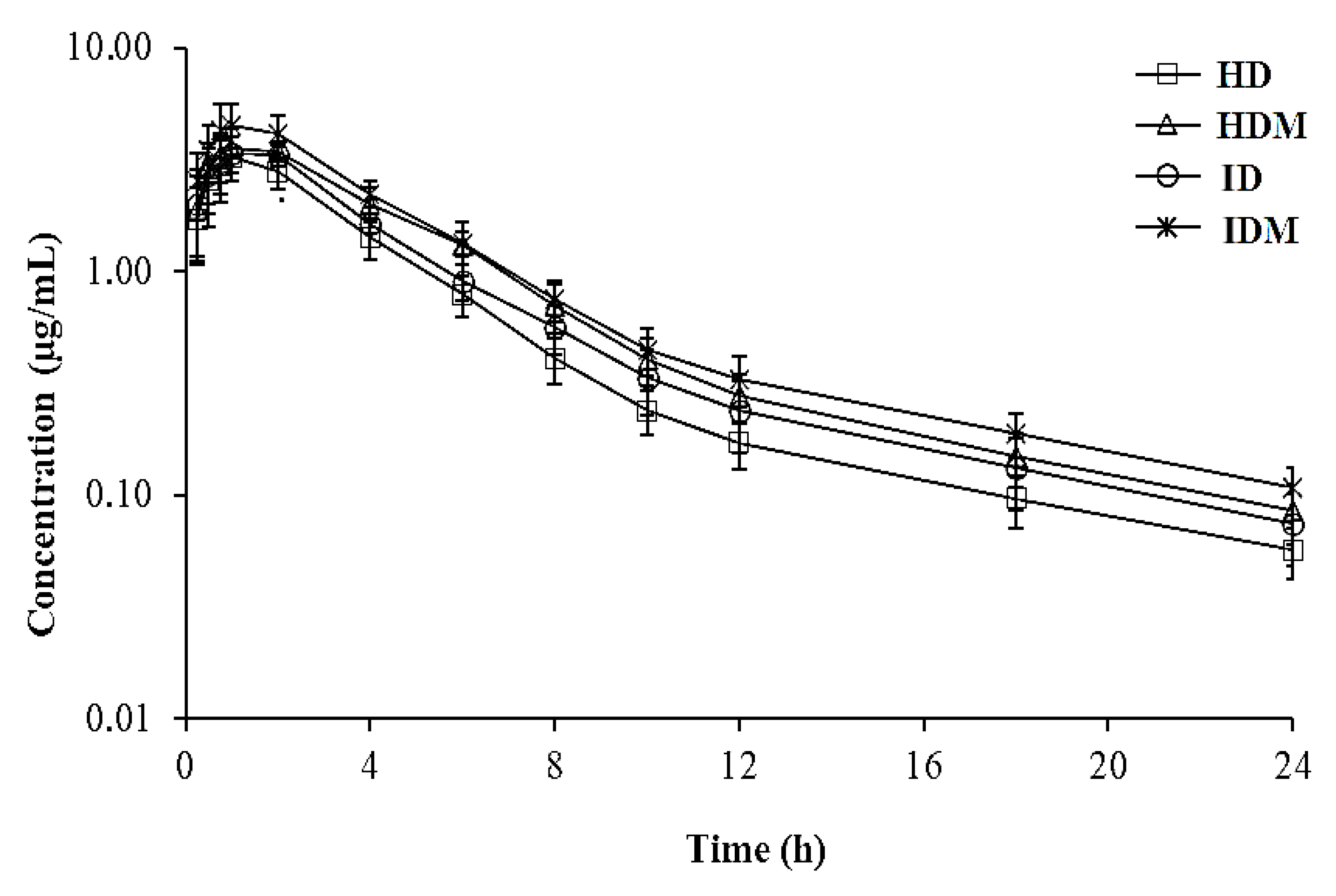
Figure 1. Semi-logarithmic plasma concentration–time curves after subcutaneous administration of danofloxacin alone or co-administered with meloxicam in healthy lambs (n = 6) and lambs with respiratory infections (n = 7). HD, healthy group who received danofloxacin alone; HDM, healthy group who received danofloxacin and meloxicam; ID, infected group who received danofloxacin alone; IDM, infected group who received danofloxacin and meloxicam. Data are presented as mean ± SD.
Antibiotics 10 01190 g001
Figure 2. Minimum inhibitory concentration (MIC) distribution of danofloxacin in the 83 strains of M. haemolytica. The MIC50 and MIC90 were set at 0.063 and 0.25 μg/mL, respectively.
Antibiotics 10 01190 g002
Table 1. The mean (±SD) age, body weight, rectal temperature, and respiratory rate of the healthy lambs (n = 6) and those with respiratory infections (n = 7) selected for the pharmacokinetic study. HD, healthy group who received danofloxacin alone; HDM, healthy group who received danofloxacin and meloxicam; ID, infected group who received danofloxacin alone; IDM, infected group who received danofloxacin and meloxicam.
ParametersHDHDMIDIDM
Age (month)3.20 ± 0.233.25 ± 0.363.50 ± 0.303.54 ± 0.51
Body weight (kg)27.50 ± 3.0328.17 ± 3.2728.86 ± 3.0827.86 ± 3.44
Rectal temperature (°C)39.77 ± 0.1539.93 ± 0.1241.01 ± 0.2241.04 ± 0.22
Respiratory rate (breath/min)42.17 ± 3.6342.83 ± 2.7961.00 ± 3.2762.43 ± 3.46
Table 2. The pharmacokinetic parameters of danofloxacin (6 mg/kg; SC) alone or co-administered with meloxicam (1 mg/kg; SC) in healthy lambs (n = 6) and lambs with respiratory infections (n = 7). t1/2ʎz, terminal half-life; ClT/F, total clearance; Vdarea/F, volume of distribution; Vdss/F, volume of distribution in a steady state; Tmax, time to reach the peak concentration; Cmax, peak plasma concentration; AUC, area under the concentration–time curve; MRT, mean residence time; HD, healthy group who received danofloxacin alone; HDM, healthy group who received danofloxacin and meloxicam; ID, infected group who received danofloxacin alone; IDM, infected group who received danofloxacin and meloxicam.
ParametersHDHDMIDIDM
t1/2ʎz (h)6.98 (5.75–8.15)6.46 (4.82–8.24)6.07 (5.01–7.48)6.73 (5.14–8.12)
ClT/F (L/h·kg)0.45 (0.39–0.51) a0.32 (0.28–0.36) b,c0.38 (0.31–0.42) b0.28 (0.21–0.31) c
Vdarea/F (L/kg)4.55 (3.52–5.40) a3.02 (2.01–4.18) b3.35 (2.98–4.20) a,b2.73 (1.94–3.44) b
Vdss/F (L/kg)2.46 (1.94–3.23) a1.88 (1.39–2.24) b2.12 (1.61–2.51) a,b1.70 (1.23–2.21) b
Tmax (h)1.01 (0.75–2.00)1.25 (1.00–2.00)1.34 (1.00–2.00)1.23 (0.75–2.00)
Cmax (µg/mL)3.36 (2.46–4.46) b3.73 (2.89–4.30) a,b3.61 (2.97–4.46) a,b4.59 (3.29–6.44) a
AUC0–24 (h.µg/mL)12.68 (11.04–14.61) c17.69 (15.94–20.32) a,b14.99 (13.59–17.97) b,c20.23 (18.37–25.93) a
AUC0–∞ (h·µg/mL)13.26 (11.63–15.26) c18.52 (16.56–20.73) a,b15.65 (13.96–19.06) b,c21.30 (19.19–27.29) a
Cmax/AUC0.25 (0.20–30)0.20 (0.15–0.25)0.23 (0.17–0.29)0.22 (0.17–27)
MRT (h)5.43 (4.54–6.27)5.81 (4.79–6.95)5.54 (3.86–6.78)6.02 (4.72–7.51)
Data are presented as the geometric mean (min–max) and were analyzed by two-way ANOVA with Tukey’s test. a,b,c Different letters in the same row are statistically different (p < 0.05).
Table 3. Protein binding ratio (%) of danofloxacin alone or in combination with meloxicam in plasma samples of healthy lambs and lambs with respiratory infections. PBHD, healthy group who was spiked with danofloxacin alone; PBHDM, healthy group who was spiked with danofloxacin and meloxicam; PBID, infected group who was spiked danofloxacin alone; PBIDM, infected group who was spiked danofloxacin and meloxicam.
Danofloxacin Concentration (µg/mL)PBHDPBHDMPBIDPBIDM
0.0536.3 ± 2.1 a24.3 ± 2.1 b36.0 ± 3.6 a22.0 ± 3.0 b
0.532.0 ± 4.6 a21.0 ± 3.0 b33.0 ± 1.0 a24.3 ± 3.8 a,b
5.027.7 ± 3.1 a,*22.6 ± 4.0 a,b29.3 ± 3.5 a18.3 ± 3.5 b
Mean ± SD32.0 ± 4.8 a22.7 ± 3.1 b32.8 ± 3.9 a21.6 ± 4.0 b
Data are presented as the mean ± SD and were analyzed by three-way ANOVA with Tukey’s test. * In PBHD group, the protein binding ratio at the concentration of 5 µg/mL is statistically lower than that at the concentration of 0.05 µg/mL (p < 0.05). a,b Different letters in the same row are statistically different (p < 0.05).
Table 4. The PK/PD surrogate indices determined using the pharmacokinetic parameters obtained from the free concentrations of danofloxacin and the MIC values determined for the bacterial strains isolated from the lambs with respiratory infections. MIC, minimum inhibitor concentration; AUC, area under the concentration–time curve; Cmax, peak plasma concentration; GM, geometric mean; ID, infected group who received danofloxacin alone; IDM, infected group who received danofloxacin and meloxicam.
Animal NoƒAUC0–24/MICƒCmax/MIC
IDIDMIDIDM
Mannheimia haemolytica, MIC90 of 0.25 µg/mL
138.4257.627.9910.32
237.8344.9810.8913.35
336.5081.319.3420.21
429.7059.9511.9911.26
550.4256.3710.4319.17
659.9758.858.6614.76
744.0860.909.1514.45
GM (min–max)41.44 (29.70–59.97)59.21 (44.98–81.31)9.69 (7.99–11.99)14.40 (10.32–20.21)
Escherichia coli, MIC of 0.125 µg/mL
176.84115.2315.9820.65
275.6689.9621.7826.69
373.01162.6318.6940.42
459.40119.8923.9822.53
5100.83112.7420.8638.33
6119.94117.7117.3129.53
788.16121.8018.3028.90
GM (min–max)82.89 (59.40–119.94)118.43 (89.96–162.63)19.39 (15.98–23.98)28.80 (20.65–40.42)
Publisher’s Note: MDPI stays neutral with regard to jurisdictional claims in published maps and institutional affiliations.