Article

Hydroxylated-Benz[a]anthracenes Induce Two Apoptosis-Related Gene Expressions in the Liver of the Nibbler Fish Girella punctata

by
Muhammad Ahya Rafiuddin
1,
Hajime Matsubara
1,*,
Kaito Hatano
2,
Masato Honda
3,
Kenji Toyota
4,
Kouhei Kuroda
2,
Keito Tsunoda
2,
Yukihiro Furusawa
5,
Yoshiaki Tabuchi
6,
Tetsushi Hirano
6,
Akihiro Sakatoku
7,
Chun-Sang Hong
8,
Ajai K. Srivastav
9,
Thumronk Amornsakun
10,
Nobuaki Shimizu
2,
Mohamed I. Zanaty
11,
Tatsuo Harumi
12,
Kohei Yamauchi
2,
Tamás Müller
13,
Ning Tang
14,
Atsuhiko Hattori
15,
Kazuichi Hayakawa
16 and
Nobuo Suzuki
2
add Show full author list remove Hide full author list
1
Noto Center for Fisheries Science and Technology, Kanazawa University, Ossaka, Noto-cho, Ishikawa 927-0552, Japan
2
Noto Marine Laboratory, Institute of Nature and Environmental Technology, Kanazawa University, Ogi, Noto-cho, Ishikawa 927-0553, Japan
3
Botanical Garden, Institute of Nature and Environmental Technology, Kanazawa University, Kanazawa, Ishikawa 920-1192, Japan
4
Department of Bioresource Science, Graduate School of Integrated Sciences for Life, Hiroshima University, 1-4-4 Kagamiyama, Higashihiroshima, Hiroshima 739-8528, Japan
5
Department of Pharmaceutical Engineering, Faculty of Engineering, Toyama Prefectural University, Imizu, Toyama 939-0398, Japan
6
Division of Molecular Genetics Research, Life Science Research Center, University of Toyama, Sugitani, Toyama 930-0194, Japan
7
School of Science, Academic Assembly, University of Toyama, Gofuku, Toyama 930-8555, Japan
8
Graduate School of Particulate Matter Management, Korea University, Seongbuk-gu, Seoul 02841, Republic of Korea
9
Department of Zoology, D.D.U. Gorakhpur University, Gorakhpur 273-009, India
10
Fisheries Technology Program, Faculty of Science and Technology, Prince of Songkla University, Pattani 94000, Thailand
11
Biotechnology and Life Sciences Department, Faculty of Postgraduate Studies for Advanced Sciences, Beni-Suef University, Beni-Suef 62511, Egypt
12
Department of Anatomy, Asahikawa Medical College, Asahikawa, Hokkaido 078-8510, Japan
13
Department of Freshwater Fish Ecology, Institute of Aquaculture and Environmental Safety, Szent István Campus, Hungarian University of Agriculture and Life Sciences, 2100 Godollo, Hungary
14
Institute of Nature and Environmental Technology, Kanazawa University, Kakuma-machi, Ishikawa 920-1192, Japan
15
Department of Sport and Wellness, College of Sport and Wellness, Rikkyo University, Niiza, Saitama 352-8558, Japan
16
Low Level Radioactivity Laboratory, Institute of Nature and Environmental Technology, Kanazawa University, Nomi, Ishikawa 923-1224, Japan
*
Author to whom correspondence should be addressed.
Toxics 2024, 12(12), 915; https://doi.org/10.3390/toxics12120915
Submission received: 20 November 2024 / Revised: 7 December 2024 / Accepted: 16 December 2024 / Published: 18 December 2024

Abstract

:
Polycyclic aromatic hydrocarbons (PAHs) are known to have toxic effects on fish. In this study, we examined the effects of benz[a]anthracene (BaA), a type of PAH, on fish liver metabolism. Nibbler fish (Girella punctata) were intraperitoneally injected with BaA (10 ng/g body weight) four times over a 10-day period. BaA significantly decreased known bone metabolism-related plasma factors such as calcium and inorganic phosphorus. Moreover, significant reductions were observed in the plasma levels of known liver metabolism-related factors, including ferrous ions, total bile acids, total bilirubin, free bilirubin, aspartate aminotransferase, and alkaline phosphatase. Interestingly, mono-hydroxylated metabolites of BaA, such as 3 hydroxylbenz[a]anthracene (3-OHBaA), were detected in the bile of BaA-injected nibbler fish. This hydroxylated form of BaA was found in its free form, rather than conjugated with glucuronic acid or sulfuric acid. Due to the lack of whole-genome sequence data for the nibbler fish, two nibbler fish-specific apoptosis-related factors (TNF receptor superfamily member 1A: tnfrsf1a and TNF superfamily member 10: tnfsf10) were isolated by De novo RNA sequencing. In a liver tissue culture, 3-OHBaA (10−6 M) significantly upregulated the expression of tnfrsf1a and tnfsf10 in the liver. These results provide the first evidence that 3-OHBaA metabolites exhibit toxic effects on the liver in teleost.

Graphical Abstract

1. Introduction

Polycyclic aromatic hydrocarbons (PAHs) are ubiquitous environmental pollutants derived from petroleum, produced through the incomplete combustion of fossil fuels, wood, and other organic materials [1]. Major oil spills, such as those from the Deepwater Horizon and sunken oil tankers (Exxon Valdez in Alaska, USA, and Nakhodka in Fukui prefecture, Japan), have caused significant ocean pollution, impacting marine organisms [2,3,4,5,6]. For over 14 years following the Exxon Valdez spill, the toxicity of PAHs from the oil persisted in marine animals [7]. The Deepwater Horizon disaster released over 636 million liters of crude oil into the Gulf of Mexico, affecting fish embryos, with PAHs in the oil causing acute and delayed mortality in fish due to cardiotoxicity [6,8].
Benz[a]anthracene (BaA), a type of PAH, has been detected in sediments from specific aquatic environments worldwide, with mass ratio ranging from 20 to 1450 ng/g of dry weight, including the Pearl River (China), Seine River (France), Mersey River (UK), Lenga River (Chile), Black Sea (Ukraine), Dar es Salaam Coast (Tanzania), Lake Superior (Canada), and Pacific Northwest lakes (USA) [9]. BaA has also been detected in the muscle tissue of several fish species [10,11,12], such as European catfish (Silurus glanis) from Italy’s Po River, with BaA levels ranging from 0.05 to 0.3 ng/g of fresh weight [10], and in the liver of juvenile coho salmon (Oncorhynchus kisutch) from Pacific Northwest lakes, with BaA levels of 2 to 18 ng/g fresh weight [13]. These findings suggest that BaA in the environment is readily accumulated by fish and accumulates in their muscles and liver.
Previously, we injected BaA intraperitoneally into marine nibbler fish (Girella punctata) over a 10-day period [14]. The results showed that high-dose BaA exposure significantly induced hypocalcemia and hypophosphatemia, along with a decrease in osteoblast (bone-forming cells) and osteoclast (bone-resorbing cells) biomarkers. Beyond disrupting bone metabolism, BaA exposure also led to a significant reduction in liver metabolic enzyme activities, including alkaline phosphatase (ALP) and aspartate aminotransferase (AST), indicating possible impacts on liver function in nibbler fish.
Previous studies have shown that 3-hydroxy BaA (3-OHBaA) can affect bone metabolism in fish [15]. Moreover, Suzuki et al. [16] demonstrated that 3-hydroxylated benzo[c]phenanthrene (3-OHBcP), another type of PAH, affects osteoblast apoptosis in fish.
Based on these findings, this study focused on BaA’s effects on liver apoptosis and investigated whether BaA is hydroxylated and how it affects the expression of liver apoptosis-related genes in nibbler fish.

2. Materials and Methods

2.1. Animals

Nibbler fish were captured from Tsukumo Bay on the Noto Peninsula (Ishikawa Prefecture, Japan) from April 2020 to August 2024. The present study was approved by the Guide for the Care and Use of Laboratory Animals at Kanazawa University (AP24-010, approved on 11 April 2024). Before 2024, as we only reared juvenile fish, a separate ethics certificate was not required.

2.2. Plasma and Bile Samples from Intraperitoneal Injection with BaA in Nibbler Fish

Immature nibbler fish (n = 23; mean weight, 74.8 ± 5.2 g) were divided into control (n = 11) and experimental (n = 12) groups. Fish were anesthetized with 2-phenoxyethanol (0.04%, FUJIFILM Wako Pure Chemical Co., Ltd., Osaka, Japan), and BaA(FUJIFILM Wako Pure Chemical Co., Ltd., Osaka, Japan) was injected intraperitoneally at a dose of 10 ng/g body weight, which was determined from the concentration in environmental water [9], the concentration in fish muscle [10,11,12,13], and the prior study [14]. The injections were administered four times, on days 1, 3, 6, and 9. BaA was initially dissolved in ethanol, with 0.9% NaCl added to reach a final ethanol concentration of 0.1%. Control fish received saline (0.9% NaCl with 0.1% ethanol) in the same manner as the experimental group. Fish were maintained at 26 °C for 10 days and fed commercial marine fish food (Marubeni Nisshin feed Co., Ltd., Tokyo, Japan) daily in the morning. Just 10 days after the beginning of the experiment, the fish were anesthetized again. Blood samples were collected from the caudal blood vessel of the anesthetized fish using a heparinized syringe. Then, bile was collected from the gallbladder with a syringe. The collected blood was placed into 1.5 mL tubes, which were then centrifuged at 5200× g for 3 min. Plasma and bile were immediately frozen and stored at −80 °C until further analysis.

2.3. Measurement of Plasma Calcium (Ca) and Inorganic Phosphorus (iP) Levels in Nibbler Fish

Plasma Ca levels (mg/dL) were determined using a Ca II assay kit (Shino-Test Corporation, Tokyo, Japan). The plasma iP levels (mg/dL) were analyzed with the IP-II assay kit provided by Kyowa MEDEX Co., Ltd. (Tokyo, Japan).

2.4. Measurement of Plasma Total Bile Acids, Total Bilirubin, Free Bilirubin, ALP, AST, and Minerals in Nibbler Fish

Plasma samples were sent to a commercial analysis laboratory (Oriental Yeast Co., Ltd., Tokyo, Japan). The plasma levels of total bile acids, total bilirubin, free bilirubin, and Fe2+, as well as the activities of ALP and AST, were measured using specific kits from FUJIFILM Wako Pure Chemical Co. Ltd. The plasma levels of Na+, K+, and Cl were quantified using an ion electrode method with a Hitachi 7180 automatic analyzer (Hitachi High Technologies Corporation, Tokyo, Japan).

2.5. Detection of 3-OHBaA in Nibbler Fish

Bile, enzymatically pretreated according to Suzuki et al. [17], was measured as follows: As outlined by Honda et al. [18], the bile samples were pretreated with the QuEChERS (Quick, Easy, Cheap, Effective, Rugged, and Safe) method using the AOAC (Association of Official Analytical Chemists) extraction packet method and 15 mL dispersive Solid-Phase Extraction for fatty samples (Agilent Technologies, Santa Clara, CA, USA). Following this, the 3-OHBaA metabolites were analyzed using an LC-MS/MS system (API-4500; AB Sciex, Milford, MA, USA) with a Kinetex F5 column (250 × 2.1 mm, 5 µm; Phenomenex, Torrance, CA, USA).

2.6. Toxic Effect of 3-OHBaAs on Cultured Liver Tissue of Nibbler Fish

Following anesthesia, the livers of non-mature nibbler fish (n = 5; mean weight, 564.0 ± 55.2 g) were carefully collected. The liver tissue pieces were then divided into four groups: two treated with 3-OHBaA (10−6 M) and two control groups. The liver tissue pieces were placed in Leibovitz’s L-15 Medium (Thermo Fisher Scientific, Waltham, MA, USA) containing 1% penicillin–streptomycin mixture (Thermo Fisher Scientific) and cultured at 15 °C for 6 h. After incubation, the liver samples were preserved in RNALaterTM (Sigma-Aldrich Inc., St. Louis, MO, USA) and stored at −80 °C.

2.7. De Novo RNA Sequencing (RNA-Seq) Analysis and Selection of Apoptosis-Related Genes

Since there are no available whole-genome sequence data for nibbler fish, we performed de novo RNA seq to identify apoptosis-related genes. Eleven immature nibbler fish (mean weight, 70.7 ± 8.2 g) were used for RNA-seq analysis to construct gene models related to apoptosis. The livers of these fish were collected under anesthesia. Liver total RNA extraction was performed using a NucleoSpin RNA II kit (Takara Bio Inc., Otsu, Shiga, Japan). Following the method of Hatano et al. [19], cDNA libraries from liver total RNA were constructed and the library was sequenced using an Illumina NovaSeq 6000 platform (Illumina, San Diego, CA, USA) with a 150 bp paired-end module. The quality of the resulting sequences was assessed using the FastQC program (version 0.11.2, available online at: http://www.bioinformatics.babraham.ac.uk/projects/fastqc (accessed on 10 September 2024). All reads were assembled using the RNA-seq de novo assembler Trinity (version 2.15.1) in paired-end mode [20]. The reference transcriptome was aligned to the NCBI protein database NR (non-redundant) using BLASTX (E-value threshold = 10−3) with the AC-DIAMOND package (version 2.0.15) [21]. The data were deposited in the DDBJ8 Sequence Read Archive under accession number DRR595512. The obtained data were analyzed using GeneSpring (Agilent Technologies, Inc., Santa Clara, CA, USA) to identify significant genes, and Ingenuity Pathway Analysis (IPA; Thermo Fisher Scientific, Inc., Waltham, MA, USA) to examine gene ontology, including biological processes, cellular components, molecular functions, and gene networks. Based on the obtained results, we in silico cloned tumor necrosis factor (TNF) receptor superfamily member 1A [22,23] tnfrsf1a and TNF superfamily member 10 [24,25,26] tnfsf10, two important key genes implicated in hepatic apoptosis [27]. Forward and reverse primers were designed to confirm the partial cDNA sequences encoding tnfrsf1a and tnfsf10 from the nibbler fish reference transcriptome DRR595512 sequence.
tnfrsf1a
Forward 5′-CACTTGAGACACTCATATGTTAC-3′
Reverse 5′-AGTGGTTGTCCAGGTGTC-3′
tnfsf10
Forward 5′-CAGTGATGACGAACTGTTCG-3′
Reverse 5′-CCCAGACCTACTTCAGACACA-3′
cDNA synthesis was then performed using the PrimeScriptTM II 1st Strand cDNA Synthesis Kit (Takara Bio Inc.) with the above liver total RNA extracted. The cDNA was amplified by PCR using 30 cycles at 94 °C for 10 s, 57.5 °C for 20 s, and 68 °C for 20 s on an automated thermal cycler (Mastercycler® nexus X2, Eppendorf, Hamburg, Germany). PCR products were sequenced with an Applied Biosystems 3730xl DNA analyzer (Thermo Fisher Scientific). The resulting cDNA sequences were submitted to GenBank under the accession numbers LC849219 (tnfrsf1a) and LC849220 (tnfsf10).

2.8. qPCR for Two Apoptosis-Related Genes in 3-OHBaA-Exposed Cultured Liver

Specific qPCR primers were designed for tnfrsf1a and tnfsf10 based on their respective partial nucleotide sequences using Primer3 software (version 2.6.1) [28].
tnfrsf1a
Forward 5′-CAGTTGCTTCTCTGACCTTC-3′
Reverse 5′-AACCTTCAGAGAAGAAGTGTCC-3′
tnfsf10
Forward 5′-TGTTCCTCCCAGCTCAG-3′
Reverse 5′-TGTTCCCGCTCAGCAG-3
ef-1a (AB874605) [14]
Forward 5′-GTATGGTCGTCACCTTTGCTC-3′
Reverse 5′-GTGGGTCGTTCTTGCTGTC-3′
Total RNA extraction and cDNA synthesis were performed as described above. PCR amplification was performed at an annealing temperature of 60 °C using the LightCycler® 96 System (Roche Diagnostics K.K., Tokyo, Japan) according to Sekimoto et al.‘s protocol [29]. The expression levels of each mRNA were normalized to the ef-1a mRNA level.

2.9. Statistical Analysis

All results are expressed as the mean ± SE. The statistical significance of the differences between the control and experimental groups was assessed using Student’s t-test or a paired t-test. In all cases, significance was set at p < 0.05.

3. Results

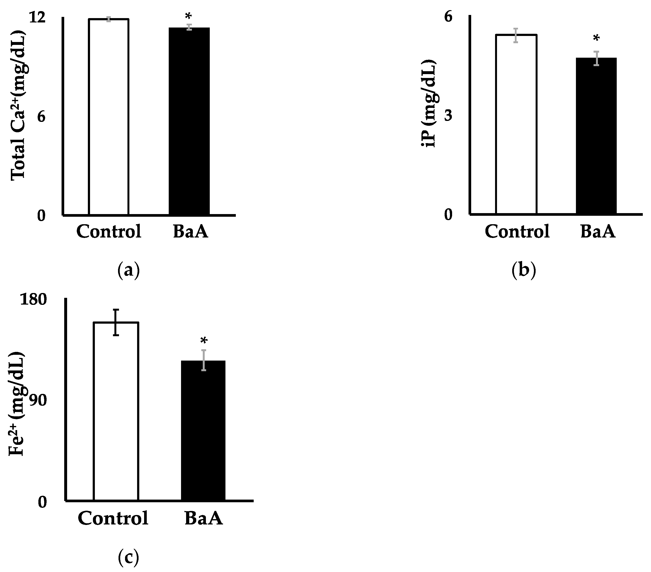
3.1. BaA Significantly Reduces Plasma Levels of Ca, iP, and Fe2+ in Nibbler Fish

Figure 1a shows that the intraperitoneal injection of BaA into nibbler fish significantly reduced plasma Ca2+ levels (11.36 ± 0.16 mg/dL) compared to the control group (11.87 ± 0.12 mg/dL). Figure 1b indicates that BaA-treated nibbler fish experienced a significant decrease in plasma iP concentration (4.71 ± 0.20 mg/dL) relative to the control group (5.40 ± 0.19 mg/dL). Figure 1c reveals a significant reduction in plasma Fe2+ levels in BaA-treated nibbler fish (125.00 ± 8.99 mg/dL) compared to controls (158.55 ± 11.62 mg/dL). Conversely, no significant differences were observed between control and BaA-treated fish in plasma levels of Na⁺ (177.45 ± 0.81 mEq/L vs. 177.83 ± 1.02 mEq/L), K+ (3.26 ± 0.16 mEq/L vs. 3.37 ± 0.06 mEq/L), or Cl (140.09 ± 1.99 mEq/L vs. 149.17 ± 2.01 mEq/L). These results suggest that BaA may induce hypocalcemia, hypophosphatemia, and iron deficiency anemia, among other conditions. Additionally, the findings imply that BaA does not affect electrolyte disorders such as hyponatremia, hypernatremia, hypokalemia, hyperkalemia, hypochloremia, or hyperchloremia, indicating a potential disruption in liver function in this species.

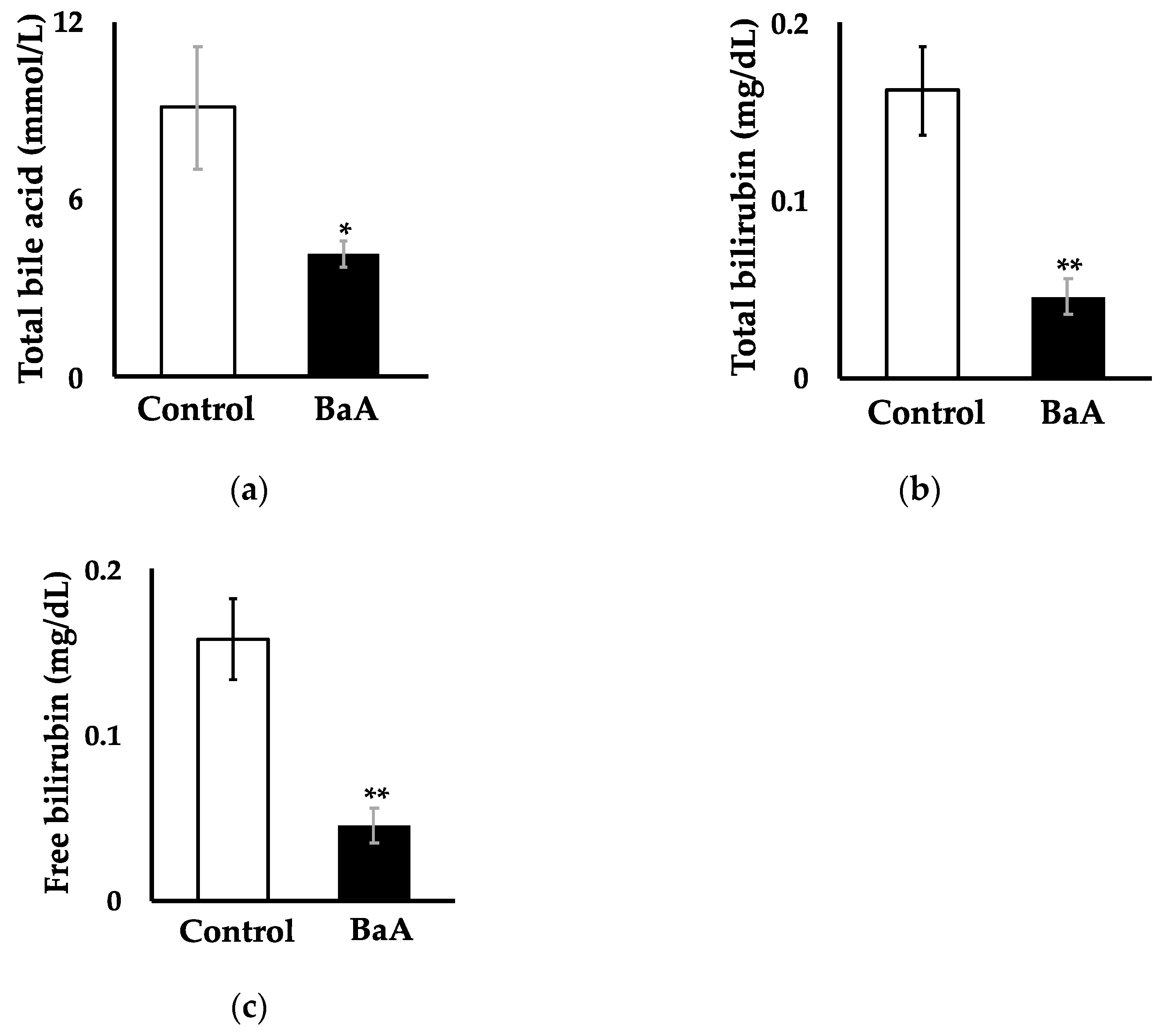
3.2. Intraperitoneal Injection of BaA in Nibbler Fish Significantly Reduces Plasma, Total Bile Acid, Total Bilirubin, and Free Bilirubin Levels

The intraperitoneal administration of BaA to nibbler fish significantly reduced plasma concentrations of total bile acid (4.17 ± 0.46 mmol/L; Figure 2a), total bilirubin (0.05 ± 0.01 mg/dL; Figure 2b), and free bilirubin (0.05 ± 0.01 mg/dL; Figure 2c) compared to the control group (9.09 ± 2.04 mmol/L, 0.16 ± 0.03 mg/dL, and 0.16 ± 0.02 mg/dL, respectively).

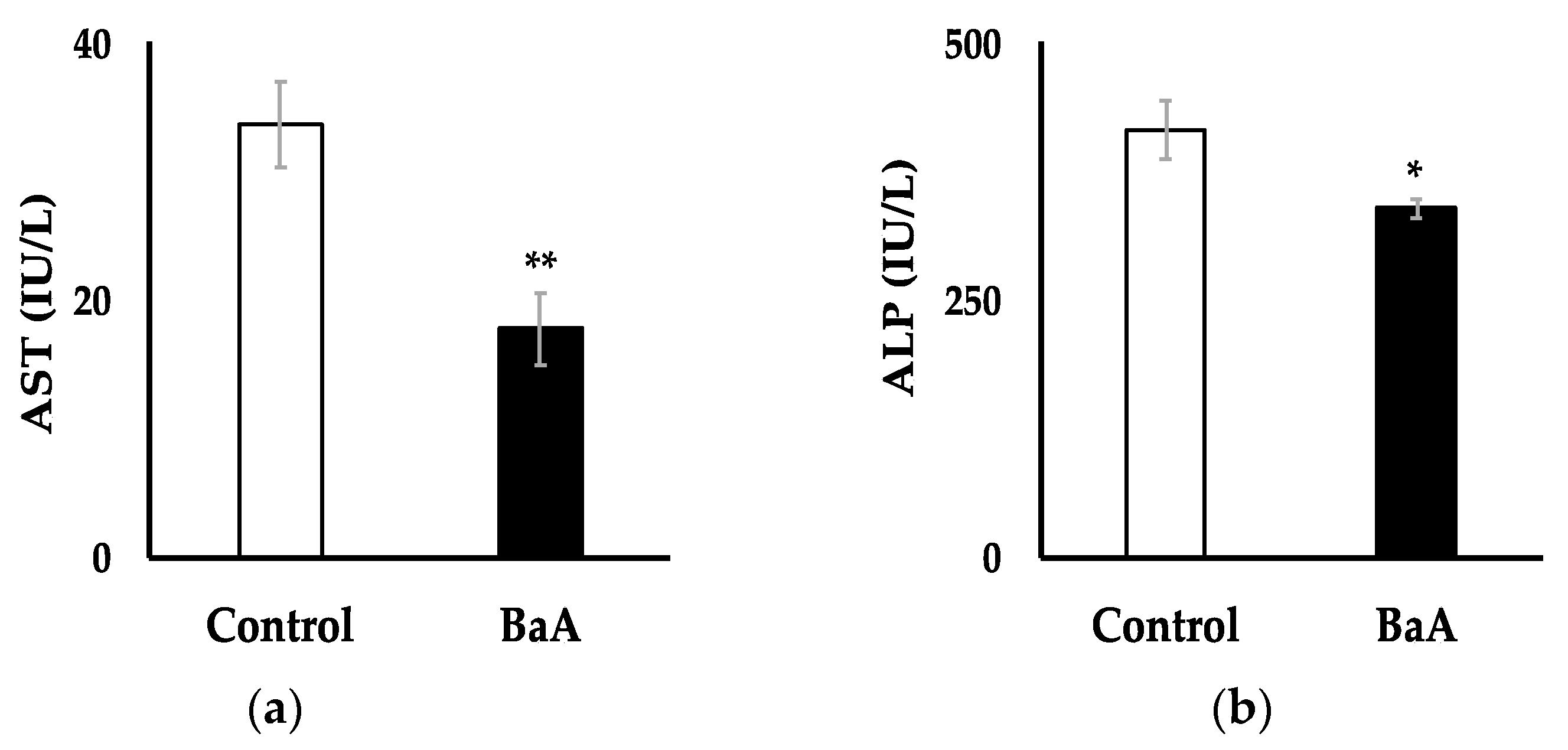
3.3. Intraperitoneal Injection of BaA in Nibbler Fish Significantly Reduces Plasma Levels of AST and ALP

The plasma levels of AST and ALP, biomarkers of liver function and injury, were significantly lower in BaA-treated nibbler fish (17.75 ± 2.79 IU/L and 339.17 ± 9.66 IU/L, respectively) compared to those in the control group (33.73 ± 3.32 IU/L and 415.45 ± 29.64 IU/L, respectively) (Figure 3). The decrease in AST was accompanied by a parallel decrease in ALT, which raises concerns about potential liver failure.

3.4. Detection of 3-OHBaA in the Bile of BaA-Treated Nibbler Fish

3-OHBaA was detected in the bile of BaA-treated nibbler fish, as shown in Figure 4. The 3-OHBaA was found to be in its free form, rather than conjugated to glucuronic acid or sulfuric acid. Specifically, 3-OHBaA was identified in the bile of BaA-treated fish, while no 3-OHBaA was detected in the bile of control fish.

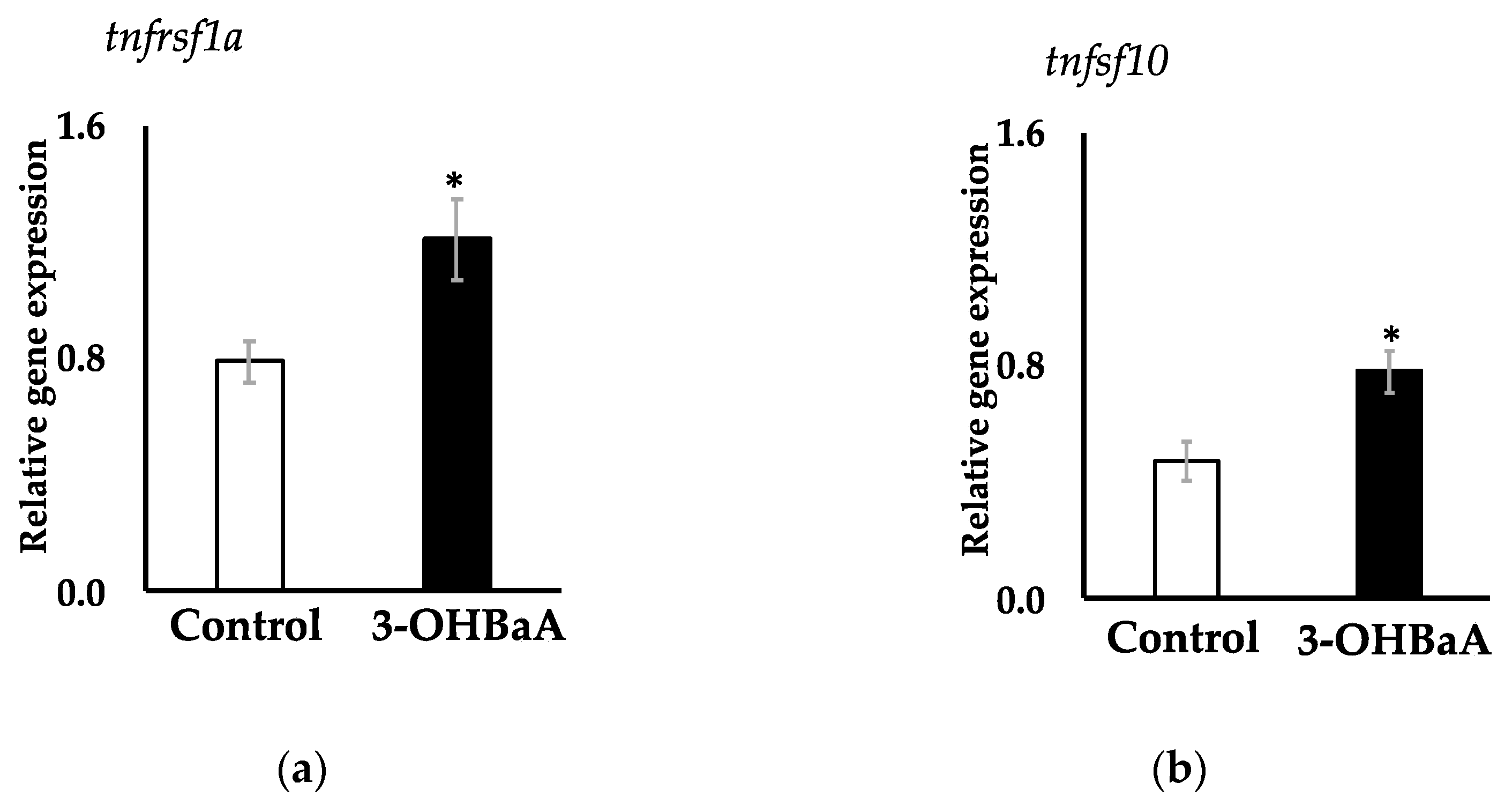
3.5. Expression of Two Apoptosis-Related Genes in 3-OHBaA-Exposed Cultured Liver

De novo RNA-seq analysis generated a reference transcriptome for the nibbler fish (DRR595512), from which two apoptosis-related genes, tnfrsf1a and tnfsf10, were identified. Partial PCR products encoding tnfrsf1a and tnfsf10 from nibbler fish exhibited complete sequence identity with the nucleotide (amino acid) sequences in the nibbler fish reference transcriptome DRR595512. Exposure to 3-OHBaA (10−6 M) significantly increased the expression of apoptosis-related genes tnfrsf1a and tnfsf10 in cultured liver cells (Figure 5). Interestingly, these increases in tnfrsf1a and tnfsf10 transcript abundance were accompanied by decreases in plasma levels of Ca2+, iP, Fe2+, total bile acids, total bilirubin, free bilirubin, AST, and ALP.

4. Discussion

BaA is a PAH commonly detected in sediments from rivers, coastal waters, and other aquatic environments worldwide. Additionally, hydroxylated PAHs are known to be metabolized from PAHs by the P4501A1 cytochrome [30] and have been detected in various organisms, including fish [2,31,32,33,34,35,36,37]. Suzuki et al. [16] found that 3-OHBcP exhibits marked toxicity by affecting bone metabolism and inducing osteoblast apoptosis in fish. In this study, we administered BaA to the marine nibbler fish (Girella punctata) and identified 3-OHBaA in its bile. Moreover, we observed an upregulation of apoptosis-related gene expression in the liver, suggesting that 3-OHBaA induces hepatic toxicity. These findings indicate that 3-OHBaA exerts toxic effects on the liver. Yang et al. [38] reported similar results in male Sprague Dawley rats, investigating the concentration of 3-OHBaA in urine and its hepatic toxicity. However, to our knowledge, no studies have directly examined the effects of hydroxylated PAHs on the liver in teleosts, making this study the first to investigate the direct toxic effects of BaA hydroxide on the teleost liver.
Hydroxylated BaA, specifically 3-OHBaA and 4-OHBaA, has been shown to inhibit the activity of both osteoclasts and osteoblasts in cultured scales of goldfish (Carassius auratus), a freshwater species, and wrasse (Pseudolabrus japonicus), a saltwater species [15]. Furthermore, Suzuki et al. [17] demonstrated that 4-OHBaA, a metabolite of BaA, inhibited osteoclastic (tartrate-resistant acid phosphatase) and osteoblastic (ALP) activities in goldfish scales, with 4-OHBaA detectable in the bile of goldfish following BaA administration. Additionally, BaA injection in goldfish induced hypocalcemia, hypophosphatemia, and reduced bone metabolism. These findings are similar to those in the current study, where BaA injection into nibbler fish also led to hypocalcemia, hypophosphatemia, and reduced liver metabolism. The detection of 3-OHBaA in the bile of BaA-treated nibbler fish suggests that 3-OHBaA is the primary metabolite and the main toxicant in this species. Hydroxylated PAHs have been implicated in reproductive toxicity [39], neurotoxicity [40], inflammatory responses [41], and cell damage and death pathways, including apoptosis [16].
Human hepatocyte apoptosis involves both extrinsic and intrinsic pathways, which are activated depending on various cellular conditions. The extrinsic pathway is triggered by death receptors, such as TNF receptor 1 (tnfrsf1a, etc.), located on the cell membrane [27]. When death ligands, like TNF (tnfsf10, etc.), bind to these receptors, this causes receptor trimerization, setting off a signaling cascade that activates caspase-8 and forms the death-inducing signaling complex [27]. On the other hand, the intrinsic pathway is activated by mitochondrial damage, which leads to the release of cytochrome c into the cytoplasm. This in turn activates caspase-9, which then activates effector caspases, furthering the apoptotic process [27]. Although the genomic information for nibbler fish is not yet available, the present de novo sequencing enabled the straightforward isolation of two important apoptosis-related factors, tnfrsf1a [22,23,27] and tnfsf10 [24,25,26,27]. This de novo RNA-seq analysis generated a reference transcriptome for the nibbler fish (DRR595512), which is expected to contribute to various fields of future research, including toxicology. The present study demonstrated that treatment with 3-OHBaA significantly increased the expression levels of these two apoptosis-related genes in the liver, both of which play crucial roles in apoptosis regulation via above caspase activation pathways. These findings suggest that 3-OHBaA likely induces cell death in liver cells of nibbler fish, potentially contributing to apoptotic processes through these molecular mechanisms.
This study indicates that 3-OHBaA may play a role in inducing apoptosis in the liver of fish. These findings highlight the importance of future research to assess the presence and concentration of 3-OHBaA in aquatic ecosystems and fish species. BaA is estimated to be present in environmental waters at concentrations ranging from 0 to 10−8 M [42]. BaA has also been detected in muscle tissues of various fish species [10,11,12]. For example, BaA mass ratio in the muscle of the freshwater fish European catfish (Silurus glanis) range from 0.05 to 0.3 ng/g (fresh weight) [10]., while the BaA mass ratio in the liver of juvenile coho salmon (Oncorhynchus kisutch) ranges from 2 to 18 ng/g (fresh weight) [13]. In teleost fish, PAHs have been reported to cause reproductive toxicity [43], immunotoxicity [44], and developmental toxicity, leading to bone deformities [45,46]. Furthermore, the hydroxylated form of BcP is 1900 times more lethal than BcP itself in fish larvae [16]. These findings suggest that 3-OHBaA may also induce apoptosis in fish at low concentrations. Apoptosis is essential for maintaining normal cellular function; however, its dysregulation can lead to various harmful effects and diseases. This study demonstrated that short-term exposure to the maximum predicted environmental concentration of 3-OHBaA in nibbler fish significantly reduced liver biomarkers and upregulated the expression of two apoptosis-related genes associated with the extrinsic pathway. Prolonged exposure to 3-OHBaA may exacerbate liver cell damage, underscoring the necessity for further research to elucidate these potential effects.
In the present study, we observed that hydroxylated PAHs induce the expression of two apoptosis-related factors in the liver of marine fish. However, our findings provide only preliminary evidence regarding the involvement of hydroxylated PAHs in apoptosis within the liver. Therefore, we anticipate that a more comprehensive analysis, including the expression of additional apoptosis-related factors and histological examination of hepatocytes, will help clarify the mechanisms by which hydroxylated PAHs are linked to apoptosis in the liver.

5. Conclusions

In conclusion, 3-OHBaA, a metabolite of BaA, was identified in the bile of the nibbler fish (Girella punctata) and was found to induce significant hepatotoxic effects. Livers exposed to 3-OHBaA exhibited an upregulated expression of two apoptosis-related genes compared to controls. These findings suggest that the first documented evidence of 3-OHBaA exhibiting biological toxicity in marine fish, indicating a potential risk of liver toxicity in other vertebrates. In this study, we propose that 3-OHBaA may adversely affect the extrinsic apoptotic pathway in the fish liver. However, further investigation is required to examine its impact on the intrinsic apoptotic pathway and other related factors.

Author Contributions

Conceptualization, H.M., M.H., A.H., and N.S. (Nobuo Suzuki); formal analysis, M.A.R., K.T. (Kenji Toyota), K.T. (Keito Tsunoda), M.H., and N.S. (Nobuo Suzuki); investigation, M.A.R., H.M., K.H. (Kaito Hatano), M.H., K.T. (Kenji Toyota), K.K., K.T. (Keito Tsunoda), Y.F., Y.T., T.H. (Tetsushi Hirano), A.S., C.-S.H., A.K.S., T.A., N.S. (Nobuaki Shimizu), M.I.Z., T.H. (Tatsuo Harumi), K.Y., T.M., N.T., A.H., K.H. (Kazuichi Hayakawa), and N.S. (Nobuaki Shimizu); resources, M.A.R., H.M., M.A.R., K.H. (Kaito Hatano), K.K., K.T. (Kenji Toyota), M.I.Z., and N.S. (Nobuo Suzuki); data curation, M.A.R., K.T. (Keito Tsunoda), M.H., Y.F., Y.T., T.H. (Tetsushi Hirano), and N.S. (Nobuo Suzuki); writing—original draft preparation, M.A.R., H.M., M.H., N.S. (Nobuo Suzuki); writing—review and editing, M.A.R., H.M., M.H., K.T. (Keito Tsunoda), A.K.S., T.M., and N.S. (Nobuo Suzuki); supervision, H.M., M.H., A.H., and N.S. (Nobuo Suzuki); project administration, H.M., M.H., A.H., and N.S. (Nobuo Suzuki); funding acquisition, M.A.R., H.M., and N.S. (Nobuo Suzuki). All authors have read and agreed to the published version of the manuscript.

Funding

This study was supported in part by grants to M.A.R. (Gant-in-Aid for SPRING Project for Doctoral Talents in Creating Novel Values [HaKaSe+ for SPRING] No. JPMJSP2135 by JST), to H.M. (Grant-in-Aid for Scientific Research [C] No. 21K05725 by JSPS and National University Management Reform Promotion Project [MEXT, Japan]), to Nobuo Suzuki (Grant-in-Aid for Scientific Research [C] No. 23K10933 by JSPS), and to T.M. (the Flagship Research Group Programme of the Hungarian University of Agriculture and Life Sciences). This work was performed under the cooperative research program of the Institute of Nature and Environmental Technology, Kanazawa University, Acceptance Nos. 23005, 23021, 23022, 23026, and 23028.

Institutional Review Board Statement

The animal study protocol was approved by the Guide for the Care and Use of Laboratory Animals at Kanazawa University (2020–2024).

Informed Consent Statement

Not applicable.

Data Availability Statement

The de novo RNA Sequence data discussed in this publication have been deposited in the DDBJ Sequence Read Archive, accession number DRR595512. The cDNA sequences obtained have been deposited in GenBank with the accession numbers LC849219 (tnfrsf1a) and LC849220 (tnfsf10).

Acknowledgments

The authors are grateful to the Shouzo Ogiso (Noto Marine Laboratory, Institute of Nature and Environmental Technology, Kanazawa University) and Arata Nagami (Noto Center for Fisheries Science and Technology, Kanazawa University) for providing the broodstock of the nibbler fish.

Conflicts of Interest

The authors declare no conflicts of interest.

References

  1. Lima, A.L.C.; Eglinton, T.I.; Reddy, C.M. High-resolution record of pyrogenic polycyclic aromatic hydrocarbon deposition during the 20th century. Environ. Sci. Technol. 2003, 37, 53–61. [Google Scholar] [CrossRef] [PubMed]
  2. Honda, M.; Suzuki, N. Toxicities of polycyclic aromatic hydrocarbons for aquatic animals. Int. J. Environ. Res. Public Health 2020, 17, 1363. [Google Scholar] [CrossRef] [PubMed]
  3. Bue, B.G.; Sharr, S.; Seeb, J.E. Evidence of damage to pink salmon populations inhabiting Prince William Sound, Alaska, two generations after the Exxon Valdez oil spill. Trans. Am. Fish. Soc. 1998, 127, 35–43. [Google Scholar] [CrossRef]
  4. Heintz, R.A.; Rice, S.D.; Wertheimer, A.C.; Bradshaw, R.F.; Thrower, F.P.; Joyce, J.E.; Short, J.W. Delayed effects on growth and marine survival of pink salmon Oncorhynchus gorbuscha after exposure to crude oil during embryonic development. Mar. Ecol. Prog. Ser. 2000, 208, 205–216. [Google Scholar] [CrossRef]
  5. Hayakawa, K.; Nomura, M.; Nakagawa, T.; Oguri, S.; Kawanishi, T.; Toriba, A.; Kizu, R.; Sakaguchi, T.; Tamiya, E. Damage to and recovery of coastlines polluted with C-heavy oil spilled from the Nakhodka. Water Res. 2006, 40, 981–989. [Google Scholar] [CrossRef]
  6. de Soysa, T.Y.; Ulrich, A.; Friedrich, T.; Pite, D.; Compton, S.L.; Ok, D.; Bernardos, R.L.; Downes, G.B.; Hsieh, S.; Stein, R.; et al. Macondo crude oil from the Deepwater Horizon oil spill disrupts specific developmental processes during zebrafish embryogenesis. BMC Biol. 2012, 10, 40. [Google Scholar] [CrossRef]
  7. Peterson, C.H.; Rice, S.D.; Short, J.W.; Esler, D.; Bodkin, J.L.; Ballachey, B.E.; Irons, D.B. Long-term ecosystem response to the Exxon Valdez oil spill. Science 2003, 302, 2082–2086. [Google Scholar] [CrossRef]
  8. Incardona, J.P.; Gardner, L.D.; Linbo, T.L.; Brown, T.L.; Esbaugh, A.J.; Mager, E.M.; Stieglitz, J.D.; French, B.L.; Labenia, J.S.; Laetz, C.A.; et al. Deepwater Horizon crude oil impacts the developing hearts of large predatory pelagic fish. Proc. Natl. Acad. Sci. USA 2014, 111, E1510–E1518. [Google Scholar] [CrossRef]
  9. Le Bihanic, F.; Sommard, V.; Perrine, d.L.; Pichon, A.; Grasset, J.; Berrada, S.; Budzinski, H.; Cousin, X.; Morin, B.; Cachot, J. Environmental concentrations of benz[a]anthracene induce developmental defects and DNA damage and impair photomotor response in Japanese medaka larvae. Ecotoxicol. Environ. Saf. 2015, 113, 321–328. [Google Scholar] [CrossRef]
  10. Squadrone, S.; Favaro, L.; Abete, M.C.; Vivaldi, B.; Prearo, M. Polycyclic aromatic hydrocarbon levels in European catfish from the upper Po River basin. Environ. Monit. Assess. 2014, 186, 2313–2320. [Google Scholar] [CrossRef]
  11. Yu, Z.; Lin, Q.; Gu, Y.; Du, F.; Wang, X.; Shi, F.; Ke, C.; Xiang, M.; Yu, Y. Bioaccumulation of polycyclic aromatic hydrocarbons (PAHs) in wild marine fish from the coastal waters of the northern South China Sea: Risk assessment for human health. Ecotoxicol. Environ. Saf. 2019, 180, 742–748. [Google Scholar] [CrossRef] [PubMed]
  12. Li, Y.; Guo, N.; Zou, X.; Li, P.; Zou, S.; Luo, J.; Yang, Y. Pollution level and health risk assessment of polycyclic aromatic hydrocarbons in marine fish from two coastal regions, the South China Sea. Mar. Pollut. Bull. 2021, 168, 112376. [Google Scholar] [CrossRef] [PubMed]
  13. Barbee, G.C.; Barich, J.; Duncan, B.; Bickham, J.W.; Matson, C.W.; Hintze, C.J.; Autenrieth, R.L.; Zhou, G.D.; McDonald, T.J.; Cizmas, L.; et al. In situ biomonitoring of PAH-contaminated sediments using juvenile coho salmon (Oncorhynchus kisutch). Ecotoxicol. Environ. Saf. 2008, 71, 454–464. [Google Scholar] [CrossRef] [PubMed]
  14. Zanaty, M.I.; Sawada, N.; Kitani, Y.; Nassar, H.F.; Mahmoud, H.M.; Hayakawa, K.; Sekiguchi, T.; Ogiso, S.; Tabuchi, Y.; Urata, M.; et al. Influence of benz[a]anthracene on bone metabolism and on liver metabolism in nibbler fish, Girella punctate. Int. J. Environ. Res. Public Health 2020, 17, 1391. [Google Scholar] [CrossRef]
  15. Suzuki, N.; Hayakawa, K.; Kameda, T.; Toriba, A.; Tang, N.; Tabata, M.J.; Takada, K.; Wada, S.; Omori, K.; Srivastav, A.K.; et al. Monohydroxylated polycyclic aromatic hydrocarbons inhibit both osteoclastic and osteoblastic activities in teleost scales. Life Sci. 2009, 84, 482–488. [Google Scholar] [CrossRef]
  16. Suzuki, N.; Honda, M.; Sato, M.; Yoshitake, S.; Kawabe, K.; Tabuchi, Y.; Omote, T.; Sekiguchi, T.; Furusawa, Y.; Toriba, A.; et al. Hydroxylated benzo[c]phenanthrene metabolites cause osteoblast apoptosis and skeletal abnormalities in fish. Ecotoxicol. Environ. Saf. 2022, 234, 113401. [Google Scholar] [CrossRef]
  17. Suzuki, N.; Nakano, J.; Kawabe, K.; Toriba, A.; Hayakawa, K.; Tang, N.; Sekiguchi, T.; Tabuchi, Y.; Ikegame, M.; Shimizu, N.; et al. Benz[a]anthracene decreases plasma calcium levels resulting from influence of scale osteoclastic and osteoblastic activities in goldfish. Int. J. Zool. Investig. 2017, 3, 72–81. [Google Scholar]
  18. Honda, M.; Hayakawa, K.; Zhang, L.; Tang, N.; Nakamura, H. Seasonal variability and risk assessment of atmospheric polycyclic aromatic hydrocarbons and hydroxylated polycyclic aromatic hydrocarbons in Kanazawa, Japan. Appl. Sci. 2022, 12, 9469. [Google Scholar] [CrossRef]
  19. Hatano, K.; Yoshida, M.A.; Hirayama, J.; Kitani, Y.; Hattori, A.; Ogiso, S.; Watabe, Y.; Sekiguchi, T.; Tabuchi, Y.; Urata, M.; et al. Deep ocean water alters the cholesterol and mineral metabolism of squid Todarodes pacificus and suppresses its weight loss. Sci. Rep. 2023, 13, 7591. [Google Scholar] [CrossRef]
  20. Grabherr, M.G.; Haas, B.J.; Yassour, M.; Levin, J.Z.; Thompson, D.A.; Amit, I.; Adiconis, X.; Fan, L.; Raychowdhury, R.; Zeng, Q.; et al. Full-length transcriptome assembly from RNA-Seq data without a reference genome. Nat. Biotechnol. 2011, 29, 644–652. [Google Scholar] [CrossRef]
  21. Mai, H.; Zhang, Y.; Li, D.; Leung, H.C.M.; Luo, R.; Wong, C.K.; Ting, H.F.; Lam, T.W. AC-DIAMOND v1: Accelerating large-scale DNA-protein alignment. Bioinformatics 2018, 34, 3744–3746. [Google Scholar] [CrossRef] [PubMed]
  22. Naudé, P.J.W.; den Boer, J.A.; Luiten, P.G.M.; Eisel, U.L.M. Tumor necrosis factor receptor cross-talk. FEBS J. 2011, 278, 888–898. [Google Scholar] [CrossRef] [PubMed]
  23. Gough, P.; Myles, I.A. Tumor Necrosis Factor Receptors: Pleiotropic Signaling Complexes and Their Differential Effects. Front. Immunol. 2020, 11, 585880. [Google Scholar] [CrossRef]
  24. Wiley, S.R.; Schooley, K.; Smolak, P.J.; Din, W.S.; Huang, C.P.; Nicholl, J.K.; Sutherland, G.R.; Smith, T.D.; Rauch, C.; Smith, C.A. Identification and characterization of a new member of the TNF family that induces apoptosis. Immunity 1995, 3, 673–682. [Google Scholar] [CrossRef]
  25. Pitti, R.M.; Marsters, S.A.; Ruppert, S.; Donahue, C.J.; Moore, A.; Ashkenazi, A. Induction of apoptosis by Apo-2 ligand, a new member of the tumor necrosis factor cytokine family. J. Biol. Chem. 1996, 271, 12687–12690. [Google Scholar] [CrossRef]
  26. Guerrache, A.; Micheau, O. TNF-Related Apoptosis-Inducing Ligand: Non-Apoptotic Signalling. Cells 2024, 13, 521. [Google Scholar] [CrossRef]
  27. Cao, L.; Quan, X.-B.; Zeng, W.-J.; Yang, X.-O.; Wang, M.-J. Mechanism of Hepatocyte Apoptosis. J. Cell Death 2016, 9, 19–29. [Google Scholar] [CrossRef]
  28. Untergasser, A.; Cutcutache, I.; Koressaar, T.; Ye, J.; Faircloth, B.C.; Remm, M.; Rozen, S.G. Primer3—New Capabilities and Interfaces. Nucleic Acids Res. 2012, 40, e115. [Google Scholar] [CrossRef]
  29. Sekimoto, A.; Ohira, T.; Shigematsu, A.; Okumura, T.; Mekuchi, M.; Toyota, K.; Mishima, H.; Kawamura, R.; Hatano, K.; Kawago, U.; et al. Functional analysis of a matrix peptide involved in calcification of the exoskeleton of the kuruma prawn, Marsupenaeus japonicus. Aquaculture 2022, 559, 738437. [Google Scholar] [CrossRef]
  30. Jaruchotikamol, A.; Jarukamjorn, K.; Sirisangtrakul, W.; Sakuma, T.; Kawasaki, Y.; Nemoto, N. Strong Synergistic Induction of CYP1A1 Expression by Andrographolide Plus Typical CYP1A Inducers in Mouse Hepatocytes. Toxicol. Sci. 2007, 224, 156–162. [Google Scholar] [CrossRef]
  31. Johnson-Restrepo, B.; Kannan, K.; Addink, R.; Adams, D.H. Polycyclic Aromatic Hydrocarbons and Their Halogenated Analogues in Fish from Florida Coastal Waters. Environ. Pollut. 2008, 151, 452–459. [Google Scholar] [CrossRef] [PubMed]
  32. Moon, H.-B.; Lee, D.H.; Lee, Y.S.; Kannan, K. Occurrence and Accumulation Patterns of Polycyclic Aromatic Hydrocarbons and Synthetic Musk Compounds in Adipose Tissues of Korean Females. Chemosphere 2012, 86, 485–490. [Google Scholar] [CrossRef]
  33. Koenig, S.; Porte, C.; Solé, M.; Sturve, J. Biliary PAH and Alkylphenol Metabolites, Biomarker Enzyme Activities, and Gene Expression Levels in the Deep-Sea Fish Alepocephalus rostratus. Environ. Sci. Technol. 2013, 47, 2854–2861. [Google Scholar] [CrossRef]
  34. Guo, Y.; Senthilkumar, K.; Alomirah, H.; Moon, H.B.; Minh, T.B.; Mohd, M.A.; Nakata, H.; Kannan, K. Concentrations and Profiles of Urinary Polycyclic Aromatic Hydrocarbon Metabolites (OH-PAHs) in Several Asian Countries. Environ. Sci. Technol. 2013, 47, 2932–2938. [Google Scholar] [CrossRef]
  35. Zhu, H.; Wang, L.; Liu, C.; Stryker, Z.; Loganathan, B.G.; Kannan, K. Phthalate Metabolites, Hydroxy-Polycyclic Aromatic Hydrocarbons, and Bisphenol Analogues in Bovine Urine Collected from China, India, and the United States. Environ. Sci. Technol. 2019, 53, 11524–11531. [Google Scholar] [CrossRef]
  36. Zhu, H.; Martinez-Moral, M.P.; Kannan, K. Variability in Urinary Biomarkers of Human Exposure to Polycyclic Aromatic Hydrocarbons and Its Association with Oxidative Stress. Environ. Int. 2021, 156, 106720. [Google Scholar] [CrossRef]
  37. Singh, V.; Negi, R.; Jacob, M.; Gayathri, A.; Rokade, A.; Sarma, H.; Kalita, J.; Tasfia, S.T.; Bharti, R.; Wakid, A.; et al. Polycyclic Aromatic Hydrocarbons (PAHs) in Aquatic Ecosystem Exposed to the 2020 Baghjan Oil Spill in Upper Assam, India: Short-Term Toxicity and Ecological Risk Assessment. PLoS ONE 2023, 18, e0293601. [Google Scholar] [CrossRef]
  38. Yang, M.; Lu, Y.; Mao, W.; Hao, L. New insight into PAH4 induced hepatotoxicity and the dose-response assessment in rats model. Chemosphere 2024, 350, 141042. [Google Scholar] [CrossRef]
  39. Luderer, U.; Christensen, F.; Johnson, W.O.; She, J.; Ip, H.S.; Zhou, J.; Alvaran, J.; Krieg, E.F., Jr.; Kesner, J.S. Associations between urinary biomarkers of polycyclic aromatic hydrocarbon exposure and reproductive function during menstrual cycles in women. Environ. Int. 2017, 100, 110–120. [Google Scholar] [CrossRef]
  40. Olasehinde, T.A.; Olaniran, A.O. Neurotoxicity of polycyclic aromatic hydrocarbons: A systematic mapping and review of neuropathological mechanisms. Toxics 2022, 10, 417. [Google Scholar] [CrossRef]
  41. Everett, C.J.; King, D.E.; Player, M.S.; Matheson, E.M.; Post, R.E.; Mainous, A.G., 3rd. Association of urinary polycyclic aromatic hydrocarbons and serum C-reactive protein. Environ. Res. 2010, 110, 79–82. [Google Scholar] [CrossRef] [PubMed]
  42. Mojiri, A.; Zhou, J.L.; Ohashi, A.; Ozaki, N.; Kindaichi, T. Comprehensive Review of Polycyclic Aromatic Hydrocarbons in Water Sources, Their Effects and Treatments. Sci. Total Environ. 2019, 696, 133971. [Google Scholar] [CrossRef] [PubMed]
  43. Hoffmann, J.L.; Oris, J.T. Altered gene expression: A mechanism for reproductive toxicity in zebrafish exposed to benzo[a]pyrene. Aquat. Toxicol. 2006, 78, 332–340. [Google Scholar] [CrossRef] [PubMed]
  44. Reynaud, S.; Deschaux, P. The effects of polycyclic aromatic hydrocarbons on the immune system of fish: A review. Aquat. Toxicol. 2006, 77, 229–238. [Google Scholar] [CrossRef] [PubMed]
  45. Barron, M.G.; Carls, M.G.; Heintz, R.; Rice, S.D. Evaluation of fish early life-stage toxicity models of chronic embryonic exposures to complex polycyclic aromatic hydrocarbon mixtures. Toxicol. Sci. 2004, 78, 60–67. [Google Scholar] [CrossRef]
  46. Billiard, S.M.; Timme-Laragy, A.R.; Wassenberg, D.M.; Cockman, C.; Di Giulio, R.T. The role of the aryl hydrocarbon receptor pathway in mediating synergistic developmental toxicity of polycyclic aromatic hydrocarbons to zebrafish. Toxicol. Sci. 2006, 92, 526–536. [Google Scholar] [CrossRef]
Figure 1. Effects of BaA on plasma Ca2+ (a), iP (b), and Fe2+ (c) levels in BaA-treated nibbler fish. Plasma Ca2+, iP and Fe2+ in the saline-treated control group (n = 11) are shown in white columns, and those in the BaA-treated experimental group (n = 12) are shown in black columns. Statistically significant differences, denoted by asterisks, were observed at p < 0.05 and were found when compared to the control group.
Toxics 12 00915 g001
Figure 2. Effects of BaA on plasma total bile acid (a), total bilirubin (b), and free bilirubin (c) levels in the BaA-treated nibbler fish. Plasma total bile acid, total bilirubin, and free bilirubin in the saline-treated control group (n = 11) are shown in white columns, and those in the BaA-treated experimental group (n = 12) are shown in black columns. Statistically significant differences, indicated by asterisks (* p < 0.05, ** p < 0.01), were found when compared to the control group.
Toxics 12 00915 g002
Figure 3. Effects of BaA on plasma AST (a) and ALP (b) activities in BaA-treated nibbler fish. Plasma activities of AST and ALP in the saline-treated control group (n = 11) are shown in white columns, and those in the BaA-treated experimental group (n = 12) are shown in black columns. Statistically significant differences, indicated by asterisks (* p < 0.05, ** p < 0.01), were observed when compared to the control group.
Toxics 12 00915 g003
Figure 4. Detection of 3-OHBaA in the bile of BaA-treated nibbler fish. 3-OHBaA in the bile was not found in controls (n = 11) and was only identified in BaA-treated fish (red: n = 12). Standard 3-OHBaA solution shown by blue line.
Toxics 12 00915 g004
Figure 5. Effects of 3-OHBaA on two apoptosis-related gene expressions in the cultured liver of nibbler fish. The expression of tnfrsf1a (a) and t tnfsf10 (b) in the liver is shown in white columns for the control group (n = 5) and black columns for the experimental group (n = 5). Statistically significant differences, indicated by asterisks, were observed at p < 0.05 compared to the control group.
Toxics 12 00915 g005
Disclaimer/Publisher’s Note: The statements, opinions and data contained in all publications are solely those of the individual author(s) and contributor(s) and not of MDPI and/or the editor(s). MDPI and/or the editor(s) disclaim responsibility for any injury to people or property resulting from any ideas, methods, instructions or products referred to in the content.

