Review

Advanced Glycation End Products: A Sweet Flavor That Embitters Cardiovascular Disease

by
Raphael S. Pinto
1,2,
Carlos A. Minanni
1,3,
Aécio Lopes de Araújo Lira
1,4 and
Marisa Passarelli
1,5,*
1
Laboratório de Lípides (LIM 10), Hospital das Clínicas (HCFMUSP) da Faculdade de Medicina da Universidade de São Paulo, São Paulo 01246-000, Brazil
2
Universidade Santa Cecília (UNISANTA), Santos 11045-907, Brazil
3
Hospital Israelita Albert Einstein (HIAE), São Paulo 05652-900, Brazil
4
Faculdade de Medicina do Centro Universitário Uninovafapi, Teresina 64073-505, Brazil
5
Programa de Pós-Graduação em Medicina, Universidade Nove de Julho (UNINOVE), São Paulo 01225-000, Brazil
*
Author to whom correspondence should be addressed.
Int. J. Mol. Sci. 2022, 23(5), 2404; https://doi.org/10.3390/ijms23052404
Submission received: 3 February 2022 / Revised: 16 February 2022 / Accepted: 17 February 2022 / Published: 22 February 2022

Abstract

:
Epidemiological studies demonstrate the role of early and intensive glycemic control in the prevention of micro and macrovascular disease in both type 1 and type 2 diabetes mellitus (DM). Hyperglycemia elicits several pathways related to the etiopathogenesis of cardiovascular disease (CVD), including the generation of advanced glycation end products (AGEs). In this review, we revisit the role played by AGEs in CVD based in clinical trials and experimental evidence. Mechanistic aspects concerning the recognition of AGEs by the advanced glycosylation end product-specific receptor (AGER) and its counterpart, the dolichyl-diphosphooligosaccharide-protein glycosyltransferase (DDOST) and soluble AGER are discussed. A special focus is offered to the AGE-elicited pathways that promote cholesterol accumulation in the arterial wall by enhanced oxidative stress, inflammation, endoplasmic reticulum stress and impairment in the reverse cholesterol transport (RCT).

1. Introduction

The last two decades have been marked by alarming data about the epidemic of diabetes mellitus (DM). In 2021, an estimated 537 million adults aged 20–79 years have DM. This represents a prevalence of 10.5% of the world’s population in this age group. This number is projected to reach 643 million by 2030, and 783 million by 2045, according to the International Diabetes Federation (IDF) [1].
DM confers a higher risk of development of chronic complications and people with DM are 2 to 3 times more likely to have cardiovascular disease (CVD) [2,3]; the prevalence of end-stage renal disease is 10 times higher [4] and amputation is 10 to 20 times more common [5] in people with DM as compared to the healthy population. In addition, diabetic retinopathy is the leading cause of vision loss in working-age adults [6].
The classic studies, namely the United Kingdom Prospective Diabetes Study (UKPDS), Action in Diabetes and Vascular Disease: Preterax and Diamicron Modified Release Controlled Evaluation (ADVANCE), Action to Control Cardiovascular Risk in Diabetes (ACCORD), and Diabetes Control and Complications Trial/Epidemiology of Diabetes Interventions and Complications (DCCT/EDIC), demonstrated that intensive glycemic therapy in patients with a recent diagnosis of type 1 and 2 diabetes (T2DM) was associated with a reduced risk in long-term complications, in particular, microvascular morbidities. Data from 30 years of the DCCT/EDIC demonstrated that intensive versus conventional glycemic treatment also has beneficial effects on macrovascular disease, especially on subclinical markers of atherosclerosis (carotid intima-media thickness and coronary calcium score) 6–12 years after the end of randomized treatment. Intensive treatment reduced aggregate cardiovascular risk by 42% and major cardiovascular events by 57% (myocardial infarction, stroke, and cardiovascular death) [7]. However, in many cases, when the glycemic control was achieved after a period of intense decompensation, the development of chronic complications was not well prevented [8,9]. Those findings seem to rely on the concept of metabolic memory or legacy effect that is based on both the permanent modification of biological macromolecules during the period of glycemic decompensation and on the accumulated risk memorized by long-term exposure to oxidative stress and advanced glycation products (AGEs), which may favor epigenetic changes. The risk of complications remains even with multifactorial interventions based on glycemic-reducing agents and antilipemic and antihypertensive drugs [8,10].
Hyperglycemia is the major etiopathogenic factor for the development of DM complications that is due to the induction of oxidative stress, considered the basis for cellular and tissue damage. Hyperglycemia favors the generation of AGEs, induces the activation of hexosamine, polyol, and protein kinase C pathways that exacerbate oxidative stress. In this review, we summarize the data on the role played by AGEs in the pathophysiology of cardiovascular disease in DM, based on human clinical studies and experimental models.

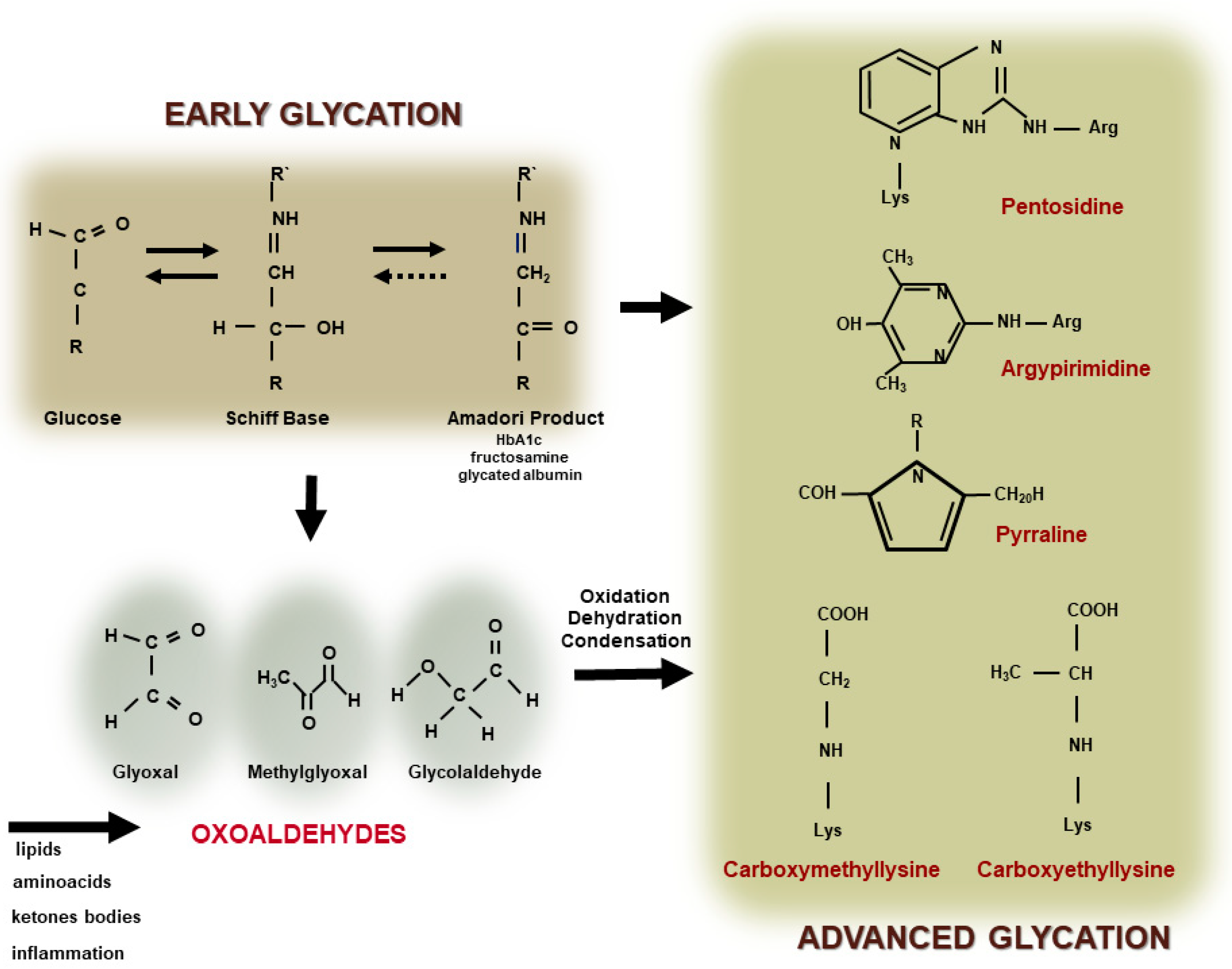
2. Formation of Advanced Glycation End Products

Louis Camille Maillard first described in 1912 the nonenzymatic reaction between reducing sugars with foods in what was called a browning reaction. Patton and Hill (1948) [11] characterized the chemical reaction that attained much more attention by the identification by Rahbar et al. (1969) [12] of a glucose-modified form of hemoglobin (glycated hemoglobin) that was found elevated in the blood of subjects with DM as compared to healthy controls. Since then, HbA1c was utilized as a parameter of metabolic control and management of DM and was associated with the development of DM complications.
The covalent and nonenzymatic reaction between reducing sugars, including glucose, with the amino-terminal portion of lysine and arginine residues in proteins, phospholipids, and nucleic acids leads to the formation of an unstable Schiff Base. The Schiff Base undergoes a more stable Amadori product or fructosyl lysine (such as glycated hemoglobin and fructosamine) and this reaction primarily depends on plasma glucose levels and the time of DM decompensation. Molecular intra and inter rearrangements of the Amadori product in most of the cases involve oxidative reactions leading to the generation of oxoaldehyde that induces a rapid and irreversible modification of proteins by advanced glycation. Oxoaldehydes are very reactive compounds, such as glyoxal (GO), methylglyoxal (MGO), glycolaldehyde, and 3-deoxyglucosone (3-DG), which are generated in DM, but also in other metabolic conditions where the carbonyl stress prevails. They rapidly interact with macromolecules leading to the fast formation of extracellular and intracellular AGEs (Figure 1).
Intracellular generated AGEs originate from the increased glucose flow through glycolysis that favor the output of reactive oxygen species (ROS) from mitochondria and the DNA-repairing enzyme poly (ADP-ribose) polymerase (PARP). Although protecting DNA from ROS-induced damage, PARP promotes poly ribosylation of the glyceraldehyde 3-phosphate dehydrogenase (GAPDH), impairing its activity. Substrate deviation from glycolysis generates MGO and, as a consequence, AGEs are formed feeding a vicious circle by inducing oxidative stress [13,14]. Hyperglycemia also induces the activation of the hexosamine, polyol and protein kinase C pathways, exacerbating the intracellular oxidative stress, which perpetuates the AGE generation [13,15] (Figure 2).
Independently of hyperglycemia, AGEs are induced during acute and chronic inflammation due to the activation of the neutrophil myeloperoxidase that induces glycolaldehyde (GAD), another very reactive oxoaldehyde. Additionally, the oxidation of certain amino acids, polyunsaturated fatty acids, and ketone bodies contributes to the generation of oxoaldehydes and then to AGE generation. In chronic kidney disease, AGEs are formed due to a reduction in the clearance of glycation reaction precursors and reduced antioxidant enzymes. Then, although the glycation reaction was firstly ascribed to a hyperglycemic milieu, it is now conceivable that it also occurs independently of glucose concentration in plasma and tissues [16]. In fact, glycation is prevalent in healthy aging individuals, mainly in association with long half-life protein, such as collagen and crystalline. Although this process takes place continuously in the body during aging, it is extremely accelerated in DM and in other carbonyl stress conditions.
Exogenous sources of oxoaldehydes and AGEs include diet and tobacco, representing unpredictable sources of the AGEs that contribute the body’s AGE pool, although the exact mechanisms that regulate AGE absorption are not well known. The impact of AGE-containing diets on human health is under intensive investigation. High heating, dry cooking, and long-time processing increase AGEs in baked, broiled, grilled, and fried foods. Cooking in an acidic medium containing wine, vinegar, or citrus juice helps to prevent AGE formation in food. A recent publication demonstrated an endocytic pathway via scavenger receptors mediating the intestinal absorption of dietary carboxymethyllysine in C. elegans [17]. In healthy humans, about 10% of dietary AGEs are absorbed and transported in circulation in association with albumin and lipoproteins, and 30% is eliminated in the urine. On the other hand, in individuals with kidney disease, only 5% of dietary AGE is excreted in the urine [18]. A high-AGE diet results in significant elevations in serum AGE and induces oxidative stress [19]. Oxoaldehydes increase in the postprandial states and during glycemic excursions inducing a fast generation of AGEs in circulation [20]. Glucose fluctuations increase plasma levels of glyceraldehyde-derived advanced glycation end-products and relate to the severity of cardiovascular disease in DM [21].
Advanced glycation end products are very heterogeneous, making difficult their assessment in blood, tissues, and cells. Some of them induces protein crosslinking and/or emit fluorescence [22]. Glyoxal and GAD render carboxymethyl lysine (CML) the most abundant AGE found in the body [23,24], particularly in the target tissues of DM late complications, such as kidneys, eyes, skin, and vessels [25,26,27,28,29]. Methylglyoxal renders to carboxyethyl lysine, MGO-derived hydroimidazolone 1 (MGO-H1), and MGO-lysine dimer, although other structures are also described in plasma and tissues, such as pentosidine, pirralyne, argypirimidine, and GO- lysine dimer.
AGEs are recognized by several receptors, although the signaling pathway elicited is better described for the advanced glycosylation end product-specific receptor, (alias RAGE). In fact, it is demonstrated that AGER mediates the biological effects of AGEs in many cell types. AGER is a multiligand receptor of the immunoglobulin superfamily, a member of pattern-recognition receptors that recognize AGEs, but also calgranulins, high mobility group protein B1 (HMGB1), lipopolysaccharides (LPS), sheet fibrils, and phosphatidylserine on the surface of apoptotic cells. The receptor for AGEs actively participates in diabetic vascular complications, as well as in the interface of innate and adaptive immunity and in inflammation. In this sense, there is a lot of evidence supporting the concept that AGEs and AGERs play an active role in the development and progression of cardiovascular disease in DM [30].
A sequence of 394 amino acids in a single hydrophobic transmembrane domain (19 amino acids) and a highly charged C-terminal cytosolic tail (43 amino acids) that mediates intracellular signaling pathways characterizes the AGER structure. The extracellular portion contains an N-terminal immunoglobulin (Ig) V-type ligand-binding domain and two Ig C-type domains (V-C-C′). The protein diaphanous homolog 1 (DIAPH1) mediates the activation of NADPH oxidase 4 after AGE binding to the AGER. The canonical pathway involves ROS generation and the transactivation of the AGER gene and others related in the production of inflammatory cytokines, adhesion molecules, chemokines, growth factors, and scavenger receptors [31,32,33]. Other signaling pathways stimulated by the AGE–AGER interaction include extracellular signal-regulated kinase 1/2 (ERK 1/2) and p38-mitogen-activated protein kinase (MAPK), PI3K/AKT, Rho GTPases, and cross talking with Toll-like receptors [34,35]. Interestingly, it is observed that the expression of the AGER is low in vascular cells, but constitutively activated in DM and inflammation [36].
Soluble isoforms of AGERs lacking intracellular domains bind to AGEs, but are unable to trigger cell signaling. There are two isoforms of soluble AGER, although their regulation is not well known. The first isoform is produced by the action proteases at cell surface, such as disintegrin and metalloproteinase domain-containing protein 10, (ADAM10) and matrix metalloproteinases (MMPs). The second isoform called endogenous secretory, esAGER, is generated by alternative splicing of the AGER gene. The measurement of a soluble AGER (sAGER) in circulation was proposed as a biomarker of CVD in DM. However, data in the literature are still controversial.
The dolichyl-diphosphooligosaccharide-protein glycosyltransferase (DDOST, alias AGER1) binds AGEs, although the intracellular signaling elicited by their interaction is not well known. It is encoded by the DDOST gene and is expressed in different cell types, including macrophages [36,37,38]. In humans, the concentration of DDOST correlates inversely with intracellular concentrations of AGEs and directly with urinary AGEs [39]. The dolichyl-diphosphooligosaccharide-protein glycosyltransferase mediates the uptake and degradation of AGEs and inhibits the activity of NADPH oxidase, which prevents the activation of NF-KB [36,40]. In addition, DDOST also prevents the epidermal growth factor receptor activation via AGEs, in the presence of the oxidative insult, and may play an important role in restricting the activity of other G protein-coupled receptors [36,37]. In mice, high concentrations of DDOST prevented the formation of atheroma induced by a high-fat diet or by DM [41]. It is still unknown if the determination of the AGER/DDOST ratio can discriminate the susceptibility of different tissues to hyperglycemia-induced complications.
Many other AGE receptors and soluble binding proteins interacting with AGEs are described, including macrophage scavenger receptors types I and II (MSR-I), class B member 1 (SRB1), galectin-3-binding protein, FEEL-1 and 2 (stabilin-1 and stabilin-2) [42], and Toll-like receptors [35].
The degradation of AGEs seems to involve lysozyme and lactoferrin-like polypeptide, while amadoriases and other enzymes mediate the degradation of intermediates of the glycation reaction, although the role of those proteins in the AGE homeostasis is not fully understood. The enzymes glyoxalase (GLO) 1 and 2 play key roles, by detoxifying MGO into SD-lactoylgluthatione and D-lactate, respectively, reducing the AGE burden [43,44].

3. AGEs as Indicators of Cardiovascular Burden: Where Are We?

The body pool of AGEs is determined by carbonyl and inflammatory stress together with kidney function that drives endogenous AGEs formation. In addition, exogenous sources that are in most cases unpredictable and variable add to this pool worsening chronic complications elicited by the AGE accumulation (Figure 3).
Markers of early glycation, such as HbA1c, fructosamine, and glycated albumin, were associated with vascular outcomes and mortality in the community-based Atherosclerosis Risk in Communities (ARIC) study [45]. Additionally, AGEs independently relate to atherosclerosis in both DM and non-DM subjects [46,47,48,49]. Strong evidence demonstrates that in DM patients [50] or DM patients with micro and/or macrovascular complications [51,52] and individuals with chronic kidney disease [53], circulating AGEs are elevated and associated with the progression of complications. Particularly, CML levels predict arterial stiffness [30] and the severity of vascular obstruction [54].
In the CORDIOPREV study, higher levels of MGO were found in T2DM subjects with severe endothelial dysfunction and increased intima-media thickness that are subclinical markers of atherosclerosis. Independently of the endothelial dysfunction, plasma CML levels were increased in people with established T2DM as compared to newly diagnosed DM individuals [55].
In a 64-year-old man with poorly controlled T2DM, obesity, smoking, hypertension, and dyslipidemia over 15 years, the absence of DM-related complications was related to low levels of AGEs (CEL and MG-H1) and sAGERs in plasma. These findings suggest that the AGE/RAGE axis may sensitize to DM-related complications [56].
The Japan Assessment of Pitavastatin and Atorvastatin in Acute Coronary Syndrome (JAPAN-ACS) did not show a correlation between the AGE and soluble AGER (sAGER) levels with atherosclerotic plaque volume. Despite the baseline levels of the AGE and sAGER were similar between the DM and non-DM subjects, the higher AGE levels were associated with plaque progression [57].
Although sAGERs seem to play a protective role in neutralizing the toxic action of AGEs, there are divergent data in the literature regarding its role in CV disease. Those conflicting results may be related to the possible distinct role of the two different isoforms of sAGERs.
In individuals diagnosed with pre-diabetes (HbA1c 5.7–6.4%) subclinical cardiac alterations related to the left atrium volume, the atrial filling velocity was independently associated with the soluble sAGER [58]. In addition, sAGERs and calgranulin S-100B were considered important biomarkers for early hemorrhagic and ischemic stroke differentiation [59]. Soluble AGER was increased in patients who underwent coronary artery bypass graft with the worst outcome as compared to those with better clinical results [60].
In normal glucose tolerance subjects, one-hour post-load glycemia was associated with low sAGERs and with increased S100A12 calgranulin levels, pulse wave velocity, and intima-media thickness [61]. Interestingly, the offspring of patients with early onset of CVD presented lower levels of sAGERs in comparison to age-matched healthy controls, suggesting that the sAGER measurement might be a valuable predictor for the stratification of CV risk [62].
A decrease in the sAGER concentration was demonstrated in T2DM subjects and associated with increased oxidative stress and endothelial dysfunction in DM [63,64]. In T1DM subjects, decreased sAGERs were found and was inversely associated with the progression of the atherosclerotic lesion. However, a positive correlation between sAGERs and CVD mortality was demonstrated in these individuals [65].
In T2DM, the ADVANCE study demonstrated a positive correlation between sAGERs and circulating AGEs, being both associated with genesis and progression of nephropathy. Additionally, sAGERs were correlated with all causes mortality [66].
The serum levels of glycated albumin and sAGERs (endogenous secretory AGERs) were, respectively, positive and negatively associated with coronary artery remodeling in DM 2 [67]. In addition, the sAGER independently correlates with the atherosclerotic lesion in patients with hypertension [68] and is an independent predictor of the worst prognosis in heart failure [69]. Contrastingly, Nakamura et al. (2007) [70] demonstrated an increase in sAGERs in T2DM patients when compared to non-DM subjects, and this correlated with the presence of CVD. Corroborating these findings, in a prospective study it was found that in T2DM individuals the high sAGER concentration was a predictor of CV events [71]. In a 12-year follow-up of a general population in Germany, AGEs and sAGERs were not associated with all-cause mortality in both men and women [72].
Reichert et al. followed 886 patients with CVD (acute myocardial infarction, stroke/transient ischemic attack-TIA) for 3 years, and observed that the presence of sAGERs in the upper quartile correlated with an increased incidence of CVD recurrence (24.9% vs. 13.1%, p < 0.0001), which was confirmed even after multivariate regression for possible confounders [73]. Basta et al. (2009) showed in a symptomatic patient who underwent carotid endarterectomy elevated sAGER concentrations in plasma as compared to asymptomatic subjects, suggesting that the sAGER may be an indicator of a higher degree of vascular inflammation [74]. Recently, our group demonstrated an inverse association between the concentrations of plasma sAGERs with oxysterols in advanced carotid atherosclerotic lesions. Furthermore, plasma fluorescent AGEs were positively related to oxysterol content and markers of cholesterol synthesis and absorption. These data indicate that sAGERs and plasma AGEs can be used as a tool to infer sterol accumulation in arteries, helping to prevent and manage acute vascular complications [70].
Despite the controversies, sAGERs are investigated as a therapeutic tool to counteract the deleterious effects of AGEs in experimental models. In addition, Ager silencing or Ager knockout animal models are protected from DM complications [75,76]. In diabetic Apoe knockout mice, treatment with sAGERs decreased atherosclerotic lesion, regardless of plasma glucose and lipid concentration, suggesting that AGER may be an important therapeutic target for diabetic macrovascular disease [77,78]. In atherosclerotic plaques, even in the absence of DM, there is a higher content of AGER ligands, such as AGEs, HMGB1, and S100/calgranulins, strengthening the idea of participation in the AGER signaling in the etiopathogenesis of atherosclerosis [79,80].
Skin tissue AGEs, assessed by SAF, are associated with the development of macrovascular events after a 5-year follow-up in T2DM [81] and are considered as good indicators of atherosclerotic burden [82]. In addition, independently of DM and CV risk factors, SAF was associated with carotid atherosclerosis in the elderly [83]. Despite not all being fluorescents, tissue AGEs are well correlated with skin autofluorescence (SAF). In skin-collagen biopsies from T1DM subjects enrolled in the DCCT/EDIC study, pentosidine, MG-H1, and glucosepane were found in association with the intima-media thickness (IMT) [84].

4. Sweet AGEs Driving the Bitter Pathophysiology of CVD

Although firstly described in association with long half-life proteins, such as crystalline and collagen, advanced glycation also occurs in long and short half-life proteins, creating a large spectrum of biological derangements. Apolipoproteins and some phospholipids that are constituents of lipoproteins are susceptible to advanced glycation compromising their biological functions. Chylomicrons and very-low-density lipoproteins (VLDL) are less susceptible to the lipoprotein lipase-mediated hydrolysis of triglycerides favoring hypertriglyceridemia. Due to the enhanced affinity of cholesteryl ester transfer protein for glycated lipoproteins and due to hypertriglyceridemia, an enhanced exchange of esterified cholesterol and triglycerides can be observed between TG-rich lipoproteins and low (LDLs) and high-density lipoproteins (HDLs). This favors the formation of small dense LDLs that are atherogenic, being more susceptible to oxidation and to enter the arterial wall compartment. Glycated LDLs are not well recognized by the LDL receptor (B-E receptor), allowing this particle to reach the arterial wall, where they are taken up by macrophage receptors (AGERs, Toll-like receptors, and scavenger receptors class A and B) leading to intracellular cholesterol accumulation and foam cell formation.
Contrary to LDLs, which have a longer half-life when glycated, glucose-modified HDLs are faster removed from the plasma. Considering the role of this lipoprotein in the prevention of atherosclerosis, there are several investigators dealing with HDL glycation and the consequences to its function.
An HDL mediates the reverse cholesterol transport a unique system that promotes excess cholesterol exportation from arterial wall macrophages to the liver, allowing its excretion in feces. Lipid poor-apoA-I or nascent HDL particles (pre-beta HDLs) interact with the phospholipid-transporting ATPase ABCA1 (ABCA-1) that is expressed in macrophages by the liver X receptor (LXR) activation by oxysterols. Free cholesterol is esterified by the phosphatidylcholine-sterol acyltransferase (alias, lecithin cholesterol acyltransferase; LCAT) being transferred to the hydrophobic HDL core that assumes a spherical format. Larger HDL particles, especially HDL2, remove more cell cholesterol by the interaction with the ATP binding cassette transporter G-1 (ABGA-1) that also exports toxic oxysterols outside macrophages. Esterified cholesterol is transferred to apo B-containing lipoproteins (chylomicrons, VLDL and LDL) by the action of CETP; allowing for the uptake of these lipoproteins by the hepatic receptors B-E and the prolow-density lipoprotein receptor-related protein 1 (LRP-1). The scavenger receptor class B member 1 (SR-B1) in the liver selectively removes esterified cholesterol in HDLs. Cholesterol can be eliminated in bile in its free form or after conversion into cholic and deoxycholic acids, by the action of, respectively, oxysterol 7-alpha-hydroxylase and sterol 27-hydroxylase, and then excreted in feces.
Early and advanced glycation compromises many steps of the RCT contributing to atherogenesis in DM. The LCAT activity on glycated HDLs is reduced, while the CETP activity is enhanced favoring the accumulation of cholesterol in atherogenic lipoproteins (LDLs, VLDLs, and chylomicrons) [85]. In addition, advanced glycated HDLs remove less cholesterol from macrophages, and the esterified cholesterol delivery to the liver mediated by SRB1 is reduced [86] (Figure 4).
Albumin is the major serum protein modified by advanced glycation and it is clinically utilized to infer glycemic control. Our group focused on analyzing the role of advanced glycated albumin (AGE-albumin) in lipid macrophage homeostasis. Albumin modified by AGEs is taken up by macrophages via AGERs and induces ROS generation by the NADPH oxidase 4 and the mitochondria [87]. In addition, the AGE–AGER elicits the production of inflammatory cytokines that together with ROS and the accumulation of toxic oxysterols (mainly 7-ketocholesterol) induce endoplasmic reticulum (ER) stress [88,89,90]. The ER stress is linked to proteasomal degradation of proteins that induces the degradation of ABCA-1 in macrophages incubated with AGE-albumin produced in vitro by the incubation with oxoaldehydes or isolated from DM individuals serum [91,92,93]. Ultimately, these events lead to the intracellular accumulation of cholesterol and oxysterols contributing to atherogenesis (Figure 4).
The intracellular accumulation of 7-ketocholesterol and the ER stress are related to plaque instability and rupture [94] due to cell necrosis and apoptosis [95]. Additionally, in vivo glycated albumin induces extracellular matrix derangement and the expression of adhesion molecules related to vascular damage [96]. All these data reinforce the clinical observation that AGE-albumin in serum is a marker for DM complications, especially CV disease.
Macrophages incubated with albumin isolated from poorly controlled DM subjects showed reduced cholesterol efflux, increased the secretion of inflammatory cytokines induced by LPS, and increased the ABCA-1 degradation rate [92,93]. Those events were normalized when those cells were incubated with albumin from the same subjects after improvement of the glycemic control [97]. Moreover, the effects of AGE-albumin in impairing cholesterol removal and inducing inflammation were sustained in cells even after the removal of AGE-albumin from the incubating medium, reflecting the long-lasting effects of advanced glycated albumin on macrophage lipid homeostasis derangements and inflammation [85]. Recent studies [98,99] demonstrate that albumin and HDLs isolated from patients with diabetes and chronic kidney disease, with decreased glomerular filtration, diminishes the efflux of cholesterol mediated by HDLs in macrophages, which may be a result of glycation and the carbamoylation that occurs in these patients with diabetes and CKD. Furthermore, hyperglycemia, by inducing ROS, promotes epigenetic changes that favor NF-KB activation and cholesterol efflux down-regulation [100].
Interestingly, AGER silencing as well the in vitro treatment of macrophages with AGE-blockers and antioxidants were able to prevent disturbances in the RCT [101,102,103]. In other studies, AGER knockdown or treatment with sAGERs were able to prevent atherogenesis in dyslipidemic animal models [77,104].
In a non-DM dyslipidemic animal model (Apoe knockout mouse), it was observed that AGE-albumin chronically injected in mice peritoneum lead to accelerated lipid deposition in the aortic arch as compared to non-glycated albumin. The amount of CML, AGER, and 4-hydroxynonenal, a marker of lipid peroxidation, greatly increased in the arterial wall of AGE-albumin-treated animals. Then, even in the absence of DM, AGEs by themselves triggered atherogenesis in dyslipidemic mice [105].
Finally, it is noteworthy that in addition to being greatly induced by hyperglycemia and inflammation that prevail in DM, AGEs by themselves can induce DM by diminishing peripheral insulin sensitivity and beta-cell insulin secretion [106]. We recently demonstrated that in healthy rats, AGEs that were chronically injected increased body weight and induced whole-body insulin resistance by reducing the expression of the Scl2a4 gene and its product, glucose transporter 4 (Glut4), in peri-epidydimal adipocytes [107] and skeletal muscle. In the soleus muscle, an increased amount of NFKB (p50) in the cell nucleus and the elevated cell content of the ER stress/unfolded response marker [106] were observed. These findings are particularly relevant when considering that Western-type diets are enriched in AGEs and can impact human health [108,109].

5. AGEs: Where Are We Going?

Advanced glycation end products (AGEs) have emerged as promising new biomarkers for the development of CVD in DM and other chronic conditions, such as chronic kidney disease (CKD), inflammatory diseases, and obesity [30,110,111]. This is based on their pathophysiological involvement and/or prediction of lipid and glucose metabolism derangements, as well as oxidative and inflammatory stress that aggravate cholesterol accumulation in the arterial wall.
However, there are some pitfalls regarding its use in clinical practice that must be circumvented. The complexity of the high heterogeneous AGE structure and their rapid interconversion make it difficult to access their specific role in CV disease. Antibody-based AGE detection, including enzyme-linked immunosorbent assay and immunoblot, are used to measure specific AGEs in plasma, tissues, and cell culture lysates, although those techniques are quite variable due to a great variety of epitopes that can be detected. On the other hand, liquid chromatography/mass spectrometry analysis can provide a more reliable profile of different AGE structures, despite being a costly and laborious method, making it difficult to analyze a large number of samples. A great variety of AGEs can be noninvasively detected by skin autofluorescence (SAF) [112]. SAF correlates with plasma and tissue AGEs, although confounders, such as ethnicity, age, self-browning creams, and skin adiposity [113], may oppose its use in the clinical prediction of DM complications.
The nature of the cross-sectional design of the majority of studies has limited conclusive elucidations regarding the role of AGEs and soluble AGERs in the development of complications associated with chronic diseases. Finally, it is difficult to address the impact of high-AGE-containing foods on health, considering the ethical limitations for long-term studies, the intrinsic presence of high fat and other nutrients fairly related to CV risk, and the presence of comorbidities.

6. Conclusions

Many clinical studies have pointed to AGEs as independent risk markers for CVD, which have been supported by studies in cell culture and animal models. All those data have added to our growing knowledge of AGE structures, measurement, stability, and biological actions opening new routes for future therapies based on the AGE inhibition and AGE–AGER axis blocking. Many methodological techniques have been intended to establish AGEs as a biomarker for CVD, which should be proven in larger longitudinal designed studies.

Author Contributions

Conceptualization, M.P.; writing—original draft preparation, C.A.M., A.L.d.A.L., R.S.P., M.P.; writing—review and editing, C.A.M., A.L.d.A.L., R.S.P., M.P.; funding acquisition, M.P. All authors have read and agreed to the published version of the manuscript.

Funding

This research was funded by Fundação de Amparo à Pesquisa do Estado de São Paulo, FAPESP grants number [# 2016/15603-0 to MP and #2018/00172-0 to RSP].

Institutional Review Board Statement

Not applicable.

Informed Consent Statement

Not applicable.

Data Availability Statement

Not applicable.

Acknowledgments

The authors would like to thank the financial support from the Fundação de Amparo à Pesquisa do Estado de São Paulo, FAPESP (grants # 2016/15603-0 to MP and #2018/00172-0 to RSP). M.P. is the recipient of a research award from the Conselho Nacional de Desenvolvimento Científico e Tecnológico, CNPq, Brazil.

Conflicts of Interest

The authors declare have no conflict of interest.

References

  1. Sun, H.; Saeedi, P.; Karuranga, S.; Pinkepank, M.; Ogurtsova, K.; Duncan, B.B.; Stein, C.; Basit, A.; Chan, J.C.N.; Mbanya, J.C.; et al. IDF Diabetes Atlas: Global, Regional and Country-Level Diabetes Prevalence Estimates for 2021 and Projections for 2045. Diabetes Res. Clin. Pract. 2021, 183, 109119. [Google Scholar] [CrossRef] [PubMed]
  2. Danaei, G.; Lawes, C.M.M.; Vander Hoorn, S.; Murray, C.J.L.; Ezzati, M. Global and Regional Mortality from Ischaemic Heart Disease and Stroke Attributable to Higher-than-Optimum Blood Glucose Concentration: Comparative Risk Assessment. Lancet 2006, 368, 1651–1659. [Google Scholar] [CrossRef]
  3. Emerging Risk Factors Collaboration; Sarwar, N.; Gao, P.; Seshasai, S.R.K.; Gobin, R.; Kaptoge, S.; Di Angelantonio, E.; Ingelsson, E.; Lawlor, D.A.; Selvin, E.; et al. Diabetes Mellitus, Fasting Blood Glucose Concentration, and Risk of Vascular Disease: A Collaborative Meta-Analysis of 102 Prospective Studies. Lancet 2010, 375, 2215–2222. [Google Scholar] [CrossRef] [Green Version]
  4. Collins, A.J.; Foley, R.N.; Gilbertson, D.T.; Chen, S.-C. United States Renal Data System Public Health Surveillance of Chronic Kidney Disease and End-Stage Renal Disease. Kidney Int. Suppl. 2015, 5, 2–7. [Google Scholar] [CrossRef] [PubMed] [Green Version]
  5. Moxey, P.W.; Gogalniceanu, P.; Hinchliffe, R.J.; Loftus, I.M.; Jones, K.J.; Thompson, M.M.; Holt, P.J. Lower Extremity Amputations—A Review of Global Variability in Incidence. Diabet. Med. 2011, 28, 1144–1153. [Google Scholar] [CrossRef] [PubMed]
  6. Congdon, N.G.; Friedman, D.S.; Lietman, T. Important Causes of Visual Impairment in the World Today. JAMA 2003, 290, 2057–2060. [Google Scholar] [CrossRef]
  7. Lachin, J.M.; Orchard, T.J.; Nathan, D.M.; DCCT/EDIC Research Group. Update on Cardiovascular Outcomes at 30 Years of the Diabetes Control and Complications Trial/Epidemiology of Diabetes Interventions and Complications Study. Diabetes Care 2014, 37, 39–43. [Google Scholar] [CrossRef] [PubMed] [Green Version]
  8. Nathan, D.M.; Cleary, P.A.; Backlund, J.-Y.C.; Genuth, S.M.; Lachin, J.M.; Orchard, T.J.; Raskin, P.; Zinman, B.; Diabetes Control and Complications Trial/Epidemiology of Diabetes Interventions and Complications (DCCT/EDIC) Study Research Group. Intensive Diabetes Treatment and Cardiovascular Disease in Patients with Type 1 Diabetes. N. Engl. J. Med. 2005, 353, 2643–2653. [Google Scholar] [CrossRef]
  9. Gaede, P.; Lund-Andersen, H.; Parving, H.-H.; Pedersen, O. Effect of a Multifactorial Intervention on Mortality in Type 2 Diabetes. N. Engl. J. Med. 2008, 358, 580–591. [Google Scholar] [CrossRef] [Green Version]
  10. Ceriello, A.; Gallo, M.; Armentano, V.; Perriello, G.; Gentile, S.; De Micheli, A.; Associazione Medici Diabetologi. Personalizing Treatment in Type 2 Diabetes: A Self-Monitoring of Blood Glucose Inclusive Innovative Approach. Diabetes Technol. Ther. 2012, 14, 373–378. [Google Scholar] [CrossRef]
  11. Patton, A.R.; Hill, E.G. Inactivation of Nutrients by Heating With Glucose. Science 1948, 107, 68–69. [Google Scholar] [CrossRef] [PubMed]
  12. Rahbar, S.; Blumenfeld, O.; Ranney, H.M. Studies of an Unusual Hemoglobin in Patients with Diabetes Mellitus. Biochem. Biophys. Res. Commun. 1969, 36, 838–843. [Google Scholar] [CrossRef]
  13. Brownlee, M. Biochemistry and Molecular Cell Biology of Diabetic Complications. Nature 2001, 414, 813–820. [Google Scholar] [CrossRef] [PubMed]
  14. Schalkwijk, C.G.; Stehouwer, C.D.A. Methylglyoxal, a Highly Reactive Dicarbonyl Compound, in Diabetes, Its Vascular Complications, and Other Age-Related Diseases. Physiol. Rev. 2020, 100, 407–461. [Google Scholar] [CrossRef] [PubMed]
  15. Mora-Fernández, C.; Domínguez-Pimentel, V.; de Fuentes, M.M.; Górriz, J.L.; Martínez-Castelao, A.; Navarro-González, J.F. Diabetic Kidney Disease: From Physiology to Therapeutics. J. Physiol. 2014, 592, 3997–4012. [Google Scholar] [CrossRef]
  16. Thornalley, P.J.; Rabbani, N. Progress in Uremic Toxin Research: Highlights and Hotspots of Protein Glycation in End-Stage Renal Disease. Semin. Dial. 2009, 22, 400–404. [Google Scholar] [CrossRef]
  17. Dubois, C.; Litke, R.; Rianha, S.; Paul-Constant, C.; Lo Guidice, J.M.; Taront, S.; Tessier, F.J.; Boulanger, E.; Fradin, C. Exposure of Caenorhabditis elegans to Dietary Nε-Carboxymethyllysine Emphasizes Endocytosis as a New Route for Intestinal Absorption of Advanced Glycation End Products. Nutrients 2021, 13, 4398. [Google Scholar] [CrossRef]
  18. Koschinsky, T.; He, C.J.; Mitsuhashi, T.; Bucala, R.; Liu, C.; Buenting, C.; Heitmann, K.; Vlassara, H. Orally absorbed reactive glycation products (glycotoxins): An environmental risk factor in diabetic nephropathy. Proc. Natl. Acad. Sci. USA 1997, 94, 6474–6479. [Google Scholar] [CrossRef] [Green Version]
  19. Cai, W.; Gao, Q.D.; Zhu, L.; Peppa, M.; He, C.; Vlassara, H. Oxidative stress-inducing carbonyl compounds from common foods: Novel mediators of cellular dysfunction. Mol. Med. 2002, 8, 337–346. [Google Scholar] [CrossRef]
  20. Maessen, D.E.; Hanssen, N.M.; Scheijen, J.L.; van der Kallen, C.J.; van Greevenbroek, M.M.; Stehouwer, C.D.; Schalkwijk, C.G. Post-Glucose Load Plasma α-Dicarbonyl Concentrations Are Increased in Individuals With Impaired Glucose Metabolism and Type 2 Diabetes: The CODAM Study. Diabetes Care 2015, 38, 913–920. [Google Scholar] [CrossRef] [Green Version]
  21. Watanabe, M.; Kawai, Y.; Kitayama, M.; Akao, H.; Motoyama, A.; Wakasa, M.; Saito, R.; Aoki, H.; Fujibayashi, K.; Tsuchiya, T.; et al. Diurnal glycemic fluctuation is associated with severity of coronary artery disease in prediabetic patients: Possible role of nitrotyrosine and glyceraldehyde-derived advanced glycation end products. J. Cardiol. 2017, 69, 625–631. [Google Scholar] [CrossRef] [PubMed] [Green Version]
  22. Genuth, S.; Sun, W.; Cleary, P.; Gao, X.; Sell, D.R.; Lachin, J.; DCCT/EDIC Research Group; Monnier, V.M. Skin Advanced Glycation End Products Glucosepane and Methylglyoxal Hydroimidazolone Are Independently Associated with Long-Term Microvascular Complication Progression of Type 1 Diabetes. Diabetes 2015, 64, 266–278. [Google Scholar] [CrossRef] [PubMed] [Green Version]
  23. Ahmed, M.U.; Thorpe, S.R.; Baynes, J.W. Identification of N Epsilon-Carboxymethyllysine as a Degradation Product of Fructoselysine in Glycated Protein. J. Biol. Chem. 1986, 261, 4889–4894. [Google Scholar] [CrossRef]
  24. Vlassara, H.; Palace, M.R. Diabetes and Advanced Glycation Endproducts. J. Intern. Med. 2002, 251, 87–101. [Google Scholar] [CrossRef] [PubMed]
  25. Baidoshvili, A.; Niessen, H.W.M.; Stooker, W.; Huybregts, R.A.J.M.; Hack, C.E.; Rauwerda, J.A.; Meijer, C.J.L.M.; Eijsman, L.; van Hinsbergh, V.W.M.; Schalkwijk, C.G. N(Omega)-(Carboxymethyl)Lysine Depositions in Human Aortic Heart Valves: Similarities with Atherosclerotic Blood Vessels. Atherosclerosis 2004, 174, 287–292. [Google Scholar] [CrossRef]
  26. Nerlich, A.G.; Schleicher, E.D. N(Epsilon)-(Carboxymethyl)Lysine in Atherosclerotic Vascular Lesions as a Marker for Local Oxidative Stress. Atherosclerosis 1999, 144, 41–47. [Google Scholar] [CrossRef]
  27. Thomas, C.J.; Cleland, T.P.; Sroga, G.E.; Vashishth, D. Accumulation of Carboxymethyl-Lysine (CML) in Human Cortical Bone. Bone 2018, 110, 128–133. [Google Scholar] [CrossRef]
  28. Charlton, A.; Garzarella, J.; Jandeleit-Dahm, K.A.M.; Jha, J.C. Oxidative Stress and Inflammation in Renal and Cardiovascular Complications of Diabetes. Biology 2020, 10, 18. [Google Scholar] [CrossRef]
  29. Nowotny, K.; Jung, T.; Höhn, A.; Weber, D.; Grune, T. Advanced Glycation End Products and Oxidative Stress in Type 2 Diabetes Mellitus. Biomolecules 2015, 5, 194–222. [Google Scholar] [CrossRef] [Green Version]
  30. Fishman, S.L.; Sonmez, H.; Basman, C.; Singh, V.; Poretsky, L. The Role of Advanced Glycation End-Products in the Development of Coronary Artery Disease in Patients with and without Diabetes Mellitus: A Review. Mol. Med. 2018, 24, 59. [Google Scholar] [CrossRef]
  31. Wautier, M.-P.; Guillausseau, P.-J.; Wautier, J.-L. Activation of the Receptor for Advanced Glycation End Products and Consequences on Health. Diabetes Metab. Syndr. 2017, 11, 305–309. [Google Scholar] [CrossRef] [PubMed]
  32. Shekhtman, A.; Ramasamy, R.; Schmidt, A.M. Glycation & the RAGE Axis: Targeting Signal Transduction through DIAPH1. Expert Rev. Proteom. 2017, 14, 147–156. [Google Scholar] [CrossRef] [Green Version]
  33. MacLean, M.; Derk, J.; Ruiz, H.H.; Juranek, J.K.; Ramasamy, R.; Schmidt, A.M. The Receptor for Advanced Glycation End Products (RAGE) and DIAPH1: Implications for Vascular and Neuroinflammatory Dysfunction in Disorders of the Central Nervous System. Neurochem. Int. 2019, 126, 154–164. [Google Scholar] [CrossRef]
  34. Van Zoelen, M.A.D.; Yang, H.; Florquin, S.; Meijers, J.C.M.; Akira, S.; Arnold, B.; Nawroth, P.P.; Bierhaus, A.; Tracey, K.J.; van der Poll, T. Role of Toll-like Receptors 2 and 4, and the Receptor for Advanced Glycation End Products in High-Mobility Group Box 1-Induced Inflammation in Vivo. Shock 2009, 31, 280–284. [Google Scholar] [CrossRef] [PubMed] [Green Version]
  35. Wang, Y.; Luo, W.; Han, J.; Khan, Z.A.; Fang, Q.; Jin, Y.; Chen, X.; Zhang, Y.; Wang, M.; Qian, J.; et al. MD2 Activation by Direct AGE Interaction Drives Inflammatory Diabetic Cardi.......iom.m.myopathy. Nat. Commun. 2020, 11, 2148. [Google Scholar] [CrossRef]
  36. Cai, W.; Torreggiani, M.; Zhu, L.; Chen, X.; He, J.C.; Striker, G.E.; Vlassara, H. AGER1 Regulates Endothelial Cell NADPH Oxidase-Dependent Oxidant Stress via PKC-Delta: Implications for Vascular Disease. Am. J. Physiol. Cell Physiol. 2010, 298, C624–C634. [Google Scholar] [CrossRef] [Green Version]
  37. Cai, W.; He, J.C.; Zhu, L.; Lu, C.; Vlassara, H. Advanced Glycation End Product (AGE) Receptor 1 Suppresses Cell Oxidant Stress and Activation Signaling via EGF Receptor. Proc. Natl. Acad. Sci. USA 2006, 103, 13801–13806. [Google Scholar] [CrossRef] [Green Version]
  38. Lu, C.; He, J.C.; Cai, W.; Liu, H.; Zhu, L.; Vlassara, H. Advanced Glycation Endproduct (AGE) Receptor 1 Is a Negative Regulator of the Inflammatory Response to AGE in Mesangial Cells. Proc. Natl. Acad. Sci. USA 2004, 101, 11767–11772. [Google Scholar] [CrossRef] [PubMed] [Green Version]
  39. Vlassara, H.; Cai, W.; Goodman, S.; Pyzik, R.; Yong, A.; Chen, X.; Zhu, L.; Neade, T.; Beeri, M.; Silverman, J.M.; et al. Protection against Loss of Innate Defenses in Adulthood by Low Advanced Glycation End Products (AGE) Intake: Role of the Antiinflammatory AGE Receptor-1. J. Clin. Endocrinol. Metab. 2009, 94, 4483–4491. [Google Scholar] [CrossRef] [Green Version]
  40. Wautier, M.P.; Chappey, O.; Corda, S.; Stern, D.M.; Schmidt, A.M.; Wautier, J.L. Activation of NADPH Oxidase by AGE Links Oxidant Stress to Altered Gene Expression via RAGE. Am. J. Physiol. Endocrinol. Metab. 2001, 280, E685–E694. [Google Scholar] [CrossRef]
  41. Torreggiani, M.; Liu, H.; Wu, J.; Zheng, F.; Cai, W.; Striker, G.; Vlassara, H. Advanced Glycation End Product Receptor-1 Transgenic Mice Are Resistant to Inflammation, Oxidative Stress, and Post-Injury Intimal Hyperplasia. Am. J. Pathol. 2009, 175, 1722–1732. [Google Scholar] [CrossRef] [PubMed] [Green Version]
  42. Tamura, Y.; Adachi, H.; Osuga, J.; Ohashi, K.; Yahagi, N.; Sekiya, M.; Okazaki, H.; Tomita, S.; Iizuka, Y.; Shimano, H.; et al. FEEL-1 and FEEL-2 Are Endocytic Receptors for Advanced Glycation End Products. J. Biol. Chem. 2003, 278, 12613–12617. [Google Scholar] [CrossRef] [PubMed] [Green Version]
  43. Thornalley, P.J. The Enzymatic Defence against Glycation in Health, Disease and Therapeutics: A Symposium to Examine the Concept. Biochem. Soc. Trans. 2003, 31, 1341–1342. [Google Scholar] [CrossRef]
  44. Yumnam, S.; Subedi, L.; Kim, S.Y. Glyoxalase System in the Progression of Skin Aging and Skin Malignancies. Int. J. Mol. Sci. 2021, 22, 310. [Google Scholar] [CrossRef] [PubMed]
  45. Selvin, E.; Rawlings, A.M.; Lutsey, P.L.; Maruthur, N.; Pankow, J.S.; Steffes, M.; Coresh, J. Fructosamine and Glycated Albumin and the Risk of Cardiovascular Outcomes and Death. Circulation 2015, 132, 269–277. [Google Scholar] [CrossRef] [Green Version]
  46. Blaauw, J.; Smit, A.J.; van Pampus, M.G.; van Doormaal, J.J.; Aarnoudse, J.G.; Rakhorst, G.; Graaff, R. Skin Autofluorescence, a Marker of Advanced Glycation End Products and Oxidative Stress, Is Increased in Recently Preeclamptic Women. Am. J. Obstet. Gynecol. 2006, 195, 717–722. [Google Scholar] [CrossRef] [PubMed]
  47. Chiang, K.-H.; Huang, P.-H.; Huang, S.-S.; Wu, T.-C.; Chen, J.-W.; Lin, S.-J. Plasma Levels of Soluble Receptor for Advanced Glycation End Products Are Associated with Endothelial Function and Predict Cardiovascular Events in Nondiabetic Patients. Coron. Artery Dis. 2009, 20, 267–273. [Google Scholar] [CrossRef]
  48. Baumann, M.; Richart, T.; Sollinger, D.; Pelisek, J.; Roos, M.; Kouznetsova, T.; Eckstein, H.-H.; Heemann, U.; Staessen, J.A. Association between Carotid Diameter and the Advanced Glycation End Product N-Epsilon-Carboxymethyllysine (CML). Cardiovasc. Diabetol. 2009, 8, 45. [Google Scholar] [CrossRef] [Green Version]
  49. Semba, R.D.; Bandinelli, S.; Sun, K.; Guralnik, J.M.; Ferrucci, L. Plasma Carboxymethyl-Lysine, an Advanced Glycation End Product, and All-Cause and Cardiovascular Disease Mortality in Older Community-Dwelling Adults. J. Am. Geriatr. Soc. 2009, 57, 1874–1880. [Google Scholar] [CrossRef] [Green Version]
  50. Odani, H.; Iijima, K.; Nakata, M.; Miyata, S.; Kusunoki, H.; Yasuda, Y.; Hiki, Y.; Irie, S.; Maeda, K.; Fujimoto, D. Identification of N(Omega)-Carboxymethylarginine, a New Advanced Glycation Endproduct in Serum Proteins of Diabetic Patients: Possibility of a New Marker of Aging and Diabetes. Biochem. Biophys. Res. Commun. 2001, 285, 1232–1236. [Google Scholar] [CrossRef]
  51. Sampathkumar, R.; Balasubramanyam, M.; Rema, M.; Premanand, C.; Mohan, V. A Novel Advanced Glycation Index and Its Association with Diabetes and Microangiopathy. Metabolism 2005, 54, 1002–1007. [Google Scholar] [CrossRef] [PubMed]
  52. Guerin-Dubourg, A.; Cournot, M.; Planesse, C.; Debussche, X.; Meilhac, O.; Rondeau, P.; Bourdon, E. Association between Fluorescent Advanced Glycation End-Products and Vascular Complications in Type 2 Diabetic Patients. Biomed. Res. Int. 2017, 2017, 7989180. [Google Scholar] [CrossRef]
  53. Kratochvilová, M.; Zakiyanov, O.; Kalousová, M.; Kříha, V.; Zima, T.; Tesař, V. Associations of Serum Levels of Advanced Glycation End Products with Nutrition Markers and Anemia in Patients with Chronic Kidney Disease. Ren Fail. 2011, 33, 131–137. [Google Scholar] [CrossRef] [PubMed]
  54. Kiuchi, K.; Nejima, J.; Takano, T.; Ohta, M.; Hashimoto, H. Increased Serum Concentrations of Advanced Glycation End Products: A Marker of Coronary Artery Disease Activity in Type 2 Diabetic Patients. Heart 2001, 85, 87–91. [Google Scholar] [CrossRef] [PubMed]
  55. De la Cruz-Ares, S.; Cardelo, M.P.; Gutiérrez-Mariscal, F.M.; Torres-Peña, J.D.; García-Rios, A.; Katsiki, N.; Malagón, M.M.; López-Miranda, J.; Pérez-Martínez, P.; Yubero-Serrano, E.M. Endothelial Dysfunction and Advanced Glycation End Products in Patients with Newly Diagnosed Versus Established Diabetes: From the CORDIOPREV Study. Nutrients 2020, 12, 238. [Google Scholar] [CrossRef] [PubMed] [Green Version]
  56. Nakamura, T.; Tsujimoto, T.; Yasuda, K.; Chujo, D.; Ohsugi, M.; Tanabe, A.; Ueki, K.; Kajio, H. Poorly Controlled Type 2 Diabetes with No Progression of Diabetes-Related Complications and Low Levels of Advanced Glycation End Products: A Case Report. Medicine 2019, 98, e16573. [Google Scholar] [CrossRef] [PubMed]
  57. Fukushima, Y.; Daida, H.; Morimoto, T.; Kasai, T.; Miyauchi, K.; Yamagishi, S.; Takeuchi, M.; Hiro, T.; Kimura, T.; Nakagawa, Y.; et al. Relationship between Advanced Glycation End Products and Plaque Progression in Patients with Acute Coronary Syndrome: The JAPAN-ACS Sub-Study. Cardiovasc. Diabetol. 2013, 12, 5. [Google Scholar] [CrossRef] [Green Version]
  58. Di Pino, A.; Mangiafico, S.; Urbano, F.; Scicali, R.; Scandura, S.; D’Agate, V.; Piro, S.; Tamburino, C.; Purrello, F.; Rabuazzo, A.M. HbA1c Identifies Subjects With Prediabetes and Subclinical Left Ventricular Diastolic Dysfunction. J. Clin. Endocrinol. Metab. 2017, 102, 3756–3764. [Google Scholar] [CrossRef] [PubMed]
  59. Montaner, J.; Mendioroz, M.; Delgado, P.; García-Berrocoso, T.; Giralt, D.; Merino, C.; Ribó, M.; Rosell, A.; Penalba, A.; Fernández-Cadenas, I.; et al. Differentiating Ischemic from Hemorrhagic Stroke Using Plasma Biomarkers: The S100B/RAGE Pathway. J. Proteom. 2012, 75, 4758–4765. [Google Scholar] [CrossRef]
  60. Simm, A.; Philipp, C.; Friedrich, I.; Scheubel, R.J.; Hofmann, H.-S.; Meibodi, K.H.; Sablotzki, A.; Silber, R.-E.; Börgermann, J. Intraoperative SRAGE Kinetics. A New Age-Related Outcome Predictor of Cardiac Surgery. Z. Gerontol. Geriatr. 2014, 47, 666–672. [Google Scholar] [CrossRef]
  61. Di Pino, A.; Urbano, F.; Scicali, R.; Di Mauro, S.; Filippello, A.; Scamporrino, A.; Piro, S.; Purrello, F.; Rabuazzo, A.M. 1 h Postload Glycemia Is Associated with Low Endogenous Secretory Receptor for Advanced Glycation End Product Levels and Early Markers of Cardiovascular Disease. Cells 2019, 8, 910. [Google Scholar] [CrossRef] [PubMed] [Green Version]
  62. Karnosová, P.; Mateřánková, M.; Seidlerová, J.; Mayer, O.; Filipovský, J.; Karnos, V. Soluble RAGEs and Cardiovascular Risk Factors in Adult Offspring of Patients with Premature Coronary Heart Disease. Blood Press 2020, 29, 87–94. [Google Scholar] [CrossRef] [PubMed]
  63. Devangelio, E.; Santilli, F.; Formoso, G.; Ferroni, P.; Bucciarelli, L.; Michetti, N.; Clissa, C.; Ciabattoni, G.; Consoli, A.; Davì, G. Soluble RAGE in Type 2 Diabetes: Association with Oxidative Stress. Free Radic. Biol. Med. 2007, 43, 511–518. [Google Scholar] [CrossRef] [PubMed]
  64. Katakami, N.; Matsuhisa, M.; Kaneto, H.; Matsuoka, T.; Sakamoto, K.; Yasuda, T.; Umayahara, Y.; Kosugi, K.; Yamasaki, Y. Serum Endogenous Secretory RAGE Level Is an Independent Risk Factor for the Progression of Carotid Atherosclerosis in Type 1 Diabetes. Atherosclerosis 2009, 204, 288–292. [Google Scholar] [CrossRef]
  65. Thomas, M.C.; Söderlund, J.; Lehto, M.; Mäkinen, V.-P.; Moran, J.L.; Cooper, M.E.; Forsblom, C.; Groop, P.-H.; FinnDiane Study Group. Soluble Receptor for AGE (RAGE) Is a Novel Independent Predictor of All-Cause and Cardiovascular Mortality in Type 1 Diabetes. Diabetologia 2011, 54, 2669–2677. [Google Scholar] [CrossRef]
  66. Thomas, M.C.; Woodward, M.; Neal, B.; Li, Q.; Pickering, R.; Marre, M.; Williams, B.; Perkovic, V.; Cooper, M.E.; Zoungas, S.; et al. Relationship between Levels of Advanced Glycation End Products and Their Soluble Receptor and Adverse Outcomes in Adults with Type 2 Diabetes. Diabetes Care 2015, 38, 1891–1897. [Google Scholar] [CrossRef] [Green Version]
  67. Du, R.; Zhang, R.Y.; Lu, L.; Shen, Y.; Pu, L.J.; Zhu, Z.B.; Zhang, Q.; Hu, J.; Yang, Z.K.; Ding, F.H.; et al. Increased Glycated Albumin and Decreased EsRAGE Levels in Serum Are Related to Negative Coronary Artery Remodeling in Patients with Type 2 Diabetes: An Intravascular Ultrasound Study. Cardiovasc. Diabetol. 2018, 17, 149. [Google Scholar] [CrossRef] [Green Version]
  68. Yoon, K.H.; Steinberg, H.; Teng, R.; Golm, G.T.; Lee, M.; O’Neill, E.A.; Kaufman, K.D.; Goldstein, B.J. Efficacy and Safety of Initial Combination Therapy with Sitagliptin and Pioglitazone in Patients with Type 2 Diabetes: A 54-Week Study. Diabetes Obes. Metab. 2012, 14, 745–752. [Google Scholar] [CrossRef]
  69. Paradela-Dobarro, B.; Agra, R.M.; Álvarez, L.; Varela-Román, A.; García-Acuña, J.M.; González-Juanatey, J.R.; Álvarez, E.; García-Seara, F.J. The Different Roles for the Advanced Glycation End Products Axis in Heart Failure and Acute Coronary Syndrome Settings. Nutr. Metab. Cardiovasc. Dis. 2019, 29, 1050–1060. [Google Scholar] [CrossRef]
  70. Nakamura, K.; Yamagishi, S.; Adachi, H.; Kurita-Nakamura, Y.; Matsui, T.; Yoshida, T.; Sato, A.; Imaizumi, T. Elevation of Soluble Form of Receptor for Advanced Glycation End Products (SRAGE) in Diabetic Subjects with Coronary Artery Disease. Diabetes Metab. Res. Rev. 2007, 23, 368–371. [Google Scholar] [CrossRef]
  71. Colhoun, H.M.; Betteridge, D.J.; Durrington, P.; Hitman, G.; Neil, A.; Livingstone, S.; Charlton-Menys, V.; Bao, W.; Demicco, D.A.; Preston, G.M.; et al. Total Soluble and Endogenous Secretory Receptor for Advanced Glycation End Products as Predictive Biomarkers of Coronary Heart Disease Risk in Patients with Type 2 Diabetes: An Analysis from the CARDS Trial. Diabetes 2011, 60, 2379–2385. [Google Scholar] [CrossRef] [Green Version]
  72. Ebert, H.; Lacruz, M.E.; Kluttig, A.; Simm, A.; Greiser, K.H.; Tiller, D.; Kartschmit, N.; Mikolajczyk, R. Association between Advanced Glycation End Products, Their Soluble Receptor, and Mortality in the General Population: Results from the CARLA Study. Exp. Gerontol. 2020, 131, 110815. [Google Scholar] [CrossRef] [PubMed]
  73. Reichert, S.; Triebert, U.; Santos, A.N.; Hofmann, B.; Schaller, H.-G.; Schlitt, A.; Schulz, S. Soluble Form of Receptor for Advanced Glycation End Products and Incidence of New Cardiovascular Events among Patients with Cardiovascular Disease. Atherosclerosis 2017, 266, 234–239. [Google Scholar] [CrossRef] [PubMed]
  74. Basta, G.; Castagnini, M.; Del Turco, S.; Epistolato, M.C.; Righini, P.; Sangiorgi, G.M.; De Caterina, R.; Tanganelli, P. High Plasma Levels of the Soluble Receptor for Advanced Glycation Endproducts in Patients with Symptomatic Carotid Atherosclerosis. Eur. J. Clin. Investig. 2009, 39, 1065–1072. [Google Scholar] [CrossRef]
  75. Tan, A.L.Y.; Sourris, K.C.; Harcourt, B.E.; Thallas-Bonke, V.; Penfold, S.; Andrikopoulos, S.; Thomas, M.C.; O’Brien, R.C.; Bierhaus, A.; Cooper, M.E.; et al. Disparate Effects on Renal and Oxidative Parameters Following RAGE Deletion, AGE Accumulation Inhibition, or Dietary AGE Control in Experimental Diabetic Nephropathy. Am. J. Physiol. Renal Physiol. 2010, 298, F763–F770. [Google Scholar] [CrossRef] [PubMed] [Green Version]
  76. Machado-Lima, A.; López-Díez, R.; Iborra, R.T.; de Souza Pinto, R.; Daffu, G.; Shen, X.; Nakandakare, E.R.; Machado, U.F.; Corrêa-Giannella, M.L.C.; Schmidt, A.M.; et al. RAGE Mediates Cholesterol Efflux Impairment in Macrophages Caused by Human Advanced Glycated Albumin. Int. J. Mol. Sci. 2020, 21, 7265. [Google Scholar] [CrossRef]
  77. Park, L.; Raman, K.G.; Lee, K.J.; Lu, Y.; Ferran, L.J.; Chow, W.S.; Stern, D.; Schmidt, A.M. Suppression of Accelerated Diabetic Atherosclerosis by the Soluble Receptor for Advanced Glycation Endproducts. Nat. Med. 1998, 4, 1025–1031. [Google Scholar] [CrossRef]
  78. Eckel, R.H.; Bornfeldt, K.E.; Goldberg, I.J. Cardiovascu.ul.l.l.lar Disease in Diabetes, beyond Glucose. Cell Metab. 2021, 33, 1519–1545. [Google Scholar] [CrossRef]
  79. Miyata, T.; Hori, O.; Zhang, J.; Yan, S.D.; Ferran, L.; Iida, Y.; Schmidt, A.M. The Receptor for Advanced Glycation End Products (RAGE) Is a Central Mediator of the Interaction of AGE-Beta2microglobulin with Human Mononuclear Phagocytes via an Oxidant-Sensitive Pathway. Implications for the Pathogenesis of Dialysis-Related Amyloidosis. J. Clin. Investig. 1996, 98, 1088–1094. [Google Scholar] [CrossRef]
  80. Inoue, K.; Kawahara, K.; Biswas, K.K.; Ando, K.; Mitsudo, K.; Nobuyoshi, M.; Maruyama, I. HMGB1 Expression by Activated Vascular Smooth Muscle Cells in Advanced Human Atherosclerosis Plaques. Cardiovasc. Pathol. 2007, 16, 136–143. [Google Scholar] [CrossRef]
  81. Yozgatli, K.; Lefrandt, J.D.; Noordzij, M.J.; Oomen, P.H.N.; Brouwer, T.; Jager, J.; Castro Cabezas, M.; Smit, A.J. Accumulation of Advanced Glycation End Products Is Associated with Macrovascular Events and Glycaemic Control with Microvascular Complications in Type 2 Diabetes Mellitus. Diabet. Med. 2018, 35, 1242–1248. [Google Scholar] [CrossRef] [PubMed]
  82. Sánchez, E.; Betriu, À.; Yeramian, A.; Fernández, E.; Purroy, F.; Sánchez-de-la-Torre, M.; Pamplona, R.; Miquel, E.; Kerkeni, M.; Hernández, C.; et al. Skin Autofluorescence Measurement in Subclinical Atheromatous Disease: Results from the ILERVAS Project. J. Atheroscler. Thromb. 2019, 26, 879–889. [Google Scholar] [CrossRef] [PubMed] [Green Version]
  83. Jujić, A.; Östling, G.; Persson, M.; Engström, G.; Nilsson, P.M.; Melander, O.; Magnusson, M. Skin Autofluorescence as a Measure of Advanced Glycation End Product Levels Is Associated with Carotid Atherosclerotic Plaque Burden in an Elderly Population. Diab. Vasc. Dis. Res. 2019, 16, 466–473. [Google Scholar] [CrossRef] [PubMed]
  84. Monnier, V.M.; Sun, W.; Gao, X.; Sell, D.R.; Cleary, P.A.; Lachin, J.M.; Genuth, S.; DCCT/EDIC Research Group. Skin Collagen Advanced Glycation Endproducts (AGEs) and the Long-Term Progression of Sub-Clinical Cardiovascular Disease in Type 1 Diabetes. Cardiovasc. Diabetol. 2015, 14, 118. [Google Scholar] [CrossRef] [Green Version]
  85. Passarelli, M.; Catanozi, S.; Nakandakare, E.R.; Rocha, J.C.; Morton, R.E.; Shimabukuro, A.F.; Quintão, E.C. Plasma Lipoproteins from Patients with Poorly Controlled Diabetes Mellitus and “in Vitro” Glycation of Lipoproteins Enhance the Transfer Rate of Cholesteryl Ester from HDL to Apo-B-Containing Lipoproteins. Diabetologia 1997, 40, 1085–1093. [Google Scholar] [CrossRef] [Green Version]
  86. Matsuki, K.; Tamasawa, N.; Yamashita, M.; Tanabe, J.; Murakami, H.; Matsui, J.; Imaizumi, T.; Satoh, K.; Suda, T. Metformin Restores Impaired HDL-Mediated Cholesterol Efflux Due to Glycation. Atherosclerosis 2009, 206, 434–438. [Google Scholar] [CrossRef]
  87. De Souza Pinto, R.; Castilho, G.; Paim, B.A.; Machado-Lima, A.; Inada, N.M.; Nakandakare, E.R.; Vercesi, A.E.; Passarelli, M. Inhibition of Macrophage Oxidative Stress Prevents the Reduction of ABCA-1 Transporter Induced by Advanced Glycated Albumin. Lipids 2012, 47, 443–450. [Google Scholar] [CrossRef]
  88. Okuda, L.S.; Castilho, G.; Rocco, D.D.F.M.; Nakandakare, E.R.; Catanozi, S.; Passarelli, M. Advanced Glycated Albumin Impairs HDL Anti-Inflammatory Activity and Primes Macrophages for Inflammatory Response That Reduces Reverse Cholesterol Transport. Biochim. Biophys. Acta 2012, 1821, 1485–1492. [Google Scholar] [CrossRef]
  89. Castilho, G.; Okuda, L.S.; Pinto, R.S.; Iborra, R.T.; Nakandakare, E.R.; Santos, C.X.; Laurindo, F.R.; Passarelli, M. ER Stress Is Associated with Reduced ABCA-1 Protein Levels in Macrophages Treated with Advanced Glycated Albumin—Reversal by a Chemical Chaperone. Int. J. Biochem. Cell Biol. 2012, 44, 1078–1086. [Google Scholar] [CrossRef]
  90. Iborra, R.T.; Machado-Lima, A.; Castilho, G.; Nunes, V.S.; Abdalla, D.S.P.; Nakandakare, E.R.; Passarelli, M. Advanced Glycation in Macrophages Induces Intracellular Accumulation of 7-Ketocholesterol and Total Sterols by Decreasing the Expression of ABCA-1 and ABCG-1. Lipids Health Dis. 2011, 10, 172. [Google Scholar] [CrossRef] [Green Version]
  91. Iborra, R.T.; Machado-Lima, A.; Okuda, L.S.; Pinto, P.R.; Nakandakare, E.R.; Machado, U.F.; Correa-Giannella, M.L.; Pickford, R.; Woods, T.; Brimble, M.A.; et al. AGE-Albumin Enhances ABCA1 Degradation by Ubiquitin-Proteasome and Lysosomal Pathways in Macrophages. J. Diabetes Complicat. 2018, 32, 1–10. [Google Scholar] [CrossRef] [PubMed]
  92. Machado-Lima, A.; Iborra, R.T.; Pinto, R.S.; Sartori, C.H.; Oliveira, E.R.; Nakandakare, E.R.; Stefano, J.T.; Giannella-Neto, D.; Corrêa-Giannella, M.L.C.; Passarelli, M. Advanced Glycated Albumin Isolated from Poorly Controlled Type 1 Diabetes Mellitus Patients Alters Macrophage Gene Expression Impairing ABCA-1-Mediated Reverse Cholesterol Transport. Diabetes Metab. Res. Rev. 2013, 29, 66–76. [Google Scholar] [CrossRef] [PubMed]
  93. Machado-Lima, A.; Iborra, R.T.; Pinto, R.S.; Castilho, G.; Sartori, C.H.; Oliveira, E.R.; Okuda, L.S.; Nakandakare, E.R.; Giannella-Neto, D.; Machado, U.F.; et al. In Type 2 Diabetes Mellitus Glycated Albumin Alters Macrophage Gene Expression Impairing ABCA1-Mediated Cholesterol Efflux. J. Cell. Physiol. 2015, 230, 1250–1257. [Google Scholar] [CrossRef] [PubMed]
  94. Myoishi, M.; Hao, H.; Minamino, T.; Watanabe, K.; Nishihira, K.; Hatakeyama, K.; Asada, Y.; Okada, K.; Ishibashi-Ueda, H.; Gabbiani, G.; et al. Increased Endoplasmic Reticulum Stress in Atherosclerotic Plaques Associated with Acute Coronary Syndrome. Circulation 2007, 116, 1226–1233. [Google Scholar] [CrossRef] [Green Version]
  95. Wang, Z.; Bao, Z.; Ding, Y.; Xu, S.; Du, R.; Yan, J.; Li, L.; Sun, Z.; Shao, C.; Gu, W. Nε-Carboxymethyl-Lysine-Induced PI3K/Akt Signaling Inhibition Promotes Foam Cell Apoptosis and Atherosclerosis Progression. Biomed. Pharmacother. 2019, 115, 108880. [Google Scholar] [CrossRef]
  96. Paradela-Dobarro, B.; Bravo, S.B.; Rozados-Luís, A.; González-Peteiro, M.; Varela-Román, A.; González-Juanatey, J.R.; García-Seara, J.; Alvarez, E. Inflammatory Effects of in Vivo Glycated Albumin from Cardiovascular Patients. Biomed. Pharmacother. 2019, 113, 108763. [Google Scholar] [CrossRef]
  97. Minanni, C.A.; Machado-Lima, A.; Iborra, R.T.; Okuda, L.S.; de Souza Pinto, R.; de Fátima Mello Santana, M.; de Araújo Lira, A.L.; Nakandakare, E.R.; Côrrea-Giannella, M.L.C.; Passarelli, M. Persistent Effect of Advanced Glycated Albumin Driving Inflammation and Disturbances in Cholesterol Efflux in Macrophages. Nutrients 2021, 13, 3633. [Google Scholar] [CrossRef]
  98. Santana, M.F.M.; Lira, A.L.A.; Pinto, R.S.; Minanni, C.A.; Silva, A.R.M.; Sawada, M.I.B.A.C.; Nakandakare, E.R.; Correa-Giannella, M.L.C.; Queiroz, M.S.; Ronsein, G.E.; et al. Enrichment of Apolipoprotein A-IV and Apolipoprotein D in the HDL Proteome Is Associated with HDL Functions in Diabetic Kidney Disease without Dialysis. Lipids Health Dis. 2020, 19, 205. [Google Scholar] [CrossRef]
  99. De Araújo Lira, A.L.; de Fátima Mello Santana, M.; de Souza Pinto, R.; Minanni, C.A.; Iborra, R.T.; de Lima, A.M.S.; Correa-Giannella, M.L.; Passarelli, M.; Queiroz, M.S. Serum albumin modified by carbamoylation impairs macrophage cholesterol efflux in diabetic kidney disease. J. Diabetes Complicat. 2021, 35, 107969. [Google Scholar] [CrossRef]
  100. Xue, J.; Yuan, Z.; Wu, Y.; Liu, Y.; Zhao, Y.; Zhang, W.; Tian, Y.; Liu, W.; Liu, Y.; Kishimoto, C. High Glucose Promotes Intracellular Lipid Accumulation in Vascular Smooth Muscle Cells by Impairing Cholesterol Influx and Efflux Balance. Cardiovasc. Res. 2010, 86, 141–150. [Google Scholar] [CrossRef] [Green Version]
  101. Daffu, G.; Shen, X.; Senatus, L.; Thiagarajan, D.; Abedini, A.; Hurtado Del Pozo, C.; Rosario, R.; Song, F.; Friedman, R.A.; Ramasamy, R.; et al. RAGE Suppresses ABCG1-Mediated Macrophage Cholesterol Efflux in Diabetes. Diabetes 2015, 64, 4046–4060. [Google Scholar] [CrossRef] [PubMed] [Green Version]
  102. Kumar, P.; Raghavan, S.; Shanmugam, G.; Shanmugam, N. Ligation of RAGE with Ligand S100B Attenuates ABCA1 Expression in Monocytes. Metabolism 2013, 62, 1149–1158. [Google Scholar] [CrossRef] [PubMed]
  103. Xu, L.; Wang, Y.-R.; Li, P.-C.; Feng, B. Atorvastatin Blocks Advanced Glycation End Products Induced Reduction in Macrophage Cholesterol Efflux Mediated With ATP-Binding Cassette Transporters G 1. Circ. J. 2019, 83, 1954–1964. [Google Scholar] [CrossRef] [PubMed] [Green Version]
  104. Sun, L.; Ishida, T.; Yasuda, T.; Kojima, Y.; Honjo, T.; Yamamoto, Y.; Yamamoto, H.; Ishibashi, S.; Hirata, K.; Hayashi, Y. RAGE Mediates Oxidized LDL-Induced pro-Inflammatory Eff.f.fects and Atherosclerosis in Non-Diabetic LDL Receptor-Deficient Mice. Cardiovasc. Res. 2009, 82, 371–381. [Google Scholar] [CrossRef] [Green Version]
  105. Gomes, D.J.; Velosa, A.P.; Okuda, L.S.; Fusco, F.B.; da Silva, K.S.; Pinto, P.R.; Nakandakare, E.R.; Correa-Giannella, M.L.; Woods, T.; Brimble, M.A.; et al. Glycated Albumin Induces Lipid Infiltration in Mice Aorta Independently of DM and RAS Local Modulation by Inducing Lipid Peroxidation and Inflammation. J. Diabetes Complicat. 2016, 30, 1614–1621. [Google Scholar] [CrossRef]
  106. Pinto-Junior, D.C.; Silva, K.S.; Michalani, M.L.; Yonamine, C.Y.; Esteves, J.V.; Fabre, N.T.; Thieme, K.; Catanozi, S.; Okamoto, M.M.; Seraphim, P.M.; et al. Advanced Glycation End Products-Induced Insulin Resistance Involves Repression of Skeletal Muscle GLUT4 Expression. Sci. Rep. 2018, 8, 8109. [Google Scholar] [CrossRef] [PubMed]
  107. Da Silva, K.S.; Pinto, P.R.; Fabre, N.T.; Gomes, D.J.; Thieme, K.; Okuda, L.S.; Iborra, R.T.; Freitas, V.G.; Shimizu, M.H.M.; Teodoro, W.R.; et al. N-Acetylcysteine Counteracts Adipose Tissue Macrophage Infiltration and Insulin Resistance Elicited by Advanced Glycated Albumin in Healthy Rats. Front. Physiol. 2017, 8, 723. [Google Scholar] [CrossRef] [Green Version]
  108. Uribarri, J.; del Castillo, M.D.; de la Maza, M.P.; Filip, R.; Gugliucci, A.; Luevano-Contreras, C.; Macías-Cervantes, M.H.; Markowicz Bastos, D.H.; Medrano, A.; Menini, T.; et al. Dietary Advanced Glycation End Products and Their Role in Health and Disease. Adv. Nutr. 2015, 6, 461–473. [Google Scholar] [CrossRef]
  109. Gill, V.; Kumar, V.; Singh, K.; Kumar, A.; Kim, J.-J. Advanced Glycation End Products (AGEs) May Be a Striking Link Between Modern Diet and Health. Biomolecules 2019, 9, 888. [Google Scholar] [CrossRef] [Green Version]
  110. Kosmopoulos, M.; Drekolias, D.; Zavras, P.D.; Piperi, C.; Papavassiliou, A.G. Impact of Advanced Glycation End Products (AGEs) Signaling in Coronary Artery Disease. Biochim. Biophys. Acta Mol. Basis Dis. 2019, 1865, 611–619. [Google Scholar] [CrossRef]
  111. Corica, D.; Aversa, T.; Ruggeri, R.M.; Cristani, M.; Alibrandi, A.; Pepe, G.; De Luca, F.; Wasniewska, M. Could AGE/RAGE-Related Oxidative Homeostasis Dysregulation Enhance Susceptibility to Pathogenesis of Cardio-Metabolic Complications in Childhood Obesity? Front. Endocrinol. 2019, 10, 426. [Google Scholar] [CrossRef] [PubMed]
  112. Cavero-Redondo, I.; Soriano-Cano, A.; Álvarez-Bueno, C.; Cunha, P.G.; Martínez-Hortelano, J.A.; Garrido-Miguel, M.; Berlanga-Macías, C.; Martínez-Vizcaíno, V. Skin Autofluorescence-Indicated Advanced Glycation End Products as Predictors of Cardiovascular and All-Cause Mortality in High-Risk Subjects: A Systematic Review and Meta-Analysis. J. Am. Heart Assoc. 2018, 7, e009833. [Google Scholar] [CrossRef] [PubMed] [Green Version]
  113. Da Moura Semedo, C.; Webb, M.; Waller, H.; Khunti, K.; Davies, M. Skin Autofluorescence, a Non-Invasive Marker of Advanced Glycation End Products: Clinical Relevance and Limitations. Postgrad. Med. J. 2017, 93, 289–294. [Google Scholar] [CrossRef] [PubMed]
Figure 1. Advanced glycation end product (AGE) formation. The glycation reaction takes place by the modification of amino-terminal groups of proteins, phospholipids, or nucleic acids, by glucose or other monosaccharides leading to the generation of a Schiff Base and an Amadori product (early glycation). The auto-oxidation of glucose and rearrangements of the Schiff Base and the Amadori product, as well as the oxidation of amino acids, lipids or ketone bodies, and inflammation, promotes the generation of oxoaldehydes (such as glyoxal, methylglyoxal, and glycolaldehyde). They lead to the rapid and irreversible generation of AGEs, including heterogenous compounds, such as, pentosidine, argypirimidine, pyrraline, carboxymethyllysine, and carboxyethyllysine.
Ijms 23 02404 g001
Figure 2. Intracellular formation of AGEs. During hyperglycemia, enhanced glucose flux through glycolysis induces the generation of superoxide anion by the mitochondria. Poly (ADP- ribose) polymerase (PARP) that protects DNA from cleavage induces a posttranslational modification of the glyceraldehyde 3P-dehydrogenase (GADPH) impairing glycolysis. Substrate (glyceraldehyde 3 phosphate and dihydroxyacetone phosphate) deviation leads to the generation of methylglyoxal that induces the formation of AGEs. Moreover, inflammation, fatty acid, amino acid, and ketone body oxidation generate oxoaldehydes, including glyoxal and glycolaldehyde that promote AGE generation. Oxidative stress increases by AGEs and by other biochemical pathways elicited by hyperglycemia (hexosamine, polyol, and protein kinase C). This vicious circle feeds the formation of AGEs and oxidative stress that is the base of cellular complications in diabetes mellitus. Parts of the figure were drawn using Servier Medical Art (https://smart.servier.com/, accessed on 14 December 2021).
Ijms 23 02404 g002
Figure 3. The body pool of AGEs. Hyperglycemia, inflammation, and chronic kidney disease are major contributors to the body’s pool of AGEs. Furthermore, dietary sources of AGEs and tobacco add to the AGE pool, while detoxification systems include kidney function and the role played by soluble AGERs and DDOST that neutralize deleterious AGER signaling, along with enzyme systems, including the glyoxalase system, aldose reductase, and the lesser-known amadoriases.
Ijms 23 02404 g003
Figure 4. Major influences of glycation on lipid metabolism and reverse cholesterol transport that favor atherogenesis. Alterations in lipid and lipoprotein metabolism induced by early and advanced glycation favor atherogenesis. In blood circulation, the lipolysis of chylomicrons and VLDLs by the lipoprotein lipase (LPL) is damaged by glycation, inducing plasma triglycerides elevation. The activity of CETP is also stimulated between glycated lipoproteins enabling more cholesterol to be transferred from HDLs to atherogenic lipoproteins and reducing HDL cholesterol. Glycated LDLs are taken up by macrophage scavenger receptors and by the advanced glycosylation end product-specific receptor (AGER). Advanced glycated albumin (AGE-albumin) induces, via AGERs, oxidative stress by enhancing the activity of NADPH oxidase 4 and mitochondrial systems and inflammation. Together, oxidative stress and inflammation lead to ER stress that triggers proteasomal and lysosomal degradation of the ABCA-1 compromising the cholesterol efflux to apo A-I and the generation of HDLs. The activity of (LCAT) is impaired by HDL glycation reducing the lipoprotein maturation. The accumulation of cholesterol and toxic oxysterols in macrophages triggers oxidation, inflammation, and ER stress creating a vicious circle that contributes to atherogenesis, plaque instability, and rupture. Parts of the figure were drawn using Servier Medical Art (https://smart.servier.com/, accessed on 14 December 2021).
Ijms 23 02404 g004
Publisher’s Note: MDPI stays neutral with regard to jurisdictional claims in published maps and institutional affiliations.

