Article

Mutation Spectrum and De Novo Mutation Analysis in Stickler Syndrome Patients with High Myopia or Retinal Detachment

State Key Laboratory of Ophthalmology, Zhongshan Ophthalmic Center, Sun Yat-sen University, 54 Xianlie Road, Guangzhou 510060, China
*
Author to whom correspondence should be addressed.
Genes 2020, 11(8), 882; https://doi.org/10.3390/genes11080882
Submission received: 6 June 2020 / Revised: 16 July 2020 / Accepted: 30 July 2020 / Published: 3 August 2020
(This article belongs to the Special Issue Genetics in Ophthalmology)

Abstract

:
Stickler syndrome is a connective tissue disorder that affects multiple systems, including the visual system. Seven genes were reported to cause Stickler syndrome in patients with different phenotypes. In this study, we aimed to evaluate the mutation features of the phenotypes of high myopia and retinal detachment. Forty-two probands diagnosed with Stickler syndrome were included. Comprehensive ocular examinations were performed. A targeted gene panel test or whole exome sequencing was used to detect the mutations, and Sanger sequencing was conducted for verification and segregation analysis. Among the 42 probands, 32 (76%) presented with high myopia and 29 (69%), with retinal detachment. Pathogenic mutations were detected in 35 (83%) probands: 27 (64%) probands had COL2A1 mutations, and eight (19%) probands had COL11A1 mutations. Truncational mutations in COL2A1 were present in 21 (78%) probands. Missense mutations in COL2A1 were present in six probands, five of which presented with retinal detachment. De novo COL2A1 mutations were detected in 10 (37%) probands, with a mean paternal childbearing age of 29.64 ± 4.97 years old. The mutation features of probands with high myopia or retinal detachment showed that the probands had a high prevalence of COL2A1 mutations, truncational mutations, and de novo mutations.

1. Introduction

Stickler syndrome is a connective tissue disorder that was first reported by Stickler et al. in 1965 [1]. The characteristics of Stickler syndrome include ocular findings of myopia, cataracts, and retinal detachment; hearing loss that is both conductive and sensorineural; midfacial underdevelopment and cleft palate (either alone or as part of the Robin sequence); and mild spondyloepiphyseal dysplasia and/or precocious arthritis [2]. The estimated prevalence of Stickler syndrome among neonates is approximately 1:7500 to 1:9000 [3].
Mutations in the following genes are considered causative of Stickler syndrome: COL2A1 (OMIM, no. 120140) [4], COL11A1 (OMIM, no. 120280) [5], COL11A2 (OMIM, no. 120290) [6], COL9A1 (OMIM, no. 120210) [7], COL9A2 (OMIM, no. 120260) [8], COL9A3 (OMIM, no. 120270) [9], and LOXL3 (OMIM, no. 607163) [10]. COL2A1, COL11A1, and COL11A2 mutations are inherited as autosomal dominant traits, whereas COL9A1, COL9A2, COL9A3, and LOXL3 mutations are inherited as recessive traits [2].
Ocular involvement is the main indication for the diagnosis of Stickler syndrome [2,11]. In patients with COL2A1 mutations, myopia is the most common sign, which is present in 90% of patients, followed by vitreous changes, detected in 40% of patients, and cataracts, in 30% of patients. Retinal abnormalities, such as lattice degeneration, retinal holes, retinal detachment, or retinal tears, are present in nearly 20% of patients [12].
In this study, we recruited Stickler syndrome patients with initial symptoms of high myopia or retinal detachment to evaluate the molecular genetic spectrum and the relationship between the genetic findings and the phenotypes of high myopia and retinal detachment.

2. Materials and Methods

A total of 84 eyes of 42 probands from 42 unrelated families with Stickler syndrome were enrolled in our study. This study was performed in accordance with the guidelines of the Declaration of Helsinki and was approved by the Institutional Review Board of the Zhongshan Ophthalmic Center (approval code 2014MEKY048). Informed consent was obtained from all participants or their guardians prior to the collection of the clinical data and genomic samples.
Genomic DNA was extracted from the leukocytes of venous blood using previously reported methods [13]. Previously published diagnostic criteria for Stickler syndrome [11]—based on assigning points for clinical features, family history data, and molecular data [2]—were used as follows: orofacial abnormalities, including cleft palate (2 points) and characteristic facial features (1 point); characteristic vitreous changes or retinal abnormalities (2 points); auditory abnormalities, including high-frequency sensorineural hearing loss (2 points) and hypermobile tympanic membranes (1 point); skeletal abnormality (1 point); and family history or molecular data (1 point). If the score was ≥5 points, a diagnosis of Stickler syndrome was established. For the patients with a score <5 but vitreous abnormalities and characteristic facial features making them highly suspect for Stickler syndrome, we will consider the following criteria presented by Snead et al. [11]: (1) a congenital vitreous anomaly and, in addition, any three of the following: (2) myopia with onset before 6 years of age, usually stable; (3) retinal detachment or para-vascular pigmented lattice degeneration; (4) joint hypermobility with an abnormal Beighton score, with or without radiological evidence of joint degeneration; (5) audiometric confirmation of a sensorineural hearing defect; and (6) midline clefting.
Targeted gene panel (TGP) sequencing was performed in 24 patients who visited us from 1 January 2015 to 31 December 2017, and whole-exome sequencing (WES) was performed in 18 patients from 1 January 2018. Sanger sequencing was used to validate the detected variants, and co-segregation analysis was performed in available family members. The TGP contained 386 genes causing inherited retinal disease referred to the retinal information network (RetNet, https://sph.uth.edu/retnet/), including five genes (COL2A1, COL11A1, COL11A2, COL9A1, and COL9A2) causing Stickler syndrome. The WES and TGP sequencing were performed using the Illumina MiSeq platform (Illumina, Madison, WI, USA) with a SeqCap EZ Library SR kit (Roche NimbleGen Roche, Inc., Madison, WI, USA), a MiSeq kit v2, and a PhiX Control kit v3. The average sequencing depth was set to 100-fold. The Strand Next Generation Sequence (NGS) software version 2.0 (Strand Scientific Intelligence Inc, Miami, FL, USA) was used to align the sequencing reads to the hg19 assembly in the Genome Browser of the University of California, Santa Cruz Genomics Institute. Variation annotation was conducted using single nucleotide polymorphism (SNP) effects analysis software. SIFT (http://sift.jcvi.org/www/SIFT_enst_submit.html), Polyphen2 (http://genetics.bwh.harvard.edu/pph2/), LRT (http://www.genetics.wustl.edu/jflab/lrt_query.html), MutationTaster (http://mutationtaster.org), MutationAssessor (http://mutationassessor.org/r3/), FATHMM (http://fathmm.biocompute.org.uk), MetaSVM (www.openbioinformatics.org/annovar), and MetaLR (www.openbioinformatics.org/annovar) were used for the bioinformatics analysis. All the mutation nomenclature followed the Human Genome Variation Society guidelines for mutation descriptions (http://www.hgvs.org). The allele frequencies of the variants detected in this study were confirmed using the Exome Aggregation Consortium (ExAC; http://exac.broadinstitute.org).
All carriers underwent a complete ophthalmic investigation, including best corrected visual acuity (BCVA) tests, intraocular pressure measurements, slit lamp examinations of the anterior segment (cornea, iris, and lens), and funduscopic examination. The retinas of the affected eyes were assessed under mydriatic conditions with binocular indirect ophthalmoscopy. Ophthalmic findings at presentation included the lens and vitreous status, type and location of retinal holes, status of the macula, and extent of retinal detachment, which were recorded. Refractive errors were measured by subjective refraction combined with objective refraction. The axial length was measured with an IOLMaster (Zeiss, Berlin, Germany). An ultrasound A system (Compact Ttouch 4.0, Quantel Medical, Cournon-d’Auvergne, France) was used for young children who could not cooperate with the IOLMaster examination and patients with retinal detachment. For fundus examinations, a fundus camera (Zeiss, Oberkochen, Germany), a scanning laser ophthalmoscope (SLO, Opto Inc., Marlborough, MA, USA), or a RetCam (Natus Medical, Inc., Pleasanton, CA, USA) was used, depending on the patient’s age and cooperation ability. High myopia was defined as an axial length greater than 26 mm or a spherical equivalent less than −6.00 D in at least one eye.

3. Results

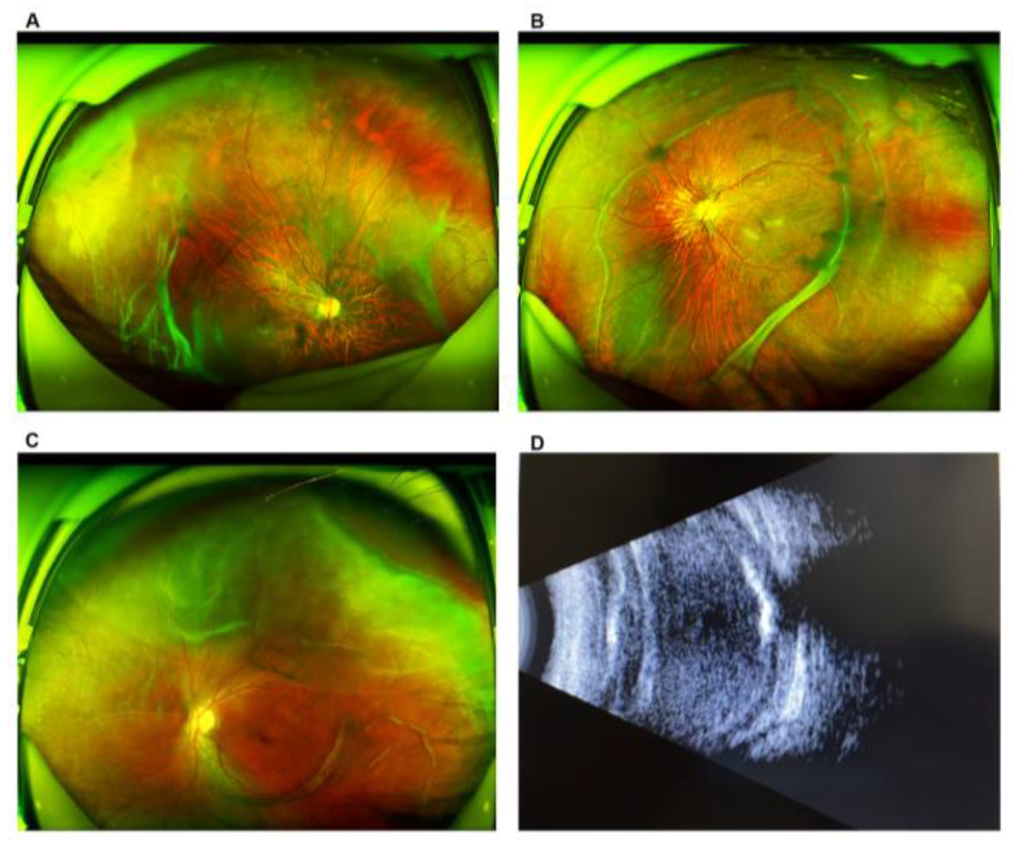
3.1. Demographic and Clinical Characteristics

A total of 84 eyes of 42 probands, 23 (55%) male and 19 (45%) female, from 42 unrelated families were included in this study. The Stickler score for each proband is listed in Supplementary Table S1. The diagnosis of Stickler syndrome was based on the sores and especially based on the typical vitreous changes and retinal abnormalities (Supplementary Figure S1). The median age at the first-time examination was eight years old. The P25 and P75 were 5 and 10.25 years, respectively. The median axial length was 27.72 mm, the P25 being 24.15 mm and the P75 being 29.04 mm. The mean spherical equivalent was −11.91 ± 5.37 D.
Twenty-seven probands had COL2A1 mutations, eight had COL11A1 mutations, and seven had no detected mutations. The first examination age was statistically different among the three groups (p = 0.048). There was no statistical difference (Kruskal–Wallis test. p = 0.260) for the onset age and retinal detachment amongst the different groups of patients. Thirty-two (32/42, 76%) probands had high myopia in total, 21 (21/27, 78%) of whom had COL2A1 mutations, and 5 (5/8, 63%) had COL11A1 mutations. Retinal detachment was present in 29 (29/42, 69%) probands, 20 (20/27, 74%) of whom had COL2A1 mutations, and 4 (4/8, 50%) had COL11A1 mutations (Table 1, Figure 1). No correlation was found between the Stickler clinical score and retinal detachment (logistic regression, B = −0.294, p = 0.507). Glaucoma was present in five (5/42, 12%) probands, all of whom had COL2A1 mutations. Cataracts were present in 10 (10/42, 24%) probands, 6 (6/27, 22%) of whom had COL2A1, and 2 (2/8, 25%) had COL11A1 mutations (Table 1). Other clinical manifestations were a midface and depressed nasal bridge (six probands), mandibular developmental delay (five probands), and cleft palate (five probands).

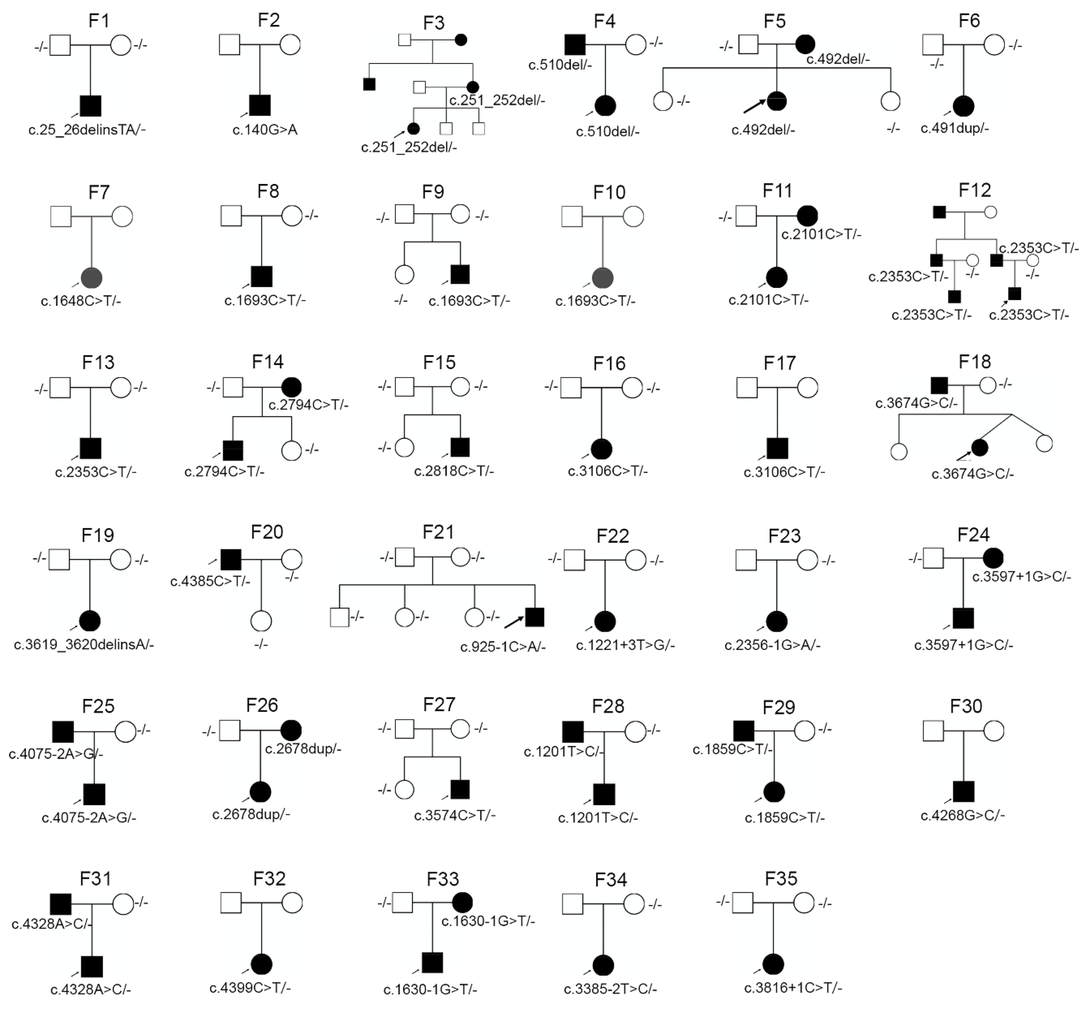
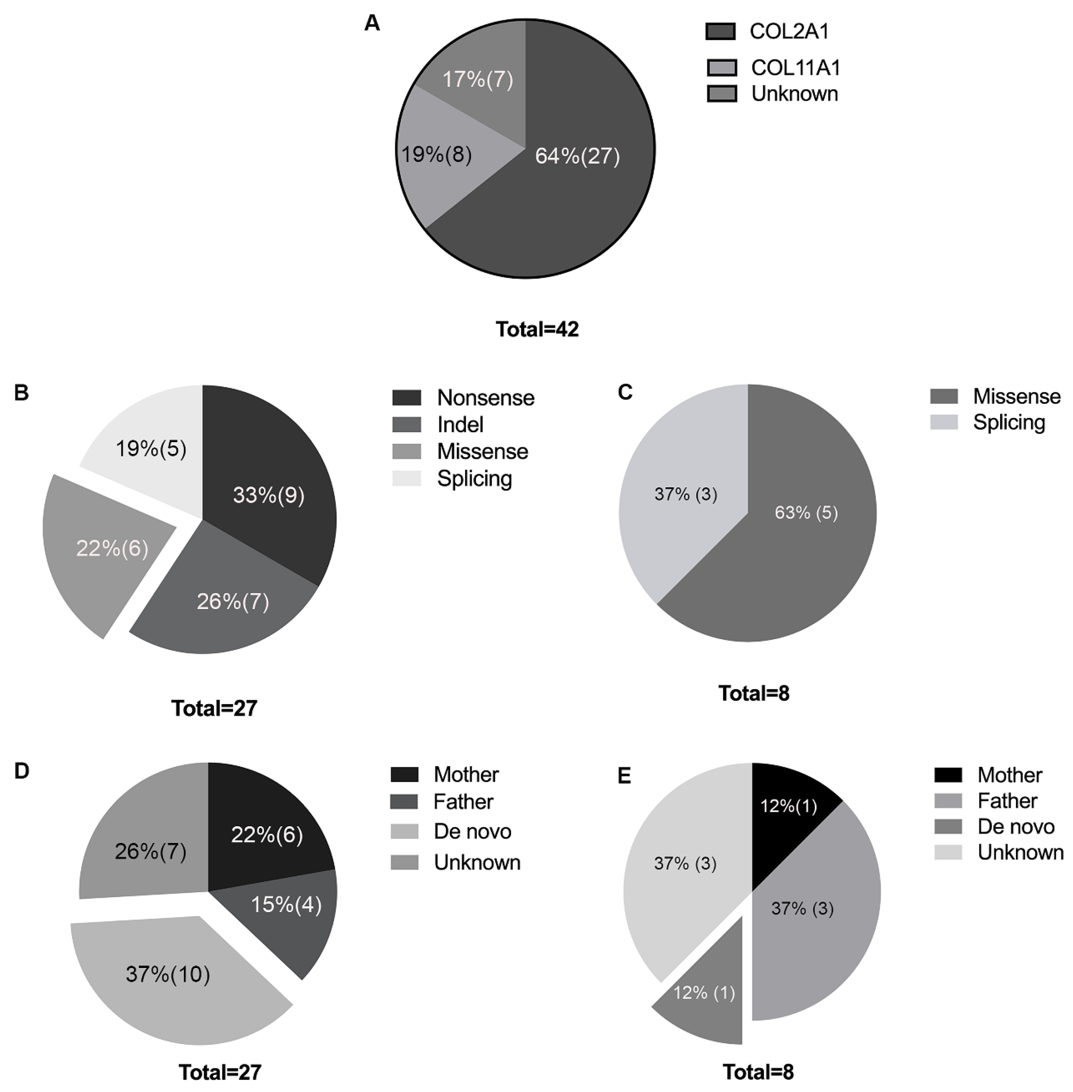
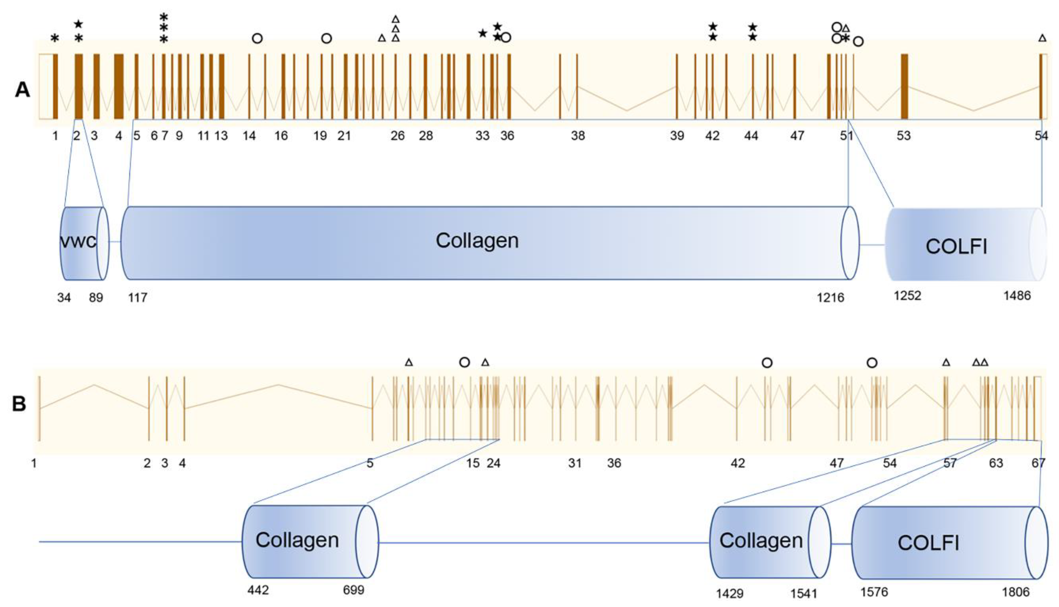
3.2. Genetic Analysis

After TGP sequencing or WES, pathogenic mutations were detected in 35 families (83%), including 27 (64%) with COL2A1 and 8 (19%) with COL11A1 mutations (Table 2, Figure 2 and Figure 3A), all in the heterozygous state (Table 2, Figure 2 and Figure 3A). A total of 23 mutations, 11 known mutations [12,14,15,16,17,18,19,20,21] and 12 novel mutations, were detected in COL2A1. These mutations were distributed among the exons with no mutation hot spots (Figure 4). Nonsense, indel, splicing, and missense mutations were detected in nine, seven, five, and six families, respectively. Of those, 21 (78%) had truncational mutations (Figure 3B). Eight mutations, two known mutations [18,22] and six novel mutations, were detected in COL11A1. Missense and splicing mutations were detected in five (63%) and three (37%) families, respectively (Figure 3C).

3.3. De Novo Mutations in COL2A1 and COL11A1

Segregation analysis in the available family members revealed de novo mutations in 37% (10/27) of the families with COL2A1 mutations (Figure 3D) and 12% (one in eight) of the families with COL11A1 mutations (Figure 3E). The family histories and family trees of these families were double-checked to verify the biological parenthood. The reproductive ages of the families with de novo mutations are displayed in Table 3. The mean paternal childbearing age was 29.64 ± 4.97 years, and the mean maternal childbearing age was 27.36 ± 4.97 years (Table 3). Except for two parents with mild myopia, all the other parents of the probands with de novo mutations had normal fundi and a BCVA better than 1.0.

3.4. Clinical Manifestation in Probands with COL2A1 Missense Mutations

Four different missense mutations (two known and two novel) were detected in six probands in COL2A1. Proband 20, with c.4385C > T (p.Arg1459Cys), developed retinal detachment in the right eye at the age of 19, following which they underwent pars plana vitrectomy (PPV), and glaucoma in their left eye at the age of 30. Proband 18, with c.3674G > C (p.Gly1225Ala), presented with bilateral leukocoria at two months of age and was subsequently diagnosed with retinal detachment bilaterally. Proband 7, with c.1648C > T (p.Arg550Cys), had high myopia and vitreous degeneration with no detected complications at the age of six. Three probands with c.1693C > T (p.Arg565Cys) mutations developed retinal detachment during early childhood bilaterally at 6, 8, and 10 years of age. One of them underwent timely PPV operations, and the other two developed ophthalmatrophia in one eye. In total, there were five out of six probands with missense COL2A1 mutations that had retinal detachment, whereas 15 out of 21 probands with truncated COL2A1 mutations had retinal detachment. There was no significant difference (Fisher’s exact test, p = 0.498) in the numbers with retinal detachment between the probands with missense and truncated COL2A1 mutations.

4. Discussion

In this study, we evaluated the mutation spectrum of patients with Stickler syndrome whose initial symptom was high myopia or retinal detachment.
To date, mutations in seven genes (COL2A1 [4], COL11A1 [5], COL11A2 [6], COL9A1 [7], COL9A2 [8], COL9A3 [9], and LOXL3 [10]) have been reported to cause Stickler syndrome. COL2A1 mutations explained the majority (80–90%) of cases [18,23], followed by COL11A1 mutations (10–20%) [23]. COL11A2, COL9A1, COL9A2, COL9A3, and LOXL3 explained only a few cases, mostly documented in case reports or case series [8,10,24,25,26]. These observations are consistent with this study’s findings. We detected mutations in only COL2A1 (64%) and COL11A2 (19%) and no mutations in the other genes. There may be several reasons for this. First, this study included patients with initial signs of high myopia or retinal detachment; however, COL11A2 mutations have been reported in non-ocular Stickler syndrome [25,27]. Second, patients with COL9A1 and COL9A3 mutations mainly present with moderate to severe myopia and rarely with retinal detachment [24,28]. Finally, the rest of the genes are inherited as autosomal recessive traits, whereas the families recruited in this study were autosomal dominant or sporadic cases. In this study, the first examination age was statistically significantly different among the three groups (p = 0.048). However, no statistically significant difference was found in the patient numbers with retinal detachment among the three groups. Perhaps longer periods of follow up and larger numbers of patients would provide additional insights into the different clinical trajectories and outcomes of COL2A1 and COL11A1 mutant patients.
Various types of mutations in COL2A1 have been reported, and truncation mutations (nonsense, frameshift, or splicing) that result in a premature stop codon are present in most patients with Stickler syndrome [1,16]. This is in line with the findings in our study, where truncation mutations were detected in 78% of the probands with COL2A1 mutations. We also detected four missense mutations in six probands, five of whom presented with retinal detachment, which is a fairly severe ocular complication.
De novo mutations in Stickler syndrome have only been reported in a few cases [29]. The proportion of cases caused by de novo pathogenic variants is unknown. In this study, we observed a large proportion of probands with de novo mutations (37% COL2A1 and 12% COL11A1) because these mutations could not be detected in the leukocyte DNA of either parent. The actual proportion of probands with de novo mutations may have been higher as 10 probands were of unknown inheritance. De novo mutations have been established as the cause of early-onset disorders ranging from rare congenital diseases to common neurodevelopment disorders [30,31,32]. Studies have demonstrated that the majority of germline de novo mutations have a paternal origin and that a higher paternal age and the number of de novo mutation increases with paternal childbearing age [33,34]. In this study, we evaluated both the paternal and maternal ages. The paternal childbearing age (29.64 ± 4.97 years old) might be one of the reasons for the high proportion of de novo mutations in this study. Further studies are needed to ascertain the origins of the mutations.
This study was a real-world single-center study. Its sample size was limited due to the rarity of Stickler syndrome and all patients being recruited in a single department (the Pediatric Ophthalmology Department of Zhongshan Ophthalmic Center). The participants presented with an initial sign of high myopia or retinal detachment, which means that the identified mutation spectrum can only be considered representative of the specific phenotypes of Stickler syndrome patients. In this study, we did not use the genomic markers to verify parentage, and the parentage of families with de novo mutations will be further confirmed by genomic markers.

5. Conclusions

We investigated the mutation features in Stickler syndrome patients with high myopia or retinal detachment. Mutations were detected only in COL2A1 (64%) and COL11A1 (19%). No mutations were detected in other genes known to be associated with Stickler syndrome. De novo mutations accounted for 37% of all COL2A1 mutations.

Supplementary Materials

The following are available online at https://www.mdpi.com/2073-4425/11/8/882/s1. Figure S1: Vitreous changes or retinal abnormalities from Scanning laser ophthalmoscopy (SLO), Table S1: Diagnose of the probands.

Author Contributions

Data curation, L.H., C.C., Z.W., L.S., S.L., T.Z., and X.L.; funding acquisition, L.H. and X.D.; investigation, X.D.; writing—original draft, L.H.; writing—review and editing, C.C. and X.D. All authors have read and agreed to the published version of the manuscript.

Funding

This study was supported by research funds of Sun Yat-sen University (15ykjxc22d); the Science and Technology Program, Guangzhou, China (201803010031); the National Natural Science Foundation of China (81700879); and the Guangdong Natural Science Foundation (grant no. 2017A030310275).

Acknowledgments

The authors are grateful to the patients for their participation.

Conflicts of Interest

The authors declare no conflict of interest.

References

  1. Stickler, G.B.; Belau, P.G.; Farrell, F.J.; Jones, J.D.; Pugh, D.G.; Steinberg, A.G.; Ward, L.E. Hereditary Progressive Arthro-Ophthalmopathy. Mayo Clin. Proc. 1965, 40, 433–455. [Google Scholar] [PubMed]
  2. Robin, N.H.; Moran, R.T.; Ala-Kokko, L. Stickler Syndrome. In GeneReviews®; Adam, M.P., Ardinger, H.H., Pagon, R.A., Wallace, S.E., Bean, L.J.H., Stephens, K., Amemiya, A., Eds.; University of Washington: Seattle, WA, USA, 1993. [Google Scholar]
  3. Liberfarb, R.M.; Goldblatt, A. Prevalence of mitral-valve prolapse in the Stickler syndrome. Am. J. Med. Genet. 1986, 24, 387–392. [Google Scholar] [CrossRef] [PubMed]
  4. Francomano, C.A.; Liberfarb, R.M.; Hirose, T.; Maumenee, I.H.; Streeten, E.A.; Meyers, D.A.; Pyeritz, R.E. The Stickler syndrome: Evidence for close linkage to the structural gene for type II collagen. Genomics 1987, 1, 293–296. [Google Scholar] [CrossRef]
  5. Richards, A.J.; Yates, J.R.; Williams, R.; Payne, S.J.; Pope, F.M.; Scott, J.D.; Snead, M.P. A family with Stickler syndrome type 2 has a mutation in the COL11A1 gene resulting in the substitution of glycine 97 by valine in alpha 1 (XI) collagen. Hum. Mol. Genet. 1996, 5, 1339–1343. [Google Scholar] [CrossRef] [PubMed] [Green Version]
  6. Melkoniemi, M.; Brunner, H.G.; Manouvrier, S.; Hennekam, R.; Superti-Furga, A.; Kaariainen, H.; Pauli, R.M.; van Essen, T.; Warman, M.L.; Bonaventure, J.; et al. Autosomal recessive disorder otospondylomegaepiphyseal dysplasia is associated with loss-of-function mutations in the COL11A2 gene. Am. J. Hum. Genet. 2000, 66, 368–377. [Google Scholar] [CrossRef] [Green Version]
  7. Van Camp, G.; Snoeckx, R.L.; Hilgert, N.; van den Ende, J.; Fukuoka, H.; Wagatsuma, M.; Suzuki, H.; Smets, R.M.; Vanhoenacker, F.; Declau, F.; et al. A new autosomal recessive form of Stickler syndrome is caused by a mutation in the COL9A1 gene. Am. J. Hum. Genet. 2006, 79, 449–457. [Google Scholar] [CrossRef] [Green Version]
  8. Baker, S.; Booth, C.; Fillman, C.; Shapiro, M.; Blair, M.P.; Hyland, J.C.; Ala-Kokko, L. A loss of function mutation in the COL9A2 gene causes autosomal recessive Stickler syndrome. Am. J. Med. Genet. Part A 2011, 155A, 1668–1672. [Google Scholar] [CrossRef]
  9. Nakashima, E.; Kitoh, H.; Maeda, K.; Haga, N.; Kosaki, R.; Mabuchi, A.; Nishimura, G.; Ohashi, H.; Ikegawa, S. Novel COL9A3 mutation in a family with multiple epiphyseal dysplasia. Am. J. Med. Genet. Part A 2005, 132A, 181–184. [Google Scholar] [CrossRef]
  10. Alzahrani, F.; Al Hazzaa, S.A.; Tayeb, H.; Alkuraya, F.S. LOXL3, encoding lysyl oxidase-like 3, is mutated in a family with autosomal recessive Stickler syndrome. Hum. Genet. 2015, 134, 451–453. [Google Scholar] [CrossRef]
  11. Snead, M.P.; Yates, J.R. Clinical and Molecular genetics of Stickler syndrome. J. Med. Genet. 1999, 36, 353–359. [Google Scholar]
  12. Hoornaert, K.P.; Vereecke, I.; Dewinter, C.; Rosenberg, T.; Beemer, F.A.; Leroy, J.G.; Bendix, L.; Bjorck, E.; Bonduelle, M.; Boute, O.; et al. Stickler syndrome caused by COL2A1 mutations: Genotype-phenotype correlation in a series of 100 patients. Eur. J. Hum. Genet. EJHG 2010, 18, 872–880. [Google Scholar] [CrossRef] [PubMed]
  13. Tang, M.; Ding, X.; Li, J.; Hu, A.; Yuan, M.; Yang, Y.; Zhan, Z.; Li, Z.; Lu, L. Novel mutations in FZD4 and phenotype-genotype correlation in Chinese patients with familial exudative vitreoretinopathy. Mol. Vis. 2016, 22, 917–932. [Google Scholar] [PubMed]
  14. Barat-Houari, M.; Dumont, B.; Fabre, A.; Them, F.T.; Alembik, Y.; Alessandri, J.L.; Amiel, J.; Audebert, S.; Baumann-Morel, C.; Blanchet, P.; et al. The expanding spectrum of COL2A1 gene variants IN 136 patients with a skeletal dysplasia phenotype. Eur. J. Hum. Genet. EJHG 2016, 24, 992–1000. [Google Scholar] [CrossRef] [PubMed] [Green Version]
  15. Sun, W.; Huang, L.; Xu, Y.; Xiao, X.; Li, S.; Jia, X.; Gao, B.; Wang, P.; Guo, X.; Zhang, Q. Exome Sequencing on 298 Probands With Early-Onset High Myopia: Approximately One-Fourth Show Potential Pathogenic Mutations in RetNet Genes. Investig. Ophthalmol. Vis. Sci. 2015, 56, 8365–8372. [Google Scholar] [CrossRef] [Green Version]
  16. Richards, A.J.; Laidlaw, M.; Whittaker, J.; Treacy, B.; Rai, H.; Bearcroft, P.; Baguley, D.M.; Poulson, A.; Ang, A.; Scott, J.D.; et al. High efficiency of mutation detection in type 1 stickler syndrome using a two-stage approach: Vitreoretinal assessment coupled with exon sequencing for screening COL2A1. Hum. Mutat. 2006, 27, 696–704. [Google Scholar] [CrossRef]
  17. Savasta, S.; Salpietro, V.; Sparta, M.V.; Foiadelli, T.; Laino, D.; Lobefalo, L.; Marseglia, G.L.; Verrotti, A. Stickler syndrome associated with epilepsy: Report of three cases. Eur. J. Pediatr. 2015, 174, 697–701. [Google Scholar] [CrossRef] [PubMed]
  18. Annunen, S.; Korkko, J.; Czarny, M.; Warman, M.L.; Brunner, H.G.; Kaariainen, H.; Mulliken, J.B.; Tranebjaerg, L.; Brooks, D.G.; Cox, G.F.; et al. Splicing mutations of 54-bp exons in the COL11A1 gene cause Marshall syndrome, but other mutations cause overlapping Marshall/Stickler phenotypes. Am. J. Hum. Genet. 1999, 65, 974–983. [Google Scholar] [CrossRef] [Green Version]
  19. Kondo, H.; Matsushita, I.; Nagata, T.; Hayashi, T.; Kakinoki, M.; Uchio, E.; Kondo, M.; Ohji, M.; Kusaka, S. Novel mutations in the COL2A1 gene in Japanese patients with Stickler syndrome. Hum. Genome. Var. 2016, 3, 16018. [Google Scholar] [CrossRef]
  20. Zechi-Ceide, R.M.; Jesus Oliveira, N.A.; Guion-Almeida, M.L.; Antunes, L.F.; Richieri-Costa, A.; Passos-Bueno, M.R. Clinical evaluation and COL2A1 gene analysis in 21 Brazilian families with Stickler syndrome: Identification of novel mutations, further genotype/phenotype correlation, and its implications for the diagnosis. Eur. J. Med. Genet. 2008, 51, 183–196. [Google Scholar] [CrossRef]
  21. Richards, A.J.; Martin, S.; Yates, J.R.; Scott, J.D.; Baguley, D.M.; Pope, F.M.; Snead, M.P. COL2A1 exon 2 mutations: Relevance to the Stickler and Wagner syndromes. Br. J. Ophthalmol. 2000, 84, 364–371. [Google Scholar] [CrossRef] [Green Version]
  22. Ala-Kokko, L.; Shanske, A.L. Mosaicism in Marshall Syndrome. Am. J. Med. Genet. Part A 2009, 149, 1327–1330. [Google Scholar] [CrossRef] [PubMed]
  23. Acke, F.R.; Malfait, F.; Vanakker, O.M.; Steyaert, W.; De Leeneer, K.; Mortier, G.; Dhooge, I.; De Paepe, A.; De Leenheer, E.M.; Coucke, P.J. Novel pathogenic COL11A1/COL11A2 variants in Stickler syndrome detected by targeted NGS and exome sequencing. Mol. Genet. Metab. 2014, 113, 230–235. [Google Scholar] [CrossRef] [PubMed]
  24. Nikopoulos, K.; Schrauwen, I.; Simon, M.; Collin, R.W.; Veckeneer, M.; Keymolen, K.; Van Camp, G.; Cremers, F.P.; van den Born, L.I. Autosomal recessive Stickler syndrome in two families is caused by mutations in the COL9A1 gene. Investig. Ophthalmol. Vis. Sci. 2011, 52, 4774–4779. [Google Scholar] [CrossRef] [PubMed] [Green Version]
  25. Iwasa, Y.; Moteki, H.; Hattori, M.; Sato, R.; Nishio, S.Y.; Takumi, Y.; Usami, S. Non-ocular Stickler syndrome with a novel mutation in COL11A2 diagnosed by massively parallel sequencing in Japanese hearing loss patients. Ann. Otol. Rhinol. Laryngol. 2015, 124 (Suppl. 1), 111S–117S. [Google Scholar] [CrossRef]
  26. Faletra, F.; D’Adamo, A.P.; Bruno, I.; Athanasakis, E.; Biskup, S.; Esposito, L.; Gasparini, P. Autosomal recessive Stickler syndrome due to a loss of function mutation in the COL9A3 gene. Am. J. Med. Genet. Part A 2014, 164A, 42–47. [Google Scholar] [CrossRef]
  27. Admiraal, R.J.; Brunner, H.G.; Dijkstra, T.L.; Huygen, P.L.; Cremers, C.W. Hearing loss in the nonocular Stickler syndrome caused by a COL11A2 mutation. Laryngoscope 2000, 110, 457–461. [Google Scholar] [CrossRef]
  28. Nixon, T.R.W.; Alexander, P.; Richards, A.; McNinch, A.; Bearcroft, P.W.P.; Cobben, J.; Snead, M.P. Homozygous Type IX collagen variants (COL9A1, COL9A2, and COL9A3) causing recessive Stickler syndrome-Expanding the phenotype. Am. J. Med. Genet. Part A 2019, 179, 1498–1506. [Google Scholar] [CrossRef]
  29. Spickett, C.; Hysi, P.; Hammond, C.J.; Prescott, A.; Fincham, G.S.; Poulson, A.V.; McNinch, A.M.; Richards, A.J.; Snead, M.P. Deep Intronic Sequence Variants in COL2A1 Affect the Alternative Splicing Efficiency of Exon 2, and May Confer a Risk for Rhegmatogenous Retinal Detachment. Hum. Mutat. 2016, 37, 1085–1096. [Google Scholar] [CrossRef] [Green Version]
  30. Acuna-Hidalgo, R.; Veltman, J.A.; Hoischen, A. New insights into the generation and role of de novo mutations in health and disease. Genome Biol. 2016, 17, 241. [Google Scholar] [CrossRef] [Green Version]
  31. Ng, S.B.; Bigham, A.W.; Buckingham, K.J.; Hannibal, M.C.; McMillin, M.J.; Gildersleeve, H.I.; Beck, A.E.; Tabor, H.K.; Cooper, G.M.; Mefford, H.C.; et al. Exome sequencing identifies MLL2 mutations as a cause of Kabuki syndrome. Nat. Genet. 2010, 42, 790–793. [Google Scholar] [CrossRef] [Green Version]
  32. de Ligt, J.; Willemsen, M.H.; van Bon, B.W.; Kleefstra, T.; Yntema, H.G.; Kroes, T.; Vulto-van Silfhout, A.T.; Koolen, D.A.; de Vries, P.; Gilissen, C.; et al. Diagnostic exome sequencing in persons with severe intellectual disability. N. Engl. J. Med. 2012, 367, 1921–1929. [Google Scholar] [CrossRef] [PubMed] [Green Version]
  33. Goldmann, J.M.; Wong, W.S.; Pinelli, M.; Farrah, T.; Bodian, D.; Stittrich, A.B.; Glusman, G.; Vissers, L.E.; Hoischen, A.; Roach, J.C.; et al. Parent-of-origin-specific signatures of de novo mutations. Nat. Genet. 2016, 48, 935–939. [Google Scholar] [CrossRef] [PubMed]
  34. Kong, A.; Frigge, M.L.; Masson, G.; Besenbacher, S.; Sulem, P.; Magnusson, G.; Gudjonsson, S.A.; Sigurdsson, A.; Jonasdottir, A.; Jonasdottir, A.; et al. Rate of de novo mutations and the importance of father’s age to disease risk. Nature 2012, 488, 471–475. [Google Scholar] [CrossRef] [PubMed] [Green Version]
Figure 1. Scanning laser ophthalmoscopy (SLO) and B-scans of patients with Stickler syndrome. (A) and (B) are from the same proband. (A) Vitreous degeneration, multiple retinal holes, and retinal detachment in the right eye. (B) Vitreous degeneration and retinal holes in the left eye. (C,D) are from the same proband. (C) Retinal detachment of the left eye. (D) B-scan showing retinal detachment, vitreous opacity, and ophthalmatrophia.
Genes 11 00882 g001
Figure 2. Pedigrees of families with COL2A1 and COL11A1 mutations. F1 to F27 are families with COL2A1 mutations, and F28 to F35 are families with COL11A1 mutations. The arrows indicate probands; the squares indicate men; the circles indicate women; filled symbols indicate affected individuals; open symbols indicate unaffected individuals.
Genes 11 00882 g002
Figure 3. (A) Mutation spectrum of the study cohort. (B) Composition of COL2A1 mutation types. (C) Composition of COL11A1 mutation types. (D) Mutation inheritance of the COL2A1 gene. (E) Mutation inheritance of COL11A1.
Genes 11 00882 g003
Figure 4. Distribution of COL2A1 and COL11A1 mutations. The circles indicate splicing mutations; the triangles indicate missense mutations; the asterisks indicate indel mutations; the pentagrams indicate nonsense mutations. (A) Mutations in COL2A1. The upper line shows the exons of COL2A1, and the lower line shows the domains of the COL2A1-encoded protein. (B). Mutations in COL11A1. The upper line shows the exons of COL11A1, and the lower line shows the domains of the COL2A1 encoded protein.
Genes 11 00882 g004
Table 1. Demographic and clinical data for this study.
Variable TotalCOL2A1COL11A1Unknown
Number (total)4227 (64%)8 (19%)7 (21%)
Male (n, %)23 (55%)14 (52%)4 (50%)5 (71%)
Female (n, %)19 (45%)13 (48%)4 (50%)2 (29%)
Age (years) (median (P25, P75))8 (5, 10.25)7 (5, 11)10 (5.75, 20.25)12 (4, 18)
AL (mm) (median (P25, P75)) 27.20 (24.15, 29.04)27.06 (24.99, 28.59)24.96 (23.58, 29.16)28.71 (21.30, 29.83)
SE (D) (Mean ± SD)−11.91 ± 5.37−11.64 ± 4.95−9.43 ± 7.27−15.58 ± 3.74
HM (n, %)32 (76%)21 (78%)5 (63%)6 (86%)
RD (n, %)29 (69%)20 (74%)4 (50%)5 (71%)
RD (eyes, %)37 (44%)25 (46%)5 (31%)7 (50%)
Glaucoma (n, %)5 (12%)5 (19%)00
Cataract (n, %)10 (24%)6 (22%)2 (25%)2 (29%)
Note: Age, Age at first examination; HM, High myopia; AL, Axial length; SE, Spherical equivalent; RD, Retinal detachment.
Table 2. Mutations detected in this study.
Family IDSequenceGeneExonMutation TypeLocationDNA ChangeAmino Acid Change ExAC—AllExAC—East AsianReference
1TGPCOL2A11indelchr12: 48398079c.25_26delinsTAp.Thr9*00novel
2TGPCOL2A12nonsensechr12:48393854c.140G > Ap.Trp47*00novel
3TGPCOL2A12indelchr12:48393736c.251_252delp.Glu84Valfs*800novel
4TGPCOL2A17indelchr12:48391409c.510delp.Gly171Valfs*2800[21]
5WESCOL2A17indelchr12:48393736c.492delp.Gly165Valfs*3400novel
6TGPCOL2A17indelchr12:48391428c.491dupp.Gly165Trpfs*2400novel
7TGPCOL2A125missensechr12:48379543c.1648C > Tp.Arg550Cys00[14]
8WESCOL2A126missensechr12:48379358c.1693C > Tp.Arg565Cys00[15]
9TGPCOL2A126missensechr12:48379358c.1693C > Tp.Arg565Cys00[15]
10TGPCOL2A126missensechr12:48379358c.1693C > Tp.Arg565Cys00[15]
11WESCOL2A133nonsensechr12:48376723 c.2101C > Tp.Arg701*00[16]
12TGPCOL2A135nonsensechr12:48375892c.2353C > Tp.Arg785*00[21]
13WESCOL2A135nonsensechr12:48375892c.2353C > Tp.Arg785*00[21]
14TGPCOL2A142nonsensechr12:48372481c.2794C > Tp.Arg932*00[15]
15WESCOL2A142nonsensechr12:48372457 c.2818C > Tp.Arg940*00[19]
16TGPCOL2A144nonsensechr12:48371798c.3106C > Tp.Arg1036*1/1148020/8542[17]
17TGPCOL2A144nonsensechr12:48371798c.3106C > Tp.Arg1036*1/1148020/8542[17]
18TGPCOL2A151missensechr12:48369312c.3674G > Cp.Gly1225Ala00novel
19WESCOL2A151indelchr12:48369362 c.3619_3620delinsAp.Pro1207Thrfs*2000novel
20TGPCOL2A154missensechr12:48367279c.4385C > Tp.Arg1459Cys3/1214100/8654novel
21WESCOL2A1Intro 14splicingchr12:48387286c.925-1C > A_00[18]
22WESCOL2A1Intro 19splicingchr12:48381391c.1221+3T > G_00novel
23TGPCOL2A1Intro 35splicingchr12:48375613c.2356-1G > A 00novel
24TGPCOL2A1Intro 50splicingchr12:48369745c.3597+1G > C_00novel
25WESCOL2A1Intro 52splicingchr12:48368116c.4075-2A > G 00Novel
26WESCOL2A140indelchr12:48373792c.2678dupp.Pro893fs00[12]
27WESCOL2A150nonsensechr12:48369769c.3574C > Tp.Arg1192*00[20]
28TGPCOL11A18missensechr1:103488342c.1201T > Cp.Phe401Leu1/1199521/8594novel
29WESCOL11A119missensechr1:103470204 c.1859C > Tp.Pro620Leu00novel
30WESCOL11A157missensechr1:103363724c.4268G > Cp.Gly1423Ala00novel
31WESCOL11A158missensechr1:103356035c.4328A > Cp.Lys1443Thr4/1027222/7546novel
32TGPCOL11A159missensechr1:103355112c.4399C > Tp.Gln1467*2/1207302/8600novel
33WESCOL11A1intro 14splicingchr1:103474073 c.1630-1G > T 00novel
34TGPCOL11A1intro 43splicingchr1:103404646c.3385-2T > C_00[18]
35TGPCOL11A1intro 50splicingchr1:103381186c.3816+1C > T_00[22]
Note: TGP, Targeted gene pane; WES, Whole exome sequencing. *, stop codon.
Table 3. Childbearing ages of the parents whose children had de novo mutations.
Family IDGeneDNA ChangesAmino Acid ChangesPaternal Childbearing Age (Years)Maternal Childbearing Age (Years)
1COL2A1c.25_26delinsTAp.Thr9*3031
6COL2A1c.491dupp.Gly165Trpfs*243030
9COL2A1c.1693C > Tp.Arg565Cys2921
13COL2A1c.2353C > Tp.Arg785*2423
15COL2A1c.2818C > Tp.Arg940*3433
16COL2A1c.3106C > Tp.Arg1036*2623
19COL2A1c.3619_3620delinsAp.Pro1207Thrfs*203333
21COL2A1c.925-1C > A_3828
22COL2A1c.1221 + 3T > G_3430
27COL2A1c.3574C > Tp.R1192*2119
35COL11A1c.3816 + 1C > T_2730
Mean ± SD 29.64 ± 4.9727.36 ± 4.97
Note: *, stop codon.

