Article

A 3D Printing-Based Transcatheter Pulmonary Valve Replacement Simulator: Development and Validation

Department of Cardiovascular Surgery, Xijing Hospital, Air Force Medical University, Xi’an 710032, China
*
Authors to whom correspondence should be addressed.
These authors contributed equally to this work.
Bioengineering 2025, 12(4), 344; https://doi.org/10.3390/bioengineering12040344
Submission received: 13 February 2025 / Revised: 24 March 2025 / Accepted: 25 March 2025 / Published: 26 March 2025
(This article belongs to the Special Issue The New Frontiers of Artificial Organs Engineering)

Abstract

:
Background: Severe pulmonary regurgitation (PR) often occurs after treatment of tetralogy of Fallot with a valve ring patch, leading to enlargement and diverse morphological characteristics of the native right ventricular outflow tract (nRVOT), which increases the difficulty of transcatheter pulmonary valve replacement (TPVR). The purpose of this study was to use the TPVR simulator to help doctors improve their surgical skills by simulating the surgical process in vitro. Methods: The TPVR simulator was developed using three-dimensional (3D) printing technology under computer-aided design. In this study, the TPVR simulator was used for preoperative simulation training and teaching. First, 10 specialists were equally divided into a 3D-printed group and a non-3D-printed group, each performing one TPVR; then, another six specialists and six young surgeons were selected to complete three TPVR simulations. Results: For the 3D-printed simulation group, the over-flap time (5.22 min (range: 4.85–5.87 min) vs. 6.72 min (range: 6.12–7.70 min), p = 0.016), fluoroscopy time (15.00 min (range: 13.50–16.50 min) vs. 19.00 min (range: 17.50–21.50 min), p = 0.012), and total operative time for the five surgeons (57.00 min (range: 54.00–62.50 min) vs. 67.00 min (range: 62.00–69.50 min), p = 0.036) were shorter. In addition, the results showed significant reductions in the median over-flap time and total time required in both the expert panel and young surgeon groups (all p < 0.05). Conclusions: The reliability and validity of the TPVR simulator was initially demonstrated and has the potential to be a teaching and training tool for surgeons.

1. Introduction

Severe pulmonary regurgitation (PR) represents the most prevalent lesion in the aftermath of tetralogy of Fallot treatment, manifesting in both pediatric and adult patients. The clinical presentation is characterized by right ventricular dilatation and dysfunction, the occurrence of arrhythmias, and limitations in exercise capacity [1,2]. Transcatheter pulmonary valve replacement (TPVR) has been demonstrated to be a promising treatment option for PR. It has been shown to reduce the need for additional surgical intervention in PR patients [3,4,5,6]. It is unfortunate that approximately 80% of patients undergoing surgical RVOT reconstruction receive either retained valve repair or transannular patching [7]. Not only does this treatment lead to severe PR later in the patient’s life, but it also results in a variety of morphological features of the nRVOT that increase the surgical difficulty of TPVR [8].
As medical visualization continues to evolve, there is an increasing demand for accuracy in digital modeling. This demand is particularly pronounced in the field of cardiovascular disease, where precision is of the essence [9]. In recent years, three-dimensional (3D) printing has emerged as a technology that is both user-friendly and versatile. This is a technology application that prints a corresponding model that exactly matches the reconstructed model, layer by layer, based on 3D reconstruction data formed by medical imaging scans, providing surgeons with a personalized model that helps them understand anatomy at a glance [10]. As 3D printing technology continues to improve, there are anecdotal reports that it can help in the implementation of TPVR [11,12]. Furthermore, patient-specific 3D-printed models can be used to simulate preoperative conditions to assess the risk of complications such as coronary compression and perivalvular leakage, and to develop a personalized preoperative plan that can help improve the safety and effectiveness of surgery [13,14].
PR is a major disease that affects the quality of life of patients. Improvements in its treatment are crucial to improving patient outcomes. The objective of this research is to utilize a TPVR simulator to help doctors improve their surgical skills by simulating the in vitro surgical process, while filling the current research gap in the application of 3D printing technology in TPVR training. By converting patient CT data into an actionable 3D model, a realistic simulation training environment is provided for young cardiovascular specialists, thereby improving surgical outcomes and the overall prognosis of patients. The research methods mainly include the production of 3D-printed models and their application in surgical training, with a focus on evaluating the effectiveness of this technology in surgical training and comparing it with traditional training methods. Through rigorous research design and data analysis, this study hopes to provide an empirical basis for the application of 3D printing technology in TPVR training.

2. Materials and Methods

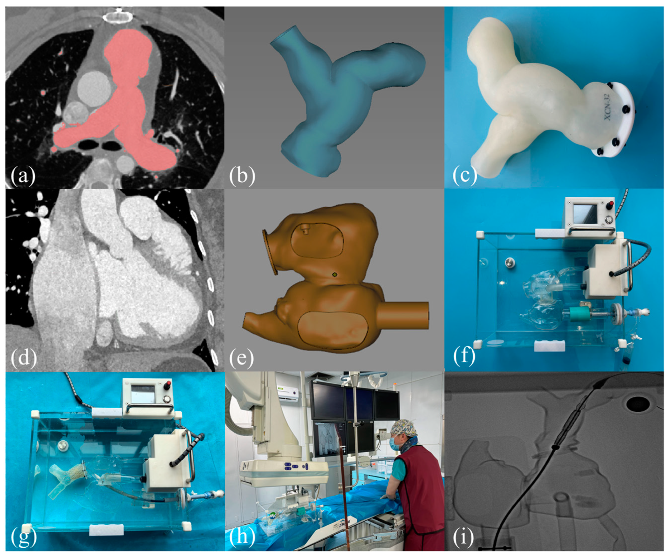
2.1. 3D Printing the RVOT Model Process and Building the TPVR Simulator

The Digital Imaging and Communications in Medicine (DICOM) format of the computed tomography (CT) data of patients with PR was imported into Materialise Mimics Version 21.0 software (Leuven, Belgium). Utilizing the threshold segmentation feature, a three-dimensional reconstruction of the RVOT was generated at the conclusion of the contraction phase. Subsequently, the resulting 3D model was further processed in Materialise 3-Matic Version 13.0 software (Leuven, Belgium), where it underwent extraction, cropping, smoothing, and repairs to accurately restore the anatomical structures in a one-to-one representation. Further, the standard triangular language files of the 3D reconstructions were processed. The model was exported to a Stratasys Polyjet 850 multi-material full-color 3D printer. Specific right ventricle–pulmonary artery (RV-PA) models operate by printing right ventricular and pulmonary artery tissue using rubber-like flexible resin material (tensile strength: 1.95 MPa, elongation at break: 190%, compression strength: 0.300 MPa, shore hardness: 24 Shore A, Stratasys, Israel) and printing calcified tissue using rigid resin material (tensile strength: 31.7 MPa, elongation at break: 25%, flexural strength: 41.4 MPa, yield compression stress: 37.6 MPa, shore hardness: 82 Shore D, Stratasys, Israel).
To construct the TPVR simulator, the patient’s CT data were first divided into three parts—inferior vena cava, right atrium, and right ventricle—to measure the internal diameter and the corresponding curvature. Utilizing the acquired measurements, specialized engineers proceeded to fabricate the respective components of the 3D reconstructed model and meticulously designed the grommets to ensure a secure fit for each segment. The simulator primarily comprised two components: (1) the operational part, which encompassed the transfemoral vein along with the 3D-printed RV-PA model, and (2) the driving part, which featured the circulatory pump, the fully integrated connection loop, and the control mechanism. By creating a pulsatile simulation that mimics pressure–pulsatile blood flow, the 3D-printed RV-PA model, together with the surrounding tissues, can simulate movement under realistic pathophysiological conditions (Figure 1).

2.2. Guidance for 3D Printing

For the professional development of young cardiovascular specialists, simulation training is essential. With the rapid evolution of computer graphics, bioengineering, and digital modeling technologies, 3D printing-based TPVR training systems will lead to more simulation-based training options. Trainees can practice several major pre-dural steps (such as crossing the valve, exchanging threads, positioning the stent) by using specific 3D-printed models to improve their manipulation skills and proficiency for an improved learning curve. It can also help doctors select valves by simulating placement to assess the risk of coronary artery compression, valve migration, and vascular complications.

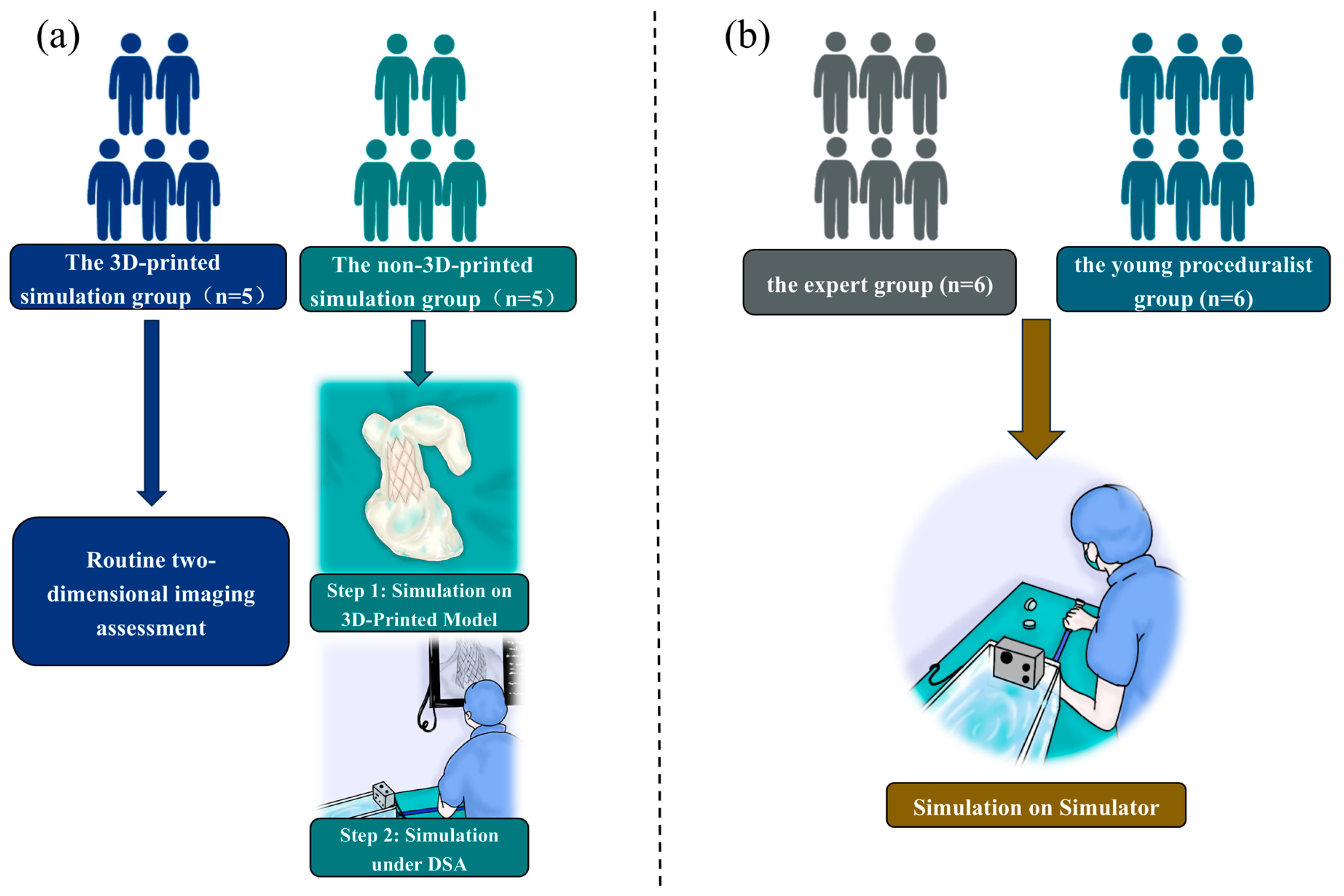
2.3. The 3D-Printed Group Versus the Non-3D-Printed Group

The flow diagram of the entire study is shown in Figure 2. A total of ten patients diagnosed with severe PR were systematically categorized into two groups: a 3D-printed simulation group and a non-3D-printed simulation group. Each group consisted of five patients. In parallel, ten specialists were selected and evenly divided into two cohorts. Within the 3D-printed simulation group, five proceduralists practiced the procedural steps through a bench test prior to TPVR. Conversely, the proceduralists in the non-3D-printed simulation group proceeded with TPVR following standard preoperative assessments. In this study, the Venus P-valve™ (Venus Medtech, Hangzhou, China) was used for both preoperative simulation and intraoperative use in TPVR. Each expert completed one operation, and the crossing-valve time, fluoroscopy time, total time, and the occurrence of the major postoperative complications were recorded and statistically analyzed. This study was reviewed and approved by the Ethics Committee of Xijing Hospital (KY20192138-C1) and conducted in accordance with the principles of the Declaration of Helsinki. The ethical registration was entered in the ClinicalTrials.gov Protocol Registration System (NCT02917980; 27 September 2016). All activities mentioned above were performed within the context of structural heart disease, specifically concentrating on valvular heart disease.

2.4. Experts Versus Young Surgeons in the Simulator

In addition, a total of six experts and six junior proceduralists were chosen and allocated into two distinct groups: one comprising the experts and the other consisting of young proceduralists. Each group contained six individuals. Every participant, whether an expert or a junior proceduralist, underwent three simulation sessions, during which the time taken to cross the valve and the overall duration of the simulations were meticulously documented. The outcomes from the first, second, and third simulation sessions were subsequently compared in pairs and subjected to statistical analysis.

2.5. Statistical Analysis

All statistical evaluations were conducted utilizing the Statistical Package for the Social Sciences (SPSS, Chicago, IL, USA), version 26.0. Continuous variables were expressed as the mean, standard deviation, or median within the interquartile range. Normally distributed data were compared using t-tests and non-normally distributed data were compared using non-parametric tests. A two-sided p-value of less than 0.05 was deemed statistically significant.

3. Results

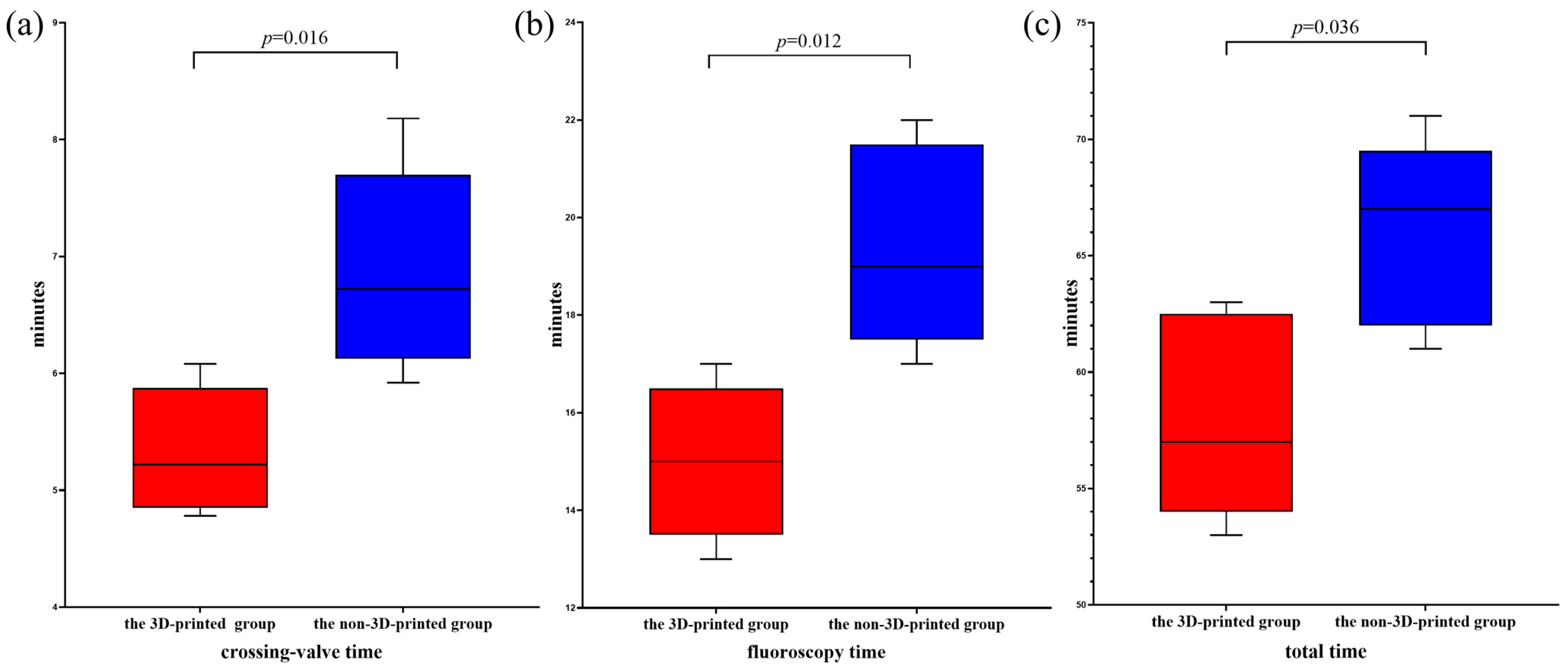
3.1. The 3D-Printed Group Versus the Non-3D-Printed Group

A cohort of ten patients diagnosed with pulmonary valve regurgitation was selected from Xijing Hospital for this study. This group comprised four males and six females, with an average age of 30.50 years (range: 21.25–43.50 years). The baseline characteristics of these patients are detailed in Table 1. Table 2 presents the baseline characteristics of the proceduralists involved in this study. Within the 3D-printed simulation cohort, the average age was recorded at 42.00 years (range: 38.50–44.50 years), while the mean years of professional experience were 9.00 years (range: 7.50–10.50 years) for interventions. Conversely, in the non-3D-printed simulation cohort, the average age was 42.00 years (range: 38.50–44.00 years), with an intervention experience of 8.00 years (range: 7.50–10.50 years). All ten specialists successfully performed one procedure each, with intraoperative data presented in Table 3. In the non-3D-printed simulation cohort, the mean over-the-valve time for the five surgeons was 6.72 min (range: 6.12–7.70 min), the fluoroscopy time averaged 19.00 min (range: 17.50–21.50 min), and the total procedural time was 67.00 min (range: 62.00–69.50 min). In contrast, the five surgeons utilizing the 3D-printed simulation methodology achieved a reduced over-the-valve time of 5.22 min (range: 4.85–5.87 min), with a fluoroscopy duration of 15.00 min (range: 13.50–16.50 min), and a cumulative time of 57.00 min (range: 54.00–62.50 min) (Figure 3). Notably, there were no significant postoperative complications reported in the 3D-printed simulation group, including serious events such as coronary artery compression or valve embolization. However, in the non-3D-printed simulation cohort, two patients experienced hemoptysis postoperatively.

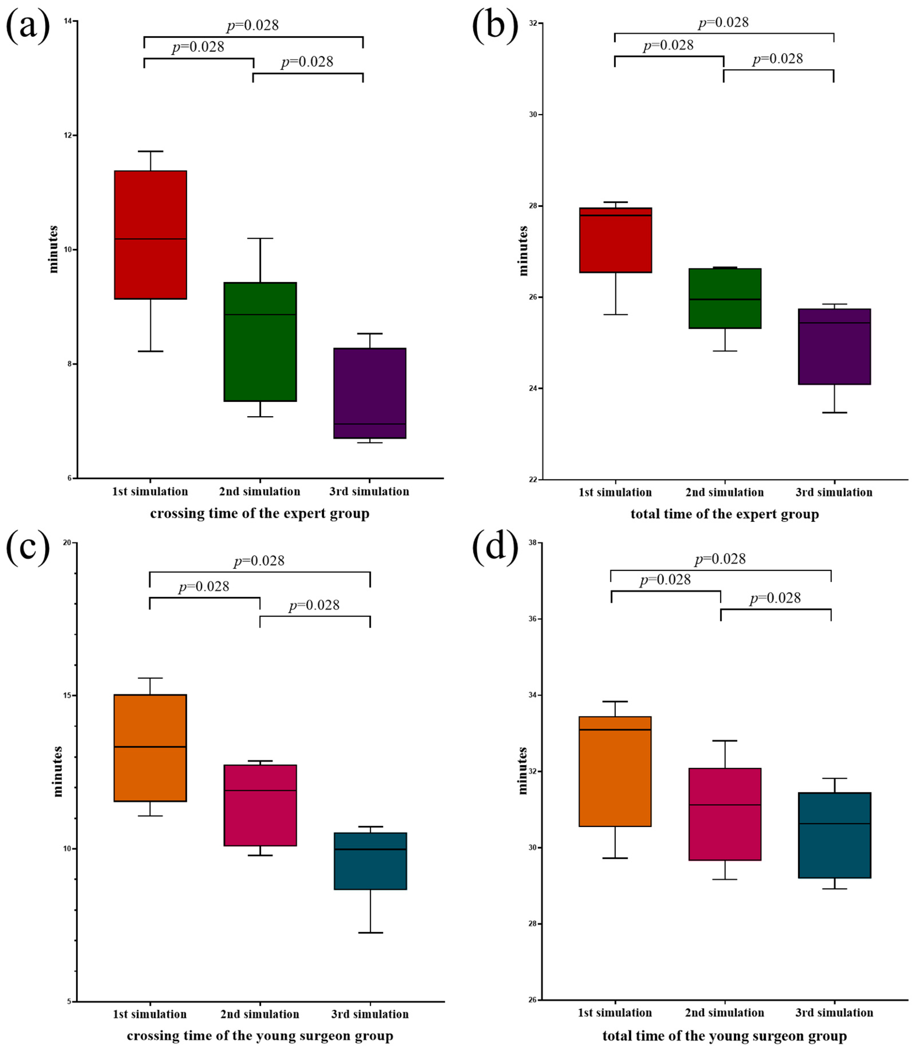
3.2. Experts Versus Young Surgeons in the Simulator

Table 4 presents the baseline characteristics of the proceduralists involved in this study. Within the expert cohort, the average age was recorded at 42.00 years (range: 40.50–45.50 years), accompanied by an average professional experience of 12.50 years (range: 11.75–17.50 years), with 10.00 years (range: 8.50–11.25 years) specifically for interventional procedures. Conversely, in the young proceduralist cohort the mean age was noted to be 30.50 years (range: 29.75–31.50 years), with an average of 3.00 years (range: 2.00–4.00 years) of mean working experience and 1.50 years (range: 1.00–2.00 years) of experience in interventions, respectively. Six of the experts and six of the young procedural experts finished the three simulations successfully. The duration required for completing the simulations is detailed in Table 5. In the expert group, the crossing-valve time required for the first simulation was 10.19 min (range: 9.12–11.39 min), which decreased to 8.87 min (range: 7.34–9.44 min) and 6.95 min (range: 6.69–8.28 min) in the next two simulations. The total time for the three simulations showed a decreasing trend to 27.79 min (range: 26.53–27.96 min), 25.95 min (range: 25.31–26.64 min), and 25.44 min (range: 24.08–25.75 min), respectively. (Figure 4a,b). For the young proceduralist group, there was also a gradual trend of decreasing crossing-valve time and total time in the three simulations, with crossing-valve times of 13.33 min (range: 11.53–15.05 min), 11.91 min (range: 10.08–12.76 min), and 9.99 min (range: 8.65–10.54 min), and total times of 33.09 min (range: 30.55–33.45 min), 31.13 min (range: 29.66–32.10 min), and 30.64 min (range: 29.19–31.45 min), respectively (Figure 4c,d). From the above results, we observed a significant decrease in both crossing-valve and total time as the number of simulation training sessions increased. The reduction in time was independent of the experience level of the trainees.

4. Discussion

TPVR has been proven to be a safe, reliable, and effective treatment option for PR. For patients with severe PR after TOF treatment, TPVR can reduce the number of extracorporeal circulation procedures that these patients need throughout their lives, reduce the burden on patients, and improve their quality of life, and is expected to increase their life expectancy [15,16]. However, challenges remain due to the variability in the morphology and dimensions of the nRVOT after early TOF treatment. A study by Schievano et al. [17] performed 3D reconstruction and morphological classification of 83 patients with RVOT dysfunction, ultimately identifying five main RVOT morphological types. The study suggests that the understanding of RVOT morphology should be enhanced and combined with outflow tract diameter and compliance to optimize the selection process for patients undergoing TPVR. In this way, the proportion of patients suitable for transcatheter treatment can be increased and their clinical prognosis improved. At the same time, cardiovascular 3D printing has also made great progress. The use of multiple materials (silicone, resin, polyethylene, rubber of different hardness) to print the RV-PA complex can effectively restore the complex nRVOT structure of patients with PR and achieve highly accurate anatomical reduction. Studies have shown that personalized 3D-printed patient-specific RV-PA models can visualize anatomical structures, enabling more accurate surgical strategy formulation and valve model selection [18,19]. In addition, 3D-printed RV-PA models can be used to predict surgical risks, such as coronary artery compression and valve displacement [13,14].
Research has demonstrated that the advantages of simulation-based training have been substantiated through extensive meta-analyses, suggesting that such training can consistently influence outcomes associated with the knowledge and skills of trainees. Etami et al. [20] used 3D printing technology combined with flexible and transparent resin materials to create a coronary artery intubation simulator with high precision and realism. The simulator was evaluated by 12 experts and its applicability in all aspects was recognized, especially in education and safety. This result provides a new solution for cardiac intervention training, which can effectively reduce the risk during the learning process and improve the practical ability of students. Barabas et al. [21] used 3D printing technology to create hands-on surgical training tools in the education of congenital coarctation of the aorta, which can significantly improve medical students’ understanding of coarctation of the aorta and its surgical treatment options. This study highlights the potential of 3D printing technology in medical education, especially in the training of complex operations, to provide students with a more intuitive learning experience. Torres et al. [22] used 3D printing to develop a patient-specific simulator that is feasible in training endovascular repair of abdominal aortic aneurysms and significantly improves surgical performance and confidence in surgical residents. This study highlights the potential application of 3D printing technology in surgical training, which may provide a more economical and effective training method for future medical education. In short, the construction of an in vitro simulation platform based on 3D printing can greatly help improve the quality of training and technical skills of young doctors. However, the application of 3D printing technology in TPVR is currently mostly used as an intuitive and convenient surgical planning tool, especially in the selection of biological prostheses [23]. At present, there is a lack of sufficient evidence in the research on TPVR simulation training.
In this study, our innovation lies in being the first application of 3D printing technology-based simulation training in TPVR training. The findings of this study indicated that the group utilizing 3D printing simulation experienced a notable enhancement in several metrics, including operating duration, crossing-valve interval, DSA duration, radiation exposure, and incidence of complications, when contrasted with the group that did not employ 3D printing simulation. In addition, this study conducted a comparative study based on simulation training and found that with the increase in the number of simulations, the simulation proficiency of experts and young proceduralists improved, and the time required was significantly shortened. In conclusion, the findings of this research indicate that a three-dimensional (3D)-printed in vitro simulation platform serves as a valuable practical training resource for TPVR. The application of 3D printing technology in simulations has been widely used in the perioperative evaluation of TPVR, significantly contributing to the refinement and advancement of surgical methodologies. The patient-specific RV-PA model used in this study can also be used by surgeons in the preoperative treatment stage of clinical practice to better understand the patient’s anatomy. The application of this clinical implementation has the potential to decrease the occurrence of unexpected intraoperative complications, thereby leading to improved surgical outcomes. The swift advancement of imaging and 3D printing technologies has enabled the creation of 3D-printed models that can precisely depict a patient’s anatomical structure. This innovation provides surgeons with invaluable visual information in a controlled environment and has proven to be highly effective in addressing complex congenital heart diseases, valvular disorders, and various other medical conditions [24,25,26]. Customized models have the potential to enhance the interaction between surgeons and patients. Additionally, proceduralists and medical students could utilize in vitro simulations to replicate the surgical procedure [27]. It now takes only 6 h to reconstruct and print a patient’s specific RV-PA model, making it easy for the surgeon to assess each patient requiring TPVR preoperatively. In addition, for complex cases the simulator only needs to replace the RV-PA model of that patient to make preoperative simulation contacts to improve the success rate of surgery. This approach has the potential to not only educate surgeons and medical trainees but also to create individualized preoperative strategies for patients. Such personalized plans are crucial in enhancing both the safety and success rates of surgical procedures.
The limitations of this study are mainly reflected in the small sample size and the lack of clinical validation analysis. Although the results show the effectiveness of the TPVR simulator in training, the small sample size may lead to a decrease in the statistical power of the results, which affects the general applicability and promotion value. Therefore, future studies should consider increasing the sample size and conducting clinical validation to further confirm the role and impact of simulators in surgical training. Moreover, under the present circumstances the characteristics of polymer materials employed in 3D printing continue to pose challenges in achieving an optimal balance between elasticity and toughness. This limitation hampers the precision of forecasting certain complications during simulations. Nonetheless, there is no doubt that materials research will also be a major area of focus as safety requirements are constantly on the increase. In addition, although the TPVR simulator uses a circulatory pump to simulate the patient’s cardiac blood flow state, the current circulatory pump cannot meet the requirements of cardiac hemodynamics assessment, and the simulator needs to be further adjusted to meet higher clinical needs [28]. In recent years, significant advancements have been achieved in the field of 3D bioprinting [29]. As interdisciplinary collaboration advances within the realms of bioengineering, materials science, biology, and computer science, significant progress is anticipated in cardiovascular three-dimensional (3D) printing for simulation training and educational purposes. Despite the existing disparity between present capabilities and clinical applications, ongoing advancements are expected to yield novel breakthroughs in this domain.

5. Conclusions

This investigation introduces a novel training tool aimed at enhancing the instructional and training methodologies associated with TPVR. Patient-specific models can be used to effectively plan for the later stages of pre-surgery, and the performance of trained physicians after training not only improved, but was significantly better than untrained physicians, while helping to reduce radiation exposure and surgical risk. The utilization of three-dimensional (3D)-printed models in medical training significantly bolsters physicians’ comprehension of anatomical configurations and the pathophysiological characteristics associated with cardiovascular disorders. Additionally, it enhances their skills in procedural tasks, augments the overall training efficacy, reduces the learning curve, and enriches practical experience. While the current simulator may not provide a wholly authentic simulation experience, its dependable simulation quality indicates a strong potential for future clinical applications.

Author Contributions

Conceptualization, Y.L. (Yang Liu) and J.Y.; Methodology, Y.M.; Validation, Y.L. (Yuanzhang Liu); Formal Analysis, Y.L. (Yuanzhang Liu), Y.W. and M.Z.; Investigation, Y.L. (Yuanzhang Liu); Resources, J.Y.; Data Curation, P.J.; Writing—Original Draft Preparation, Y.L. (Yuanzhang Liu); Writing—Review and Editing, Y.L. (Yuanzhang Liu), Y.M. and Y.W.; Supervision, Y.L. (Yang Liu) and J.Y.; Funding Acquisition, J.Y. All authors have read and agreed to the published version of the manuscript.

Funding

Development and Transformation of New Technology and Construction of Precision Diagnosis and Treatment System for Transcatheter Interventional Diagnosis and Treatment of Structural Heart Diseases: 2022YFC2503400; National Natural Science Foundation: 82370375; Research on Key Techniques of Minimally Invasive Treatment for Valvular Heart Diseases: 2023-YBSF-105; Xijing Hospital Booster Foundation: XJZT24LY42.

Institutional Review Board Statement

This research was performed in alignment with the principles outlined in the Declaration of Helsinki and received endorsement from the Ethics Committee of Xijing Hospital (Approval Number: KY-20192138-C-1). Additionally, it was registered with the ClinicalTrials.gov Protocol Registration System (NCT02917980).

Informed Consent Statement

Written informed consent was obtained from the individual for the publication of any potentially identifiable images or data included in this article.

Data Availability Statement

The data presented in this study are available on request from the corresponding author.

Acknowledgments

We would like to thank Make Medical Technology Co., Ltd. (Xi’an, China) for supplying the 3D-printed models.

Conflicts of Interest

The authors declare no conflicts of interest.

References

  1. Basquin, A.; Pineau, E.; Galmiche, L.; Bonnet, D.; Sidi, D.; Boudjemline, Y. Transcatheter Valve Insertion in a Model of Enlarged Right Ventricular Outflow Tracts. J. Thorac. Cardiovasc. Surg. 2010, 139, 198–208. [Google Scholar] [CrossRef]
  2. Latus, H.; Stammermann, J.; Voges, I.; Waschulzik, B.; Gutberlet, M.; Diller, G.P.; Schranz, D.; Ewert, P.; Beerbaum, P.; Kühne, T.; et al. Impact of Right Ventricular Pressure Load after Repair of Tetralogy of Fallot. J. Am. Heart Assoc. 2022, 11, e022694. [Google Scholar]
  3. Álvarez-Fuente, M.; Toledano, M.; Garrido-Lestache, E.; Sánchez, I.; Molina, I.; Rivero, N.; García-Ormazábal, I.; Del Cerro, M.J. Balloon-Expandable Pulmonary Valves for Patched or Native Right Ventricular Outflow Tracts. Pediatr. Cardiol. 2023, 44, 1285–1292. [Google Scholar]
  4. Jalal, Z.; Valdeolmillos, E.; Malekzadeh-Milani, S.; Eicken, A.; Georgiev, S.; Hofbeck, M.; Sieverding, L.; Gewillig, M.; Ovaert, C.; Bouvaist, H.; et al. Mid-Term Outcomes Following Percutaneous Pulmonary Valve Implantation Using the “Folded Melody Valve” Technique. Circ. Cardiovasc. Interv. 2021, 14, e009707. [Google Scholar]
  5. Martin, M.H.; Meadows, J.; McElhinney, D.B.; Goldstein, B.H.; Bergersen, L.; Qureshi, A.M.; Shahanavaz, S.; Aboulhosn, J.; Berman, D.; Peng, L.; et al. Safety and Feasibility of Melody Transcatheter Pulmonary Valve Replacement in the Native Right Ventricular Outflow Tract: A Multicenter Pediatric Heart Network Scholar Study. JACC Cardiovasc. Interv. 2018, 11, 1642–1650. [Google Scholar]
  6. Guzeltas, A.; Tanidir, I.C.; Gokalp, S.; Topkarci, M.A.; Sahin, M.; Ergul, Y. Implantation of the Edwards Sapien Xt and Sapien 3 Valves for Pulmonary Position in Enlarged Native Right Ventricular Outflow Tract. Anatol. J. Cardiol. 2021, 25, 96–103. [Google Scholar]
  7. Tannous, P.; Nugent, A. Transcatheter Pulmonary Valve Replacement in Native and Nonconduit Right Ventricle Outflow Tracts. J. Thorac. Cardiovasc. Surg. 2021, 162, 967–970. [Google Scholar]
  8. Park, W.Y.; Kim, G.B.; Lee, S.Y.; Kim, A.Y.; Choi, J.Y.; Jang, S.I.; Kim, S.H.; Cha, S.G.; Wang, J.K.; Lin, M.T.; et al. The Adaptability of the Pulsta Valve to the Diverse Main Pulmonary Artery Shape of Native Right Ventricular Outflow Tract Disease. Catheter. Cardiovasc. Interv. 2024, 103, 587–596. [Google Scholar]
  9. Corrigan, F.E., 3rd; Gleason, P.T.; Condado, J.F.; Lisko, J.C.; Chen, J.H.; Kamioka, N.; Keegan, P.; Howell, S.; Clements, S.D., Jr.; Babaliaros, V.C.; et al. Imaging for Predicting, Detecting, and managing complications After transcatheter aortic Valve Replacement. JACC Cardiovasc. Imaging 2019, 12, 904–920. [Google Scholar] [CrossRef]
  10. Fan, Y.; Wong, R.H.L.; Lee, A.P. Three-Dimensional Printing in Structural Heart Disease and Intervention. Ann. Transl. Med. 2019, 7, 579. [Google Scholar]
  11. Pluchinotta, F.R.; Sturla, F.; Caimi, A.; Giugno, L.; Chessa, M.; Giamberti, A.; Votta, E.; Redaelli, A.; Carminati, M. 3-Dimensional Personalized Planning for Transcatheter Pulmonary Valve Implantation in a Dysfunctional Right Ventricular Outflow Tract. Int. J. Cardiol. 2020, 309, 33–39. [Google Scholar] [CrossRef]
  12. Schievano, S.; Migliavacca, F.; Coats, L.; Khambadkone, S.; Carminati, M.; Wilson, N.; Deanfield, J.E.; Bonhoeffer, P.; Taylor, A.M. Percutaneous Pulmonary Valve Implantation Based on Rapid Prototyping of Right Ventricular Outflow Tract and Pulmonary Trunk from Mr Data. Radiology 2007, 242, 490–497. [Google Scholar] [CrossRef]
  13. Han, Y.; Shao, Z.; Sun, Z.; Han, Y.; Xu, H.; Song, S.; Pan, X.; de Jaegere, P.P.T.; Fan, T.; Zhang, G. In Vitro Bench Testing Using Patient-Specific 3d Models for Percutaneous Pulmonary Valve Implantation with Venus P-Valve. Chin. Med. J. 2024, 137, 990–996. [Google Scholar] [CrossRef]
  14. Wang, Y.; Jin, P.; Meng, X.; Li, L.; Mao, Y.; Zheng, M.; Liu, L.; Liu, Y.; Yang, J. Treatment of Severe Pulmonary Regurgitation in Enlarged Native Right Ventricular Outflow Tracts: Transcatheter Pulmonary Valve Replacement with Three-Dimensional Printing Guidance. Bioengineering 2023, 10, 1136. [Google Scholar] [CrossRef]
  15. Müller, J.; Engelhardt, A.; Fratz, S.; Eicken, A.; Ewert, P.; Hager, A. Improved Exercise Performance and Quality of Life after Percutaneous Pulmonary Valve Implantation. Int. J. Cardiol. 2014, 173, 388–392. [Google Scholar] [CrossRef]
  16. Cheatham, J.P.; Hellenbrand, W.E.; Zahn, E.M.; Jones, T.K.; Berman, D.P.; Vincent, J.A.; McElhinney, D.B. Clinical and Hemodynamic Outcomes up to 7 Years after Transcatheter Pulmonary Valve Replacement in the Us Melody Valve Investigational Device Exemption Trial. Circulation 2015, 131, 1960–1970. [Google Scholar] [CrossRef]
  17. Schievano, S.; Coats, L.; Migliavacca, F.; Norman, W.; Frigiola, A.; Deanfield, J.; Bonhoeffer, P.; Taylor, A.M. Variations in Right Ventricular Outflow Tract Morphology Following Repair of Congenital Heart Disease: Implications for Percutaneous Pulmonary Valve Implantation. J. Cardiovasc. Magn. Reson. 2007, 9, 687–695. [Google Scholar] [CrossRef]
  18. Zhou, D.; Pan, W.; Jilaihawi, H.; Zhang, G.; Feng, Y.; Pan, X.; Liu, J.; Yu, S.; Cao, Q.; Ge, J. A Self-Expanding Percutaneous Valve for Patients with Pulmonary Regurgitation and an Enlarged Native Right Ventricular Outflow Tract: One-Year Results. EuroIntervention 2019, 14, 1371–1377. [Google Scholar] [CrossRef]
  19. Odemis, E.; Aka, I.B.; Ali, M.H.A.; Gumus, T.; Pekkan, K. Optimizing Percutaneous Pulmonary Valve Implantation with Patient-Specific 3d-Printed Pulmonary Artery Models and Hemodynamic Assessment. Front. Cardiovasc. Med. 2023, 10, 1331206. [Google Scholar] [CrossRef]
  20. Etami, H.V.; Rismawanti, R.I.; Hanifah, V.A.N.; Herianto, H.; Yanuar, Y.; Kuswanto, D.; Anggrahini, D.W.; Gharini, P.P.R. Ct-Derived 3d Printing for Coronary Artery Cannulation Simulator Design Manufacturing. Bioengineering 2022, 9, 338. [Google Scholar] [CrossRef]
  21. Barabas, I.J.; Vegh, D.; Bottlik, O.; Kreuter, P.; Hartyanszky, I.; Merkely, B.; Palkovics, D. The Role of 3d Technology in the Practical Education of Congenital Coarctation and Its Treatment-a Feasibility Pilot Study. BMC Med. Educ. 2024, 24, 357. [Google Scholar]
  22. Torres, I.O.; De Luccia, N. A Simulator for Training in Endovascular Aneurysm Repair: The Use of Three Dimensional Printers. Eur. J. Vasc. Endovasc. Surg. 2017, 54, 247–253. [Google Scholar]
  23. Chessa, M.; Giugno, L.; Butera, G.; Carminati, M. Multi-Modal Imaging Support in a Staging Percutaneous Pulmonary Valve Implantation. Eur. Heart J. 2016, 37, 66. [Google Scholar] [CrossRef]
  24. Qian, Z.; Wang, K.; Liu, S.; Zhou, X.; Rajagopal, V.; Meduri, C.; Kauten, J.R.; Chang, Y.H.; Wu, C.; Zhang, C.; et al. Quantitative Prediction of Paravalvular leak in Transcatheter Aortic valve Replacement Based On tissue-Mimicking 3d Printing. JACC Cardiovasc. Imaging 2017, 10, 719–731. [Google Scholar]
  25. Santoro, G.; Pizzuto, A.; Rizza, A.; Cuman, M.; Federici, D.; Cantinotti, M.; Pak, V.; Clemente, A.; Celi, S. Transcatheter Treatment of “Complex” Aortic Coarctation Guided by Printed 3d Model. JACC Case Rep. 2021, 3, 900–904. [Google Scholar] [CrossRef]
  26. Mao, Y.; Liu, Y.; Zhai, M.; Yang, J. Application of and Prospects for 3-Dimensional Printing in Transcatheter Mitral Valve Interventions. Rev. Cardiovasc. Med. 2023, 24, 61. [Google Scholar] [CrossRef]
  27. Ding, P.; Li, L.; Liu, Y.; Jin, P.; Tang, J.; Yang, J. Three-Dimensional Printing for Heart Diseases: Clinical Application Review. Biodes Manuf. 2021, 4, 675–687. [Google Scholar]
  28. Marques, D.M.C.; Silva, J.C.; Serro, A.P.; Cabral, J.M.S.; Sanjuan-Alberte, P.; Ferreira, F.C. 3d Bioprinting of Novel Κ-Carrageenan Bioinks: An Algae-Derived Polysaccharide. Bioengineering 2022, 9, 109. [Google Scholar] [CrossRef]
  29. Mayfield, C.K.; Ayad, M.; Lechtholz-Zey, E.; Chen, Y.; Lieberman, J.R. 3d-Printing for Critical Sized Bone Defects: Current Concepts and Future Directions. Bioengineering 2022, 9, 680. [Google Scholar] [CrossRef]
Figure 1. Construction of the TPVR simulator and simulation training on the bench test. (ac) The main process of 3D-printed RV-PA models; (df) The main process of the pulsatile simulator; (gi) Trainees simulated TPVR using the pulsatile simulator. TPVR, transcatheter pulmonary valve replacement; 3D, three-dimensional; RV-PA, right ventricle–pulmonary artery.
Bioengineering 12 00344 g001
Figure 2. Schematic of the flow of this study. (a) The five proceduralists in the group utilizing 3D-printed simulations sequentially performed two simulation steps prior to the TPVR. In contrast, the five proceduralists in the non-3D-printed simulation group carried out a standard two-dimensional imaging evaluation before the TPVR procedure. (b) Both the expert group (n = 6) and the young proceduralist group (n = 6) successfully conducted three rounds of simulations using the pulsatile simulator.
Bioengineering 12 00344 g002
Figure 3. Outcomes of TPVR. (a) A comparative analysis of the crossing-valve duration was conducted between the group utilizing 3D-printed simulations and the group employing non-3D-printed simulations, revealing a statistically significant difference (p = 0.016). (b) An additional comparison of the crossing-valve duration between the 3D-printed simulation cohort and the non-3D-printed simulation cohort indicated a significant difference in fluoroscopy time as well (p = 0.012). (c) Furthermore, a comparison of the overall procedural time between the 3D-printed simulation group and the non-3D-printed simulation group demonstrated a highly significant difference (p = 0.036). The p-value is from a non-parametric test.
Bioengineering 12 00344 g003
Figure 4. The outcomes of TPVR simulations. (a) A comparative analysis of the crossing-valve duration across three simulations within the expert cohort. (b) A comparative assessment of the total duration among the three simulations in the expert cohort. (c) A comparative evaluation of the crossing-valve duration across three simulations within the young surgeon cohort. (d) A comparative analysis of the overall duration among the three simulations in the young surgeon cohort. Statistical significance was determined using non-parametric tests and the corresponding p-values are reported.
Bioengineering 12 00344 g004
Table 1. Patient characteristics.
CharacteristicsPatient1Patient2Patient3Patient4Patient5Patient6Patient7 Patient8Patient9Patient10
Age, years/sex19/M30/F37/F52/F23/M48/F12/M22/F31/F42/M
Weight (kg)68626055785351685881
Height (cm)175158162156173156150170166178
NYHA functional classIIIIIIIIIVIIIIVIIIIIIVIV
PR severity grade4+4+4+4+4+4+4+4+4+4+
Peak transpulmonary valve gradient11171921171911181 710
TR severity grade1+3+3+2+3+3+2+2+3+3+
RV–PA conduit length (mm)58525755595548535662
nRVOT diameter (mm)44383736413732353843
The narrowest plane/diameter
(mm)
Distal
MPA/23
Distal
MPA/24
Mid
MPA/28
Mid
MPA/23
PA/
33
Mid
MPA/27
Distal
MPA/24
Distal
MPA/22
Mid
MPA/26
PA/30
NYHA—New York Heart Association; PR—pulmonary regurgitation; TR—tricuspid regurgitation; RV–PA—right ventricle–pulmonary artery; nRVOT—native right ventricular outflow tract.
Table 2. Baseline information of experts in the 3D-printed simulation group and the non-3D-printed simulation group (n = 10).
NumberSexAge
(Years)
Years for
Proceduralist
Years for
Intervention
Patient
Number
A1Male4616111
A2Male421372
A3Male4315103
A4Male391284
A5Male381195
B1Male421386
B2Male401097
B3Male4516128
B4Male371089
B5Male4311710
Group A represents the 3D-printed simulation group; group B represents the non-3D-printed simulation group.
Table 3. Results of the 3D-printed simulation group versus the non-3D-printed simulation group.
TimeCrossing ValveFluoroscopyTotalResidual PRComplications
A15′13″15′00″53′00″NoneNone
A24′55″14′00″57′00″NoneNone
A35′40″16′00″62′00″NoneNone
A44′47″13′00″55′00″NoneNone
A56′05″17′00″63′00″NoneNone
B15′55″17′00″61′00″NoneNone
B27′13″21′00″71′00″TraceHemoptysis
B36′43″19′00″68′00″NoneNone
B46′20″18′00″67′00″NoneNone
B58′11″22′00″63′00″NoneHemoptysis
Group A represents the 3D-printed simulation group; group B represents the non-3D-printed simulation group.
Table 4. Baseline information of proceduralists in the expert group and the young proceduralist group (n = 12).
NumberSexAge
(Years)
Years for
Proceduralist
Years for
Intervention
C1Male431210
C2Male471711
C3Male411212
C4Male451910
C5Male39139
C6Male41117
D1Male3122
D2Male3042
D3Male3331
D4Male2932
D5Male3121
D6Male3041
Group C represents the expert group; group D represents the young proceduralist group.
Table 5. Results of the expert group versus the young proceduralist group.
Rank1st2nd3rd
DeviceTotalDeviceTotalDeviceTotal
C110′35″27′49″8′56″26′07″6′37″25′43″
C29′48″26′55″9′11″25′28″7′06″25′19″
C38′13″25′37″7′26″24′49″6′43″24′17″
C411′17″27′46″8′48″26′39″8′12″25′51″
C59′26″26′50″7′05″25′47″6′48″23′28″
C611′43″28′05″10′12″26′38″8′32″25′33″
D112′49″33′19″12′03″31′26″10′29″30′51″
D211′41″30′49″9′47″29′49″7′15″29′17″
D313′50″32′53″12′43″30′49″10′22″30′25″
D411′05″29′44″10′11″29′10″9′36″28′55″
D515′34″33′50″12′52″32′48″10′43″31′49″
D614′52″33′18″11′46″31′52″9′07″31′20″
Group C represents the expert group; group D represents the young proceduralist group.
Disclaimer/Publisher’s Note: The statements, opinions and data contained in all publications are solely those of the individual author(s) and contributor(s) and not of MDPI and/or the editor(s). MDPI and/or the editor(s) disclaim responsibility for any injury to people or property resulting from any ideas, methods, instructions or products referred to in the content.

