Article

Astaxanthin Ameliorates Diabetic Retinopathy in Swiss Albino Mice via Inhibitory Processes of Neuron-Specific Enolase Activity

by
Aswinprakash Subramanian
1,2,
Jayaraman Thirunavukkarasu
2,* and
Arunachalam Muthuraman
3,*
1
Anatomy Unit, Faculty of Medicine, AIMST University, Bedong 08100, Kedah, Malaysia
2
Department of Pharmacology, Saveetha Institute of Medical & Technical Sciences (SIMATS), Saveetha University, Chennai 602105, Tamil Nadu, India
3
Pharmacology Unit, Faculty of Pharmacy, AIMST University, Bedong 08100, Kedah, Malaysia
*
Authors to whom correspondence should be addressed.
Processes 2022, 10(7), 1318; https://doi.org/10.3390/pr10071318
Submission received: 9 April 2022 / Revised: 21 May 2022 / Accepted: 23 May 2022 / Published: 5 July 2022
(This article belongs to the Special Issue Regulation and Control of Intracellular Signalling)

Abstract

:
Retinopathy is one of the most common complications of diabetes mellitus. Diabetic retinopathy (DR) occurs due to microvascular damage in retinal tissues provoked by high blood sugar levels. The available drugs for DR are limited. Astaxanthin (AST) has anti-hypertensive, anti-obesity, and anti-diabetic properties. However, the therapeutic effect of AST on DR remains elusive. The present study is designed to investigate the effects of AST on DR via inhibition of neuron-specific enolase (NSE) activity. DR was induced by the administration of streptozotocin (STZ, 35 mg/kg: intraperitoneal; and 20 μL of STZ: intravitreal) in mice. AST (10 and 20 mg/kg) was administered orally (p.o.) for 21 days. The DR associated visual changes were assessed at different time intervals via optokinetic motor response (OMR) and penta-maze (PM) tests. Blood glucose level as well as retinal catalase, lactate dehydrogenase (LDH), & neuron-specific enolase (NSE) were estimated. The reference drug i.e., dexamethasone (DEX, 10 mg/kg; p.o.) was administered for 21 days. The administration of AST showed significant ameliorative potential in DR. Hence, AST can be used as a natural medicine for the management of DR due to its potential antioxidant, anti-diabetic, and NSE inhibitory properties.

1. Introduction

Enolase is one of the metabolic glycolytic enzymes. It is also known as phosphopyruvate hydratase. This metalloenzyme is responsible for the catalytic conversion of 2-phosphoglycerate to phosphoenolpyruvate. The chemical reaction occurs in all tissue [1]. In a pathophysiological aspect, it acts as a plasminogen binding protein in hypoxic and ischemic conditions [2]. Enolase also plays multiple roles in immune tolerance, allergic responses, and growth regulation. Furthermore, the immunoglobulin production associated with enolase enhances host cell humoral and cell-mediated immune responses [3,4]. Clinically, in DR, the retinal micro vessels undergo the following conditions i.e., hypoxia & ischemia of blood vessels, hemorrhages, microaneurysms, macular edema and degenerations, lipid exudates, neovascularization (angiogenesis), and cotton-wool spots [5,6]. These pathophysiological changes are due to the alteration of multiple cellular enzymes like catalase, lactate dehydrogenase, and enolase, especially neuron-specific enolase (NSE). NSE is one of the novel biomarkers for DR and macular degeneration [7]. Chronic hyperglycemia elevates the NSE level in the blood due to damage of peripheral nerves and micro vessels [6]. It is also closely linked to the development of insulin resistance in diabetic patients. Hence, the enolase level is expected to increase during the progression of DR. The activity of enolase is enhanced in the condition of tissue injury with activated endothelial cells, neutrophils, and protease-activated receptor-2 (PAR-2) [8]. Further, it is also recognized in the detection of pathogens and activated immune cells which act as plasminogen acceptors and participate in systemic infections. Enolase and NSE are inflammatory signals that involve inflammation, infections, and autoimmunity reactions [9]. Furthermore, NSE is also linked with the expression of prion proteins like heat-shock proteins, cytoskeletal, and chromatin structural proteins in different inflammatory disorders [10]. It has been shown that patients with proliferative DR had increased levels of both vitreous and serum NSE [11].
DR is one of the major microvascular complications which is associated with diabetes mellitus. DR is managed with the administration of dexamethasone and intravitreal triamcinolone. Recently, other medications like anti-vascular endothelial growth factor drugs like aflibercept and ranibizumab are also used for the treatment of DR [12]. These drugs reduce diabetic macular edema and neovascularization of the retinal disc via inhibition of pro-angiogenic factors [13]. However, the safety and proven efficacy of these drugs remain questionable due to potential intolerable side effects like intraocular inflammation, elevated levels of intraocular tension, ocular hemorrhage, and chances of endophthalmitis [14,15]. Hence, newer and safer medications are still required in the management of DR.
Herbal medicines like Azadirachta indica, Ginkgo biloba, Gymnema sylvestre, Salvia miltiorrhiza, Stephania tetrandra, and Pinus pinaster have been used to treat DR [16,17]. The traditional Chinese plant-like Ruscus extract, Sanqi Tongshu, Xueshuantong, Xuesaitong, and Puerarin are also used for various ocular disorders [18]. The major actions of these plant-based medicines include antioxidant, anti-infection, regulation of angiogenesis, improvement of microcirculation, reduction of advanced glycation end-products, and strengthening of the integrity of capillaries membranes. Moreover, natural medicines like dendrobium alkaloids [19], curcumin [20], breviscapine [21], and saffron [22] reduce the severity of diabetic microvascular complications. AST is one of the members of the natural xanthophyll (carotenoid) compound [23]. It possesses potential antioxidant, anti-inflammatory, and neurovascular protective actions [24]. Additionally, it is also used for the management of diabetes mellitus, hypertension, and hypercholesteremia [25]. Recently, AST is known to treat various retinal diseases like uveitis, asthenopian, and cataract in experimental animals and humans via modulation of metabolic pathways, restoring cellular homeostasis due to its multi-targeted therapeutic actions [26]. In a study using STZ induced diabetic rats, AST affected the expression of oxidative and inflammatory stress mediators like acrolein, nitrotyrosine, intercellular adhesion molecule-1 and monocyte chemoattractant protein-1 in the ocular tissues [27]. AST also hinders the retinal photoreceptor cells from oxidative stress induced by high glucose level [28]. Clinically, AST has been used to manage ocular diseases owing to its antioxidant, anti-inflammatory, and anti-apoptotic actions [29]. However, the therapeutic role of AST in DR still needs to be explored with correlation to NSE activity. Hence, the present study is designed to explore the role of AST in DR with correlation to NSE activity.

2. Materials and Methods

2.1. Animals

Disease-free male Swiss albino mice (12 months old; 20–30 g) were used in the present research. All animals were maintained in the central animal house in AIMST University with a standard laboratory diet (Soon-Soon Oilmills Sdn Bhd, Pinang, Malaysia). The animals were allowed access to water and food ad libitum. The 12-h light and dark cycles were used to maintain the circadian rhythm. The temperature was maintained at 25 °C and the humidity level was maintained at 50%. This experimental protocol was approved by AIMST University Animal Ethics Committee (AUAEC/FOM 2020/21-Amendment No. 1). The care for animals was taken as per guidelines of AUAEC.

2.2. Induction of Diabetic Retinopathy (DR)

Streptozotocin is a destructive chemical on pancreatic β-cells which is commonly used for the induction of diabetic mellitus in experimental animals. This method was described by Yuan et al. [30] and Tawfik et al. [31]. DR was developed by single intraperitoneal injection of streptozotocin (STZ; 35 mg/kg) [32238-91; Nacalai Tesque Inc.]. The development of DR was accelerated by intravitreal (i.vit.) injection of STZ with a dose of 20 µL of 7% w/v of STZ stock solution on the 7th day.

2.3. Experimental Design

Five groups of male adult mice (n = 8) were employed in this study. Group-I served as a normal (naïve) control group. Group II served as the diabetic retinopathy (DR, as negative control) group. Diabetes mellitus was induced by intraperitoneal (i.p.) administration of STZ (35 mg/kg) and it is considered as day 0. The onset of diabetes was confirmed by estimation of fasting blood glucose levels (>250 mg/dL or >7 mmol/L) on day 3. Further, the progression of DR was accelerated by intravitreal (i.vit.) injection of 20 µL of 7% w/v STZ stock solution on day 7. Groups III and IV served as test compound treatment groups i.e., AST [SML0982; Sigma-Alrich] solution in 0.5% carboxymethylcellulose was administered orally for 21 consecutive days (starting from day 8) at doses of 10 and 20 mg/kg, respectively. Group V served as a reference drug treatment group i.e., dexamethasone (DEX), 10 mg/kg administered orally for 21 consecutive days (from day 8). DEX was selected as the reference drug because it has been widely used in clinical studies and proven to be efficacious in the management of DR [32,33]. In studies using a mouse model, DEX confers protective effect against retinopathy [34,35]. The blood glucose level was estimated on day 0, 3, and day 21. The behavioral responses i.e., optomotor response (OMR) and penta-maze (PM) were assessed at different time intervals i.e., day 7, 14, 21, and 28. The biochemical markers estimated include retinal tissue catalase, LDH, NSE, and total protein levels.

2.4. Assessment of DR Associated Changes of Visual Acuity Behaviours

The DR associated visual acuity changes were assessed using OMR and PM tests at different time intervals i.e., day 7, 14, 21, and 28.

2.4.1. Assessment of Visual Acuity Functions by OMR Test Device

The DR associated visual acuity changes was assessed by an OMR test device as described by Prusky et al. [36] with a slight modification from Kretschmer et al. [37]. Briefly, the OMR device consists of two concentric circular chambers. It is covered with an opaque square box. The inner side of the square box was made dark. The white light-emitting diode (LED) strip was placed on the inner wall of the square box. The inner wall of the big circular (30 cm diameter) chamber was sticked with alternating black and transparent stripes vertically. This inner chamber was allowed to rotate at 10-rpm with a synchronous motor. The non-revolving, smaller, transparent, circular (20 cm diameter) chamber was placed at the center of the OMR device. Each animal was placed in this smaller chamber and could see the movement of alternating black and transparent stripes. The visual acuity test was assessed based on the animal’s movement against the clockwise and counter-clockwise movements of the outer chamber. The animal movement was recorded for 4 min for the assessment of spatial frequency threshold (SFT; whole animal circular movement against grid movement). The assessment was conducted in triplicate.

2.4.2. Assessment of Spatial Imagery Transformation Function by PM Test Device

The spatial imagery transformation function of mice was assessed by using the ‘Penta-maze’ test device as a described method of Rondi-Reig et al. [38] with minor modification from Vorhees and Williams [39]. Briefly, it looks like a star-shaped maze. A PM device is built with polyacrylic material. It is in circular shape and consists of five-way water channels with solid central pentagon objects. These five waterway channels are radiated equally from the center. The walls of the PM device are made with uniform color. The maze was filled with water and made opaque with non-toxic dye. One arm of the PM device is considered as a starting point and other arms are used as two pathway processes: (1) allocentric (object-to-object representation) pathway and (2) egocentric (self-to-object representation) pathway, leading to different destination points. These destination points were cued with red and green LED illumination on a platform. Each pathway contains unique imaginary cues. The analysis of allocentric navigation pathway entry i.e., mice reaching the individual destination point, is also called as the absolute location i.e., a submerged platform with the presence of extra wall cues in alternative pathways. The allocentric navigation training was given on day 22. On day 23, an egocentric response was recorded by placing the animal in the nearest arm from the destination points of the PM device. The platforms were available in destination and starting points. During egocentric response recording, the wall navigation cues were removed and the induvial destination point was clearly visible. The egocentric response was recorded as the number of error entries (NE) within the observation period of 5 min. The strategies of spatial imagery transformation function were described by Fouquet et al. [40]. The assessment was conducted in triplicate.

2.5. Biochemical Estimations

On day 28, animals were anesthetized with diethyl ether [1709-50; R&M Chemicals]. Blood samples were collected from the tail vein and fasting blood glucose levels were estimated by using a commercial Accu-Chek Active glucometer device. Thereafter, animals were sacrificed and retinal tissues were collected for the estimation of tissue biomarkers like catalase, LDH, NSE, and total protein levels. The details of biomarkers estimation are explained in the following sections.

2.5.1. Estimation of Catalase as an Indication of Oxidative Stress

Catalase enzyme is a primary endogenous antioxidant enzyme in the biological system. Retinal tissue catalase activity was estimated as a described method by Hadwan, [41]. Briefly, 0.5 mL of aliquot was mixed with 0.5 mL of distilled water and 1 mL of 10 mM hydrogen peroxide solution [1477-80; R&M Chemicals] (pH 7.0, 50 mM) was added. The mixture was vortexed and incubated at room temperature (37 °C) for 2 min. Then, 0.6 mL of working solution was added for the assessment of catalase activity. The working solution consists of a 1:1:1.8 ratio of cobalt (II) [255599; Sigma-Alrich] solution, Graham salt (Sodium hexametaphosphate) [71600; Sigma-Alrich] solution, and sodium bicarbonate [MO00420069; Orioner Hightech Sdn Bhd] solutions, respectively. The working solution mixture containing tubes was vortexed immediately for 5 s and then kept in the dark at 37 °C for 10 min. The olive green color chromogen i.e., carbonato-cobaltate (III) complex [Co (CO3)3]Co)] was developed, and absorbance was measured by using a spectrophotometer (DU 640B Spectrophotometer, Beckman Coulter Inc., Brea, California, USA) at 440 nm. The total catalase activity was calculated by using the following formula:
Catalase   Activity   U / ml = 2.303   t     l o g   δ   O . D .   standard δ   O . D .   test  
In this formula, ‘δ O.D.’ represents the changes in absorbance/minutes and ‘t’ represents the time. The calculated level of catalase (U/mL) value was further integrated with mg of protein. The net value of catalase activity was expressed as a unit of catalase activity per mg of protein.

2.5.2. Estimation of LDH as an Indication of Retinal Tissue Inflammation

LDH enzyme is one of the inflammatory marker enzymes of retinal tissue injury. The retinal tissue LDH activity was estimated as a described method of Vanderlinde [42]. Briefly, 25 μL of supernatant of retinal tissue homogenate was mixed with 3 mL of the reaction mixture. The reaction mixture consists of 0.1 M potassium phosphate [6482-00; R&M Chemicals] (pH 7.5), 0.33 mM sodium pyruvate [P2256; Sigma Aldrich], and 0.14 mM nicotinamide adenine dinucleotide hydrogen [10128023001; Sigma Alrich] (NADH, reduced form of NAD+). At this point, LDH [10127876001; Sigma Alrich] (0.1 enzyme unit) is ready to react with the reagent mixture. The tubes were vortexed and incubated at room temperature (37 °C) for 2 min. The changes of absorbance were recorded by using a spectrophotometer (DU 640B Spectrophotometer, Beckman Coulter Inc., Brea, California, USA) at 340 nm. The standard plot was prepared with 0, 2, 4, 6, 8, and 10 µL of the 1.25 mM NADH standard and it generates the 0, 2.5, 5, 7.5, 10, and 12.5 nmole.
The changes in absorbance reading were measured after 5 min as an initial measurement (Ti) after plate incubation at 37 °C. Thereafter, a reading was measured every 5 min. During incubation, the test samples were protected from light exposure. The measurement was stopped when the test sample reading was greater than that of the highest standard i.e., 12.5 nmole (Tf). The net value of LDH activity was calculated by using the following formula:
LDH   Activity   U / ml = B   Reaction   time   x   V   x   DF
In this formula, ‘B’ represents the amount (nmole) of NADH generated between Ti and Tf (Ti, Time of initial reading and Tf, time of final reading). Reaction time represents the = Ti-Tf. The letter ‘V’ represents the sample volume (ml). The letter ‘DF’ represents the dilution factor. The letter ‘U’ represents the unit. The calculated level of LDH (U/mL) value was further integrated with mg of protein. The net value of catalase activity was expressed as a unit of LDH activity per mg of protein.

2.5.3. Estimation of NSE as an Indication of Retinal Macular Degeneration

The retinal NSE level was estimated by using the commercial enzyme-linked immunosorbent assay (ELISA) kit [E-CL-M0076; Elabscience]. NSE is a neuronal survival (neurotrophic) factor, especially in the retinal inter-photoreceptor matrix regions of the retina [43]. Briefly, 50 μL of standard and samples were placed in appropriate wells of a microtiter plate. Then, 50 μL of antibody cocktail were added to all wells and incubated at room temperature for 1 h. Thereafter, the microtiter plate fluid was aspirated and all the wells were washed three times with 350 μL of wash buffer (1×). Finally, 100 μL of 3,3′,5,5′-tetramethylbenzidine (TMB) solution was added. The reaction of TMB and horse reddish peroxidase (HRP) enzyme was stopped by the addition of 100 μL of stop solution containing hydrochloric acid. TMB acts as a chromogenic substrate for the HRP enzyme. The colorless TMB turned to a blue color (TMB+). Further, this color turned into a yellow color (TMB2+) after the addition of a reaction stop solution. The changes in absorbance were recorded by using a spectrophotometer (DU 640B Spectrophotometer, Beckman Coulter Inc., Brea, California, USA) at 450 nm. The absorbance of blank with the substrate was recorded. The measurement of absorbance with variable standard NSE concentrations i.e., 0, 312.5, 625, 1250, 2500, 5000, 10,000, and 20,000 picograms per milliliter (pg/mL) was prepared as per commercial ELISA kit instructions. The standard curve was prepared with absorbance value (y-axis) versus each standard concentration (x-axis). The level of NSE was quantified by using the following formula:
NSE = δ   O . D .   blank   control   δ   O . D .   value   against   the   standard   curve   X   DF
In this formula, ‘δ O.D.’ represents the changes in absorbance/minutes and DF represents the dilution factor. The NSE level was noted as pg/mL.

2.5.4. Estimation of Total Protein

The retinal tissue total protein level was estimated as a method described by Lowry et al. [44]. Briefly, the 0.15 mL of tissue supernatant was diluted up to 1 mL with phosphate buffer. Then, 5 mL of Lowry’s reagent was added to all the test tubes. Lowry’s reagent consists of two major reaction mixtures i.e., (1) Reagent A: It consists of 2 g of sodium potassium tartrate; 100 g of sodium carbonate; and 500 mL of 1 N sodium hydroxide [5145-81; R&M Chemicals] in one liter of distilled water. (2) Reagent B: It consists of 2 g of sodium potassium tartrate [217255; Sigma-Alrich]; 1 g of copper sulfate [6668-70; R&M Chemicals]; 90 mL of distilled water; and 10 mL of 1N sodium hydroxide. Reagents A and B were mixed at 9:1 ratio. This reagent is called Lowry’s reagent and is used for the total protein estimation. The sample and Lowry reagent containing test tubes were mixed thoroughly and allowed to stand for 15 min at room temperature. Foiln-Ciocalteu reagent was prepared by dissolving 10 g of sodium tungstate [223336; Sigma-Alrich] and 2.5 g of sodium molybdate [243655; Sigma-Alrich] in 70 mL of distilled water followed by adding 5 mL of 85% phosphoric acid [1502-80; R&M Chemicals] and 10 mL of concentrated hydrochloric acid [1386-80; R&M Chemicals]. Then, 0.5 mL of Foiln-Ciocalteu reagent was added and vortexed vigorously. Thereafter, test tubes were incubated at room temperature for 30 min. The purple color chromogen was developed and changes of absorbance were recorded by using a spectrophotometer (DU 640B Spectrophotometer, Beckman Coulter Inc., Brea, California, USA) at 750 nm. The standard curve was prepared with 0.2–2.4 milligram of bovine serum albumin (BSA) per milliliter. The total protein level was noted as mg/mL.

2.6. Statistical Analysis

All the results were expressed as mean ± standard deviation (SD). The data of visual behaviors were statistically analyzed using two-way analysis of variance (ANOVA) followed by Bonferroni post hoc test and data of blood glucose retinal tissue biomarkers i.e., catalase, LDG, and NSE levels were analyzed using one way ANOVA followed by Tukey’s Multiple Range test using Graph pad prism version-5.0 software. The probability value i.e., p ˂ 0.05 was statistically significant.

3. Results

3.1. Effect of AST in DR Associated Visual Behavioural Changes

The administration of STZ (35 mg/kg; i.p.; and 20 µL of 7% w/v; i.vit.) showed significant (p < 0.05) visual behavioral changes along with retinal micro-anatomical changes (unpublished data) when compared to naïve animal group. The administration of AST (10 and 20 mg/kg; p.o.) significantly ameliorated the visual behavioral changes i.e., OMR, and PM tests in a dose-dependent manner when compared to the DR group. The effect of AST is similar to the reference drug i.e., DEX (10 mg/kg; p.o.). The details are described in the following section.

3.1.1. Effect of AST in DR Associated Changes of Visual Acuity Functions

The administration of STZ (35 mg/kg; i.p.; and 20 µL of 7% w/v; i.vit.) showed significant (p < 0.05) impairment of visual acuity in the OMR test, indicated by a decrease in the SFT values when compared to the normal control group. The administration of AST (10 and 20 mg/kg; p.o.) significantly ameliorated the STZ impaired OMR test responses when compared to the DR group. The ameliorative effect was similar to that of the reference drug i.e., DEX (10 mg/kg; p.o.) treated group. The results are illustrated in Figure 1.

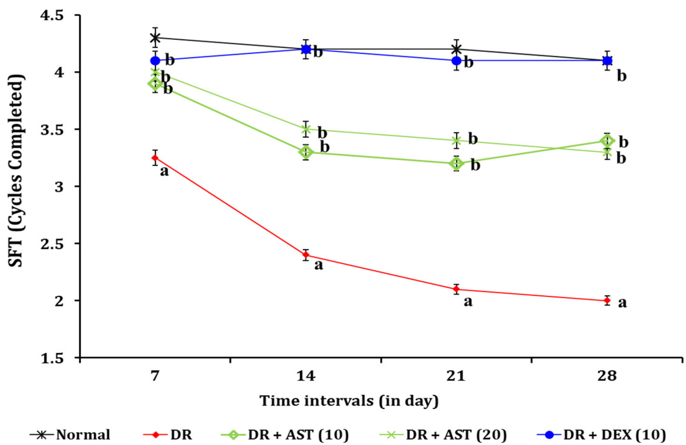
3.1.2. Effect of AST in DR Associated Changes in Spatial Imagery Transformation Function

The administration of STZ (35 mg/kg; i.p.; and 20 µL of 7% w/v; i.vit.) showed significant (p < 0.05) impairment of spatial imagery transformation in the PM test, indicated by the increased number of errors when compared to the normal control group. The administration of AST (10 and 20 mg/kg; p.o.) significantly ameliorated the STZ associated increase in error values in PM test responses when compared to the DR group. The ameliorative effect was similar to that of the reference drug i.e., DEX (10 mg/kg; p.o.) treated group. The results are illustrated in Figure 2.

3.2. Effect of AST in DR Associated Changes in Biochemical Estimations

On day 28, all the animals were anesthetized for collection of tail vein blood samples and the fasting blood glucose level was estimated by using a commercial Accu-Chek Active glucometer. Thereafter, animals were sacrificed and retinal tissues were collected immediately. They are used for the estimation of tissue biomarker changes like catalase, LDH, NSE, and total protein levels. The results of DR-induced biomarkers changes are described in the following sections.

3.2.1. Effect of AST in DR Associated Changes of Blood Glucose Level

The administration of STZ (35 mg/kg; i.p.; and 20 µL of 7% w/v; i.vit.) resulted in a significant (p < 0.05) rise in fasting blood glucose level on day 3 and day 28 when compared to the normal control group. Elevated blood glucose could enhance the progression of diabetic complications. The administration of AST (10 and 20 mg/kg; p.o.) significantly ameliorated the STZ induced elevated blood glucose level on day 28 when compared to the DR group. The administration of DEX (10 mg/kg; p.o.) partially reduced the STZ induced elevated blood glucose level. The results are illustrated in Table 1.

3.2.2. Effect of AST in DR Associated Tissue Biomarker Changes

The administration of STZ (35 mg/kg; i.p.; and 20 µL of 7% w/v; i.vit.) significantly (p < 0.05) altered the retinal tissue biomarkers i.e., catalase, LDH, and NSE when compared to the normal control group. The administration of AST (10 and 20 mg/kg; p.o.) significantly attenuated the STZ induced changes of the above tissue biomarkers when compared to the DR group. This indicates that AST has a regulatory role in these tissue biomarkers in this experimental model. These ameliorative effects were similar to those of the reference drug i.e., DEX (10 mg/kg; p.o.) treatment group. The results are tabulated in Table 2.

4. Discussion

The administration of STZ (35 mg/kg; i.p.; and 20 µL of 7% w/v; i.vit.) resulted in the development of DR. It is mainly due to the enlargement of retinal blood vessels, extravasation of blood vessels, extensive vacuolations, and disarrangement of retinal cell layers in the vitreous regions [45,46]. In addition, DR animals had visual acuity changes i.e., decrease the in SFT value in the OMR test and increase in the number of errors in the PM test. Similar results were reported [47,48]. The retinal tissue biomarkers in DR-induced animals were altered i.e., increased level of LDH and NSE and decreased the level of catalase activity, in congruent with the findings of other studies [7,49]. The administration of AST (10 and 20 mg/kg; p.o.) ameliorated the DR associated changes in visual behavior and retinal tissue biomarkers. Similar effects were observed in the reference drug i.e., DEX (10 mg/kg; p.o.) treated group.
The above results indicate that the intravitreal injection of STZ potentially causes retinopathy in diabetic mice via retinal microvascular damage. Microvascular damage is very common in diabetic conditions [50]. The abnormalities of retinal microvasculature lead to the narrowing of (generalized and focal) arteries and nicking of arteriovenous tissue [51]. These events are observed in hypertension, obesity, aging, and other disease conditions with retinopathy. Moreover, it alters the physiology of retinal blood vessels due to the development of ischemia and hypoxic events [52]. Furthermore, it causes visual field defects in the affected eye or both eyes [53]. Clinically, hypoxia-associated progression of retinopathy is observed in a diabetic patient. Chronic diffuse retinal ischemia is also known to cause visual field impairment in patients with diabetic retinopathy [46]. Our experimental research revealed similar kinds of visual defects in diabetic mice.
The changes in biomarkers like decreased catalase activity and raised LDH & NSE indicate that the development of DR is associated with the free radical generation, inflammation, and enhancement of retinal ganglionic degeneration, respectively [54]. Catalase is one of the heme enzymes and it is present in the peroxisome of all cells. Physiologically, it reduces the accumulation of free radicals via conversion of reactive oxygen species i.e., hydrogen peroxide to water and oxygen [55]. In pathological conditions, catalase mitigates the toxic effects of free radicals especially hydrogen peroxide in retinal tissue, and degeneration of retinal pigment. The reduction of catalase activity is a primary contributor of DR progression [56]. LDH enzyme also contributed to the damage of retinal tissue and retinal degeneration. LDH-A type is mainly present in photoreceptors and the inner portion of the avascular retinal tissue [57]. In disease conditions, LDH activity is elevated and contributes towards the progression of DR from stage 1 to stage 4 [58]. Experimentally, an inhibitor of LDH release, i.e., teneligliptin, protected the retinal endothelial cell from injury against the hypoxia/reoxygenation events [59]. In the present study, the results revealed that AST has the potential to reduce the LDH level and raise the catalase level in retinal tissue against the STZ toxicity on mice retina and delay the progression of DR.
NSE is found in mammals and participates in the metabolic process of carbohydrates. It is also called gamma-enolase, enolase 2 (ENO2), and phosphopyruvate hydratase. The homodimer form of this isoenzyme is found in matured neurons and various parts of the neuronal cells [60]. In the developmental stage, it changes from alpha enolase form to gamma enolase in neural tissue. NSE is a potential hall-marker in diabetic complications including diabetic peripheral neuropathy and DR [60,61]. A clinical study revealed that an elevated level of NSE in serum was found in diabetes mellitus patients. Furthermore, the expression of NSE in the retinal tissue is linked with the alteration of synaptic structures and altered sequence of retinal cell genesis in diabetic conditions [6]. Moreover, the changes in NSE level are responsible for retinal detachment. Astaxanthin (ATX) is a marine carotenoid compound that has potential antioxidant and anti-inflammatory actions [62]. Moreover, it reduces the activity of aldose reductase (AR) which is responsible for the progression of DR [63]. The natural astaxanthin has favorable effects against ischemia-reperfusion injury associated with brain damage and neurovascular inflammation [64]. Commercially, the synthetic glucocorticoid, i.e., DEX, is widely used in various inflammatory and immunological disorders including DR [65,66]. AST possesses multi-targeted actions similar to dexamethasone and it protects the retinal blood vessels in diabetic conditions [67].
There are a few limitations in this study. Firstly, qualitative data such as histology and retina photos were not generated. Secondly, our study used only male mice. There is a possibility that AST and DEX may show different responses in female mice. Therefore, qualitative data should be captured in future studies to enhance the strength of evidence. Moreover, mice of both genders and even other rodent species could be used.

5. Conclusions

The present study revealed that AST showed ameliorative effects against STZ induced DR via potential regulation of oxidative stress (catalase) and NSE level in retinal tissues as well as controlled DR progression. Since astaxanthin has been reported in the literature to have antioxidant and neuroprotective properties, it can be used in future studies investigating neurovascular disorders like dementia, stroke, and diabetic complications. This study can be extrapolated to the clinical stage once more fruitful results are reported.

Author Contributions

Conceptualization, A.M.; methodology, A.M.; validation, A.M.; formal analysis, A.S.; investigation, A.S.; resources, A.M.; data curation, J.T.; writing—original draft preparation, A.S.; writing—review and editing, J.T. and A.M.; visualization, A.S.; supervision, J.T. and A.M.; project administration, A.M.; funding acquisition, A.S. and A.M. All of the authors approved the final manuscript. All authors have read and agreed to the published version of the manuscript.

Funding

This research was funded by the Malaysian Ministry of Education through the Fundamental Research Grant Scheme, FRGS/1/2021/SKK0/AIMST/03/4.

Institutional Review Board Statement

The animal study protocol was approved by the AIMST University Animal Ethics Committee of AIMST University (No: AUAEC/FOP/2020/13), on 9 November 2020.

Informed Consent Statement

Not applicable.

Data Availability Statement

Not applicable.

Acknowledgments

The authors would like to thank AIMST University and Saveetha University for their unconditional help. We are also thankful to Jagadeesh Dhamodharan, Sohrab Akhtar Shaikh, and Rajavel Varatharajan for their support in the execution of this study.

Conflicts of Interest

The authors declare no conflict of interest.

References

  1. Ahmad, A.L.; Chan, C.Y.; Abd Shukor, S.R.; Mashitah, M.D. Recovery of Oil and Carotenes from Palm Oil Mill Effluent (POME). Chem. Eng. J. 2008, 141, 383–386. [Google Scholar] [CrossRef]
  2. Ahmed, Y.; Yaakob, Z.; Akhtar, P.; Sopian, K. Production of Biogas and Performance Evaluation of Existing Treatment Processes in Palm Oil Mill Effluent (POME). Renew. Sustain. Energy Rev. 2015, 42, 1260–1278. [Google Scholar] [CrossRef]
  3. Wang, J.; Wang, E.; Liu, T.; He, Y.; Wang, K. The Highly Conserved α-Enolase Stimulats Cross-Protective Immunity against Serotype I and II Streptococcus Iniae Infection in Channel Catfish (Ictalurus Punctatus). Aquaculture 2022, 550, 737854. [Google Scholar] [CrossRef]
  4. Ryans, K.; Omosun, Y.; McKeithen, D.N.; Simoneaux, T.; Mills, C.C.; Bowen, N.; Eko, F.O.; Black, C.M.; Igietseme, J.U.; He, Q. The Immunoregulatory Role of Alpha Enolase in Dendritic Cell Function during Chlamydia Infection. BMC Immunol. 2017, 18, 27. [Google Scholar] [CrossRef] [PubMed]
  5. Ghaib, Z.J.; Ghudhaib, K.K.; Mohsen, F.Y. Assessment of Neuron Specific Enolase Level and Some Related Biochemical Factors in Patients with Diabetic Peripheral Nerve Disorders. Indian J. Forensic Med. Toxicol. 2021, 15, 1494–1500. [Google Scholar] [CrossRef]
  6. Yu, Z.-W.; Liu, R.; Li, X.; Wang, Y.; Fu, Y.-H.; Li, H.-Y.; Yuan, Y.; Gao, X.-Y. High Serum Neuron-Specific Enolase Level Is Associated with Mild Cognitive Impairment in Patients with Diabetic Retinopathy. Diabetes Metab. Syndr. Obes. Targets Ther. 2020, 13, 1359–1365. [Google Scholar] [CrossRef] [Green Version]
  7. Li, J.; Yan, M.; Zhang, Y.; Xie, M.; Yan, L.; Chen, J. Serum Neuron-Specific Enolase Is Elevated as a Novel Indicator of Diabetic Retinopathy Including Macular Oedema. Diabet. Med. J. Br. Diabet. Assoc. 2015, 32, 102–107. [Google Scholar] [CrossRef]
  8. Grein, J.; Ohmagari, N.; Shin, D.; Diaz, G.; Asperges, E.; Castagna, A.; Feldt, T.; Green, G.; Green, M.L.; Lescure, F.-X.; et al. Compassionate Use of Remdesivir for Patients with Severe COVID-19. N. Engl. J. Med. 2020, 382, 2327–2336. [Google Scholar] [CrossRef]
  9. Cheng, J.L.; Beebe, J.D.; Nepple, K.G.; Zakharia, Y.; Mullins, R.F.; Flamme-Wiese, M.J.; Thurtell, M.J.; Han, I.C. Autoimmune Retinopathy and Optic Neuropathy Associated with Enolase-Positive Renal Oncocytoma. Am. J. Ophthalmol. Case Rep. 2018, 12, 55–60. [Google Scholar] [CrossRef]
  10. Kikuchi, A.; Yoneda, M.; Hasegawa, T.; Matsunaga, A.; Ikawa, M.; Nakamura, T.; Ezura, M.; Baba, T.; Sugeno, N.; Ishiyama, S.; et al. High Prevalence of Serum Anti-NH2-Terminal of α-Enolase Antibodies in Patients with Multiple System Atrophy and Corticobasal Syndrome. J. Neurol. 2021, 268, 4291–4295. [Google Scholar] [CrossRef]
  11. Asadova, V.; Gul, Z.; Buyukuysal, R.L.; Yalcinbayir, O. Assessment of Neuron-Specific Enolase, S100B and Malondialdehyde Levels in Serum and Vitreous of Patients with Proliferative Diabetic Retinopathy. Int. Ophthalmol. 2020, 40, 227–234. [Google Scholar] [CrossRef] [PubMed]
  12. Tricco, A.C.; Thomas, S.M.; Lillie, E.; Veroniki, A.A.; Hamid, J.S.; Pham, B.; Lee, T.; Agarwal, A.; Sharpe, J.P.; Scott, A.; et al. Anti-Vascular Endothelial Growth Factor Therapy for Age-Related Macular Degeneration: A Systematic Review and Network Meta-Analysis. Syst. Rev. 2021, 10, 315–329. [Google Scholar] [CrossRef] [PubMed]
  13. Mudaliar, S.; Hupfeld, C.; Chao, D.L. SGLT2 Inhibitor–Induced Low-Grade Ketonemia Ameliorates Retinal Hypoxia in Diabetic Retinopathy—A Novel Hypothesis. J. Clin. Endocrinol. Metab. 2021, 106, 1235–1244. [Google Scholar] [CrossRef]
  14. Chatziralli, I. Ranibizumab for the Treatment of Diabetic Retinopathy. Exp. Opin. Biol. Ther. 2021, 21, 991–997. [Google Scholar] [CrossRef]
  15. Iyer, P.G.; Albini, T.A. Drug-Related Adverse Effects of Antivascular Endothelial Growth Factor Agents. Curr. Opin. Ophthalmol. 2021, 32, 191–197. [Google Scholar] [CrossRef] [PubMed]
  16. Behl, T.; Kaur, I. Herbal Plants: A Boon in the Treatment of Diabetic Retinopathy. Pharmacologia 2015, 6, 1–10. [Google Scholar] [CrossRef] [Green Version]
  17. Chakraborty, R.; Mandal, V. Role of Phytomedicine in Alleviating Oxidative Stress-Mediated Vascular Complications in Diabetes. In Evidence Based Validation of Traditional Medicines; Mandal, S.C., Chakraborty, R., Sen, S., Eds.; Springer: Singapore, 2021; pp. 141–162. ISBN 9789811581267. [Google Scholar]
  18. Zhang, H.W.; Zhang, H.; Grant, S.J.; Wan, X.; Li, G. Single Herbal Medicine for Diabetic Retinopathy. Cochrane Database Syst. Rev. 2018, 2018, CD007939. [Google Scholar] [CrossRef]
  19. Wang, Q.; Gong, Q.; Wu, Q.; Shi, J. Neuroprotective Effects of Dendrobium Alkaloids on Rat Cortical Neurons Injured by Oxygen-Glucose Deprivation and Reperfusion. Phytomedicine 2010, 17, 108–115. [Google Scholar] [CrossRef]
  20. Saha, L.; Chakrabarti, A.; Kumari, S.; Bhatia, A.; Banerjee, D. Antiapoptotic and Neuroprotective Role of Curcumin in Pentylenetetrazole (PTZ) Induced Kindling Model in Rat. Indian J. Exp. Biol. 2016, 54, 133–141. [Google Scholar]
  21. Guo, C.; Zhu, Y.; Weng, Y.; Wang, S.; Guan, Y.; Wei, G.; Yin, Y.; Xi, M.; Wen, A. Therapeutic Time Window and Underlying Therapeutic Mechanism of Breviscapine Injection against Cerebral Ischemia/Reperfusion Injury in Rats. J. Ethnopharmacol. 2014, 151, 660–666. [Google Scholar] [CrossRef]
  22. Asadollahi, M.; Nikdokht, P.; Hatef, B.; Sadr, S.S.; Sahraei, H.; Assarzadegan, F.; Pirzad Jahromi, G. Protective Properties of the Aqueous Extract of Saffron (Crocus Sativus L.) in Ischemic Stroke, Randomized Clinical Trial. J. Ethnopharmacol. 2019, 238, 111833. [Google Scholar] [CrossRef] [PubMed]
  23. Ambati, R.; Phang, S.-M.; Ravi, S.; Aswathanarayana, R. Astaxanthin: Sources, Extraction, Stability, Biological Activities and Its Commercial Applications—A Review. Mar. Drugs 2014, 12, 128–152. [Google Scholar] [CrossRef] [PubMed]
  24. Grimmig, B.; Daly, L.; Subbarayan, M.; Hudson, C.; Williamson, R.; Nash, K.; Bickford, P.C. Astaxanthin Is Neuroprotective in an Aged Mouse Model of Parkinson’s Disease. Oncotarget 2018, 9, 10388–10401. [Google Scholar] [CrossRef] [PubMed] [Green Version]
  25. Mashhadi, N.S.; Zakerkish, M.; Mohammadiasl, J.; Zarei, M.; Mohammadshahi, M.; Haghighizadeh, M.H. Astaxanthin improves glucose metabolism and reduces blood pressure in patients with type 2 diabetes mellitus. Asia Pac. J. Clin. Nutr. 2018, 27, 341–346. [Google Scholar] [CrossRef] [PubMed]
  26. Giannaccare, G.; Pellegrini, M.; Senni, C.; Bernabei, F.; Scorcia, V.; Cicero, A.F.G. Clinical Applications of Astaxanthin in the Treatment of Ocular Diseases: Emerging Insights. Mar. Drugs 2020, 18, 239. [Google Scholar] [CrossRef] [PubMed]
  27. Yeh, P.-T.; Huang, H.-W.; Yang, C.-M.; Yang, W.-S.; Yang, C.-H. Astaxanthin Inhibits Expression of Retinal Oxidative Stress and Inflammatory Mediators in Streptozotocin-Induced Diabetic Rats. PLoS ONE 2016, 11, e0146438. [Google Scholar] [CrossRef] [PubMed] [Green Version]
  28. Lai, T.-T.; Yang, C.-M.; Yang, C.-H. Astaxanthin Protects Retinal Photoreceptor Cells against High Glucose-Induced Oxidative Stress by Induction of Antioxidant Enzymes via the PI3K/Akt/Nrf2 Pathway. Antioxidants 2020, 9, 729. [Google Scholar] [CrossRef] [PubMed]
  29. Donoso, A.; González-Durán, J.; Muñoz, A.A.; González, P.A.; Agurto-Muñoz, C. Therapeutic Uses of Natural Astaxanthin: An Evidence-Based Review Focused on Human Clinical Trials. Pharmacol. Res. 2021, 166, 105479. [Google Scholar] [CrossRef]
  30. Yuan, D.; Xu, Y.; Hang, H.; Liu, X.; Chen, X.; Xie, P.; Yuan, S.; Zhang, W.; Lin, X.; Liu, Q. Edaravone Protect against Retinal Damage in Streptozotocin-Induced Diabetic Mice. PLoS ONE 2014, 9, e99219. [Google Scholar] [CrossRef] [Green Version]
  31. Tawfik, A.; Mohamed, R.; Elsherbiny, N.; DeAngelis, M.; Bartoli, M.; Al-Shabrawey, M. Homocysteine: A Potential Biomarker for Diabetic Retinopathy. J. Clin. Med. 2019, 8, 121. [Google Scholar] [CrossRef] [Green Version]
  32. Iglicki, M.; Zur, D.; Busch, C.; Okada, M.; Loewenstein, A. Progression of Diabetic Retinopathy Severity after Treatment with Dexamethasone Implant: A 24-Month Cohort Study the ‘DR-Pro-DEX Study’. Acta Diabetol. 2018, 55, 541–547. [Google Scholar] [CrossRef] [PubMed]
  33. Altun, A.; Kanar, H.S.; Aki, S.F.; Arsan, A.; Hacisalihoglu, A. Effectiveness and Safety of Coadministration of Intravitreal Dexamethasone Implant and Silicone Oil Endotamponade for Proliferative Diabetic Retinopathy with Tractional Diabetic Macular Edema. J. Ocul. Pharmacol. Ther. 2021, 37, 131–137. [Google Scholar] [CrossRef] [PubMed]
  34. Inada, M.; Taguchi, M.; Harimoto, K.; Karasawa, Y.; Takeuchi, M.; Ito, M. Protective Effects of Dexamethasone on Hypoxia-Induced Retinal Edema in a Mouse Model. Exp. Eye Res. 2019, 178, 82–90. [Google Scholar] [CrossRef] [PubMed]
  35. Rotschild, T.; Nandgaonkar, B.N.; Yu, K.; Higgins, R.D. Dexamethasone Reduces Oxygen Induced Retinopathy in a Mouse Model. Pediatr. Res. 1999, 46, 94–100. [Google Scholar] [CrossRef] [PubMed] [Green Version]
  36. Prusky, G.T.; Douglas, R.M.; Nelson, L.; Shabanpoor, A.; Sutherland, R.J. Visual Memory Task for Rats Reveals an Essential Role for Hippocampus and Perirhinal Cortex. Proc. Natl. Acad. Sci. USA 2004, 101, 5064–5068. [Google Scholar] [CrossRef] [PubMed] [Green Version]
  37. Kretschmer, F.; Sajgo, S.; Kretschmer, V.; Badea, T.C. A System to Measure the Optokinetic and Optomotor Response in Mice. J. Neurosci. Methods 2015, 256, 91–105. [Google Scholar] [CrossRef] [Green Version]
  38. Rondi-Reig, L. Impaired Sequential Egocentric and Allocentric Memories in Forebrain-Specific-NMDA Receptor Knock-Out Mice during a New Task Dissociating Strategies of Navigation. J. Neurosci. 2006, 26, 4071–4081. [Google Scholar] [CrossRef] [Green Version]
  39. Vorhees, C.V.; Williams, M.T. Assessing Spatial Learning and Memory in Rodents. ILAR J. 2014, 55, 310–332. [Google Scholar] [CrossRef] [Green Version]
  40. Fouquet, C.; Babayan, B.M.; Watilliaux, A.; Bontempi, B.; Tobin, C.; Rondi-Reig, L. Complementary Roles of the Hippocampus and the Dorsomedial Striatum during Spatial and Sequence-Based Navigation Behavior. PLoS ONE 2013, 8, e67232. [Google Scholar] [CrossRef] [Green Version]
  41. Hadwan, M.H. Simple Spectrophotometric Assay for Measuring Catalase Activity in Biological Tissues. BMC Biochem. 2018, 19, 7. [Google Scholar] [CrossRef]
  42. Vanderlinde, R.E. Measurement of Total Lactate Dehydrogenase Activity. Ann. Clin. Lab. Sci. 1985, 15, 13–31. [Google Scholar] [PubMed]
  43. Li, A.; Lane, W.; Johnson, L.; Chader, G.; Tombran-Tink, J. Neuron-Specific Enolase: A Neuronal Survival Factor in the Retinal Extracellular Matrix? J. Neurosci. 1995, 15, 385–393. [Google Scholar] [CrossRef]
  44. Lowry, O.H.; Rosebrough, N.J.; Farr, A.L.; Randall, R.J. Protein Measurement with the Folin Phenol Reagent. J. Biol. Chem. 1951, 193, 265–275. [Google Scholar] [CrossRef]
  45. McAllister, I.L.; Vijayasekaran, S.; Zhang, D.; McLenachan, S.; Chen, F.K.; Yu, D.-Y. Neuronal Degeneration and Associated Alterations in Cytokine and Protein in an Experimental Branch Retinal Venous Occlusion Model. Exp. Eye Res. 2018, 174, 133–146. [Google Scholar] [CrossRef] [PubMed] [Green Version]
  46. Kovacova, A.; Shotliff, K. Eye Problems in People with Diabetes: More than Just Diabetic Retinopathy. Pract. Diabetes 2022, 39, 34. [Google Scholar] [CrossRef]
  47. Alam, N.M.; Douglas, R.M.; Prusky, G.T. Treatment of Age-Related Visual Impairment with a Peptide Acting on Mitochondria. Dis. Models Mech. 2022, 15, dmm048256. [Google Scholar] [CrossRef] [PubMed]
  48. Chuang, J.-Z.; Yang, N.; Nakajima, N.; Otsu, W.; Fu, C.; Yang, H.H.; Lee, M.P.; Akbar, A.F.; Badea, T.C.; Guo, Z.; et al. Retinal Pigment Epithelium-Specific CLIC4 Mutant Is a Mouse Model of Dry Age-Related Macular Degeneration. Nat. Commun. 2022, 13, 374. [Google Scholar] [CrossRef]
  49. Obrosova, I.G.; Drel, V.R.; Kumagai, A.K.; Szábo, C.; Pacher, P.; Stevens, M.J. Early Diabetes-Induced Biochemical Changes in the Retina: Comparison of Rat and Mouse Models. Diabetologia 2006, 49, 2525–2533. [Google Scholar] [CrossRef] [Green Version]
  50. Kermorvant-Duchemin, E.; Pinel, A.C.; Lavalette, S.; Lenne, D.; Raoul, W.; Calippe, B.; Behar-Cohen, F.; Sahel, J.-A.; Guillonneau, X.; Sennlaub, F. Neonatal Hyperglycemia Inhibits Angiogenesis and Induces Inflammation and Neuronal Degeneration in the Retina. PLoS ONE 2013, 8, e79545. [Google Scholar] [CrossRef] [Green Version]
  51. Wong, T.Y.; Klein, R.; Couper, D.J.; Cooper, L.S.; Shahar, E.; Hubbard, L.D.; Wofford, M.R.; Sharrett, A.R. Retinal Microvascular Abnormalities and Incident Stroke: The Atherosclerosis Risk in Communities Study. Lancet 2001, 358, 1134–1140. [Google Scholar] [CrossRef]
  52. Ansari, P.; Tabasumma, N.; Snigdha, N.N.; Siam, N.H.; Panduru, R.V.N.R.S.; Azam, S.; Hannan, J.M.A.; Abdel-Wahab, Y.H.A. Diabetic Retinopathy: An Overview on Mechanisms, Pathophysiology and Pharmacotherapy. Diabetology 2022, 3, 159–175. [Google Scholar] [CrossRef]
  53. Bro, T.; Andersson, J. The Effects of Visual-Field Loss from Panretinal Photocoagulation of Proliferative Diabetic Retinopathy on Performance in a Driving Simulator. Eye 2022, 1–6. [Google Scholar] [CrossRef] [PubMed]
  54. Liu, H.; Hu, F.-Y.; Xu, P.; Wu, J.-H. Regulation of Mitophagy by Metformin Improves the Structure and Function of Retinal Ganglion Cells Following Excitotoxicity-Induced Retinal Injury. Exp. Eye Res. 2022, 217, 108979. [Google Scholar] [CrossRef]
  55. Al-Hajaya, Y.; Karpinska, B.; Foyer, C.H.; Baker, A. Nuclear and Peroxisomal Targeting of Catalase. Plant Cell Environ. 2022, 45, 1096–1108. [Google Scholar] [CrossRef] [PubMed]
  56. Huang, S.; Liu, C.-H.; Wang, Z.; Fu, Z.; Britton, W.R.; Blomfield, A.K.; Kamenecka, T.M.; Dunaief, J.L.; Solt, L.A.; Chen, J. REV-ERBα Regulates Age-Related and Oxidative Stress-Induced Degeneration in Retinal Pigment Epithelium via NRF2. Redox Biol. 2022, 51, 102261. [Google Scholar] [CrossRef] [PubMed]
  57. Kim, M.H.; Kim, D.-H.; Yang, S.G.; Kim, D.Y. Improved Effect of a Mitochondria-Targeted Antioxidant on Hydrogen Peroxide-Induced Oxidative Stress in Human Retinal Pigment Epithelium Cells. BMC Pharmacol. Toxicol. 2021, 22, 7. [Google Scholar] [CrossRef] [PubMed]
  58. Borțea, C.I.; Stoica, F.; Boia, M.; Iacob, E.R.; Dinu, M.; Iacob, R.; Iacob, D. Risk Factors Associated with Retinopathy of Prematurity in Very and Extremely Preterm Infants. Medicina 2021, 57, 420. [Google Scholar] [CrossRef] [PubMed]
  59. Zhang, Z.; Jin, X.; Yang, C.; Li, Y. Teneligliptin Protects against Hypoxia/Reoxygenation-Induced Endothelial Cell Injury. Biomed. Pharmacother. 2019, 109, 468–474. [Google Scholar] [CrossRef] [PubMed]
  60. Archibong, V.B.; Ekanem, T.; Igiri, A.; Ofutet, E.O.; Ifie, J.E. The Effect of Codeine Administration on Oxidative Stress Biomarkers and the Expression of the Neuron-Specific Enolase in the Brain of Wistar Rats. Naunyn-Schmiedeberg’s Arch. Pharmacol. 2021, 394, 1665–1673. [Google Scholar] [CrossRef]
  61. Fujita, Y.; Murakami, T.; Nakamura, A. Recent Advances in Biomarkers and Regenerative Medicine for Diabetic Neuropathy. Int. J. Mol. Sci. 2021, 22, 2301. [Google Scholar] [CrossRef]
  62. Fakhri, S.; Dargahi, L.; Abbaszadeh, F.; Jorjani, M. Astaxanthin Attenuates Neuroinflammation Contributed to the Neuropathic Pain and Motor Dysfunction Following Compression Spinal Cord Injury. Brain Res. Bull. 2018, 143, 217–224. [Google Scholar] [CrossRef] [PubMed]
  63. Benlarbi-Ben Khedher, M.; Hajri, K.; Dellaa, A.; Baccouche, B.; Hammoum, I.; Boudhrioua-Mihoubi, N.; Dhifi, W.; Ben Chaouacha-Chekir, R. Astaxanthin Inhibits Aldose Reductase Activity in Psammomys Obesus, a Model of Type 2 Diabetes and Diabetic Retinopathy. Food Sci. Nutr. 2019, 7, 3979–3985. [Google Scholar] [CrossRef] [PubMed]
  64. Cakir, E.; Cakir, U.; Tayman, C.; Turkmenoglu, T.T.; Gonel, A.; Turan, I.O. Favorable Effects of Astaxanthin on Brain Damage Due to Ischemia- Reperfusion Injury. Comb. Chem. High Throughput Screen. 2020, 23, 214–224. [Google Scholar] [CrossRef] [PubMed]
  65. Kanda, A.; Hirose, I.; Noda, K.; Murata, M.; Ishida, S. Glucocorticoid-transactivated TSC22D3 Attenuates Hypoxia- and Diabetes-induced Müller Glial Galectin-1 Expression via HIF-1α Destabilization. J. Cell. Mol. Med. 2020, 24, 4589–4599. [Google Scholar] [CrossRef] [Green Version]
  66. Bhandari, S.; Gabrielle, P.-H.; Nguyen, V.; Daien, V.; Viola, F.; Bougamha, W.; Young, S.; Romero-Nuñez, B.; Figueras-Roca, M.; Zarranz-Ventura, J.; et al. Dexamethasone Implant for Diabetic Macular Oedema: 1-Year Treatment Outcomes from the Fight Retinal Blindness! Registry. Ophthalmol. Ther. 2022, 11, 797–810. [Google Scholar] [CrossRef]
  67. Yang, M.; Zhao, T.; Deng, T.; Wang, Z. Protective Effects of Astaxanthin against Diabetic Retinal Vessels and Pro-Inflammatory Cytokine Synthesis. Int. J. Clin. Exp. Med. 2019, 12, 4725–4734. [Google Scholar]
Figure 1. Effect of AST on DR associated visual acuity function in OMR test. Digits in parenthesis indicate dose in mg/kg. Data were expressed as mean ± SD, n = 8 mice per group. a p < 0.05 vs. normal group; b p < 0.05 DR group. Abbreviation: AST, astaxanthin; DEX, dexamethasone; DR, diabetic retinopathy; and SFT, spatial frequency threshold.
Processes 10 01318 g001
Figure 2. Effect of AST on DR associated spatial imagery transformation in PM test. Digits in parenthesis indicate a dose of mg/kg. Data were expressed as mean ± SD, n = 8 mice per group. a p < 0.05 vs. normal group; b p < 0.05 DR group. Abbreviation: AST, astaxanthin; DEX, dexamethasone; and DR, diabetic retinopathy.
Processes 10 01318 g002
Table 1. Effect of AST on DR associated changes of blood glucose level.
GroupsDay 0 (mmol/L)Day 3 (mmol/L)Day 28 (mmol/L)
Normal6.1 ± 0.46.4 ± 0.86.3 ± 0.4
DR6.3 ± 0.824.3 ± 1.4 a25.1 ± 1.5 a
DR + AST (10)6.2 ± 0.625.2 ± 0.9 a13.4 ± 1.4 b
DR + AST (20)6.0 ± 0.725.1 ± 1.3 a14.6 ± 0.5 b
STZ + DEX (10)6.1 ± 0.523.3 ± 1.6 a23.1 ± 0.6 a
Digits in parenthesis indicate dose in mg/kg Data were expressed as mean ± SD, n = 8 mice per group a p < 0.05 vs. normal group; b p < 0.05 DR group Abbreviation: AST, astaxanthin; DEX, dexamethasone; and DR, diabetic retinopathy.
Table 2. Effect of AST in DR associated tissue biomarker changes.
GroupsCatalase (U/mg of Protein)LDH (U/mg of Protein)NSE (pg/mL)
Normal14.4 ± 1.36.7 ± 1.017.3 ± 1.5
DR (35)3.6 ± 1.1 a12.9 ± 1.2 a84.7 ± 2.9 a
DR + AST (10)9.3 ± 0.9 b8.1 ± 0.9 b32.2 ± 1.2 b
DR + AST (20)11.6 ± 0.6 b7.6 ± 0.4 b23.4 ± 1.6 b
DR + DEX (10)13.9 ± 1.2 b7.2 ± 0.8 b19.2 ± 2.2 b
Digits in parenthesis indicate dose in mg/kg Data were expressed as mean ± SD, n = 8 mice per group a p < 0.5 vs. normal group b p < 0.5 vs. DR group Abbreviation: AST, astaxanthin DEX, dexamethasone DR, diabetic retinopathy LDH, lactate dehydrogenase and NSE, neuron-specific enolase.
Publisher’s Note: MDPI stays neutral with regard to jurisdictional claims in published maps and institutional affiliations.

