
Cysteinyl Leukotriene Pathway and Cancer
by
 , , , , , ,
Ming-Ju Tsai
1,2,3 ,
,
Wei-An Chang
1,2,3 ,
,
Cheng-Hao Chuang
1,4,
Kuan-Li Wu
1,4,
Chih-Hung Cheng
1,4,
Chau-Chyun Sheu
1,2,3,
Ya-Ling Hsu
4 and
Jen-Yu Hung
1,2,3,5,* 1
Division of Pulmonary and Critical Care Medicine, Department of Internal Medicine, Kaohsiung Medical University Hospital, Kaohsiung Medical University, Kaohsiung 807, Taiwan
2
School of Medicine, College of Medicine, Kaohsiung Medical University, Kaohsiung 807, Taiwan
3
Department of Respiratory Care, College of Medicine, Kaohsiung Medical University, Kaohsiung 807, Taiwan
4
Graduate Institute of Medicine, College of Medicine, Kaohsiung Medical University, Kaohsiung 807, Taiwan
5
Department of Internal Medicine, Kaohsiung Municipal Ta-Tung Hospital, Kaohsiung Medical University, Kaohsiung 807, Taiwan
*
Author to whom correspondence should be addressed.
Int. J. Mol. Sci. 2022, 23(1), 120; https://doi.org/10.3390/ijms23010120
Submission received: 19 November 2021
/
Revised: 18 December 2021
/
Accepted: 21 December 2021
/
Published: 23 December 2021
(This article belongs to the Collection Feature Papers in Molecular Oncology)
Abstract
:Cancer remains a leading cause of death worldwide, despite many advances being made in recent decades. Changes in the tumor microenvironment, including dysregulated immunity, may contribute to carcinogenesis and cancer progression. The cysteinyl leukotriene (CysLT) pathway is involved in several signal pathways, having various functions in different tissues. We summarized major findings of studies about the roles of the CysLT pathway in cancer. Many in vitro studies suggested the roles of CysLTs in cell survival/proliferation via CysLT1 receptor (CysLT1R). CysLT1R antagonism decreased cell vitality and induced cell death in several types of cancer cells, such as colorectal, urological, breast, lung and neurological malignancies. CysLTs were also associated with multidrug resistance of cancer, and CysLT1R antagonism might reverse chemoresistance. Some animal studies demonstrated the beneficial effects of CysLT1R antagonist in inhibiting tumorigenesis and progression of some cancer types, particularly colorectal cancer and lung cancer. The expression of CysLT1R was shown in various cancer tissues, particularly colorectal cancer and urological malignancies, and higher expression was associated with a poorer prognosis. The chemo-preventive effects of CysLT1R antagonists were demonstrated in two large retrospective cohort studies. In summary, the roles of the CysLT pathway in cancer have been delineated, whereas further studies are still warranted.
1. Unmet Need of Cancer Treatment
Cancer is a major public health issue/concern worldwide and is the leading cause of death in many countries, including Taiwan [1,2]. Many types of procedures and medications are now available for cancer treatment, including surgery, radiotherapy, chemotherapeutic agents and targeted therapy. The development of immunotherapy seems to open a new era of cancer treatment. Check point inhibitor, one of the different types of immunotherapeutic agents, has been demonstrated to treat certain types of cancer alone or in combination with chemotherapeutic agents [3]. Although many anti-cancer treatments are available, drug resistance usually develops sooner or later, and some patients had poor or even no response to the treatment at the beginning. As a result, high cancer mortality remains, suggesting the urgent need of a large improvement in cancer treatment.
The tumor microenvironment is composed of the extracellular matrix and basement membrane, endothelial cells, cancer-associated fibroblasts, neuroendocrine cells, adipose cells, pericytes and tumor-infiltrating immune cells [4]. A tumor and its microenvironment interact constantly and influence each other. Such interactions begin at the very early phase of tumor formation, and continue during primary growth, local invasion, intravasation and establishment at the metastatic site. The role of the tumor microenvironment has been demonstrated to modulate the aggressiveness, motility, dissemination and colonization of cancer cells in distal organs in the past two decades [5]. Inflammation, including the leukotriene pathway, has been proposed as one of the mechanisms in tumor initiation, progression and metastasis [6]. Cancer-associated immune dysregulation is also a key contributor for tumor progression and metastasis.
By understanding the role of tumor microenvironment and cancer immunology in cancer progression, as well as the underlying detailed molecular mechanisms, we will have a better chance of developing novel diagnostic modalities and therapeutic agents for cancer diagnosis and treatment.
2. Cysteinyl Leukotriene Pathway
2.1. Leukotrienes
Leukotrienes were initially identified in the late 1970s, as a family of inflammatory lipid mediators synthesized from arachidonic acid (AA) in different cells, including mast cells, eosinophils, neutrophils, basophils and macrophages [7].
While the cell membrane encounters stimulation or injury, phospholipase A2 catalyzes the hydrolysis of AA from phospholipids of cell membrane (Figure 1). The stimulation could be inflammatory or immunologic processes, such as immediate hypersensitivity, platelet activating factor or calcium ionophore. The liberated free AA will then be dehydrated by 5-lipoxygenase (5-LO) in concert with 5-lipoxygenase-activating protein (FLAP) and become unstable epoxide leukotriene A4 (LTA4) [8,9]. In the next step, the unstable epoxide LTA4 is converted by LTA4 hydrolase into leukotriene B4 (LTB4) and by leukotriene C4 (LTC4) synthase into LTC4 [10]. After transporting into the extracellular milieu, LTC4 will be converted to leukotriene D4 (LTD4) [11]. Finally, dipeptidase deprives LTD4’s glycine residue, turning LTD4 into leukotriene E4 (LTE4) [12].
Due to lacking the peptide side chain of cysteinyl leukotrienes, LTB4 was not classified as cysteinyl leukotriene [13]. As a classical chemoattractant, LTB4 could trigger aggregation and adherence of leukocytes to the endothelium. Furthermore, it also regulates the immune responses associated with host-defense against infections. LTB4 is involved in many inflammatory diseases, including dermatitis, arthritis, nephritis and chronic obstructive pulmonary disease [10].
2.2. Cysteinyl Leukotrienes
The cysteinyl leukotrienes (CysLTs) include LTC4, LTD4 and LTE4 (Figure 1). Among them, LTE4 is the most stable, which can be measured in the urine. The urinary LTE4 level could therefore be used as a marker of ‘whole body’ leukotriene synthesis [14]. LTC4 is an 18-kDa membrane protein. It is a noncovalent homodimer. Mg2+ could augment the enzyme activity of LTC4; on the other hand, Co2+ and the function of FLAP inhibitor MK886 could inhibit it [15]. LTD4 increased cytosolic calcium and activated the MAPK pathway in THP-1 cells, a human monocytic leukemia cell line [16,17]. Activation of PKC inhibited LTC4 synthase activity and attenuates production of CysLTs in an eosinophilic sub-strain of human myeloid leukemia cell line HL-60 [18]. CysLTs are well-known for their role in inflammation, and have been reported to have pro-angiogenic activities [19,20,21].
There are three groups of cysteinyl leukotriene receptors (CysLTRs), including CysLT1R, CysLT2R and CysLT3R. The affinity of CysLTs to their receptors are different:
- the affinity of CysLTs to CysLT1R: LTD4 > LTC4 >> LTE4;
- the affinity of CysLTs to CysLT2R: LTD4 = LTC4 >> LTE4;
- the affinity of CysLTs to CysLT3R: LTC4 > LTD4 [22].
2.3. CysLT1 Receptor (CysLT1R)
CysLT1R distributes in various human tissues, including the respiratory system, peripheral blood leukocytes, gastrointestinal system and brain [7]. Smooth muscle cells, epithelial cells, interstitial lung macrophages and basophils accumulating in the airways of asthma patients could express CysLT1R [7,23,24]. The expression of CysLT1R could also be found in peripheral blood leukocytes, including monocytes, macrophages, eosinophils, pregranulocytic CD34+ cells, neutrophils and some B lymphocytes [7]. Tumors of the colon and brain also express CysLT1R [22].
CysLT1R is involved in several signal pathways. LTD4 is able to induce elevation of intracellular free Ca2+ concentration and phosphatidylinositol metabolism [25]. Via non-voltage gated channels, LTD4 could lead to constriction of the small bronchioles [26]. Activating CysLT1R induces phosphorylation of mitogen-activated protein kinase (MAPK) through a Gi/o-protein in mesangial cells, airway smooth muscle cells and human mast cells [22].
STAT-1 was demonstrated to be involved in the signal transduction mechanism of the CysLT1R; phosphorylation of STAT-1, through protein kinase C (PKC) and ERK1/2 activation, causes expression of ICAM-1 and increased eosinophil adhesion [24]. In studies of human asthma smooth muscle cells, LTD4 stimulation of Gi/o-coupled CysLT1Rs leads to the transactivation of the epidermal growth factor receptors (EGFRs) through the intervention of PI3K and ROS, followed by the classical Ras-ERK1/2 signaling pathway. It could then control G1 progression and cell proliferation [23]. CysLT1R also plays an important role in Alzheimer’s disease. In the study of primary cultured neurons, LTD4 was demonstrated to cause the production of Aβ by enhancement of β- or γ-secretase resulting from activating the CysLT1R-mediated NF-κB signaling pathway [27].
2.4. CysLT2 Receptor (CysLT2R)
The mRNA of CysLT2R mRNA expression is high in the heart, interstitial macrophages of lung, brain, spleen, lymph nodes and peripheral blood [28]. The expression level of CysLT2R mRNA is particularly high in the hypothalamus, thalamus, putamen, pituitary gland and medulla of brain. In the peripheral blood, eosinophils particularly express very high level of CysLT2R [28]. On the cell surface of human umbilical vein endothelial cells, CysLT2R is highly expressed and might be involved in leukotriene-dependent vascular reactions [29].
CysLT2R induced microglia M1 polarization through activating the NF-κB pathway, and might promote inflammation and neuronal damage [30]. Aspirin-exacerbated respiratory disease is associated with idiosyncratic CysLT- and mast cells-driven reactions to aspirin. In aspirin-exacerbated respiratory disease, CysLT2R signaling on platelets could use RAGE/HMGB1 as a link to the downstream type 2 respiratory immunopathology and IL-33-dependent mast cell activation typical of aspirin-exacerbated respiratory disease [31].
2.5. CysLT3 Receptor (CysLT3R)
CysLT3R has high-affinity for LTE4 [32]. CysLT3R was demonstrated to be expressed on murine airway epithelial cells and to mediate goblet cell mucin release in response to exogenous LTE4 [33]. CysLT3R could also regulate the numbers of goblet cells in the nasal mucosa. The aeroallergen and LTE4-elicited CysLT3R-dependent type 2 lung inflammation could be attenuated by blocking IL-25. Therefore, CysLT3R may have the potential to be a therapeutic target for inflammatory lung disease [34].
2.6. Cysteinyl Leukotriene Pathway Antagonists
In daily clinical practice, medications targeting the CysLT pathway are implicated in the treatment of asthma and allergic rhinitis. In patients with asthma, a chronic inflammatory lung disease characterized by airway hyperreactivity, CysLTs increased vascular permeability and smooth-muscle contraction, which are the causes of patients’ symptoms [35]. The pharmacological mechanism of montelukast, zafirlukast and pranlukast in treating asthma is antagonism of CysLT1Rs (Table 1). The other drug, zileuton, can be used for treating asthma by inhibiting 5-LO. However, the responses to these pharmacologic blockades remain heterogenous [35].
3. In Vitro Studies about the Roles of Cysteinyl Leukotriene Pathway in Cancer
Major findings of in vitro studies about the roles of the CysLT pathway in cancer are summarized in Table A1 in Appendix A.
3.1. Hematologic Malignancies
Studies about physiological roles of CysLTs in cancer cells have been initiated since 1980s, mainly in leukemia cells [36,37]. Increased LTC4 production was noted in chronic myeloid leukemia (CML) [37,38]. Some early studies using leukemia cell lines further enriched our knowledge about the role of CysLTs in cell biology [16,17,18].
In line with clinical practice, tyrosine kinase inhibitors, such as imatinib, dasatinib and nilotinib, inhibited cell growth of CML cells, and montelukast further reduced cell proliferation in a dose-dependent manner as key proteins of the leukotriene pathway were expressed in these CML cells [39]. A subsequent study revealed that montelukast, through a CysLT1R-dependent pathway, induced apoptosis of CML cells by inducing Bax overexpression, cytochrome C release, PARP-1 cleavage and caspase-3 activation, which could be additive to the pro-apoptotic effect of imatinib; montelukast also altered Wnt/β-catenin signaling, inducing phosphorylation of β-catenin and downregulating the downstream target c-myc [40].
In terms of chronic lymphocytic leukemia (CLL), two CLL cell lines (EHEB and MEC-1 cells) expressed high levels of CysLT1R and low level of CysLT2R [41]. LTD4 induced CysLT1R-mediated calcium fluxes, actin polymerization, chemotaxis and activation of MAPK pathway in CLL cells [41]. CysLT1R antagonists (MK571 and LY171883) reduced viability and increased apoptosis of CLL cells [41].
CysLT1R expression was demonstrated in human primary mediastinal B-cell lymphoma cell lines (Med-B1, Karpas-1106P); CysLTs induced a calcium signal in Med-B1 cells, which could be blocked by zafirlukast [42]. Some of the human Hodgkin lymphoma cell lines, including L1236 and KMH2 cells, expressed functional CysLT1R, responding with a robust calcium signal upon LTD4 challenge, which could be blocked by zafirlukast [43].
3.2. Colorectal Cancer
The role of the CysLT pathway has been extensively studied in intestinal epithelium and associated tumors. The expression of CysLT1R was demonstrated in intestine 407 (Int 407), a non-transformed human embryonic intestinal epithelial cell line and two human colon cancer cell lines, Caco-2 and SW-480 [44].
Several studies have demonstrated the role of the CysLT pathway in cell survival of colorectal cancer cells. Overexpression of CysLT1R increased cell viability in Caco-2 cells [44]. A study using Int 407 found that LTD4 was capable of preventing apoptosis induced by NS-398, a COX-2 inhibitor [45]. Further study revealed that LTD4 not only reversed the apoptosis induced by COX-2 inhibition but also reduced the apoptotic potential by lowering the basal level of caspase 8 activation and truncated Bid generation [46]. LTD4 enhanced proliferation via a distinct Ras-independent, PKCɛ-dependent activation of Erk-1/2 and a parallel Ras-dependent signaling pathway [47]. A study using Int 407 and Caco-2 cells demonstrated that LTD4 induced upregulation of COX-2 and Bcl-2 through a pertussis toxin sensitive G-protein and MAPK pathway [48]. In both Int 407 and Caco-2 cells, LTD4 stimulation induced activation and nuclear translocation of cytosolic phospholipase A2α, an important regulator of colon tumor growth, via a calcium-dependent mechanism involving activation of PKC and the MAPK pathway [49]. LTD4 increased the level of free β-catenin in Int 407 cells; the increased free β-catenin translocated to the nucleus where it activated TCF/LEF transcription factors; the increased free β-catenin also translocated to the mitochondria where it associated to the antiapoptotic protein Bcl-2 [50]. Further studies showed that LTD4 increased mitochondrial metabolic activity and gene transcription and increased reactive oxygen species levels and subsequent activations of the p65 subunit of NF-κB, presumably through β-catenin accumulation in the mitochondria [51]. Collectively, these findings lend credence to the idea that the CysLT pathway potentially provides intrinsic oncogenic signals involving cell survival and anti-apoptosis.
Furthermore, LTD4 increased the motility of Int 407 cells via a PI3K/Rac signaling pathway [52]. LTD4 increased β-catenin level in colon cancer cells [53]. LTD4 induced nuclear translocation of β-catenin, upregulated β-catenin target genes and increased the proliferation and migration in HCT116 cells, but not in HT29 cells; the effect could be prevented by pretreatment with ZM198,615, a CysLT1R antagonist [53].
CysLT1Rs were found in the plasma membrane and outer nuclear membrane in both Int 407 and Caco-2 cells [54]. The colorectal carcinoma cell line, Caco-2 cells, appeared to have greater intracellular formation of CysLTs and more CysLT1Rs in both plasma membrane and outer nuclear membrane than the non-tumor cell line, Int 407 cells [54]. Another study found that the basal level of CysLT1R was higher in several colon cancer cell lines (HT-29, SW-480, Caco-2 and HCT-116) compared to Int 407 cells [55]. LTD4 significantly increased CysLT1R expression in Int 407 cells, but not in colon cancer cell lines; LTD4 induced upregulation of CysLT2R in several colon cancer cell lines [55]. LTD4 could also induce nuclear translocation of CysLT1Rs from the plasma membrane to the nucleus in Int 407 cells, but not in Caco-2 cells [54].
A study using several intestinal epithelial cell lines found the autocrine pattern of endogenously produced CysLTs which mediated the survival and proliferation of intestinal epithelial cells via CysLT1R signaling [56]. MK571, a CysLT1R antagonist, induced apoptosis and dose-dependent proliferation inhibition in two non-tumor cell lines (Int 407 and IEC-6 cells), but only led to proliferation reduction without apoptosis in the tumor intestinal cell lines (Caco-2, SW480, HCT-116 and HT-29 cells); the presence of nuclear CysLT1Rs in intestinal cancer cells, which are inaccessible to the receptor antagonist, might provide a clue to the finding [56]. In a study with HT-29 and SW-480, montelukast prevented LTD4-induced colony formation and disrupted colonospheres as well as downregulation of cancer stem cell markers (ALDH1 and DCLK1), suggesting the beneficial effect in minimizing cancer stem cells of CysLT1R inhibition [57]. In addition, 1,4-dihydroxy quininib reduced clonal formation and gene silencing of CysLT1R significantly reduced expression of angiogenic marker calpain-2, which further confirmed the importance of CysLT1R in cancer progression and angiogenesis [58].
The expression of low-affinity CysLT2R, compared with CysLT1R, was higher in a non-tumor cell line (Int 407) but was lower in two colon cancer cell lines (Caco-2 and SW480) [59]. Similar to CysLT1R, CysLT2R was found to be located both at the plasma membrane and the nuclear membrane [59]. Although CysLT2R signaling had no effect on cell proliferation or apoptosis of Caco-2 cells, LTC4 increased the activity of alkaline phosphatase and aminopeptidase N, suggesting the role of CysLT2R in cellular differentiation [59]. A subsequent study showed that IFN-α could upregulate CysLT2R in Caco-2 cells [60]. LTC4 induced expression of mucin-2, and the effect could be blocked by AP 100984 (a specific CysLT2R antagonist) but not by montelukast (a specific CysLT1R antagonist) [60]. CysLT2R signaling was able to suppress cell migration induced by epidermal growth factor (EGF) signaling in Int 407 cells [60]. All-trans retinoic acid (ATRA) treatment increased CysLT2R expression without affecting CysLT1R level, and upregulated LTC4 synthase in SW480 cells; the effect was not observed in HCT-116 cells, which was ATRA-resistant [61]. ATRA did not affect cell proliferation or induce apoptosis of SW480 cells [61]. ATRA induced MUC-2 expression and alkaline phosphatase activity in SW480 cells, and AP 100984 reduced the effect [61].
3.3. Pancreatic Cancer and Hepatoma
A comprehensive study revealed CysLT1R expression in several pancreatic cancer cell lines (PA-TU-8988T, SUIT-2 and PANC1 cells, but not in MIAPaCa-2 cells); LTD4 promoted the proliferation of pancreatic cancer cells, whereas treatment with montelukast caused cell cycle arrest at G0/G1 phase without inducing apoptosis [62].
In a recent study using human hepatoma cell lines, CysLT antagonists (pranlukast and montelukast) inhibited ADAM9 activity and upregulated level of membrane-bound MHC class I-related chain A (mMICA), which might facilitate natural killer cell-mediated cytotoxicity, suggesting the potential of using leukotriene receptor antagonists along with regorafenib in the treatment of hepatoma [63].
3.4. Urological Malignancies
Several studies have demonstrated CysLT1R expression in human cancer cells of renal cell carcinoma, bladder cancer, prostate cancer and testicular cancer [64,65,66]. Treatment with montelukast downregulated CysLT1R expression, reduced cell viability and induced early apoptosis in human prostate cancer cell lines (LNCaP, PC3, DU-145) [64,67], a human renal cell carcinoma cell line (Caki-1) [64,68], a human bladder cancer cell line (T24) [64,65] and a testicular cancer cell line (NEC-8) [64,66]. The effects were not observed in normal stromal prostate cell lines [67], normal proximal tubular endothelial cells (PRTEC) [68]. Montelukast also inhibited hypoxia-induced HIF-1α activation in prostate cancer cells, but the effect was not shown by pranlukast and zafirlukast, suggesting this effect was not mediated by the CysLT1R pathway [69]. MK591, a 5-LO inhibitor developed to inhibit leukotriene biosynthesis, induced apoptosis in LNCaP cells [70]. Inhibition of 5-LO by MK886 completely blocked 5-HETE production, inducing massive apoptosis in both hormone-responsive (LNCaP) and hormone-nonresponsive (PC3) human prostate cancer cells [71].
3.5. Breast Cancer
LTB4 and LTD4 inhibited MCF-7 breast cancer cell growth, and a leukotriene antagonist (LY171883) and a 5-LO inhibitor (MK886) further lifted the inhibitory effect, suggesting that LT-receptors mediated the growth-inhibitory effect of LTB4 or LTD4 [72]. CysLTR antagonists, montelukast and zafirlukast reduced cell viability of MDAMB-231, a triple-negative breast cancer cell line, in a dose-dependent manner; zafirlukast mainly exerted its action on cell cycle, while montelukast mainly induced apoptosis [73].
Similar to the findings in colon cancer, activating CysLT2R signaling with LTC4 (preferentially binds to CysLT2R, rather than CysLT1R) did not affect cell proliferation or apoptosis of breast cancer cells but reduced cell migration [74].
3.6. Lung Cancer
Inhibition of 5-LO resulted in interruption of 5-LO-mediated growth factor signaling, resulting in significant growth reduction and enhanced apoptosis in a number of lung cancer cell lines [75]. Furthermore, 5-LO inhibitors (AA861 or ETH 615-139) and zafirlukast (a CysLT1R antagonist) blocked the release of organic osmolytes (taurine, meAIB) and the concomitant cell volume restoration following hypoosmotic swelling of A549 cells; inhibition of 5-LO or CysLT1R did not affect caspase-3 activity during hypoxia [76].
Montelukast inhibited the viability/proliferation of several lung cancer cell lines (A549, H460, H1299, CL1-0 and CL1-5), and induced cell death via nuclear translocation of apoptosis-inducing factor (AIF) [77].
3.7. Neurological Malignancies
A study examined several neuroblastoma cell lines (SH-SY5Y, SK-N-BE2, SK-N-SH, SK-N-AS, SK-N-FI, SK-N-DZ, IMR-32) and found that all of them expressed 5-LO, CysLT1R and CysLT2R [78]. Neuroblastoma cells endogenously produced leukotrienes [78]. LTD4 significantly increased cell viability and proliferation of neuroblastoma cells, and montelukast induced cell cycle arrest and apoptosis [78].
Montelukast and zafirlukast inhibited proliferation and induced apoptosis of glioblastoma cells (A172 and U-87 MG cells) in a concentration-dependent manner; both medications decreased Bcl-2 expression without affecting Bax level [79]. Montelukast induced more apoptosis than zafirlukast in A172 cells, but not in U-87 MG cells; zafirlukast caused cell cycle arrest at G0/G1 phase by upregulating the expression of p53 and p21 and showed a greater antiproliferative effect than montelukast [79]. Montelukast and zafirlukast, but not zileuton, significantly inhibited migration and invasion of glioblastoma cells, as well as inhibiting the expression and activities of MMP-2 and MMP-9 in glioblastoma cells and primary human astrocytes, suggesting the important role of CysLT1R in migration and invasion of glioblastoma [80].
3.8. Other Malignancies
Uveal melanoma cell lines derived from primary (Mel285, Mel270) and metastatic (OMM2.5) uveal melanoma expressed CysLT1R and CysLT2R [81]. Montelukast, quininib and 1,4-dihydroxy quininib significantly inhibited uveal melanoma cells in a time- and dose-dependent manner, whereas a CysLT2-selective antagonist, HAMI 3379, did not show growth inhibition effect [81]. Quininib significantly inhibited long-term proliferation, altered the cancer secretome of inflammatory and angiogenic factors and inhibited oxidative phosphorylation [81].
LTC4 mediated the second wave of Rac1 activation and cell migration; treatment with 5-LO inhibitors (AA861 and BU-4664L) or CysLT1R antagonists (MK571 and montelukast), as well as knockdown of CysLT1R, suppressed cell migration of A431 cells, an epidermoid carcinoma cell line, through downregulating EGF-induced expression of T cell lymphoma invasion and metastasis-inducing protein 1 (Tiam1) [82].
3.9. Drug Resistance and Cysteinyl Leukotriene Pathway
The LTD4 receptor has been highly associated with multidrug resistance protein (MRP), a membrane transporter of multiple anticancer drugs. MK571, a CysLT1R antagonist, modulated MRP-associated multidrug resistance in HL60/AR and GLC4/ADR cells, MRP-overexpressing multidrug resistant human leukemia and human small cell lung cancer cell lines, respectively, in a dose-dependent manner [83]. Similarly, increased expression of multidrug-resistance-associated protein 1 (MRP1) was observed in multidrug resistant phenotype of prostate cancer, and adding non-toxic doses of MK571, zafirlukast or buthionine sulfoximine significantly increased the sensitivity of the MDR models to cytotoxic drugs [84].
ONO-1078, a peptide LTD4 receptor antagonist, inhibited the transporting activity of MRP and increased vincristine uptake, resulting in increased sensitivity to vincristine of multidrug-resistant CV60 and its parental drug-sensitive KB-3-1 cell line [85]. Similarly, ONO-1078 enhanced the sensitivity of lung cancer NCI-H520 cells to several chemotherapeutic agents, including vincristine, doxorubicin and etoposide, through inhibiting the function of MRP [86].
To investigate the role of CysLT1R in chemoresistance, a study established 5-FU-resistant colon cancer cell lines by culturing with increasing concentration of 5-FU over a period of 6–8 months [87]. The 5-FU-resistant colon cancer cell lines expressed increased CysLT1R, which regulated 5-FU resistance via β-catenin activation and promoted 5-FU-resistance-derived stemness [87]. Montelukast restricted the motility of 5-FU-resistant colon cancer cells, sensitized them to 5-FU and decreased 5-FU-resistance-derived stemness, suggesting that inhibition of CysLT1R signaling with montelukast might reverse drug resistance of colon cancer [87].
4. Animal Studies about the Roles of Cysteinyl Leukotriene Pathway in Cancer
Major findings of animal studies about the roles of the CysLT pathway in cancer are summarized in Table A2 in Appendix A.
4.1. The Role of Vascular Permeability Mediated by Cysteinyl Leukotrienes
CysLTs may modulate vascular permeability, and therefore may affect drug delivery or tumor metastasis. An animal study using a rat C6 glioma model showed that LTE4 selectively opened the blood-tumor barrier and increased the tumor uptake of intravenously injected methotrexate [88].
In contrast, CysLTR antagonists may inhibit tumor metastasis by inhibiting capillary permeability. An animal study using transplantable rat colon adenocarcinoma RCN9 cells implanted via the cisterna magna of male Fisher rats showed that pranlukast, but not montelukast, could inhibit tumor cell migration through brain capillary [89]. Using Lewis lung carcinoma metastasis model in mice, the effect of both pranlukast and montelukast on inhibiting tumor cell migration through peripheral capillary was demonstrated [89].
A study using male C57BL/6 mice, including wild type, Cysltr1−/− and Cysltr2−/−, implanted with Matrigel plugs or subcutaneously injected with Lewis lung carcinoma cells demonstrated the important role of CysLT2R in regulating tumor angiogenesis, metastasis and endothelial cell dysregulation [90]. CysLT2R-regulated angiogenesis was shown in isolated mouse endothelial cells and in Matrigel implants [90]. The growth and metastases of implanted Lewis lung carcinoma cells were significantly reduced in CysLT2R-null mice, compared with wild-type or CysLT1R-null mice. In wild-type mice subcutaneously implanted with Lewis lung carcinoma cells, the expression of CysLT2R, but not CysLT1R, was increased in tumor vasculature, while BayCysLT2 (a selective CysLT2R antagonist), but not MK571 (a CysLT1R antagonist), reduced tumor growth, angiogenesis and lung metastasis of tumor cells [90].
4.2. Colorectal Cancer
The role of CysLTs in colon cancer have been investigated in several studies using a xenograft model of nude mouse subcutaneously injected with human colon cancer cells. LTD4 promoted cancer-initiating cells in initiating tumor growth by allowing immuno-modulation of the tumor microenvironment [91]. The important role of CysLT1R in colon tumorigenesis was shown in a study using a colitis-associated colon cancer mice model induced by azoxymethane/dextran sulfate sodium, showing higher relative body weight, reduction in inflammation and polyps with lower-grade dysplasia and decreased nuclear expression of β-catenin and COX-2 in mice with global disruption of CYSLTR1 gene expression [92]. In studies using a xenograft model, CysLT1R antagonists (montelukast and ZM198,615) inhibited proliferation and induced apoptosis of tumor cells, resulting in reduced size of the inoculated tumor [57,93]. Montelukast also significantly decreased amounts of cancer-stem cells, as well as macrophage infiltration and decreased expression of ALDH1A1, DCLK1 and BCL2 in the tumor tissue [57]. As CysLTs might involve in angiogenesis, 1,4-dihydroxy quininib significantly reduced tumor volume and the expression of angiogenic marker calpain-2 [58].
4.3. Pancreatic Cancer
The chemo-preventive effect and therapeutic potential of montelukast on pancreatic cancer was studied with a Syrian golden hamster model, using N-nitrosobis (2-oxopropyl) amine (BOP) to induce pancreatic ductal carcinomas, which were shown to be similar to those in humans morphologically and molecularly; montelukast suppressed pancreatic carcinogenesis by suppressing cell proliferation via the LTD4-CysLT1R axis [62].
4.4. Lung Cancer
The effects of leukotriene pathway inhibitors (zafirlukast, MK886 and Zileuton) on preventing lung cancer were investigated with a mice model of lung tumors induced by vinyl carbamate injection; leukotriene pathway inhibitors (zafirlukast, MK886 and zileuton) prevented lung tumor formation and slowed the growth and progression of adenomas to carcinoma [94]. A later study, using an orthotopic immunocompetent mouse model of lung cancer with Lewis lung carcinoma cells injected into lungs of syngeneic mice, found increased production of leukotrienes (LTB4, LTC4, LTD4 and LTE4), in dependent on cytosolic phospholipase A2, in the tumor microenvironment [95]. Our study using mice with Lewis lung carcinoma cells injected subcutaneously (to monitor the tumor growth on a daily basis) showed that montelukast significantly hampered the tumor growth, and the tumor tissue of the montelukast group showed markedly decreased Ki-67 expression and markedly increased terminal deoxynucleotidyl transferase dUTP nick end labeling (TUNEL)-positive cells [77].
4.5. Neurological Malignancies
An animal study using experimental brain tumor of intracerebral C6 glioma in a Sprague-Dawley rat model found that pretreatment with LTC4 significantly extends survival in rats treated with cisplatin [96].
5. Clinical Studies about the Roles of Cysteinyl Leukotriene Pathway in Cancer
Major findings of clinical studies about the roles of the CysLT pathway in cancer are summarized in Table A3 in Appendix A.
5.1. Hematological Malignancies
A study analyzing blood samples from 17 CML patients in the chronic phase and 15 healthy medication-free volunteers found that mature CML CD16 (+) neutrophils had aberrantly increased expression of LTC4 synthase, which might be responsible for stimulating the proliferation of human bone marrow-derived myeloid progenitor cells [97]. A subsequent study showed that urinary excretion of LTE4 was significantly higher in CML patients than healthy controls, supporting the increased LTC4 synthase activity in CML patients; they further found that neutrophilic LTC4 synthase expression and activity were markedly elevated and were normalized with imatinib mesylate treatment [98]. A study using multiplex single cell polymerase chain reaction analyzed the expression of the mediators of the leukotriene pathway in bone marrow BCR-ABL+CD34+CD38− cells at diagnosis found the majority of cells expressed CysLT1R and CysLT2R, but not ALOX5; treatment with zileuton or montelukast failed to suppress cell growth, suggesting that targeting the CysLT pathway may not be a promising strategy to eradicate leukemia stem cells in CML patients [99].
The lymph node biopsy specimens from 57 non-Hodgkin lymphoma patients were assessed using immunohistochemical staining, and it was found that primary mediastinal B-cell lymphoma was the only type showing CysLT1R expression in tumor cells, while other types of lymphoma included in the study showed no CysLT1R expression [42]. The same study group further assessed biopsy specimens from 29 Hodgkin lymphoma patients with immunohistochemical studies and assessed the micro-dissected Hodgkin Reed–Sternberg cells with microarray analysis, showing that CysLT1R was expressed by primary Hodgkin Reed–Sternberg cells, which were surrounded by CysLT-producing eosinophils, macrophages and mast cells; the findings might suggest that CysLTs were important mediators in the pathogenesis of Hodgkin lymphoma, contributing to the aberrant cytokine network [43].
5.2. Colorectal Cancer
Several studies have analyzed tissue samples from colorectal cancer patients with immunohistochemical staining, in situ hybridization and tissue microarrays, showing higher CysLT1R and 5-LO expression, particular in the nuclei, in the cancer tissues than in normal colon tissues [54]. Nuclear accumulation of CysLT1R was strongly correlated with stronger staining of proliferative marker Ki-67 [54]. Higher expression of CysLT1R, particularly in the nucleus, was associated with a poorer prognosis [44,100], whereas higher nuclear expression of CysLT2R was associated with an earlier stage and better prognosis [59,100]. Aggressive tumors generally expressed less CysLT2R and IFN-α receptor and more EGFR, with a negative correlation between CysLT2R and EGFR expression, suggesting a potential protective role of CysLT2R against tumor progression [60]. In addition, 1,4-dihydroxy quininib significantly decreased the secretion of both angiogenic factor TIE-2 and adhesion molecule VCAM-1 in human ex vivo colorectal cancer tumor explants [58].
5.3. Esophageal Cancer and Gastric Cancer
A study assessed esophageal biopsy specimens with specific enzyme immunoassays found higher expression levels of LTB4 and CysLTs in esophageal adenocarcinoma tissue compared to Barrett’s metaplasia and control tissues [101]. However, another study analyzing esophageal biopsy samples with immunohistochemistry and quantitative reverse transcription-polymerase chain reaction showed significantly lower expression levels of CysLT1R and CysLT2R in esophageal cancer tissues than in control squamous epithelium [102].
Assessing with immunohistochemistry, gastric cancer tissue showed significantly increased immunoreactive score of CysLT1R than tumor-free gastric mucosa; the intestinal type had more CysLT1R and CysLT2R expression than the diffuse type [103].
5.4. Pancreatic Cancer and Hepatoma
In a study enrolling 92 hepatocellular carcinoma patients and 20 healthy control subjects, remarkably higher circulating LTD4 level was noted in patients of hepatocellular carcinoma, particular in those with chronic hepatitis B infection or metastasis [104].
Immunohistochemical analyses of 108 pancreatic ductal adenocarcinoma tissues revealed that high CysLT1R expression was associated with worse overall survival [62].
5.5. Urological Malignancies
Strong CysLT1R expression was noted in several urological cancer samples, including renal cell carcinoma [64,68], bladder cancer (transitional cell carcinoma) [64,65], prostate cancer and prostatic intraepithelial neoplasia [64,67] and testicular cancer (including seminoma, embryonal carcinoma, yolk sac tumors, choriocarcinoma and teratoma) [64,66], than in the corresponding normal tissue, and more extensive and intense expression was noted in cancer with a higher grade or an advanced stage. Very weak CysLT1R expression was observed in relatively normal tissues (normal kidney tissue, normal bladder tissue, benign prostatic hyperplasia, normal prostate tissue and normal testis tissue) [64].
5.6. Breast Cancer
A study investigating 144 breast cancer specimens with tissue microarray and immunohistochemistry found that breast cancers with higher CysLT1R and lower CysLT2R expression levels were associated with higher histological grade and worse overall survival [74].
5.7. Other Malignancies
CysLT1R staining was evident in neuroblastoma surgical specimens and the adjacent vasculature, suggesting a potential treatment target [78].
A study assessing data of 80 primary uveal melanoma samples in The Cancer Genome Atlas (TCGA) showed that higher expression of CYSLTR1 and CYSLTR2 genes were significantly associated with poorer disease-free survival and overall survival [81]. However, the study group also examined the tissue from 52 patients of primary uveal melanoma using tissue microarray and only found poorer overall survival in those with higher CysLT1R expression, whereas CysLT2R expression was not associated with survival [81].
5.8. Chemopreventive Effects of Cysteinyl Leukotriene Inhibition
A large nationwide retrospective cohort study using the Taiwan National Health Insurance Research Database investigated the effect of CysLTR antagonists (LTRAs), including montelukast and zafirlukast, in reducing cancer incidence in asthma patients [105]. Comparing 4185 LTRA users with 20,925 LTRA non-users, we found that using LTRA decreased cancer risk in a dose-dependent manner, and the chemo-preventive effect of LTRA was markedly observed in terms of lung, colorectal, liver and breast cancer [105]. Another national retrospective cohort study used data from the Department of Veteran Affairs to investigate the association between leukotriene inhibition and lung cancer incidence among U.S. Veterans with asthma [106]. The researchers found that 23,730 patients with leukotriene pathway inhibiting medication (montelukast, zafirlukast, or zileuton) exposure, compared with 534,736 patients without exposure, had significantly reduced risk of lung cancer [106].
6. Future Perspectives
In summary, inflammation plays important roles in tumor initiation, progression and metastasis [6], and the role of the CysLT pathway in cancer has been demonstrated in several cancers using different approaches. Increased 5-LO expression and downstream CysLT pathway have been recognized as inflammation to shape the tumor microenvironment and are associated with cancer proliferation, migration and invasion.
Serial change in the expression level of CysLT1R within the spectrum from normal tissue to low-grade and then advanced malignancies has been documented in several cancers. For example, very weak CysLT1R expression was observed in relatively normal tissues or benign lesions of urological organs [64]. In contrast, strong CysLT1R expression was noted in several urological cancer samples, with higher expression associated with a higher tumor grade or an advanced stage [64,65,66,67,68]. Similarly, higher CysLT1R and 5-LO expression was noted in colon cancer tissues than in normal colon tissues, with higher expression level of CysLT1R associated with higher tumor Ki-67 level and poorer prognosis [44,54,100]. The role of CysLT1R in colon tumorigenesis was demonstrated in a study using a colitis-associated colon cancer mice model, showing polyps with lower-grade dysplasia in mice with disruption of CYSLTR1 expression [92]. These findings demonstrated the important role of the CysLT pathway in carcinogenesis and tumor progression.
While the findings still appear somewhat heterogenous, further studies are warranted to clarify the role of the CysLT pathway in cancer biology, including cancer cells and their microenvironment, of different cancers. In our opinion, the following aspects require further investigation: (1) the roles of CysLTs and CysLTRs in proliferation, stemness, invasion and migration of cancer cells, as well as their secretomes and interactions with other cells in the tumor microenvironment; (2) the roles of CysLTs and CysLTRs in the cells in tumor microenvironment, particularly tumor-associated immune cells, which establish a cancer-promoting milieu; (3) the effects of CysLT inhibition, particularly CysLTR antagonists, in chemoprevention; and (4) the effects of CysLT inhibition, particularly CysLTR antagonists, as adjunct for the currently available standard anti-cancer treatment modalities. With more understanding about the role of the CysLT pathway in cancer, we will have better chance to provide precision care, including diagnostic and therapeutic approaches, for cancer patients.
Author Contributions
Conceptualization, M.-J.T. and J.-Y.H.; data curation, M.-J.T., W.-A.C. and C.-H.C. (Cheng-Hao Chuang); writing—original draft preparation, M.-J.T., W.-A.C. and J.-Y.H.; writing—review and editing, K.-L.W., C.-H.C. (Chih-Hung Cheng), Y.-L.H. and C.-C.S. All authors have read and agreed to the published version of the manuscript.
Funding
This research was funded by the Ministry of Science and Technology (grant number: MOST-104-2314-B-037-034-MY3 and MOST-108-2314-B-037-097-MY3), Kaohsiung Municipal Ta-Tung Hospital (kmtth-109-R012), and Kaohsiung Medical University Hospital (KMUH108-8R15).
Institutional Review Board Statement
Not applicable.
Informed Consent Statement
Not applicable.
Data Availability Statement
Not applicable.
Conflicts of Interest
The authors declare no conflict of interest related to this article.
Appendix A

Table A1.
Selected in vitro studies investigating the cysteinyl leukotriene pathway in cancer.
| Author, Year | Cancer Cells | Major Findings |
|---|---|---|
| Hematologic malignancies | ||
| Stenke L, 1988 [37] | chronic myelogenous leukemia cell | Increased LTC4 synthase activity with increased LTC4 production in chronic myelogenous leukemia cells. |
| Stenke L, 1990 [38] | chronic myelogenous leukemia cell | Increased LTC4, rather than LTB4, producing capacity in patients of chronic myelogenous leukemia. |
| Chan CC, 1994 [16] | THP-1, a human monocytic leukemia cell line | LTD4 induced increased cytosolic calcium in THP-1 cells. |
| Hoshino M, 1998 [17] | THP-1, a human monocytic leukemia cell line | LTD4 activated the MAPK pathway. |
| Yektaei-Karin E, 2017 [39] | chronic myeloid leukemia (CML) cell lines (K562, KCL22, KU812), primary CD34 + blood cells from two CML patients, human colon carcinoma cell lung (HCT-116), lung fibroblast cell line (WI-38) | Montelukast and BWA4C (a 5-LO inhibitor) reduced cell proliferation of CML cells in a dose dependent manner. |
| Zovko A, 2018 [40] | chronic myeloid leukemia cell lines (K562 and JURL-MK1) | Montelukast, through a CysLT1R-dependent pathway, induced apoptosis of CML cells by inducing Bax overexpression, cytochrome c release, PARP-1 cleavage, and caspase-3 activation, as well as altered Wnt/β-catenin signaling. |
| Drost AC, 2012 [41] | chronic lymphocytic leukemia cell lines (EHEB, MEC-1); primary chronic lymphocytic leukemia cells from 54 patients; CD19 + (B cells) and CD19-cells from 8 healthy individuals | EHEB and MEC-1 cells expressed high levels of CysLT1R and low level of CysLT2R. LTD4 induced CysLT1R-mediated calcium fluxes, actin polymerization, chemotaxis and activation of MAPK pathway in chronic lymphocytic leukemia (CLL) cells. CysLT1R antagonists (MK571 and LY171883) reduced viability and increased apoptosis of CLL cells. |
| Schain F, 2008 [42] | human primary mediastinal B-cell lymphoma cell lines (Med-B1, Karpas-1106P) and a Hodgkin lymphoma cell line (L1236) | Med-B1, Karpas-1106P and L1236 cells expressed CysLT1R. LTC4 and LTD4 induced a calcium signal in Med-B1 cells, which could be blocked by zafirlukast. |
| Schain F, 2008 [43] | human Hodgkin lymphoma cell line cell lines (L1236, HDLM2, KMH2, L428, L591) | L1236 and KMH2 cells expressed functional CysLT1R, responding with a robust calcium signal upon LTD4 challenge, which could be blocked by zafirlukast. |
| Colorectal cancer | ||
| Ohd JF, 2003 [44] | Int 407 and two human colon cancer cell lines (Caco-2 and SW-480) | Int 407, Caco-2 and SW-480 expressed CysLT1R. The viability of Caco-2 cells increased with overexpression of CysLT1R. |
| Nielsen CK, 2003 [45] | Intestine 407 (Int 407), a human embryonic intestinal epithelial cell line | LTD4 prevented apoptosis of Int 407 cells induced by NS-398, a COX-2 inhibitor. |
| Wikström K, 2003 [46] | Intestine 407 (Int 407), a human embryonic intestinal epithelial cell line | LTD4 prevented apoptosis of Int 407 cells induced by NS-398, a COX-2 inhibitor. LTD4 also reduced the apoptotic potential by preventing caspase 8 activation and Bid cleavage. |
| Paruchuri S, 2002 [47] | Intestine 407 (Int 407), a human embryonic intestinal epithelial cell line | LTD4 enhance proliferation of Int 407 cells via two distinct signaling pathways (a Ras-independent and a Ras-dependent). |
| Wikström K, 2003 [48] | a human embryonic intestinal epithelial cell line (Int 407) and a human colorectal carcinoma cell line (Caco-2) | LTD4 upregulated COX-2 and Bcl-2 through a pertussis toxin sensitive G-protein and MAPK pathway in Int 407 and Caco-2 cells. |
| Parhamifar L, 2005 [49] | a human embryonic intestinal epithelial cell line (Int 407) and a human colorectal carcinoma cell line (Caco-2) | LTD4 stimulation induced cytosolic phospholipase A2α activation and nuclear translocation via a calcium-dependent mechanism involving activation of PKC and the MAPK pathway in both Int 407 and Caco-2 cells. |
| Mezhybovska M, 2006 [50] | Intestine 407 (Int 407), a human embryonic intestinal epithelial cell line | LTD4 induced β-catenin signaling in Int 407 cells, which activated TCF/LEF transcription factors in the nucleus and increased the association between β-catenin with Bcl-2 in the mitochondria. |
| Mezhybovska M, 2009 [51] | a human embryonic intestinal epithelial cell line (Int 407) and a human colorectal carcinoma cell line (Caco-2) | LTD4 increased mitochondrial metabolic activity and gene transcription and increased reactive oxygen species levels and subsequent activations of the p65 subunit of NF-κB, presumably through β-catenin accumulation in the mitochondria. |
| Paruchuri S, 2005 [52] | Intestine 407 (Int 407), a human embryonic intestinal epithelial cell line | LTD4 trigger a motile response of Int 407 cells via a PI3K/Rac signaling pathway. |
| Salim T, 2014 [53] | Human colorectal adenocarcinoma cell lines (HCT116, HT29) | LTD4 increased β-catenin level in colon cancer cells. In HCT116 cells but not in HT29 cells, LTD4 induced nuclear translocation of β-catenin, upregulated β-catenin target genes, and enhanced proliferation and migration; the effect could be prevented by pretreatment with ZM198,615, a CysLT1R antagonist. |
| Nielsen CK, 2005 [54] | a human embryonic intestinal epithelial cell line (Int 407) and a human colorectal carcinoma cell line (Caco-2) | CysLT1Rs were found in the outer nuclear membrane in Int 407 and Ca-co-2 cells. LTD4 induced nuclear translocation of the CysLT1Rs from the plasma membrane to the nucleus in Int 407 cells. |
| Yudina Y, 2008 [55] | Int 407 and 4 colon cancer cell lines (HT-29, SW-480, Caco-2, HCT-116) | The basal level of CysLT1R was higher in colon cancer cells compared to Int 407 cells. LTD4 significantly increased CysLT1R expression in Int 407 cells, but not in colon cancer cell lines; LTD4 induced upregulation of CysLT2R in colon cancer cell lines. |
| Paruchuri S, 2006 [56] | non-tumor intestinal epithelial cell lines (Int 407 and IEC-6) and tumor intestinal cell lines (Caco-2, SW480, HCT-116 and HT-29) | Constitutive CysLT1R signaling, which was maintained in an autocrine pattern, mediated both survival and proliferation of intestinal cells. A CysLT1R antagonist (MK571) induced apoptosis in non-tumor intestinal cells, but not in tumor-derived intestinal cell lines. |
| Bellamkonda K, 2018 [57] | human colon adenocarcinoma-derived cell lines (HT-29, SW-480) | Montelukast prevented LTD4-induced colony formation and disrupted colonospheres, as well as downregulation of cancer stem cell markers (ALDH1 and DCLK1). |
| Magnusson C, 2007 [59] | Int 407 and two human colon cancer cell lines (Caco-2 and SW-480) | The expression of CysLT2R, compared with CysLT1R, was higher in Int 407 but lower in two colon cancer cell lines (Caco-2 and SW480). CysLT2R was found to be located both at the plasma membrane and the nuclear membrane. CysLT2R signaling led to terminal differentiation of Caco-2 cells but had no effect on cell proliferation or apoptosis. |
| Magnusson C, 2011 [60] | Int 407 and 2 colon cancer cell lines (Caco-2, SW-480) | IFN-α could upregulate CysLT2R in Caco-2 cells. LTC4 induced expression mucin-2, and the effect could be blocked by AP 100984 (a specific CysLT2R antagonist) but not by montelukast (a specific CysLT1R antagonist). CysLT2R signaling was able to suppress cell migration induced by EGF signaling in Int 407 cells. |
| Bengtsson AM, 2013 [61] | colon cancer cell lines (Caco-2, SW-480, HCT-116) | All-trans retinoic acid (ATRA) treatment increased CysLT2R expression without affecting CysLT1R level, and upregulated LTC4 synthase in SW480 cells; the effect was not observed in HCT-116 cells. ATRA did not affect cell proliferation or induce apoptosis of SW480 cells. ATRA induced MUC-2 expression and alkaline phosphatase activity in SW480 cells, and a CysLT2R antagonist (AP 100984) reduced the effect. |
| Butler CT, 2019 [58] | HT29-Luc2 Bioware Ultra human colorectal cancer cell line | 1,4-dihydroxy quininib reduced clonal formation of HT29-Luc2. Gene silencing of CysLT1R in HT29-Luc2 cells significantly reduced expression of angiogenic marker calpain-2. |
| Pancreatic cancer and hepatoma | ||
| Kachi K, 2021 [62] | human pancreatic cancer cell lines (PA-TU-8988T, MIAPaCa-2, SUIT-2, PANC1) | CysLT1R was expressed in PA-TU-8988T, SUIT-2 and PANC1 cells, but not in MIAPaCa-2 cells. LTD4 promoted pancreatic cancer cell proliferation, whereas treatment with montelukast caused cell cycle arrest at G0/G1 phase without inducing apoptosis. |
| Arai J, 2021 [63] | human hepatoma cell lines (HepG2, PLC/PRF/5) | Pranlukast and montelukast inhibited ADAM9 activity and upregulated level of mem-brane-bound MHC class I-related chain A (mMICA). |
| Urological malignancies | ||
| Matsuyama M, 2010 [64] | a human RCC cell line (Caki-1), normal prostate stromal cells, a human bladder cancer cell line (T24), human prostate cancer cell lines (LNCaP, PC3, DU-145), a testicular cancer cell line (NEC-8), normal proximal tubular endothelial cells | All tested cancer cell lines showed CysLT1R expression, and treatment with montelukast downregulated CysLT1R expression, reduced cell viability, and induced early apoptosis in these cells. |
| Matsuyama M, 2009 [65] | T24, a human transitional cell carcinoma cell line | T24 cells expressed CysLT1R; montelukast induced apoptosis of T24 cells. |
| Matsuyama M, 2009 [66] | NEC-8, a human testicular cancer cell line | NEC-8 expressed CysLT1R. Montelukast reduced cell viability and induced apoptosis of NEC-8 cells. |
| Matsuyama M, 2007 [67] | human prostate cancer cell lines (LNCaP, PC3, DU-145) and normal stromal prostate cell lines | Montelukast significantly inhibited the proliferation and induced apoptosis of prostate cancer cell lines, but not normal stromal prostate cell lines. |
| Funao K, 2008 [68] | human renal cell carcinoma cell line (Caki-1) and normal proximal tubular endothelial cells | Montelukast significantly inhibited the proliferation and induced apoptosis of Caki-1, but not normal proximal tubular endothelial cells. |
| Tang C, 2018 [69] | human prostate cancer cell lines (LNCaP, PC-3, DU-145) and normal stromal prostate cell lines (NPCs) | Montelukast inhibited hypoxia-induced HIF-1α activation in prostate cancer cells, and inhibited their proliferation. Montelukast also induced downregulation of HIF-1α under hypoxic conditions, but the effect was not shown by pranlukast and zafirlukast. |
| Sarveswaran S, 2010 [70] | LNCaP, a human prostate cancer cell line | MK591, a 5-LO inhibitor developed to inhibit leukotriene biosynthesis, induced apoptosis in LNCaP cells. |
| Ghosh J, 1998 [71] | Hormone-responsive (LNCaP) and -nonresponsive (PC3) human prostate cancer cells | Inhibition of 5-LO by MK886 induced massive apoptosis in human prostate cancer cells, LNCaP and PC3. |
| Breast cancer | ||
| Przylipiak A, 1998 [72] | MCF-7, a human mammary cancer cell line | LTB4 and LTD4 inhibited growth of MCF-7 cells, and leukotriene antagonist LY171883 and 5-LO inhibitor MK886 further lifted the inhibitory effect. |
| Suknuntha K, 2018 [73] | MDAMB-231, a triple-negative breast cancer cell line | Montelukast and zafirlukast reduced cell viability of MDAMB-231 in a dose-dependent manner. Montelukast mainly induced apoptosis, while zafirlukast mainly exerted its action on cell cycle. |
| Magnusson C, 2011 [74] | breast cancer cell lines (MCF-7, MDA-MB-231) | Activating CysLT2R signaling with LTC4 did not affect cell proliferation or apoptosis of breast cancer cells but reduced cell migration. |
| Lung cancer | ||
| Avis IM, 1996 [75] | human small cell lung cancer cell lines (H209, H345, H82, N417) and human non-small cell lung cancer cell lines (H1155, H23, A549) | Inhibition of 5-LO resulted in enhanced levels of programmed cell death and significant growth reduction for a number of lung cancer cell lines. |
| Holm JB, 2013 [76] | A549, a human adenocarcinoma cell line | 5-LO inhibitors (AA861 or ETH 615-139) and zafirlukast (a CysLT1R antagonist) blocked the release of organic osmolytes (taurine, meAIB) and the concomitant cell volume restoration following hypoosmotic swelling of A549 cells; inhibition of 5-LO or CysLT1R did not affect caspase-3 activity during hypoxia. |
| Tsai MJ, 2017 [77] | human lung cancer cell lines (A549, H460, H1299, CL1-0, CL1-5) and mouse Lewis lung carcinoma cell line (LLC1) | Montelukast inhibited the viability/proliferation of lung cancer cells and induced cell death via nuclear translocation of apoptosis-inducing factor. |
| Neurological malignancies | ||
| Sveinbjörnsson B, 2008 [78] | human neuroblastoma cell lines (SH-SY5Y, SK-N-BE2, SK-N-SH, SK-N-AS, SK-N-FI, SK-N-DZ, IMR-32) and a human myelocytic cell line (U937) | All neuroblastoma cell lines expressed CysLT1R and CysLT2R. Neuroblastoma cells endogenously produced leukotrienes. LTD4 significantly increased cell viability and proliferation of neuroblastoma cells. Montelukast induced cell cycle arrest and apoptosis of neuroblastoma cells. |
| Piromkraipak P, 2018 [79] | glioblastoma cell lines (A172, U-87 MG) | Montelukast and zafirlukast inhibited proliferation and induced apoptosis in a concentration-dependent manner in glioblastoma cells; both medications decreased Bcl-2 expression without affecting Bax level. Montelukast induced more apoptosis than zafirlukast in A172 cells, but not in U-87 MG cells; zafirlukast caused cell cycle arrest at G0/G1 phase by upregulating the expression of p53 and p21 and showed a greater antiproliferative effect than montelukast. |
| Piromkraipak P, 2018 [80] | glioblastoma cell lines (A172, U373) and primary human astrocyte | Montelukast and zafirlukast, but not zileuton, significantly inhibited migration and invasion of glioblastoma cells, as well as inhibited the expression and activities of MMP-2 and MMP-9. |
| Other malignancies | ||
| Slater K, 2020 [81] | uveal melanoma cell lines derived from primary (Mel285, Mel270) and metastatic (OMM2.5) uveal melanoma | Uveal melanoma cell lines expressed CysLT1R and CysLT2R. Montelukast, quininib, and 1,4-dihydroxy quininib significantly inhibited uveal melanoma cells in a time- and dose- dependent manner, whereas a CysLT2-selective antagonist, HAMI 3379, did not show growth inhibition effect. Quininib significantly inhibited long-term proliferation, altered the cancer secretome of inflammatory and angiogenic factors, and inhibited oxidative phosphorylation. |
| Magi S, 2013 [82] | A431, an epidermoid carcinoma cell line | LTC4 mediated the second wave of Rac1 activation and cell migration; treatment with 5-LO inhibitors (AA861 and BU-4664L) or CysLT1R antagonists (MK571 and montelukast), as well as knockdown of CysLT1R, suppressed cell migration of A431 cells. |
| Association between cysteinyl leukotriene pathway and drug resistance | ||
| Gekeler V, 1995 [83] | multidrug resistant human leukemia cell line (HL60/AR) and human small cell lung cancer cell line (GLC4/ADR) | MK571 modulated MRP-associated multidrug resistance in HL60/AR and GLC4/ADR cells. |
| Brussel JP, 2004 [84] | PC3 and DU-145, human prostate cancer cell lines | Prostate cancer cell with multidrug resistant phenotype had increased expression of multidrug-resistance-associated protein 1 (MRP1) and adding MK571, zafirlukast or buthionine sulfoximine significantly increased the sensitivity to cytotoxic drugs. |
| Nagayama S, 1998 [85] | human epidermoid carcinoma cell line (KB-3-1) and its multidrug resistant subclone (CV 60) | ONO-1078, a LTD4 receptor antagonist, inhibited the transporting activity of MRP and increased the sensitivity to vincristin of CV 60 and KB-3-1 cells. |
| Nakano R, 1998 [86] | NCI-H520, a human lung cancer cell line | ONO-1078 enhanced the sensitivity of lung cancer NCI-H520 cells to vincristine, doxorubicin and etoposide through inhibiting the function of MRP. |
| Satapathy SR, 2020 [87] | human colon cancer cell lines (HCT116, HT-29) and their 5-FU-resistant (5-FU-R) cell lines | The 5-FU-R colon cancer cell lines expressed increased CysLT1R, which regulated 5-FU resistance via β-catenin activation and promoted 5-FU-R-derived stemness. Montelukast restricted the motility of 5-FU-R colon cancer cells, sensitized them to 5-FU, and decreased 5-FU-R-derived stemness. |
Table A2.
Selected animal studies investigating the cysteinyl leukotriene pathway in cancer.
| Author, Year | Animal Model of Cancer | Major Findings |
|---|---|---|
| The role of vascular permeability mediated by cysteinyl leukotrienes | ||
| Chio CC, 1995 [88] | Rat C6 glioma model | LTE4 selectively opened the blood-tumor barrier and increased the tumor uptake of intravenously injected methotrexate. |
| Nozaki M, 2010 [89] | male Fisher rats, transplantable rat colon adenocarcinoma RCN9 cells implanted via the cisterna magna; Lewis lung carcinoma metastasis model in mice | Pranlukast, but not montelukast, inhibited tumor cell migration through brain capillary; both pranlukast and montelukast inhibited tumor cell migration through peripheral capillary. |
| Duah E, 2019 [90] | male C57BL/6 mice, including wild type, Cysltr1−/− and Cysltr2−/−, implanted with Matrigel plugs or subcutaneously injected with Lewis lung carcinoma (LLC) cells | CysLT2R regulated angiogenesis in isolated mouse endothelial cells and in Matrigel implants in mice. The growth and metastases of implanted LLC cells were significantly reduced in CysLT2R-null mice than in wild-type or CysLT1R-null mice. In wild-type mice, the expression of CysLT2R, but not CysLT1R, was increased in tumor vasculature, and BayCysLT2 (a selective CysLT2R antagonist), but not MK571 (a CysLT1R antagonist), reduced tumor growth, angiogenesis and lung metastasis of Lewis lung carcinoma cells in wild-type mice. |
| Colorectal cancer | ||
| Bellamkonda K, 2018 [57] | a xenograft model of nude mice subcutaneously injected with human colon adenocarcinoma-derived cell lines (HT-29, SW-480) | Montelukast significantly inhibited tumor growth and decreased amounts of cancer-stem cells. |
| Bellamkonda K, 2016 [91] | a nude mouse xenograft model with subcutaneous injection of human HCT-116 colon cancer cells | LTD4 promoted cancer-initiating cells in initiating tumor growth by allowing immuno-modulation of the tumor microenvironment. |
| Osman J, 2017 [92] | a colitis-associated colon cancer mice model induced by azoxymethane/dextran sulfate sodium | The mice with global disruption of CYSLTR1 gene expression had higher relative body weight, reduction in inflammation, and polyps with lower-grade dysplasia and decreased nuclear expression of β-catenin and COX-2. |
| Savari S, 2013 [93] | a xenograft model of nude mice inoculated with HCT-116 colon cancer cells | CysLT1R antagonists (montelukast and ZM198,615) inhibited proliferation and induced apoptosis of tumor cells, resulting in reduced size of the inoculated tumor. |
| Butler CT, 2019 [58] | HT29-Luc2 cells in immunocompromised mice (Balb/C nu/nu) | In the tumor xenograft model, 1,4-dihydroxy quininib significantly reduced tumor volume and the expression of angiogenic marker calpain-2. |
| Pancreatic cancer | ||
| Kachi K, 2021 [62] | a Syrian golden hamster model, using N-nitrosobis (2-oxopropyl) amine (BOP) to induce pancreatic ductal carcinomas | Montelukast suppressed pancreatic carcinogenesis by suppressing cell proliferation via the LTD4-CysLT1R axis. |
| Lung cancer | ||
| Tsai MJ, 2017 [77] | Mice with Lewis lung carcinoma injected subcutaneously | Montelukast significantly delayed the tumor growth, as well as decreased Ki-67 expression and increased terminal deoxynucleotidyl transferase dUTP nick end labeling (TUNEL)-positive cells in the tumor tissue. |
| Gunning WT, 2002 [94] | vinyl carbamate injection to induce lung tumors in mice | Leukotriene pathway inhibitors (zafirlukast, MK886, and Zileuton) prevented lung tumor formation and slowed the growth and progression of adenomas to carcinoma. |
| Poczobutt JM, 2013 [95] | orthotopic immunocompetent mouse model of lung cancer with Lewis lung carcinoma cells injected into lungs of syngeneic mice | A later study, using an orthotopic immunocompetent mouse model of lung cancer with Lewis lung carcinoma cells injected into lungs of syngeneic mice, found increased production of leukotrienes (LTB4, LTC4, LTD4 and LTE4), in dependent on cytosolic phospholipase A2, in the tumor microenvironment. |
| Neurological malignancies | ||
| Black P, 1998 [96] | intracerebral C6 glioma in Sprague-Dawley rats | Pretreatment with LTC4 did significantly extend survival in rats treated with cisplatin |
Table A3.
Selected clinical studies investigating the cysteinyl leukotriene pathway in cancer.
| Author, Year | Patients/Specimens | Major Findings |
|---|---|---|
| Hematological malignancies | ||
| Schain F, 2008 [42] | lymph node biopsy specimens from 57 non-Hodgkin lymphoma patients | Primary mediastinal B-cell lymphoma was the only type showing CysLT1R expression in tumor cells, while other included lymphomas showed no CysLT1R expression. |
| Schain F, 2008 [43] | Specimens from 29 Hodgkin lymphoma patients | CysLT1R was expressed by primary Hodgkin Reed–Sternberg cells. |
| Sjölinder M, 2000 [97] | 17 patients in the chronic phase of Ph chromosome-positive (Ph+) chronic myeloid leukemia (CML) and 15 healthy medication-free volunteers | Mature CML CD16 (+) neutrophils had aberrantly increased expression of LTC4 synthase. |
| Roos C, 2008 [98] | 23 chronic myeloid leukemia (CML) patients and 18 healthy controls | Urinary excretion of LTE4 was significantly higher in CML patients than healthy controls. Neutrophilic LTC4 synthase expression and activity were markedly elevated and were normalized with imatinib mesylate treatment. |
| Dolinska M, 2017 [99] | bone marrow aspirates collected from 10 chronic myeloid leukemia (CML) patients and healthy adult volunteers | The majority of the single leukemic BCR-ABL+CD34+CD38− cells expressed CysLT1R and CysLT2R, but not ALOX5; treatment with zileuton or montelukast failed to suppress cell growth. |
| Colorectal cancer | ||
| Ohd JF, 2003 [44] | colorectal cancer samples from 84 patients | Using immunohistochemistry and in situ hybridization performed on tissue arrays showed that cysLT1R expression was significantly associated with poorer survival. |
| Nielsen CK, 2005 [54] | specimens from 44 patients with colorectal cancer | Colorectal cancer tissue showed higher CysLT1R and 5-LO immunohistochemical staining, particular in the nuclei, compared to normal colon tissue; the nuclear accumulation of CysLT1R was strongly correlated with stronger staining of proliferative marker Ki-67. |
| Magnusson C, 2007 [59] | tissues from 77 patients of colon cancer | Colon cancer tissue had a significantly lower CysLT2R expression than paired normal tissue; lower expression of CysLT2R in colon cancer was associated with later stage and poorer prognosis. |
| Magnusson C, 2011 [60] | specimens from 78 colon cancer patients | The tissue microarray showed that aggressive tumors generally expressed less IFN-α receptor and more EGFR, with a negative correlation between CysLT2R and EGFR expression. |
| Magnusson C, 2010 [100] | tissue microarray of 329 colorectal cancer patients | Higher nuclear expression of CysLT1R was associated with a poorer prognosis, whereas higher nuclear expression of CysLT2R was associated with a better prognosis. |
| Butler CT, 2019 [58] | human ex vivo colorectal tumor explants representing Dukes’ stage A, B and C colorectal cancer | In human ex vivo colorectal cancer tumor explants, 1,4-dihydroxy quininib significantly decreased the secretion of both angiogenic factor TIE-2 and adhesion molecule VCAM-1. |
| Esophageal cancer and gastric cancer | ||
| Shutt JD, 2012 [101] | esophageal biopsy specimens from 14 Barrett’s metaplasia, 2 high-grade dysplasia, 11 esophageal adenocarcinoma and 11 squamous control | Increased expression levels of LTB4 and CysLTs were found in esophageal adenocarcinoma tissue than in Barrett’s metaplasia and control tissues. |
| Venerito M, 2016 [102] | 19 patients with esophageal squamous cell cancer and 9 sex- and age-matched patients with functional dyspepsia | Significantly decreased expression levels of CysLT1R and CysLT2R were noted in esophageal cancer tissues compared with control squamous epithelium. |
| Venerito M, 2011 [103] | 35 gastric cancer tissue, 29 tumor-free tissue | Gastric cancer tissue showed significantly increased immunoreactive score of CysLT1R than tumor-free gastric mucosa; intestinal type had more CysLT1R and CysLT2R expression than the diffuse type. |
| Zhou Y, 2011 [104] | 92 hepatocellular carcinoma (HCC) patients and 20 health controls | Remarkably higher circulating LTD4 level was noted in HCC patients versus healthy subjects, especially in patients with chronic hepatitis B infection or metastasis. |
| Pancreatic cancer and hepatoma | ||
| Kachi K, 2021 [62] | 108 samples of pancreatic ductal adenocarcinoma tissues | Immunohistochemical analyses on pancreatic ductal adenocarcinoma tissues revealed that high CysLT1R expression was associated with worse overall survival. |
| Urological malignancies | ||
| Matsuyama M, 2010 [64] | tissue specimens from: 58 patients with renal cell carcinoma, paired with normal kidney tissue from 20 patients; 90 patients with bladder cancer, paired with normal bladder tissue from 30 patients; 151 patients with prostate cancer, 20 patients with prostatic intraepithelial neoplasia, 20 patients with benign prostatic hyperplasia, paired with normal prostate tissue from 20 patients; 30 patients with testicular cancer, paired with normal testis tissue from 10 patients | Immunohistochemistry revealed strong CysLT1R expression in all cancer samples, and more extensive and intense expression was noted in cancer with a higher grade or an advanced stage. Very weak CysLT1R expression was observed in relatively normal tissues. |
| Matsuyama M, 2009 [65] | tissue specimens from 90 transitional cell carcinoma (TCC) patients and 30 patients with normal bladder | CysLT1R expression was significantly more extensive and intense in TCC specimens, but not in normal bladder tissue; the expression level was greater in higher grade and advanced-stage cancer. |
| Matsuyama M, 2009 [66] | tissue specimens from 30 patients with testicular cancer and 10 patients with normal testes | Strong CysLT1R expression was observed in testicular cancer specimens, in contrast to weak expression in normal testes. |
| Matsuyama M, 2007 [67] | Tumor specimens were obtained from patients with prostate cancer (n = 151), prostatic intraepithelial neoplasia (n = 20), benign prostatic hyperplasia (n = 20), and normal prostate (n = 20) | Prostate cancer tissue had a significantly more extensive and intense CysLT1R expression, in nuclei and cytoplasm, than other groups, and the expression was stronger in those with higher Gleason score. |
| Funao K, 2008 [68] | Specimens from 58 patients with renal cell carcinoma and 20 patients with normal kidney tissues | Significantly more extensive and intense CysLT1R expression, in nuclei and cytoplasm, was observed in renal cell carcinoma tissues, compared with normal kidney tissue; the expression of CysLT1R was stronger in cancer tissues with higher grade. |
| Breast cancer | ||
| Magnusson C, 2011 [74] | specimens from 144 breast cancer patients | Breast cancers with higher CysLT1R and lower CysLT2R expression levels were associated with higher histological grade and worse overall survival. |
| Other malignancies | ||
| Sveinbjörnsson B, 2008 [78] | 27 neuroblastoma surgical specimens, 3 childhood ganglioneuromas, 3 samples of nonmalignant adrenals from children | CysLT1R staining was evident in all clinical tumor specimens and the adjacent vasculature. |
| Slater K, 2020 [81] | data of 80 primary uveal melanoma samples in The Cancer Genome Atlas (TCGA) and tissue from 52 patients of primary uveal melanoma in the Liverpool Ocular Oncology Centre | The data from TCGA showed that higher expression of CYSLTR1 and CYSLTR2 genes were significantly associated with poorer disease-free survival and overall survival. However, analysis with tissue microarray of 52 patients only showed poorer overall survival in those with higher CysLT1R expression but CysLT2R expression was not associated with survival. |
| Chemopreventive effects of cysteinyl leukotriene inhibition | ||
| Tsai MJ, 2016 [105] | 4185 CysLTR antagonist (LTRA) users and 20,925 LTRA non-users from Taiwan National Health Insurance Research Database | LTRA use decreased cancer risk in a dose-dependent manner in asthma patients. The chemopreventive effect of LTRA was markedly observed in terms of lung, colorectal, liver and breast cancer. |
| Sutton SS, 2021 [106] | 23,730 patients with leukotriene pathway inhibiting medication exposure and 534,736 patients from the data from the Department of Veteran Affairs | Patients with leukotriene pathway inhibiting medication (montelukast, zafirlukast, or zileuton) exposure had a significantly reduced risk of lung cancer than those without exposure. |
References
- Hsiao, A.J.; Chen, L.H.; Lu, T.H. Ten leading causes of death in Taiwan: A comparison of two grouping lists. J. Formos Med. Assoc. 2015, 114, 679–680. [Google Scholar] [CrossRef]
- World Health Organization. Cancer. Available online: https://www.who.int/news-room/fact-sheets/detail/cancer (accessed on 15 November 2021).
- Robert, C. A decade of immune-checkpoint inhibitors in cancer therapy. Nat. Commun. 2020, 11, 3801. [Google Scholar] [CrossRef] [PubMed]
- Balkwill, F.R.; Capasso, M.; Hagemann, T. The tumor microenvironment at a glance. J. Cell Sci. 2012, 125, 5591–5596. [Google Scholar] [CrossRef] [PubMed] [Green Version]
- Kurose, K.; Hoshaw-Woodard, S.; Adeyinka, A.; Lemeshow, S.; Watson, P.H.; Eng, C. Genetic model of multi-step breast carcinogenesis involving the epithelium and stroma: Clues to tumour-microenvironment interactions. Hum. Mol. Genet. 2001, 10, 1907–1913. [Google Scholar] [CrossRef] [PubMed] [Green Version]
- Tian, W.; Jiang, X.; Kim, D.; Guan, T.; Nicolls, M.R.; Rockson, S.G. Leukotrienes in Tumor-Associated Inflammation. Front. Pharmacol. 2020, 11, 1289. [Google Scholar] [CrossRef] [PubMed]
- Peters-Golden, M.; Gleason, M.M.; Togias, A. Cysteinyl leukotrienes: Multi-functional mediators in allergic rhinitis. Clin. Exp. Allergy 2006, 36, 689–703. [Google Scholar] [CrossRef] [Green Version]
- Ophir, J.; Brenner, S.; Kivity, S. Leukotrienes. Int. J. Dermatol. 1985, 24, 199–203. [Google Scholar] [CrossRef] [PubMed]
- Peters-Golden, M.; Brock, T.G. 5-lipoxygenase and FLAP. Prostaglandins Leukot. Essent. Fat. Acids 2003, 69, 99–109. [Google Scholar] [CrossRef]
- Haeggstrom, J.Z.; Kull, F.; Rudberg, P.C.; Tholander, F.; Thunnissen, M.M. Leukotriene A4 hydrolase. Prostaglandins Other Lipid Mediat. 2002, 68-69, 495–510. [Google Scholar] [CrossRef]
- Peters-Golden, M.; Henderson, W.R., Jr. Leukotrienes. N. Engl. J. Med. 2007, 357, 1841–1854. [Google Scholar] [CrossRef]
- Al-Azzam, N.; Elsalem, L. Leukotriene D4 role in allergic asthma pathogenesis from cellular and therapeutic perspectives. Life Sci. 2020, 260, 118452. [Google Scholar] [CrossRef] [PubMed]
- Crooks, S.W.; Stockley, R.A. Leukotriene B4. Int. J. Biochem. Cell Biol. 1998, 30, 173–178. [Google Scholar] [CrossRef]
- Singh, R.K.; Gupta, S.; Dastidar, S.; Ray, A. Cysteinyl leukotrienes and their receptors: Molecular and functional characteristics. Pharmacology 2010, 85, 336–349. [Google Scholar] [CrossRef]
- Lam, B.K.; Austen, K.F. Leukotriene C4 synthase: A pivotal enzyme in cellular biosynthesis of the cysteinyl leukotrienes. Prostaglandins Other Lipid Mediat. 2002, 68, 511–520. [Google Scholar] [CrossRef]
- Chan, C.C.; Ecclestone, P.; Nicholson, D.W.; Metters, K.M.; Pon, D.J.; Rodger, I.W. Leukotriene D4-induced increases in cytosolic calcium in THP-1 cells: Dependence on extracellular calcium and inhibition with selective leukotriene D4 receptor antagonists. J. Pharmacol. Exp. Ther. 1994, 269, 891–896. [Google Scholar]
- Hoshino, M.; Izumi, T.; Shimizu, T. Leukotriene D4 activates mitogen-activated protein kinase through a protein kinase Calpha-Raf-1-dependent pathway in human monocytic leukemia THP-1 cells. J. Biol. Chem. 1998, 273, 4878–4882. [Google Scholar] [CrossRef] [Green Version]
- Ali, A.; Ford-Hutchinson, A.W.; Nicholson, D.W. Activation of protein kinase C down-regulates leukotriene C4 synthase activity and attenuates cysteinyl leukotriene production in an eosinophilic substrain of HL-60 cells. J. Immunol. 1994, 153, 776–788. [Google Scholar]
- Murphy, A.G.; Casey, R.; Maguire, A.; Tosetto, M.; Butler, C.T.; Conroy, E.; Reynolds, A.L.; Sheahan, K.; O’Donoghue, D.; Gallagher, W.M.; et al. Preclinical validation of the small molecule drug quininib as a novel therapeutic for colorectal cancer. Sci. Rep. 2016, 6, 34523. [Google Scholar] [CrossRef] [Green Version]
- Burke, L.; Butler, C.T.; Murphy, A.; Moran, B.; Gallagher, W.M.; O’Sullivan, J.; Kennedy, B.N. Evaluation of Cysteinyl Leukotriene Signaling as a Therapeutic Target for Colorectal Cancer. Front. Cell Dev. Biol. 2016, 4, 103. [Google Scholar] [CrossRef] [Green Version]
- Tsopanoglou, N.E.; Pipili-Synetos, E.; Maragoudakis, M.E. Leukotrienes C4 and D4 promote angiogenesis via a receptor-mediated interaction. Eur. J. Pharmacol. 1994, 258, 151–154. [Google Scholar] [CrossRef]
- Rovati, G.E.; Capra, V. Cysteinyl-leukotriene receptors and cellular signals. Sci. World J. 2007, 7, 1375–1392. [Google Scholar] [CrossRef]
- Ravasi, S.; Citro, S.; Viviani, B.; Capra, V.; Rovati, G.E. CysLT1 receptor-induced human airway smooth muscle cells proliferation requires ROS generation, EGF receptor transactivation and ERK1/2 phosphorylation. Respir Res. 2006, 7, 42. [Google Scholar] [CrossRef] [Green Version]
- Profita, M.; Sala, A.; Bonanno, A.; Siena, L.; Ferraro, M.; Di Giorgi, R.; Montalbano, A.M.; Albano, G.D.; Gagliardo, R.; Gjomarkaj, M. Cysteinyl leukotriene-1 receptor activation in a human bronchial epithelial cell line leads to signal transducer and activator of transcription 1-mediated eosinophil adhesion. J. Pharmacol. Exp. Ther. 2008, 325, 1024–1030. [Google Scholar] [CrossRef] [Green Version]
- Winkler, J.D.; Sarau, H.M.; Foley, J.J.; Crooke, S.T. Leukotriene D4-induced homologous desensitization in basal and differentiated U-937 cells: Characterization with the partial agonist leukotriene E4 and assessment of receptor reserve. J. Pharmacol. Exp. Ther. 1988, 247, 54–62. [Google Scholar]
- Snetkov, V.A.; Hapgood, K.J.; McVicker, C.G.; Lee, T.H.; Ward, J.P. Mechanisms of leukotriene D4-induced constriction in human small bronchioles. Br. J. Pharmacol. 2001, 133, 243–252. [Google Scholar] [CrossRef] [Green Version]
- Wang, X.Y.; Tang, S.S.; Hu, M.; Long, Y.; Li, Y.Q.; Liao, M.X.; Ji, H.; Hong, H. Leukotriene D4 induces amyloid-beta generation via CysLT(1)R-mediated NF-kappaB pathways in primary neurons. Neurochem. Int. 2013, 62, 340–347. [Google Scholar] [CrossRef]
- Heise, C.E.; O’Dowd, B.F.; Figueroa, D.J.; Sawyer, N.; Nguyen, T.; Im, D.S.; Stocco, R.; Bellefeuille, J.N.; Abramovitz, M.; Cheng, R.; et al. Characterization of the human cysteinyl leukotriene 2 receptor. J. Biol. Chem. 2000, 275, 30531–30536. [Google Scholar] [CrossRef] [Green Version]
- Sjostrom, M.; Johansson, A.S.; Schroder, O.; Qiu, H.; Palmblad, J.; Haeggstrom, J.Z. Dominant expression of the CysLT2 receptor accounts for calcium signaling by cysteinyl leukotrienes in human umbilical vein endothelial cells. Arterioscler. Thromb. Vasc. Biol. 2003, 23, e37–e41. [Google Scholar] [CrossRef] [Green Version]
- Zhao, R.; Ying, M.; Gu, S.; Yin, W.; Li, Y.; Yuan, H.; Fang, S.; Li, M. Cysteinyl Leukotriene Receptor 2 is Involved in Inflammation and Neuronal Damage by Mediating Microglia M1/M2 Polarization through NF-kappaB Pathway. Neuroscience 2019, 422, 99–118. [Google Scholar] [CrossRef]
- Liu, T.; Barrett, N.A.; Kanaoka, Y.; Buchheit, K.; Laidlaw, T.M.; Garofalo, D.; Lai, J.; Katz, H.R.; Feng, C.; Boyce, J.A. Cysteinyl leukotriene receptor 2 drives lung immunopathology through a platelet and high mobility box 1-dependent mechanism. Mucosal Immunol. 2019, 12, 679–690. [Google Scholar] [CrossRef]
- Kanaoka, Y.; Maekawa, A.; Austen, K.F. Identification of GPR99 protein as a potential third cysteinyl leukotriene receptor with a preference for leukotriene E4 ligand. J. Biol. Chem. 2013, 288, 10967–10972. [Google Scholar] [CrossRef] [Green Version]
- Bankova, L.G.; Lai, J.; Yoshimoto, E.; Boyce, J.A.; Austen, K.F.; Kanaoka, Y.; Barrett, N.A. Leukotriene E4 elicits respiratory epithelial cell mucin release through the G-protein-coupled receptor, GPR99. Proc. Natl. Acad. Sci. USA 2016, 113, 6242–6247. [Google Scholar] [CrossRef] [Green Version]
- Bankova, L.G.; Dwyer, D.F.; Yoshimoto, E.; Ualiyeva, S.; McGinty, J.W.; Raff, H.; von Moltke, J.; Kanaoka, Y.; Frank Austen, K.; Barrett, N.A. The cysteinyl leukotriene 3 receptor regulates expansion of IL-25-producing airway brush cells leading to type 2 inflammation. Sci. Immunol. 2018, 3, eaat9453. [Google Scholar] [CrossRef] [Green Version]
- Godson, C. Balancing the Effect of Leukotrienes in Asthma. N. Engl. J. Med. 2020, 382, 1472–1475. [Google Scholar] [CrossRef]
- Mong, S.; Wu, H.L.; Wong, A.; Sarau, H.M.; Crooke, S.T. Leukotriene D4 receptor-mediated phosphoinositol hydrolysis and calcium mobilization in rat basophilic leukemic cells. J. Pharmacol. Exp. Ther. 1988, 247, 803–813. [Google Scholar]
- Stenke, L.; Reizenstein, P.; Lindgren, J.A. Increased leukotriene C4 production in chronic myelogenous leukemia. Acta Oncol. 1988, 27, 803–805. [Google Scholar] [CrossRef] [Green Version]
- Stenke, L.; Samuelsson, J.; Palmblad, J.; Dabrowski, L.; Reizenstein, P.; Lindgren, J.A. Elevated white blood cell synthesis of leukotriene C4 in chronic myelogenous leukaemia but not in polycythaemia vera. Br. J. Haematol. 1990, 74, 257–263. [Google Scholar] [CrossRef]
- Yektaei-Karin, E.; Zovko, A.; Nilsson, A.; Nasman-Glaser, B.; Kanter, L.; Radmark, O.; Wallvik, J.; Ekblom, M.; Dolinska, M.; Qian, H.; et al. Modulation of leukotriene signaling inhibiting cell growth in chronic myeloid leukemia. Leuk. Lymphoma 2017, 58, 1903–1913. [Google Scholar] [CrossRef]
- Zovko, A.; Yektaei-Karin, E.; Salamon, D.; Nilsson, A.; Wallvik, J.; Stenke, L. Montelukast, a cysteinyl leukotriene receptor antagonist, inhibits the growth of chronic myeloid leukemia cells through apoptosis. Oncol. Rep. 2018, 40, 902–908. [Google Scholar] [CrossRef] [Green Version]
- Drost, A.C.; Seitz, G.; Boehmler, A.; Funk, M.; Norz, K.P.; Zipfel, A.; Xue, X.; Kanz, L.; Mohle, R. The G protein-coupled receptor CysLT1 mediates chemokine-like effects and prolongs survival in chronic lymphocytic leukemia. Leuk. Lymphoma 2012, 53, 665–673. [Google Scholar] [CrossRef]
- Schain, F.; Schain, D.; Mahshid, Y.; Liu, C.; Porwit, A.; Xu, D.; Claesson, H.E.; Sundstrom, C.; Bjorkholm, M.; Sjoberg, J. Differential expression of cysteinyl leukotriene receptor 1 and 15-lipoxygenase-1 in non-Hodgkin lymphomas. Clin. Lymphoma Myeloma 2008, 8, 340–347. [Google Scholar] [CrossRef]
- Schain, F.; Tryselius, Y.; Sjoberg, J.; Porwit, A.; Backman, L.; Malec, M.; Xu, D.; Vockerodt, M.; Baumforth, K.R.; Wei, W.; et al. Evidence for a pathophysiological role of cysteinyl leukotrienes in classical Hodgkin lymphoma. Int. J. Cancer 2008, 123, 2285–2293. [Google Scholar] [CrossRef]
- Ohd, J.F.; Nielsen, C.K.; Campbell, J.; Landberg, G.; Lofberg, H.; Sjolander, A. Expression of the leukotriene D4 receptor CysLT1, COX-2, and other cell survival factors in colorectal adenocarcinomas. Gastroenterology 2003, 124, 57–70. [Google Scholar] [CrossRef]
- Nielsen, C.K.; Ohd, J.F.; Wikstrom, K.; Massoumi, R.; Paruchuri, S.; Juhas, M.; Sjolander, A. The leukotriene receptor CysLT1 and 5-lipoxygenase are upregulated in colon cancer. Adv. Exp. Med. Biol. 2003, 525, 201–204. [Google Scholar]
- Wikstrom, K.; Juhas, M.; Sjolander, A. The anti-apoptotic effect of leukotriene D4 involves the prevention of caspase 8 activation and Bid cleavage. Biochem. J. 2003, 371, 115–124. [Google Scholar] [CrossRef] [Green Version]
- Paruchuri, S.; Hallberg, B.; Juhas, M.; Larsson, C.; Sjolander, A. Leukotriene D(4) activates MAPK through a Ras-independent but PKCepsilon-dependent pathway in intestinal epithelial cells. J. Cell Sci. 2002, 115, 1883–1893. [Google Scholar] [CrossRef]
- Wikstrom, K.; Ohd, J.F.; Sjolander, A. Regulation of leukotriene-dependent induction of cyclooxygenase-2 and Bcl-2. Biochem. Biophys. Res. Commun. 2003, 302, 330–335. [Google Scholar] [CrossRef]
- Parhamifar, L.; Jeppsson, B.; Sjolander, A. Activation of cPLA2 is required for leukotriene D4-induced proliferation in colon cancer cells. Carcinogenesis 2005, 26, 1988–1998. [Google Scholar] [CrossRef] [Green Version]
- Mezhybovska, M.; Wikstrom, K.; Ohd, J.F.; Sjolander, A. The inflammatory mediator leukotriene D4 induces beta-catenin signaling and its association with antiapoptotic Bcl-2 in intestinal epithelial cells. J. Biol. Chem. 2006, 281, 6776–6784. [Google Scholar] [CrossRef] [Green Version]
- Mezhybovska, M.; Yudina, Y.; Abhyankar, A.; Sjolander, A. Beta-catenin is involved in alterations in mitochondrial activity in non-transformed intestinal epithelial and colon cancer cells. Br. J. Cancer 2009, 101, 1596–1605. [Google Scholar] [CrossRef] [Green Version]
- Paruchuri, S.; Broom, O.; Dib, K.; Sjolander, A. The pro-inflammatory mediator leukotriene D4 induces phosphatidylinositol 3-kinase and Rac-dependent migration of intestinal epithelial cells. J. Biol. Chem. 2005, 280, 13538–13544. [Google Scholar] [CrossRef] [Green Version]
- Salim, T.; Sand-Dejmek, J.; Sjolander, A. The inflammatory mediator leukotriene D4 induces subcellular beta-catenin translocation and migration of colon cancer cells. Exp. Cell Res. 2014, 321, 255–266. [Google Scholar] [CrossRef]
- Nielsen, C.K.; Campbell, J.I.; Ohd, J.F.; Morgelin, M.; Riesbeck, K.; Landberg, G.; Sjolander, A. A novel localization of the G-protein-coupled CysLT1 receptor in the nucleus of colorectal adenocarcinoma cells. Cancer Res. 2005, 65, 732–742. [Google Scholar]
- Yudina, Y.; Parhamifar, L.; Bengtsson, A.M.; Juhas, M.; Sjolander, A. Regulation of the eicosanoid pathway by tumour necrosis factor alpha and leukotriene D4 in intestinal epithelial cells. Prostaglandins Leukot. Essent. Fat. Acids 2008, 79, 223–231. [Google Scholar] [CrossRef]
- Paruchuri, S.; Mezhybovska, M.; Juhas, M.; Sjolander, A. Endogenous production of leukotriene D4 mediates autocrine survival and proliferation via CysLT1 receptor signalling in intestinal epithelial cells. Oncogene 2006, 25, 6660–6665. [Google Scholar] [CrossRef] [Green Version]
- Bellamkonda, K.; Satapathy, S.R.; Douglas, D.; Chandrashekar, N.; Selvanesan, B.C.; Liu, M.; Savari, S.; Jonsson, G.; Sjolander, A. Montelukast, a CysLT1 receptor antagonist, reduces colon cancer stemness and tumor burden in a mouse xenograft model of human colon cancer. Cancer Lett. 2018, 437, 13–24. [Google Scholar] [CrossRef]
- Butler, C.T.; Kennedy, S.A.; Buckley, A.; Doyle, R.; Conroy, E.; Gallagher, W.M.; O’Sullivan, J.; Kennedy, B.N. 1,4-dihydroxy quininib attenuates growth of colorectal cancer cells and xenografts and regulates the TIE-2 signaling pathway in patient tumours. Oncotarget 2019, 10, 3725–3744. [Google Scholar] [CrossRef] [Green Version]
- Magnusson, C.; Ehrnstrom, R.; Olsen, J.; Sjolander, A. An increased expression of cysteinyl leukotriene 2 receptor in colorectal adenocarcinomas correlates with high differentiation. Cancer Res. 2007, 67, 9190–9198. [Google Scholar] [CrossRef] [Green Version]
- Magnusson, C.; Bengtsson, A.M.; Liu, M.; Liu, J.; Ceder, Y.; Ehrnstrom, R.; Sjolander, A. Regulation of cysteinyl leukotriene receptor 2 expression--a potential anti-tumor mechanism. PLoS ONE 2011, 6, e29060. [Google Scholar] [CrossRef] [Green Version]
- Bengtsson, A.M.; Jonsson, G.; Magnusson, C.; Salim, T.; Axelsson, C.; Sjolander, A. The cysteinyl leukotriene 2 receptor contributes to all-trans retinoic acid-induced differentiation of colon cancer cells. BMC Cancer 2013, 13, 336. [Google Scholar] [CrossRef] [Green Version]
- Kachi, K.; Kato, H.; Naiki-Ito, A.; Komura, M.; Nagano-Matsuo, A.; Naitoh, I.; Hayashi, K.; Kataoka, H.; Inaguma, S.; Takahashi, S. Anti-Allergic Drug Suppressed Pancreatic Carcinogenesis via Down-Regulation of Cellular Proliferation. Int. J. Mol. Sci. 2021, 22, 7444. [Google Scholar] [CrossRef] [PubMed]
- Arai, J.; Goto, K.; Otoyama, Y.; Nakajima, Y.; Sugiura, I.; Kajiwara, A.; Tojo, M.; Ichikawa, Y.; Uozumi, S.; Shimozuma, Y.; et al. Leukotriene receptor antagonists enhance HCC treatment efficacy by inhibiting ADAMs and suppressing MICA shedding. Cancer Immunol. Immunother. 2021, 70, 203–213. [Google Scholar] [CrossRef]
- Matsuyama, M.; Yoshimura, R. Cysteinyl-leukotriene1 receptor is a potent target for the prevention and treatment of human urological cancer. Mol. Med. Rep. 2010, 3, 245–251. [Google Scholar] [CrossRef] [PubMed]
- Matsuyama, M.; Funao, K.; Hayama, T.; Tanaka, T.; Kawahito, Y.; Sano, H.; Takemoto, Y.; Nakatani, T.; Yoshimura, R. Relationship between cysteinyl-leukotriene-1 receptor and human transitional cell carcinoma in bladder. Urology 2009, 73, 916–921. [Google Scholar] [CrossRef] [PubMed]
- Matsuyama, M.; Funao, K.; Kawahito, Y.; Sano, H.; Chargui, J.; Touraine, J.L.; Nakatani, T.; Yoshimura, R. Expression of cysteinylLT1 receptor in human testicular cancer and growth reduction by its antagonist through apoptosis. Mol. Med. Rep. 2009, 2, 163–167. [Google Scholar] [CrossRef] [PubMed] [Green Version]
- Matsuyama, M.; Hayama, T.; Funao, K.; Kawahito, Y.; Sano, H.; Takemoto, Y.; Nakatani, T.; Yoshimura, R. Overexpression of cysteinyl LT1 receptor in prostate cancer and CysLT1R antagonist inhibits prostate cancer cell growth through apoptosis. Oncol. Rep. 2007, 18, 99–104. [Google Scholar] [CrossRef] [Green Version]
- Funao, K.; Matsuyama, M.; Naganuma, T.; Kawahito, Y.; Sano, H.; Nakatani, T.; Yoshimura, R. The cysteinylLT1 receptor in human renal cell carcinoma. Mol. Med. Rep. 2008, 1, 185–189. [Google Scholar]
- Tang, C.; Lei, H.; Zhang, J.; Liu, M.; Jin, J.; Luo, H.; Xu, H.; Wu, Y. Montelukast inhibits hypoxia inducible factor-1alpha translation in prostate cancer cells. Cancer Biol. Ther. 2018, 19, 715–721. [Google Scholar] [CrossRef] [Green Version]
- Sarveswaran, S.; Myers, C.E.; Ghosh, J. MK591, a leukotriene biosynthesis inhibitor, induces apoptosis in prostate cancer cells: Synergistic action with LY294002, an inhibitor of phosphatidylinositol 3′-kinase. Cancer Lett. 2010, 291, 167–176. [Google Scholar] [CrossRef]
- Ghosh, J.; Myers, C.E. Inhibition of arachidonate 5-lipoxygenase triggers massive apoptosis in human prostate cancer cells. Proc. Natl. Acad. Sci. USA 1998, 95, 13182–13187. [Google Scholar] [CrossRef] [Green Version]
- Przylipiak, A.; Hafner, J.; Przylipiak, J.; Runnebaum, B.; Rabe, T.; Kohn, F.M. Influence of leukotrienes on in vitro growth of human mammary carcinoma cell line MCF-7. Eur. J. Obstet. Gynecol. Reprod. Biol. 1998, 77, 61–65. [Google Scholar] [CrossRef]
- Suknuntha, K.; Yubolphan, R.; Krueaprasertkul, K.; Srihirun, S.; Sibmooh, N.; Vivithanaporn, P. Leukotriene Receptor Antagonists Inhibit Mitogenic Activity in Triple Negative Breast Cancer Cells. Asian Pac. J. Cancer Prev. 2018, 19, 833–837. [Google Scholar] [CrossRef] [PubMed]
- Magnusson, C.; Liu, J.; Ehrnstrom, R.; Manjer, J.; Jirstrom, K.; Andersson, T.; Sjolander, A. Cysteinyl leukotriene receptor expression pattern affects migration of breast cancer cells and survival of breast cancer patients. Int. J. Cancer 2011, 129, 9–22. [Google Scholar] [CrossRef] [PubMed]
- Avis, I.M.; Jett, M.; Boyle, T.; Vos, M.D.; Moody, T.; Treston, A.M.; Martinez, A.; Mulshine, J.L. Growth control of lung cancer by interruption of 5-lipoxygenase-mediated growth factor signaling. J. Clin. Investig. 1996, 97, 806–813. [Google Scholar] [CrossRef] [Green Version]
- Holm, J.B.; Grygorczyk, R.; Lambert, I.H. Volume-sensitive release of organic osmolytes in the human lung epithelial cell line A549: Role of the 5-lipoxygenase. Am. J. Physiol. Cell Physiol. 2013, 305, C48–C60. [Google Scholar] [CrossRef] [Green Version]
- Tsai, M.J.; Chang, W.A.; Tsai, P.H.; Wu, C.Y.; Ho, Y.W.; Yen, M.C.; Lin, Y.S.; Kuo, P.L.; Hsu, Y.L. Montelukast Induces Apoptosis-Inducing Factor-Mediated Cell Death of Lung Cancer Cells. Int. J. Mol. Sci. 2017, 18, 1353. [Google Scholar] [CrossRef] [Green Version]
- Sveinbjornsson, B.; Rasmuson, A.; Baryawno, N.; Wan, M.; Pettersen, I.; Ponthan, F.; Orrego, A.; Haeggstrom, J.Z.; Johnsen, J.I.; Kogner, P. Expression of enzymes and receptors of the leukotriene pathway in human neuroblastoma promotes tumor survival and provides a target for therapy. FASEB J. Off. Publ. Fed. Am. Soc. Exp. Biol. 2008, 22, 3525–3536. [Google Scholar] [CrossRef] [Green Version]
- Piromkraipak, P.; Parakaw, T.; Phuagkhaopong, S.; Srihirun, S.; Chongthammakun, S.; Chaithirayanon, K.; Vivithanaporn, P. Cysteinyl leukotriene receptor antagonists induce apoptosis and inhibit proliferation of human glioblastoma cells by downregulating B-cell lymphoma 2 and inducing cell cycle arrest. Can. J. Physiol. Pharmacol. 2018, 96, 798–806. [Google Scholar] [CrossRef]
- Piromkraipak, P.; Sangpairoj, K.; Tirakotai, W.; Chaithirayanon, K.; Unchern, S.; Supavilai, P.; Power, C.; Vivithanaporn, P. Cysteinyl Leukotriene Receptor Antagonists Inhibit Migration, Invasion, and Expression of MMP-2/9 in Human Glioblastoma. Cell Mol. Neurobiol. 2018, 38, 559–573. [Google Scholar] [CrossRef]
- Slater, K.; Heeran, A.B.; Garcia-Mulero, S.; Kalirai, H.; Sanz-Pamplona, R.; Rahman, A.; Al-Attar, N.; Helmi, M.; O’Connell, F.; Bosch, R.; et al. High Cysteinyl Leukotriene Receptor 1 Expression Correlates with Poor Survival of Uveal Melanoma Patients and Cognate Antagonist Drugs Modulate the Growth, Cancer Secretome, and Metabolism of Uveal Melanoma Cells. Cancers 2020, 12, 2950. [Google Scholar] [CrossRef]
- Magi, S.; Takemoto, Y.; Kobayashi, H.; Kasamatsu, M.; Akita, T.; Tanaka, A.; Takano, K.; Tashiro, E.; Igarashi, Y.; Imoto, M. 5-Lipoxygenase and cysteinyl leukotriene receptor 1 regulate epidermal growth factor-induced cell migration through Tiam1 upregulation and Rac1 activation. Cancer Sci. 2013, 105, 290–296. [Google Scholar] [CrossRef]
- Gekeler, V.; Ise, W.; Sanders, K.H.; Ulrich, W.R.; Beck, J. The leukotriene LTD4 receptor antagonist MK571 specifically modulates MRP associated multidrug resistance. Biochem. Biophys. Res. Commun. 1995, 208, 345–352. [Google Scholar] [CrossRef]
- van Brussel, J.P.; Oomen, M.A.; Vossebeld, P.J.; Wiemer, E.A.; Sonneveld, P.; Mickisch, G.H. Identification of multidrug resistance-associated protein 1 and glutathione as multidrug resistance mechanisms in human prostate cancer cells: Chemosensitization with leukotriene D4 antagonists and buthionine sulfoximine. BJU Int. 2004, 93, 1333–1338. [Google Scholar] [CrossRef] [PubMed]
- Nagayama, S.; Chen, Z.S.; Kitazono, M.; Takebayashi, Y.; Niwa, K.; Yamada, K.; Tani, A.; Haraguchi, M.; Sumizawa, T.; Furukawa, T.; et al. Increased sensitivity to vincristine of MDR cells by the leukotriene D4 receptor antagonist, ONO-1078. Cancer Lett. 1998, 130, 175–182. [Google Scholar] [CrossRef]
- Nakano, R.; Oka, M.; Nakamura, T.; Fukuda, M.; Kawabata, S.; Terashi, K.; Tsukamoto, K.; Noguchi, Y.; Soda, H.; Kohno, S. A leukotriene receptor antagonist, ONO-1078, modulates drug sensitivity and leukotriene C4 efflux in lung cancer cells expressing multidrug resistance protein. Biochem. Biophys. Res. Commun. 1998, 251, 307–312. [Google Scholar] [CrossRef] [PubMed]
- Satapathy, S.R.; Sjolander, A. Cysteinyl leukotriene receptor 1 promotes 5-fluorouracil resistance and resistance-derived stemness in colon cancer cells. Cancer Lett. 2020, 488, 50–62. [Google Scholar] [CrossRef]
- Chio, C.C.; Lin, S.J.; Lin, M.T. Leukotriene E4 selectively increase the delivery of methotrexate to the C6 gliomas in rats. J. Neuro-Oncol. 1995, 25, 89–95. [Google Scholar] [CrossRef]
- Nozaki, M.; Yoshikawa, M.; Ishitani, K.; Kobayashi, H.; Houkin, K.; Imai, K.; Ito, Y.; Muraki, T. Cysteinyl leukotriene receptor antagonists inhibit tumor metastasis by inhibiting capillary permeability. Keio J. Med. 2010, 59, 10–18. [Google Scholar] [CrossRef] [Green Version]
- Duah, E.; Teegala, L.R.; Kondeti, V.; Adapala, R.K.; Keshamouni, V.G.; Kanaoka, Y.; Austen, K.F.; Thodeti, C.K.; Paruchuri, S. Cysteinyl leukotriene 2 receptor promotes endothelial permeability, tumor angiogenesis, and metastasis. Proc. Natl. Acad. Sci. USA 2019, 116, 199–204. [Google Scholar] [CrossRef] [Green Version]
- Bellamkonda, K.; Chandrashekar, N.K.; Osman, J.; Selvanesan, B.C.; Savari, S.; Sjolander, A. The eicosanoids leukotriene D4 and prostaglandin E2 promote the tumorigenicity of colon cancer-initiating cells in a xenograft mouse model. BMC cancer 2016, 16, 1–14. [Google Scholar] [CrossRef] [Green Version]
- Osman, J.; Savari, S.; Chandrashekar, N.K.; Bellamkonda, K.; Douglas, D.; Sjolander, A. Cysteinyl leukotriene receptor 1 facilitates tumorigenesis in a mouse model of colitis-associated colon cancer. Oncotarget 2017, 8, 34773. [Google Scholar] [CrossRef] [PubMed] [Green Version]
- Savari, S.; Liu, M.; Zhang, Y.; Sime, W.; Sjolander, A. CysLT(1)R antagonists inhibit tumor growth in a xenograft model of colon cancer. PLoS ONE 2013, 8, e73466. [Google Scholar] [CrossRef] [PubMed] [Green Version]
- Gunning, W.T.; Kramer, P.M.; Steele, V.E.; Pereira, M.A. Chemoprevention by lipoxygenase and leukotriene pathway inhibitors of vinyl carbamate-induced lung tumors in mice. Cancer Res. 2002, 62, 4199–4201. [Google Scholar] [PubMed]
- Poczobutt, J.M.; Gijon, M.; Amin, J.; Hanson, D.; Li, H.; Walker, D.; Weiser-Evans, M.; Lu, X.; Murphy, R.C.; Nemenoff, R.A. Eicosanoid profiling in an orthotopic model of lung cancer progression by mass spectrometry demonstrates selective production of leukotrienes by inflammatory cells of the microenvironment. PLoS ONE 2013, 8, e79633. [Google Scholar] [CrossRef] [Green Version]
- Black, P.; Hand, C.M.; Vender, J.R.; Finkelstein, S.D. Chemotherapy in experimental brain tumor, part 2: Pretreatment with leukotriene C4 prolongs survival. J. Neuro-Oncol. 1998, 36, 7–19. [Google Scholar] [CrossRef]
- Sjolinder, M.; Stenke, L.; Nasman-Glaser, B.; Widell, S.; Doucet, J.; Jakobsson, P.J.; Lindgren, J.A. Aberrant expression of active leukotriene C(4) synthase in CD16(+) neutrophils from patients with chronic myeloid leukemia. Blood 2000, 95, 1456–1464. [Google Scholar] [CrossRef]
- Roos, C.; Stenke, L.; Ohm, L.; Widell, S.; Kumlin, M.; Lindgren, J.A.; Tornhamre, S. Clinical imatinib mesylate treatment induces early normalisation of aberrant neutrophil leukotriene C4 synthase expression and activity in chronic myeloid leukaemia. Br. J. Haematol. 2008, 142, 992–995. [Google Scholar] [CrossRef]
- Dolinska, M.; Piccini, A.; Wong, W.M.; Gelali, E.; Johansson, A.S.; Klang, J.; Xiao, P.; Yektaei-Karin, E.; Stromberg, U.O.; Mustjoki, S.; et al. Leukotriene signaling via ALOX5 and cysteinyl leukotriene receptor 1 is dispensable for in vitro growth of CD34(+)CD38(-) stem and progenitor cells in chronic myeloid leukemia. Biochem. Biophys. Res. Commun. 2017, 490, 378–384. [Google Scholar] [CrossRef] [PubMed]
- Magnusson, C.; Mezhybovska, M.; Lorinc, E.; Fernebro, E.; Nilbert, M.; Sjolander, A. Low expression of CysLT1R and high expression of CysLT2R mediate good prognosis in colorectal cancer. Eur. J. Cancer 2010, 46, 826–835. [Google Scholar] [CrossRef]
- Shutt, J.D.; Boger, P.; Neale, J.R.; Patel, P.; Sampson, A.P. Activity of the leukotriene pathway in Barrett’s metaplasia and oesophageal adenocarcinoma. Inflamm. Res. 2012, 61, 1379–1384. [Google Scholar] [CrossRef]
- Venerito, M.; Helmke, C.; Jechorek, D.; Wex, T.; Rosania, R.; Antweiler, K.; Weigt, J.; Malfertheiner, P. Leukotriene receptor expression in esophageal squamous cell cancer and non-transformed esophageal epithelium: A matched case control study. BMC Gastroenterol. 2016, 16, 85. [Google Scholar] [CrossRef] [PubMed] [Green Version]
- Venerito, M.; Kuester, D.; Harms, C.; Schubert, D.; Wex, T.; Malfertheiner, P. Upregulation of leukotriene receptors in gastric cancer. Cancers 2011, 3, 3156–3168. [Google Scholar] [CrossRef] [PubMed] [Green Version]
- Zhou, Y.; Guo, D.; Li, H.; Jie, S. Circulating LTD4 in patients with hepatocellular carcinoma. Tumor Biol. 2011, 32, 139–144. [Google Scholar] [CrossRef]
- Tsai, M.J.; Wu, P.H.; Sheu, C.C.; Hsu, Y.L.; Chang, W.A.; Hung, J.Y.; Yang, C.J.; Yang, Y.H.; Kuo, P.L.; Huang, M.S. Cysteinyl Leukotriene Receptor Antagonists Decrease Cancer Risk in Asthma Patients. Sci. Rep. 2016, 6, 23979. [Google Scholar] [CrossRef]
- Sutton, S.S.; Magagnoli, J.; Cummings, T.H.; Hardin, J.W. Leukotriene inhibition and the risk of lung cancer among U.S. veterans with asthma. Pulm. Pharmacol. Ther. 2021, 71, 102084. [Google Scholar] [CrossRef] [PubMed]
Figure 1.
The cysteinyl leukotriene pathway. Leukotriene A4 (LTA4), originated from arachidonic acid, is converted into leukotriene C4 (LTC4) by LTC4 synthase. After transporting into the extracellular milieu, LTC4 is converted to leukotriene D4 (LTD4) and then leukotriene E4 (LTE4). The cysteinyl leukotrienes, including LTC4, LTD4 and LTE4, bind to cysteinyl leukotriene receptors, mainly CysLT1R and CysLT2R, to activate signaling pathways in the target cells.
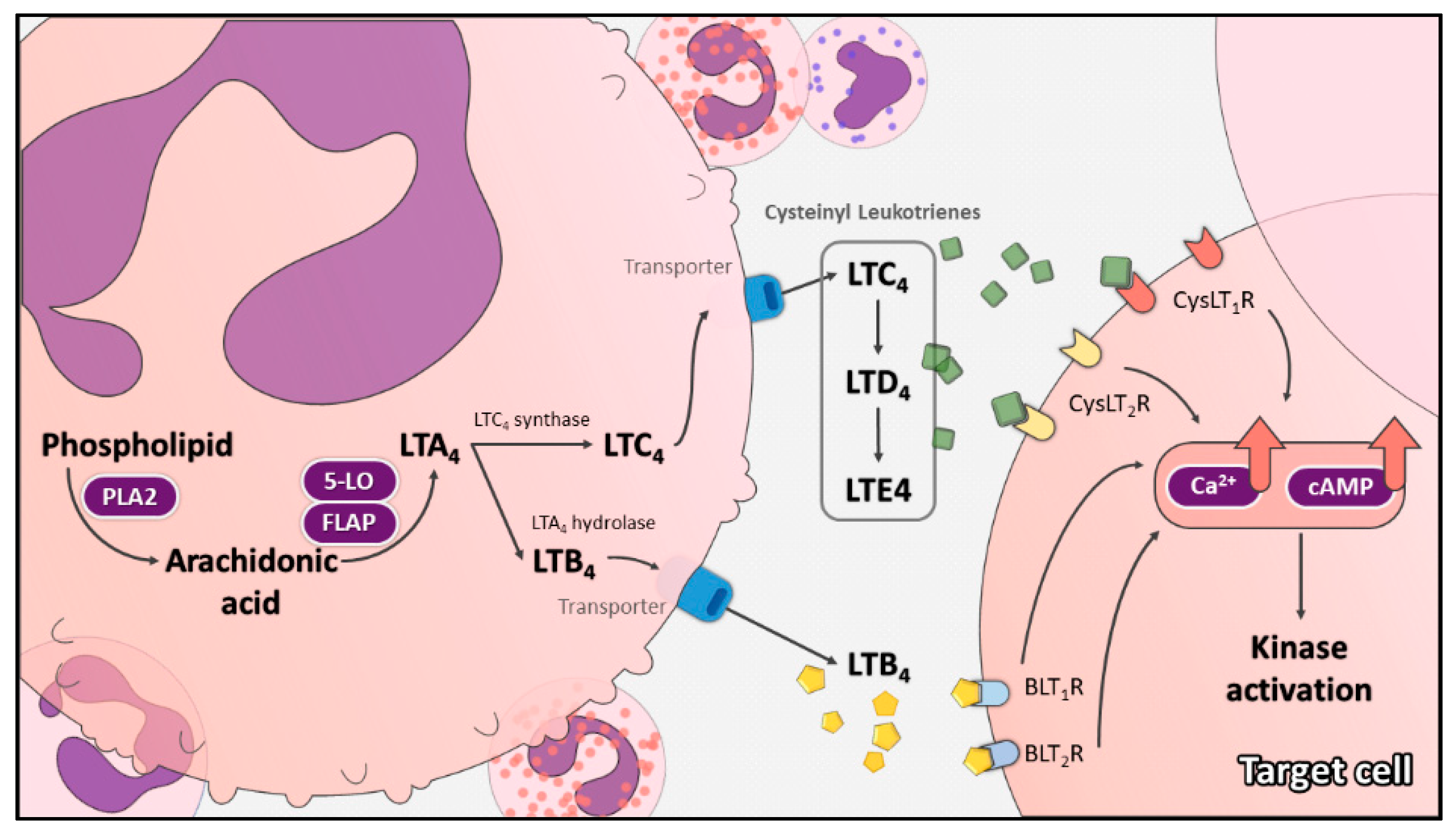
Table 1.
Cysteinyl leukotriene pathway antagonists.
| Medication | Pharmacologic Effect | Remark |
|---|---|---|
| Zileuton | 5-LO inhibitor | Clinically available * |
| AA861 | 5-LO inhibitor | |
| BU-4664L | 5-LO inhibitor | |
| BWA4C | 5-LO inhibitor | |
| ETH 615-139 | 5-LO inhibitor | |
| MK591 | 5-LO inhibitor | |
| MK886 | 5-LO inhibitor | |
| Montelukast | Selective CysLT1R antagonist | Clinically available * |
| Zafirlukast | Selective CysLT1R antagonist | Clinically available * |
| Pranlukast | Selective CysLT1R antagonist | Clinically available * |
| MK571 | Selective CysLT1R antagonist | |
| ZM198,615 | Selective CysLT1R antagonist | |
| AP 100984 | Selective CysLT2R antagonist | |
| BayCysLT2 | Selective CysLT2R antagonist | |
| HAMI 3379 | Selective CysLT2R antagonist | |
| Quininib 1,4-dihydroxy quininib | Selective CysLT1R (and CysLT2R) antagonist |
Abbreviations: 5LO = 5-lipoxygenase; CysLT1R = cysteinyl leukotriene 1 receptor; CysLT2R = cysteinyl leukotriene 2 receptor. * Approved by the US Food and Drug Administration.
Publisher’s Note: MDPI stays neutral with regard to jurisdictional claims in published maps and institutional affiliations. |
© 2021 by the authors. Licensee MDPI, Basel, Switzerland. This article is an open access article distributed under the terms and conditions of the Creative Commons Attribution (CC BY) license (https://creativecommons.org/licenses/by/4.0/).
Share and Cite
MDPI and ACS Style
Tsai, M.-J.; Chang, W.-A.; Chuang, C.-H.; Wu, K.-L.; Cheng, C.-H.; Sheu, C.-C.; Hsu, Y.-L.; Hung, J.-Y. Cysteinyl Leukotriene Pathway and Cancer. Int. J. Mol. Sci. 2022, 23, 120. https://doi.org/10.3390/ijms23010120
AMA Style
Tsai M-J, Chang W-A, Chuang C-H, Wu K-L, Cheng C-H, Sheu C-C, Hsu Y-L, Hung J-Y. Cysteinyl Leukotriene Pathway and Cancer. International Journal of Molecular Sciences. 2022; 23(1):120. https://doi.org/10.3390/ijms23010120
Chicago/Turabian StyleTsai, Ming-Ju, Wei-An Chang, Cheng-Hao Chuang, Kuan-Li Wu, Chih-Hung Cheng, Chau-Chyun Sheu, Ya-Ling Hsu, and Jen-Yu Hung. 2022. "Cysteinyl Leukotriene Pathway and Cancer" International Journal of Molecular Sciences 23, no. 1: 120. https://doi.org/10.3390/ijms23010120
Note that from the first issue of 2016, this journal uses article numbers instead of page numbers. See further details here.