Review

Pancreatic Cystic Lesions: A Focused Review on Cyst Clinicopathological Features and Advanced Diagnostics

1
Department of Pathology, The Ohio State University Wexner Medical Center, Columbus, OH 43210, USA
2
School of Medicine, The Ohio State University, Columbus, OH 43210, USA
3
Division of Gastroenterology, Department of Internal Medicine, The Ohio State University Wexner Medical Center, Columbus, OH 43210, USA
*
Author to whom correspondence should be addressed.
Diagnostics 2023, 13(1), 65; https://doi.org/10.3390/diagnostics13010065
Submission received: 16 November 2022 / Revised: 13 December 2022 / Accepted: 19 December 2022 / Published: 26 December 2022
(This article belongs to the Special Issue Diagnosis and Management of Pancreatic Cysts)

Abstract

:
Macroscopic, endomicroscopic, and histologic findings and correlation are an integral part of the diagnostic evaluation of pancreatic cystic lesions (PCLs), as complementing morphologic features seen by different specialties are combined to contribute to a final diagnosis. However, malignancy risk stratification of PCLs with worrisome features can still be challenging even after endoscopic ultrasound guided-fine needle aspiration (EUS-FNA) with cytological evaluation. This review aims to summarize cyst clinicopathological features from the pathologists’ perspective, coupled with knowledge from advanced diagnostics–confocal laser endomicroscopy and cyst fluid molecular analysis, to demonstrate the state-of-art risk stratification of PCLs. This review includes illustrative photos of surgical specimens, endomicroscopic and histologic images, and a summary of cyst fluid molecular markers.

1. Introduction

Pancreatic cystic lesions (PCLs) are being detected more frequently due to the increasing use of cross-sectional imaging [1]. Our knowledge of different types of PCLs has grown dramatically in recent years. A multidisciplinary approach using a combination of radiological imaging, endoscopic ultrasound (EUS), cytology, cyst fluid analysis, and molecular profiling is most helpful in diagnosing and risk stratifying PCLs [2,3,4,5]. Despite these advancements, challenges still exist in the appropriate classification of PCLs as benign versus precancerous and accurate risk-stratification of intraductal papillary mucinous neoplasm (IPMNs). Unwarranted resection of benign PCLs (up to 15% of pancreatectomies) causes unnecessary mortality and morbidity [6], and there is continued surgical overtreatment of branch duct-IPMNs where up to 50% of lesions only reveal low-grade dysplasia [7].
Various guidelines and recommendations exist for the management of PCLs [8]. The authors’ institution follows the 2017 international consensus guidelines for intraductal papillary mucinous neoplasm (IPMN) of the pancreas [1]. While the differentiation of most PCLs can be achieved through a combination of the clinical history, gender, imaging characteristics, cytology and cyst fluid analyses, malignancy risk stratification of PCLs with worrisome features can still be challenging even after EUS-FNA with cytological evaluation. This review aims to summarize cyst clinicopathological features from the pathologists’ perspective, coupled with knowledge from advanced diagnostics—confocal laser endomicroscopy and cyst fluid molecular analysis—to demonstrate the state-of-art risk stratification of PCLs.
EUS-guided needle-based confocal laser endomicroscopy (nCLE) generates real-time endomicroscopic images of the PCLs. Coupled with intravascularly injected fluorescein dye, nCLE accentuates the vascular pattern of the lesion revealing distinct epithelial features. Benefiting from a preserved interstitium, in vivo nCLE captures certain vascular features less obvious on histology (example: serous cystadenoma) and can increase the accuracy of differentiation of PCLs. Molecular analysis by next-generation sequencing (NGS) has evolved significantly in recent years with improved accuracy in the diagnosis and risk-stratification of PCLs [9].
According to recent surgical series of PCLs [10,11,12], the most resected PCLs are mucinous neoplasms: IPMNs (45% of all resected PCLs) and mucinous cystic neoplasm (MCN, 16%). The most frequently resected non-mucinous neoplasms include serous cystic neoplasms (SCN, 16%), cystic neuroendocrine tumors (cNET, 5%), and solid pseudopapillary neoplasm (SPN, 3%) [13]. In this review, for each of these and other commonly encountered PCLs, the definition, epidemiology, location, macroscopic, endomicroscopic, histologic features, and molecular alterations will be reviewed systematically to help with the differential considerations and the ultimate construction of the final diagnosis.

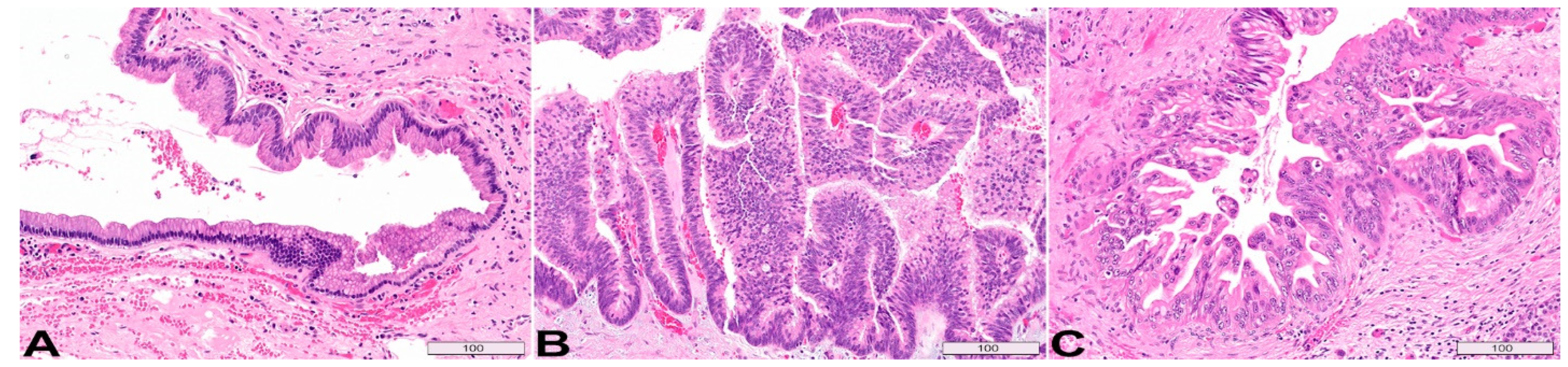
2. Intraductal Papillary Mucinous Neoplasm

Definition: Intraductal papillary mucinous neoplasm (IPMN) of the pancreas is a grossly visible (>5 mm) intraductal epithelial neoplasm arising in the main duct (MD) and/or branch-duct (BD) of the pancreas. The epithelia of IPMN feature papillary formation and mucin production.
Epidemiology: Peak incidence is between 62–67 years of age. In the USA, MD-IPMN is slightly more common in men (male:female ratio 1.1:1), while BD-IPMN is more common in women (male:female ratio 0.76:1) [14].
Location: IPMN can occur anywhere in the pancreatic ductal system, with the head of the pancreas being the most common location. Multicentricity (synchronous or metachronous lesions) is observed in up to 40% of cases; therefore, clinical follow-up of the remnant pancreas is important even if the resection margin is negative [15,16,17,18,19,20].
Macroscopic appearance: MD-IPMNs are mostly located in the head of the pancreas. If the more proximal pancreatic duct is involved, mucin may be seen extruding from the dilated ampullary orifice into the duodenum (fish-mouth papilla) [21,22]. The main duct in IPMN is by definition >5 mm in diameter and filled with tenacious mucin and lined by friable papillary formations [17]. If secondary ducts are also involved, it is classified as mixed duct-type IPMN.
BD-IPMNs mostly occur in the uncinate process and present as peripheral multilocular cystic lesions. A gross photo of a resected BD-IPMN is shown in Figure 1A–D. The cut surface of the pancreas demonstrates characteristic multilocular, Swiss cheese appearance. Some cysts have smooth lining (Figure 1A gross photo short arrow; Figure 1B histology), while others are lined by papillary neoplastic proliferation (Figure 1A gross photo long arrow; Figure 1C histology). The adjacent pancreatic parenchyma often undergoes lobular atrophy (Figure 1D between arrows).
IPMNs > 3 cm and/or with mural nodules/papillary projection are likely to harbor high-grade dysplasia and invasive carcinoma. A MD-IPMN is shown (Figure 1E,F). The cyst is lined by neoplastic epithelium and the lumen contains papillary/villous growth, partially obstructing the duct lumen (Figure 1E). This case contains both low-grade dysplasia (Figure 1F upper half) and high-grade dysplasia (Figure 1F lower half).
Endomicroscopy: EUS-nCLE of IPMN shows finger-like papillary projections composed of an outer epithelium and inner vascular core [23]. Figure 2 shows the typical appearance of IPMNs as seen during real-time in vivo EUS-nCLE procedure. Figure 2A shows a BD-IPMN with low-grade dysplasia. The epithelium is thinner and more translucent. Comparatively, Figure 2B demonstrates BD-IPMN with high-grade dysplasia with thicker and darker epithelium indicative of cellular and nuclear stratification, respectively. Both computer-aided artificial intelligence algorithms and human-interobserver studies have revealed a high diagnostic accuracy in nCLE-guided differentiation of IPMNs [24,25].

Histopathology

Histologic Subtypes: There are three histologic subtypes of IPMN, based on the predominant cell differentiation of the neoplastic epithelium [14]. The former oncocytic subtype is now considered a separate entity “Intraductal Oncocytic Papillary Neoplasm (IOPN)”, due to its different molecular underpinning and prognosis [26,27,28,29].
The three histologic subtypes of IPMNs are gastric type (~70%), intestinal type (~20%), and pancreatobiliary type (~10%) (Figure 3). Gastric type (Figure 3A) resembles gastric foveolar epithelium, composed of tall columnar cells with basally oriented small nuclei and abundant cytoplasmic mucin. Scattered goblet cells may be seen. Gastric type is often associated with low-grade dysplasia and usually occurs in branch ducts [30]. Intestinal type (Figure 3B) is the second most common histologic type, which most often resides in the main duct and is associated with high-grade dysplasia. It features hyperchromatic villous papillae, containing elongated and crowded nuclei, basophilic cytoplasm and variable amount of goblet cells [31,32,33,34]. Pancreaticobiliary type (Figure 3C) contains amphophilic to eosinophilic cytoplasm, enlarged nuclei with nucleoli, and moderate cytoplasmic mucin. It typically involves the main duct and often shows high-grade cytoarchitectural atypia. Some consider the pancreatobiliary type a variant of high-grade gastric type [31,35].
Grading of dysplasia: IPMNs is currently classified as low grade and high grade, based on the highest degree of cytoarchitectural atypia in the epithelium [14,36]. Low-grade IPMNs are characterized by mucinous epithelium with mild to moderate atypia. The epithelium may be flat (Figure 3A), show small tufting, or arrange into tall papillae but with bland cytology (Figure 4A). High-grade IPMNs feature severe atypia, characterized by papillae with irregular branching and budding, nuclear stratification with loss of polarity, cellular pleomorphism, and increased mitoses (Figure 4B–D). Notably, with increasing dysplasia, the papillae may lose the central fibrovascular core. In Figure 4B, while the bottom villous structure still has a fibrovascular core, the villi in the center of the image become slender without stromal core and the nuclei are rounded up, showing high-grade features including pleomorphism and loss of polarity.
Molecular alterations: Somatic mutations in KRAS and GNAS are the two most common genetic alterations seen in IPMN, together seen in >95% of all IPMNs [37,38]. KRAS mutation is an early event and nearly a prerequisite in the pathogenesis of ductal neoplasms, including pancreatic ductal adenocarcinomas (PDAC). Activating GNAS mutations are enriched in the intestinal subtype [39,40,41]. RNF43 somatic mutations are seen in about 50% of IPMNs [37,42]. Changes in tumor suppressor genes, such as TP53, CDKN2A and SMAD4, as well as mutations of mTOR genes (PTEN, PIK3CA, AKT1) are associated with advanced neoplasia (high-grade dysplasia and invasion) [43,44,45,46,47,48,49]. Alterations of other genes in the mitogen-activated protein kinase (MAPK) pathway have also been detected in mucinous cysts, including BRAF, ERBB2, HRAS, and MAPK1. GNAS and BRAF mutations in mucinous cysts are fairly specific for IPMN [9,46].
IPMN-associated invasive carcinoma: Overall, about a third of resected IPMN are associated with invasive carcinoma. Main-duct IPMN (>10 mm) has higher rate of associated malignancy, up to 60%, whereas branch-duct IPMN (>3 cm) has a lower rate (15–20%) [50,51,52,53,54]. The two most common IPMN-associated invasive carcinomas are tubular (ductal) adenocarcinoma and colloid adenocarcinoma. Colloid adenocarcinomas almost always arise in a background of intestinal-type IPMN, typically harbor GNAS mutations, and carry a better prognosis. Tubular adenocarcinoma is morphologically similar to conventional pancreatic ductal adenocarcinoma (PDAC) and is associated with pancreaticobiliary/gastric-type IPMN, and KRAS mutation [40,55,56]. The invasive component can be very focal, therefore thorough (if not complete) sampling of the resected pancreas is warranted.

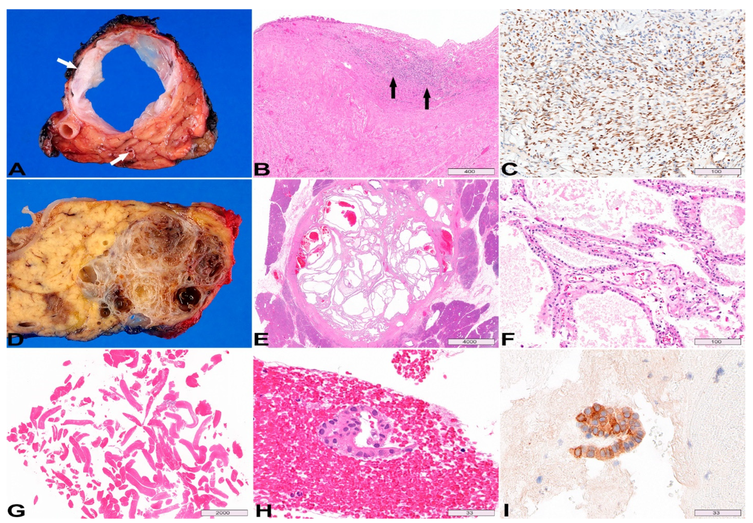
3. Mucinous Cystic Neoplasm

Definition: Mucinous cystic neoplasm (MCN) of the pancreas is a mucin-producing cystic epithelial neoplasm that is associated with characteristic ovarian-type subepithelial stroma.
Epidemiology: The mean patient age is 48 years. MCN predominantly (>98%) occurs in women [14]. Increasing age correlates with a higher risk of invasive carcinoma, suggesting that progression occurs over a period of years.
Location: Almost exclusively, MCNs occur in the body or tail of the pancreas (>98%) [57,58,59,60]. Unlike IPMN, MCN is typically solitary, without synchronous lesions.
Macroscopic appearance: The mean size of MCN is 6 cm (ranging 2 to 35 cm). MCNs with invasive carcinoma are larger (>5 cm, mean: 9 cm) [57]. MCNs can be unilocular or multilocular with a few septa. They do not communicate with the pancreatic ductal system and typically have a thick fibrous wall (≥3 mm, Figure 5A top arrow) [61]. The pathogenesis is unclear and thought to be related to ectopic embryonic remnants, which explains some “ancient” changes such as a thick fibrous wall and mural calcifications. High-grade MCNs often have mural nodules or papillary projections.
Endomicroscopy: EUS-nCLE of MCNs shows horizon-type epithelial bands (Figure 6A) of variable thickness without papillary conformation [23]. The epithelial bands are single or in multiple layers and are best observed when viewed tangentially to the nCLE probe. Further, MCNs can reveal clusters of inflammatory cells with areas of a dark background and fluorescent macrophages. Since MCNs may contain foci of atrophy and chronic inflammation, nCLE characterization of epithelial bands can sometimes be challenging during in vivo EUS. Inflammatory clusters of cells or debris can also be observed in pseudocysts, but such patients frequently have a history of pancreatitis and nCLE does not reveal any epithelial bands (Figure 6B,C).
Histopathology: The mucinous epithelium of MCNs is morphologically similar to that of IPMNs, and the dysplasia is also graded based on a two-tiered system [36]. It is not uncommon for the epithelium of MCN to be denuded in areas, sometimes extensively. Looking for the subepithelial ovarian-type stroma is helpful in such cases (Figure 5B arrows), and the presence of this distinctive stroma is required for the diagnosis of MCN [14]. However, the ovarian-type stroma may be attenuated (hypocellular/hyalinized) in large MCNs, postmenopausal patients, or around areas with advanced neoplasia [57]. Positive immunohistochemical staining for progesterone receptor (60–90%) (Figure 5C) and estrogen receptor (30%) may aid in the detection of the ovarian-type stroma [60,62].
Invasive carcinoma typically occurs in MCNs that are large (>5 cm) or with gross papillary nodules [60,63,64]. Invasion is seen in about 15% of MCNs [57,59,65]. Similar to IPMNs, the invasive component is usually tubular/ductal adenocarcinoma and can be focal, so extensive/complete sampling of the cyst wall is indicated [14,57,66].
Molecular alterations: Activating mutations in codon 12 of KRAS is seen in 50–66% of MCNs as well as loss of function in RNF43 [9,41,67]. Unlike IPMN, GNAS mutations are rarely seen in MCN [43]. The gene mutations associated with advanced neoplasia in IPMN may be observed in MCN, including TP53, CDKN2A, SMAD4, and/or mTOR genes (PTEN, PIK3CA, AKT1) [9].

4. Serous Cystadenoma

Definition: Serous cystadenoma (SCA) is a benign epithelial neoplasm composed of uniform cuboidal, glycogen-rich clear cells that often form cysts containing serous fluid.
Epidemiology: The mean age at presentation is 58 years, with a female predominance (female: male = 3:1).
Location: SCA can occur anywhere in the pancreas; mostly (50–75%) in the body or tail.
Macroscopic appearance: SCAs are well-circumscribed, multilocular cystic mass, without communication with the pancreatic ductal system. The mean size is about 4 cm with a wide range (1 to 25 cm in diameter) [68,69,70,71]. The cyst wall is typically thin (<3 mm) [61,72]. The microcystic variant shows characteristic honeycomb or sponge pattern (Figure 5D,E). A central scar with a sunburst calcification pattern is present in 30% of cases. There are also rare macrocystic (oligocystic) variant and solid variant.
Endomicroscopy: EUS-nCLE of SCA shows an intricate fern pattern of vascularity (Figure 6D,E) or also called superficial vascular network. This interstitial vascular pattern reveals parallel or inter-connected network of capillaries underneath the epithelium [23]. Trafficking red blood cells are frequently observed in the fine vascular meshwork during the in vivo EUS-nCLE procedure. There is variation in contrast (fluorescein) penetration within the capillaries; often, the capillaries are densely packed with red blood cells.
Histopathology: The lining epithelium of SCA consists of a single layer of low cuboidal epithelial cells with clear cytoplasm, due to abundant intracytoplasmic glycogen. Variably prominent capillary network is seen underneath the epithelium (Figure 5F). Nuclear atypia and mitoses are typically absent. The neoplastic epithelium is immunoreactive for inhibin, which can be helpful in small biopsy or cytology specimens. Microforceps biopsy may show very scant cuboidal epithelial cells in a background of blood (Figure 5G,H); positive inhibin staining helps to confirm the diagnosis (Figure 5I) in the appropriate clinical setting.
Molecular alternations: Germline or somatic alterations of the tumor suppressor gene VHL is present in SCAs [9,41,67,73]. In the setting of germline VHL mutation, there could be multifocal SCAs in the pancreas. Alterations in genes associated with IPMN, MCN, and PDAC, such as KRAS, GNAS, CDKN2A, and SMAD4, have not been reported in SCAs [41]. TP53 or TERT promoter mutations may be prognostically important, as they are associated with interval growth of cyst size [9].

5. Cystic Neuroendocrine Tumor

Pancreatic neuroendocrine tumors are typically well-circumscribed, solid tumors. However, hemorrhage and secondary degeneration may occur in the center of the tumor (Figure 7A,B), resulting in a grossly cystic neuroendocrine tumor (cNET) with only viable tumor cells in the cyst wall. EUS-nCLE of cNET shows dark clusters (trabeculae) of cells separated by stroma [23]. The clustering of cells can conform to various shapes, commonly in cords or groups with occasional geometric formations (Figure 6F). Histologically, cNET is composed of nests, trabeculae, or ribbons of neoplastic cells, separated by thin vascular fibrous septa (Figure 7C). MEN1 alterations are highly specific for cNETs, but the sensitivity is low (27%) [9]. Loss of ATRX/DAXX and the presence of alternative lengthening of telomeres (ALT) are associated with poor prognosis [9]. cNETs with loss of heterozygosity (LOH) of ≥3 genes tend to have distant metastasis [9].

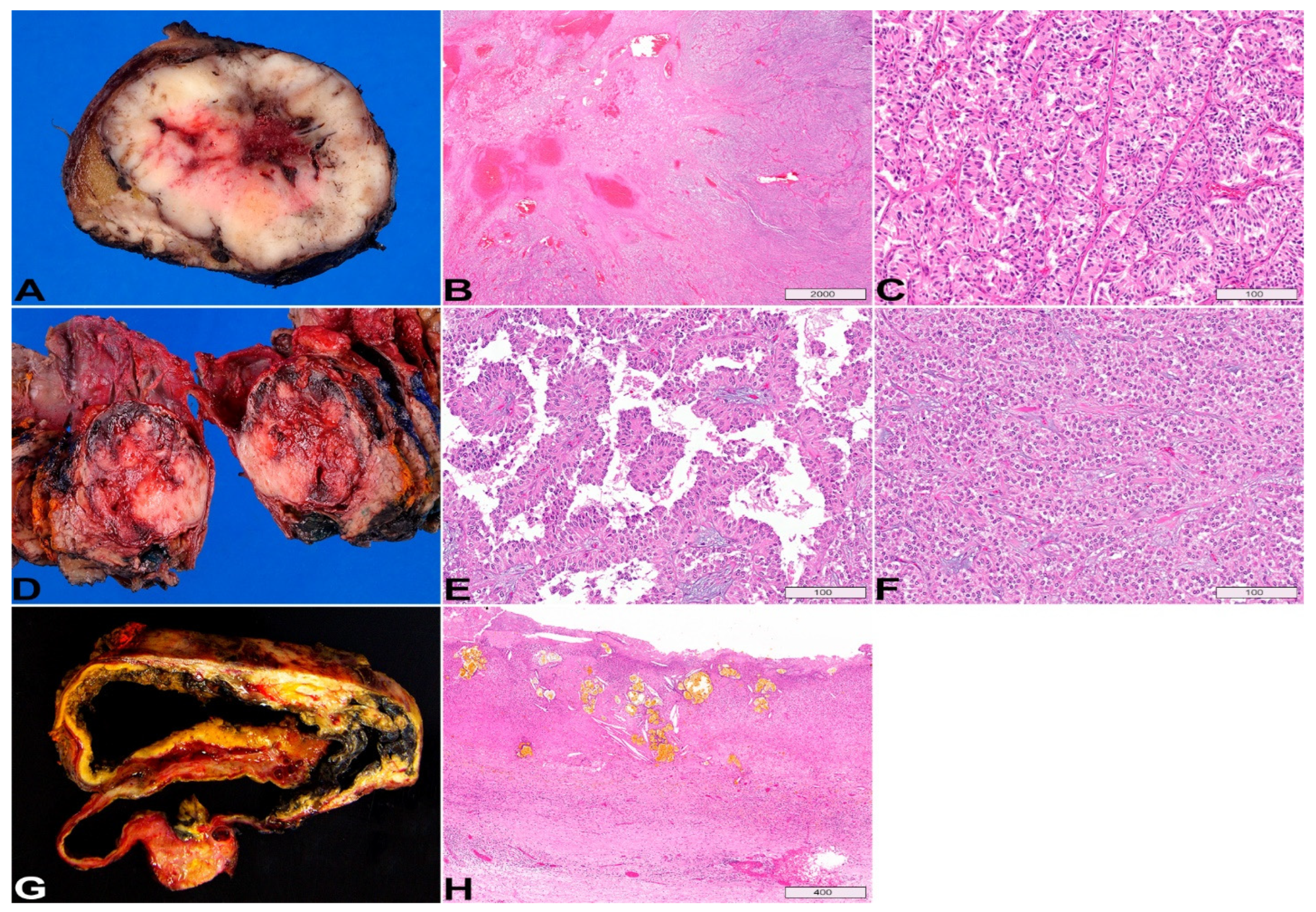
6. Solid Pseudopapillary Neoplasm

Definition: Solid pseudopapillary neoplasm (SPN) is a low-grade malignant tumor that lacks a specific line of pancreatic epithelial differentiation. It most likely arises from genital ridge cells that were translocated to the pancreas during embryogenesis [74].
Epidemiology: SPNs occur predominantly (90%) in young women. The mean age at presentation is 28 years.
Location: SPNs have a slight preference for the pancreatic tail [75,76].
Macroscopic appearance: SPNs are well-demarcated mass lesions, with variable solid and cystic components, and occasional calcifications. SPNs are typically large tumors (average size: 8 cm; range: 0.5–25.0 cm) [14]. The cystic component reflects degenerative changes secondary to hemorrhagic necrosis (Figure 7D). Not surprisingly, small tumors tend to be more solid.
Endomicroscopy: EUS-nCLE of SPN is indistinguishable from cNET revealing groups or clusters of cells separated by interstitial spaces [23].
Histopathology: SPN is composed of epithelioid cells, forming pseudopapillary structures due to perivascular growth (Figure 7E) and also crowded nests imparting a solid appearance in areas (Figure 7F). SPNs may mimic a pancreatic NET, macroscopically, endomicroscopically, and histologically. Diffuse nuclear staining of β-catenin, as well as the expression of SOX11 and TFE3 by immunohistochemistry, are helpful to confirm the diagnosis of SPN [77,78,79,80]. Of note, SPNs may be positive for synaptophysin by immunohistochemistry, but chromogranin should be negative.
Molecular alternations: Somatic activating mutation in CTNNB1 (encoding β-catenin) is the main molecular feature of SPNs [41,81,82].

7. Pseudocyst

A pseudocyst is a collection of fluid contents walled off by fibrous tissue after episode(s) of pancreatitis (Figure 7G). The location is usually outside the pancreas. EUS-nCLE of pseudocyst (Figure 6B,C) reveals a dark background due to the absence of vascular interstitium and a true epithelium-lined cyst wall. Some auto-fluorescent inflammatory cells are usually present in pseudocysts [23]. Histologically, a cyst lining is absent, and the cyst wall is composed of fibroinflammatory tissue, often with evidence of old hemorrhage and necrosis (cholesterol clefts and pigments, Figure 7H).

8. Squamous Lined Epithelial Cysts

Definition: Squamous lined epithelial cysts can be divided into three types: lymphoepithelial cysts (LEC), dermoid cysts (cystic teratomas), and epidermoid cysts in intrapancreatic accessory spleen (ECIPAS) [83]. LECs are benign, true cysts lined by mature, keratinizing squamous epithelium with abundant surrounding lymphoid tissue [83,84,85]. They are theorized to arise from misplaced branchial cleft cysts during embryogenesis [86], squamous metaplasia of obstructed intrapancreatic ducts, or inclusion of benign epithelium or ectopic pancreas into a peripancreatic lymph node [85,87]. Dermoid cysts of the pancreas, like other teratomas, are tumors composed of tissue derived from germ layers (ectoderm, mesoderm, and endoderm) [88]. ECIPAS is a cystic mass arising from intrapancreatic accessory spleen (congenital abnormality characterized by ectopic splenic tissue) [89].
Epidemiology: LECs are the most prevalent among squamous-lined epithelial cysts and account for approximately 0.5% of all PCLs. The mean patient age at presentation is 56 years, with a male predominance (80%) [84]. Dermoid cysts of the pancreas occur in a younger age group with a mean age of 23 (range 2–53 years) with no gender predominance [83]. The mean patient age for ECIPAS is 38 years [83] with a slight female predominance [90].
Location: LEC can occur anywhere in the pancreas (head, body, or tail) or can be found in an extra-pancreatic location [83,84,91,92]. Dermoid cysts can occur anywhere along the pathway of ectodermal cell migration and can been found in anywhere in the pancreas [93,94]. ECIPAS are primarily located in the tail of the pancreas [83,90].
Macroscopic Appearance: The mean size of LECs is approximately 5 cm (range 1.2–17 cm). They are often round and well-demarcated from the surrounding pancreas. They can be multilocular (60%) or unilocular (40%) in appearance. The contents of the cysts appear “cheesy” or “caseous” (signifying keratinaceous debris) or may less often be clear and serous [84]. Dermoid cysts of the pancreas contain a combination of both cystic and solid components. Ectodermal differentiation (skin, hair follicles, sweat glands, sebaceous material) is the most common, but structures from other germ layers (cartilage, bone, thyroid tissue, etc.) may be present [88]. The mean size of ECIPAS is 4.5 cm (range 2.3–6.5 cm). ECIPAS can be unilocular or multilocular. It may contain serous fluid but will lack presence of hair or skin appendages [83,95,96].
Endomicroscopy: LECs can reveal clusters of bright particles suggestive of keratinous debris otherwise can be heterogenous in appearance with a bland background (squamous epithelium) and lattice-type blood vessels [23,97]. ECIPAS can reveal cords of cells suggestive of ectopic splenic tissue [97].
Histopathology: LECs are lined by squamous epithelium and surrounded by subepithelial lymphoid tissue with germinal centers [84,98]. Other rarer features of LECs include: containing sebaceous differentiation, keratin debris and cholesterol crystals, scattered mucinous cells with features similar to goblet cells, and multinucleated giant cells [84,98,99,100,101]. Dermoid cysts can include mucinous epithelium, respiratory-type mucosa, sebaceous units, and hair. Unlike LEC, dermoid cysts lack subepithelial lymphoid tissue [83,84,93]. ECIPAS are typically lined with squamous (keratinized or nonkeratinized) epithelium surrounded by normal splenic tissue [96], although lining containing flattened cuboidal epithelium has been reported [90]. They have been reported to contain blood, cholesterol clefts, macrophages, and microcalcifications [90,96].
Cyst Fluid: Occasionally cyst fluid from LECs and rarely ECIPAS has elevated carcinoembryonic antigen (CEA) levels causing challenges with differentiation from mucinous neoplasms [84,90,91,95,101].

9. Simple Mucinous Cyst

In the absence of a visible obstructive process, mucinous cysts that are >1 cm, without any features of IPMN or MCN, are classified as simple mucinous cysts [36,102,103,104,105]. Cyst fluid analysis may show elevated CEA levels. Histologically, the mucinous epithelium is flat with rare tufting, but well-formed papillae are absent. The most common mutations include KMT2C (62%), KRAS and TP53 (15%) [52,106]. However, in 31% cases, no mutations are detected. A simple mucinous cyst is essentially a diagnosis of exclusion, and the main differential considerations include IPMN, MCN, and a retention cyst involved by pancreatic intraepithelial neoplasia.
Other rare pancreatic cystic lesions not discussed in this review include intraductal oncocytic neoplasm (IOPN), intraductal tubulopapillary neoplasm (ITPN), acinar cystic transformation of the pancreas, among others [13,52,107].

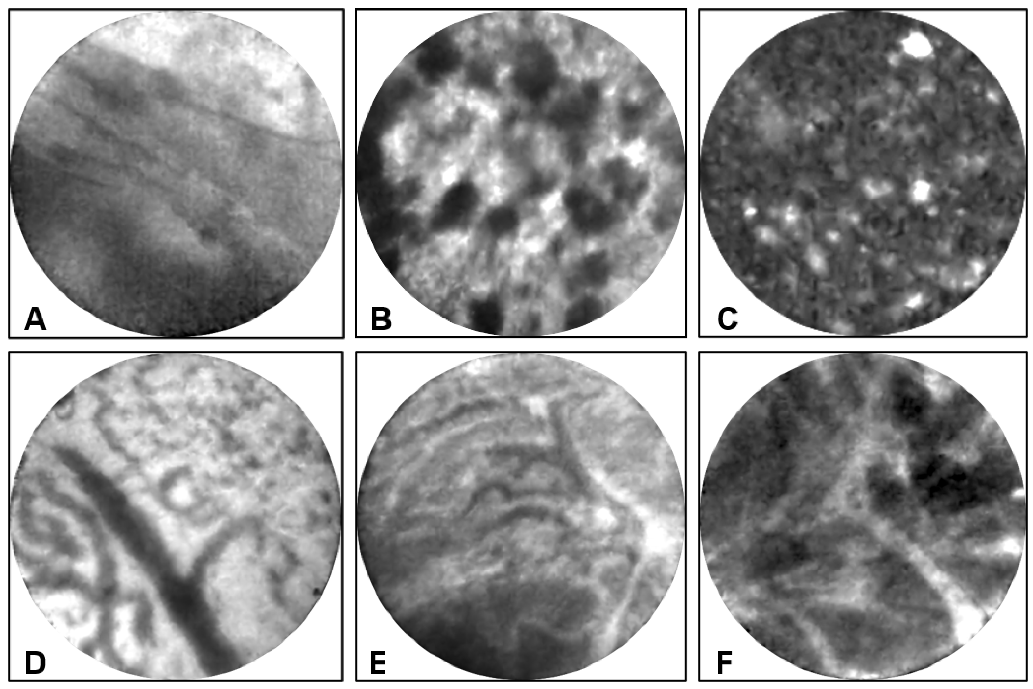
10. Advanced and Emerging Diagnostic Tools for Pancreatic Cystic Lesions

Despite our ever-increasing knowledge of PCLs, there remains continued resection of benign cysts at high rates and resection of IPMNs with low-grade dysplasia. In this review, we have described features observed in specific PCLs using two advanced diagnostics (nCLE and cyst fluid NGS). Both techniques can enhance the diagnostic accuracy of cyst type and advanced neoplasia significantly [108], and they may be considered when radiomics and EUS cyst morphology are indicative of a high-risk PCL [2]. However, these advanced diagnostics are only available in limited centers currently, and large prospective studies are needed before incorporation into guidelines. Other emerging diagnostic tools for PCLs include Mass Spectrometry and Optical Coherence Tomography (OCT).
Needle-based confocal laser endomicroscopy (nCLE) provides real time, en-face (perpendicular to histologic sectioning planes) visualization of cyst lining at the microscopic level. Through a combined assessment of interstitial vascular pattern, cyst content fluorescence characteristics, and the “negative” image/architecture of the cyst epithelium, the accuracy of nCLE diagnosis of PCLs reaches over 90% [109,110]. In addition, nCLE evaluation of the “thickness” and “darkness” of the cyst epithelia allows grading of dysplasia and thus risk stratification of IPMNs [24]. However, the lack of adequate exposure/training for the real-time interpretation of images impede the widespread use of nCLE. Artificial intelligence (AI)-aided interpretation of nCLE images may help overcome some of the challenges [25].
Next generation sequencing of the nucleic acid present in the cyst fluid, offers molecular insight into the genetic makeup of the cells shed from the neoplastic cyst epithelium. Certain genetic alternations indicate specific pancreatic cyst types, and molecular evolution with additional gene mutations is correlated with neoplastic progression and advanced neoplasia [9]. Alternations involving KRAS, GNAS, and/or BRAF indicate mucinous cyst; GNAS and BRAF mutations in mucinous cysts are fairly specific for IPMN (rare in MCN). Advanced neoplasia in mucinous cysts is associated with alterations in TP53, SMAD4, CTNNB1, CDKN2A, and/or the mTOR genes (PTEN, PIK3CA, AKT1). Cystic pancreatic NETs are associated with MEN1 mutations and LOH for multiple genes. Loss of ATRX/DAXX and the presence of alternative lengthening of telomeres (ALT) are associated with poor prognosis in pancreatic NETs. SCAs often harbor VHL mutations; TP53 and TERT promoter mutations are seen in large-sized SCAs.
EUS through-the-needle biopsy (TTNB) represents a “bite” biopsy of the cyst wall by a microforceps introduced through a standard EUS 19-gauge FNA needle. TTNB generates larger tissue fragments and boasts good diagnostic yield (69.5%) and high histological accuracy (86.7%) in a meta-analysis of 454 patients [111]. Another meta-analysis demonstrates that TTNB has a higher sensitivity and specificity than cytology [112]. However, a relatively high rate of complications has been reported with mucinous cysts or cysts with connection to pancreatic duct. In a prospective study of 101 patients who underwent TTNB, adverse event rate was 9.9% (10 patients, including 9 acute pancreatitis and 1 fatality) [113]. Therefore, TTNB should be considered for patients where the necessity of an accurate diagnosis outweighs the risks [114]. We exclusively reserve TTNB for SCAs. Only when nCLE shows SCA (or non-mucinous) pattern, we will consider TTNB.
Mass spectrometry is a useful tool that measures the mass-to-charge ratio of ions and allows for the identification of the molecular weight, structure, and chemical formula of pure substances [115,116]. Additionally, mass spectrometry has been a helpful noninvasive tool that can assist in the early diagnosis of cancer by identifying both the presence of cancer and its progression over time [117,118]. Early mass spectrometry studies analyzing protein profiles obtained from pancreatic cyst fluid found unique peptide patterns for various benign PCLs [119,120], detected pancreatic tumor markers (such as Mucin family members, S100 proteins, CEA-related proteins) [121], and were able to distinguish between benign and malignant pancreatic lesions [120]. Notably, one study combined a novel biomarker panel of four proteins with CA19-9 and was able to diagnose pancreatic carcinoma with a sensitivity of 95% and specificity of 94.1% [122]. In a more recent 2018 diagnostic study, targeted mass spectrometry taken from pancreatic cyst fluid using protein biomarkers from mucin-5AC and prostate stem-cell antigen was able to identify advanced neoplasia with an accuracy of 96%. Targeted mass spectrometry can detect 95% of malignant/severely dysplastic lesions, compared with 35% and 50% for CEA and cytology respectively. Additionally, a panel of peptides taken from mucin-5AC and mucin-2 was able to discriminate between premalignant/malignant and benign lesions with an accuracy of 97%, outperforming cyst fluid CEA (61%) and cytology (84%) [123].
Optical Coherence Tomography is an imaging modality that features a spatial resolution at the µm level. OCT imaging has been applied to the examination of human eyes [124], skin [125], and cardiovascular tissue [126]. Recently, a prototype endoscopic OCT has been reported for early diagnosis and Endo-OCT image-guided brachytherapy of pancreatic cancer and precursor lesions [127].

11. Summary

Unlike pancreatic cancers that have known risk factors such as smoking, chronic pancreatitis, diet, and diabetes, most neoplastic pancreatic cysts do not have a clear etiology. Perhaps, they may be considered genetic diseases, caused by inherited (germline) and somatic mutations. Patients with certain hereditary cancer syndromes are known to be predisposed to developing pancreatic cysts. IPMNs have been reported in patients with McCune–Albright syndrome, Peutz–Jeghers syndrome, Lynch syndrome, familial adenomatous polyposis, hereditary breast and ovarian cancer syndrome, familial atypical multiple mole melanoma, and Carney complex [128]. SCNs develop in 90% of patients with von Hippel–Lindau syndrome (VHL). Overall, 10–20% of PanNETs are associated with hereditary syndromes including multiple endocrine neoplasia type 1, VHL, neurofibromatosis type 1, tuberous sclerosis, and others [14]. It is unclear why MCNs and SPNs have a female predominance. They could be related to persistent fetal periductal mesenchyme or translocated embryonic tissue from genital ridges that respond and proliferate in response to hormonal stimulation [14].
Pancreatic cystic lesions have distinct clinicopathologic, endomicroscopic, and molecular features facilitating differential diagnosis and risk stratification (Table 1). However, there are overlaps and it remains a clinical challenge in the pre-surgical evaluation of PCLs. An integrated multidisciplinary approach is the most effective approach in managing PCLs. This allows sharing of unique insights from different specialties, ultimately fitting the pieces of the puzzle together. This review compares the clinicopathological, endomicroscopic, and molecular features of the most common PCLs, bridging information across specialties with an emphasis on macroscopic, (endo)microscopic morphology, and molecular findings. In the diagnostic workup of a new PCL, demographic information, clinical presentation, and location, can generate a working differential. Distinct image-guided (MRI/CT, and EUS) cyst morphological features (size, locularity, wall thickness, mural nodule, duct communication) can refine and narrow down the differential. Standard of care analysis of the cyst fluid (CEA, amylase, glucose, and cytology) improve the differentiation of PCLs. EUS-FNA of cyst fluid with molecular analysis and nCLE for in vivo endomicroscopy, when indicated (generally PCLs ≥ 2 cm diameter), collectively approach a high (>90%) accuracy for the diagnosis and risk stratification of PCLs [2,3,4,5,9,23].

Author Contributions

Conceptualization and Image Acquisition, W.C. Writing–Original Draft Preparation, W.C. and N.A.; Writing–Review & Editing, S.G.K. All authors have read and agreed to the published version of the manuscript.

Funding

This research received no external funding.

Institutional Review Board Statement

Not applicable.

Informed Consent Statement

Not applicable.

Data Availability Statement

Not applicable.

Acknowledgments

The authors thank Shawn Scully for his assistance with image processing.

Conflicts of Interest

Krishna SG is the principal investigator of an investigator initiated study. The study in part is funded by a grant to The Ohio State University Wexner Medical Center from Mauna Kea Technologies, Paris, France. The other authors have nothing to disclose.

References

  1. Tanaka, M.; Fernández-del Castillo, C.; Kamisawa, T.; Jang, J.Y.; Levy, P.; Ohtsuka, T.; Salvia, R.; Shimizu, Y.; Tada, M.; Wolfgang, C.L. Revisions of international consensus Fukuoka guidelines for the management of IPMN of the pancreas. Pancreatology 2017, 17, 738–753. [Google Scholar] [CrossRef] [PubMed]
  2. Ardeshna, D.R.; Cao, T.; Rodgers, B.; Onongaya, C.; Jones, D.; Chen, W.; Koay, E.J.; Krishna, S.G. Recent advances in the diagnostic evaluation of pancreatic cystic lesions. World J. Gastroenterol. 2022, 28, 624–634. [Google Scholar] [CrossRef] [PubMed]
  3. Buerlein, R.C.D.; Shami, V.M. Management of pancreatic cysts and guidelines: What the gastroenterologist needs to know. Ther. Adv. Gastrointest. Endosc. 2021, 14, 26317745211045769. [Google Scholar] [CrossRef] [PubMed]
  4. Kim, H.S.; Jang, J.-Y. Management Algorithms for Pancreatic Cystic Neoplasms. Arch. Pathol. Lab. Med. 2022, 146, 322–329. [Google Scholar] [CrossRef] [PubMed]
  5. Turner, R.C.; Melnychuk, J.T.; Chen, W.; Jones, D.; Krishna, S.G. Molecular Analysis of Pancreatic Cyst Fluid for the Management of Intraductal Papillary Mucinous Neoplasms. Diagnostics 2022, 12, 2573. [Google Scholar] [CrossRef] [PubMed]
  6. De Pretis, N.; Mukewar, S.; Aryal-Khanal, A.; Bi, Y.; Takahashi, N.; Chari, S. Pancreatic cysts: Diagnostic accuracy and risk of inappropriate resections. Pancreatology 2017, 17, 267–272. [Google Scholar] [CrossRef]
  7. Sharib, J.M.; Fonseca, A.L.; Swords, D.S.; Jaradeh, K.; Bracci, P.M.; Firpo, M.A.; Hatcher, S.; Scaife, C.L.; Wang, H.; Kim, G.E. Surgical overtreatment of pancreatic intraductal papillary mucinous neoplasms: Do the 2017 International Consensus Guidelines improve clinical decision making? Surgery 2018, 164, 1178–1184. [Google Scholar] [CrossRef]
  8. Aziz, H.; Acher, A.W.; Krishna, S.G.; Cloyd, J.M.; Pawlik, T.M. Comparison of Society Guidelines for the Management and Surveillance of Pancreatic Cysts: A Review. JAMA Surg. 2022, 157, 723–730. [Google Scholar] [CrossRef]
  9. Paniccia, A.; Polanco, P.M.; Boone, B.A.; Wald, A.I.; McGrath, K.; Brand, R.E.; Khalid, A.; Kubiliun, N.; O’Broin-Lennon, A.M.; Park, W.G.; et al. Prospective, Multi-Institutional, Real-Time Next-Generation Sequencing of Pancreatic Cyst Fluid Reveals Diverse Genomic Alterations That Improve the Clinical Management of Pancreatic Cysts. Gastroenterology 2022, 164, 117–133.e7. [Google Scholar] [CrossRef]
  10. Gaujoux, S.; Brennan, M.F.; Gonen, M.; D’Angelica, M.I.; DeMatteo, R.; Fong, Y.; Schattner, M.; DiMaio, C.; Janakos, M.; Jarnagin, W.R.; et al. Cystic Lesions of the Pancreas: Changes in the Presentation and Management of 1,424 Patients at a Single Institution over a 15-Year Time Period. J. Am. Coll. Surg. 2011, 212, 590–600. [Google Scholar] [CrossRef]
  11. Kleeff, J.; Michalski, C.; Kong, B.; Erkan, M.; Roth, S.; Siveke, J.; Friess, H.; Esposito, I. Surgery for cystic pancreatic lesions in the post-sendai era: A single institution experience. HPB Surg. 2015, 2015, 847837. [Google Scholar] [CrossRef] [PubMed] [Green Version]
  12. Valsangkar, N.P.; Morales-Oyarvide, V.; Thayer, S.P.; Ferrone, C.R.; Wargo, J.A.; Warshaw, A.L.; Fernandez-del Castillo, C. 851 resected cystic tumors of the pancreas: A 33-year experience at the Massachusetts General Hospital. Surg. 2012, 152, S4–S12. [Google Scholar] [CrossRef] [PubMed] [Green Version]
  13. Esposito, I.; Haeberle, L. Nonmucinous Cystic Lesions of the Pancreas. Arch. Pathol. Lab. Med. 2022, 146, 312–321. [Google Scholar] [CrossRef] [PubMed]
  14. WHO Classification of Tumours-IARC. In Tumours of the pancreas.Digestive System Tumours, 5th ed.; International Agency for Research on Cancer: Lyon, France, 2019; Volume 1.
  15. Adsay, N.V.; Longnecker, D.S.; Klimstra, D.S. Pancreatic tumors with cystic dilatation of the ducts: Intraductal papillary mucinous neoplasms and intraductal oncocytic papillary neoplasms. Semin. Diagn. Pathol. 2000, 17, 16–30. [Google Scholar] [PubMed]
  16. Jang, J.Y.; Kim, S.W.; Ahn, Y.J.; Yoon, Y.S.; Choi, M.G.; Lee, K.U.; Han, J.K.; Kim, W.H.; Lee, Y.J.; Kim, S.C.; et al. Multicenter analysis of clinicopathologic features of intraductal papillary mucinous tumor of the pancreas: Is it possible to predict the malignancy before surgery? Ann. Surg. Oncol. 2005, 12, 124–132. [Google Scholar] [CrossRef]
  17. Loftus, E.V., Jr.; Olivares-Pakzad, B.A.; Batts, K.P.; Adkins, M.C.; Stephens, D.H.; Sarr, M.G.; DiMagno, E.P. Intraductal papillary-mucinous tumors of the pancreas: Clinicopathologic features, outcome, and nomenclature. Members of the Pancreas Clinic, and Pancreatic Surgeons of Mayo Clinic. Gastroenterology 1996, 110, 1909–1918. [Google Scholar] [CrossRef]
  18. Pelaez-Luna, M.; Chari, S.T.; Smyrk, T.C.; Takahashi, N.; Clain, J.E.; Levy, M.J.; Pearson, R.K.; Petersen, B.T.; Topazian, M.D.; Vege, S.S.; et al. Do Consensus indications for resection in branch duct intraductal papillary mucinous neoplasm predict malignancy? A study of 147 patients. Am. J. Gastroenterol. 2007, 102, 1759–1764. [Google Scholar] [CrossRef]
  19. Sohn, T.A.; Yeo, C.J.; Cameron, J.L.; Hruban, R.H.; Fukushima, N.; Campbell, K.A.; Lillemoe, K.D. Intraductal papillary mucinous neoplasms of the pancreas: An updated experience. Ann. Surg. 2004, 239, 788–799. [Google Scholar] [CrossRef]
  20. Tanaka, M.; Sawai, H.; Okada, Y.; Yamamoto, M.; Funahashi, H.; Takeyama, H.; Manabe, T. Clinicopathologic study of intraductal papillary-mucinous tumors and mucinous cystic tumors of the pancreas. Hepatogastroenterology 2006, 53, 783–787. [Google Scholar]
  21. Klöppel, G.; Basturk, O.; Schlitter, A.M.; Konukiewitz, B.; Esposito, I. Intraductal neoplasms of the pancreas. Semin. Diagn. Pathol. 2014, 31, 452–466. [Google Scholar] [CrossRef]
  22. Yamaguchi, K.; Tanaka, M. Mucin-hypersecreting tumor of the pancreas with mucin extrusion through an enlarged papilla. Am. J. Gastroenterol. 1991, 86, 835–839. [Google Scholar] [PubMed]
  23. Krishna, S.G.; Hart, P.A.; Malli, A.; Kruger, A.J.; McCarthy, S.T.; El-Dika, S.; Walker, J.P.; Dillhoff, M.E.; Manilchuk, A.; Schmidt, C.R.; et al. Endoscopic Ultrasound-Guided Confocal Laser Endomicroscopy Increases Accuracy of Differentiation of Pancreatic Cystic Lesions. Clin. Gastroenterol. Hepatol. Off. Clin. Pract. J. Am. Gastroenterol. Assoc. 2019, 18, 432–440.e6. [Google Scholar] [CrossRef] [PubMed]
  24. Krishna, S.G.; Hart, P.A.; DeWitt, J.M.; DiMaio, C.J.; Kongkam, P.; Napoleon, B.; Othman, M.O.; Yew Tan, D.M.; Strobel, S.G.; Stanich, P.P.; et al. EUS-guided confocal laser endomicroscopy: Prediction of dysplasia in intraductal papillary mucinous neoplasms (with video). Gastrointest. Endosc. 2020, 91, 551–563.e5. [Google Scholar] [CrossRef] [PubMed]
  25. Machicado, J.D.; Chao, W.L.; Carlyn, D.E.; Pan, T.Y.; Poland, S.; Alexander, V.L.; Maloof, T.G.; Dubay, K.; Ueltschi, O.; Middendorf, D.M.; et al. High performance in risk stratification of intraductal papillary mucinous neoplasms by confocal laser endomicroscopy image analysis with convolutional neural networks (with video). Gastrointest. Endosc. 2021, 94, 78–87.e2. [Google Scholar] [CrossRef] [PubMed]
  26. Adsay, N.V.; Adair, C.F.; Heffess, C.S.; Klimstra, D.S. Intraductal oncocytic papillary neoplasms of the pancreas. Am. J. Surg. Pathol. 1996, 20, 980–994. [Google Scholar] [CrossRef] [PubMed]
  27. Basturk, O.; Chung, S.M.; Hruban, R.H.; Adsay, N.V.; Askan, G.; Iacobuzio-Donahue, C.; Balci, S.; Zee, S.Y.; Memis, B.; Shia, J.; et al. Distinct pathways of pathogenesis of intraductal oncocytic papillary neoplasms and intraductal papillary mucinous neoplasms of the pancreas. Virchows Arch. 2016, 469, 523–532. [Google Scholar] [CrossRef] [Green Version]
  28. Basturk, O.; Tan, M.; Bhanot, U.; Allen, P.; Adsay, V.; Scott, S.N.; Shah, R.; Berger, M.F.; Askan, G.; Dikoglu, E.; et al. The oncocytic subtype is genetically distinct from other pancreatic intraductal papillary mucinous neoplasm subtypes. Mod. Pathol. 2016, 29, 1058–1069. [Google Scholar] [CrossRef] [Green Version]
  29. Marchegiani, G.; Mino-Kenudson, M.; Ferrone, C.R.; Warshaw, A.L.; Lillemoe, K.D.; Fernández-del Castillo, C. Oncocytic-type intraductal papillary mucinous neoplasms: A unique malignant pancreatic tumor with good long-term prognosis. J. Am. Coll. Surg. 2015, 220, 839–844. [Google Scholar] [CrossRef]
  30. Ban, S.; Naitoh, Y.; Mino-Kenudson, M.; Sakurai, T.; Kuroda, M.; Koyama, I.; Lauwers, G.Y.; Shimizu, M. Intraductal papillary mucinous neoplasm (IPMN) of the pancreas: Its histopathologic difference between 2 major types. Am. J. Surg. Pathol. 2006, 30, 1561–1569. [Google Scholar] [CrossRef]
  31. Adsay, N.V.; Conlon, K.C.; Zee, S.Y.; Brennan, M.F.; Klimstra, D.S. Intraductal papillary-mucinous neoplasms of the pancreas: An analysis of in situ and invasive carcinomas in 28 patients. Cancer 2002, 94, 62–77. [Google Scholar] [CrossRef]
  32. Adsay, N.V.; Merati, K.; Basturk, O.; Iacobuzio-Donahue, C.; Levi, E.; Cheng, J.D.; Sarkar, F.H.; Hruban, R.H.; Klimstra, D.S. Pathologically and biologically distinct types of epithelium in intraductal papillary mucinous neoplasms: Delineation of an “intestinal” pathway of carcinogenesis in the pancreas. Am. J. Surg. Pathol. 2004, 28, 839–848. [Google Scholar] [CrossRef] [PubMed]
  33. Furukawa, T.; Klöppel, G.; Volkan Adsay, N.; Albores-Saavedra, J.; Fukushima, N.; Horii, A.; Hruban, R.H.; Kato, Y.; Klimstra, D.S.; Longnecker, D.S.; et al. Classification of types of intraductal papillary-mucinous neoplasm of the pancreas: A consensus study. Virchows Arch. 2005, 447, 794–799. [Google Scholar] [CrossRef] [PubMed]
  34. Nakamura, A.; Horinouchi, M.; Goto, M.; Nagata, K.; Sakoda, K.; Takao, S.; Imai, K.; Kim, Y.S.; Sato, E.; Yonezawa, S. New classification of pancreatic intraductal papillary-mucinous tumour by mucin expression: Its relationship with potential for malignancy. J. Pathol. 2002, 197, 201–210. [Google Scholar] [CrossRef] [PubMed]
  35. Jabłońska, B.; Szmigiel, P.; Mrowiec, S. Pancreatic intraductal papillary mucinous neoplasms: Current diagnosis and management. World J. Gastrointest. Oncol. 2021, 13, 1880–1895. [Google Scholar] [CrossRef] [PubMed]
  36. Basturk, O.; Hong, S.M.; Wood, L.D.; Adsay, N.V.; Albores-Saavedra, J.; Biankin, A.V.; Brosens, L.A.; Fukushima, N.; Goggins, M.; Hruban, R.H.; et al. A Revised Classification System and Recommendations From the Baltimore Consensus Meeting for Neoplastic Precursor Lesions in the Pancreas. Am. J. Surg. Pathol. 2015, 39, 1730–1741. [Google Scholar] [CrossRef] [PubMed]
  37. Amato, E.; Molin, M.D.; Mafficini, A.; Yu, J.; Malleo, G.; Rusev, B.; Fassan, M.; Antonello, D.; Sadakari, Y.; Castelli, P.; et al. Targeted next-generation sequencing of cancer genes dissects the molecular profiles of intraductal papillary neoplasms of the pancreas. J. Pathol. 2014, 233, 217–227. [Google Scholar] [CrossRef] [PubMed]
  38. Ren, R.; Krishna, S.G.; Chen, W.; Frankel, W.L.; Shen, R.; Zhao, W.; Avenarius, M.R.; Garee, J.; Caruthers, S.; Jones, D. Activation of the RAS pathway through uncommon BRAF mutations in mucinous pancreatic cysts without KRAS mutation. Mod. Pathol. 2021, 34, 438–444. [Google Scholar] [CrossRef]
  39. Molin, M.D.; Matthaei, H.; Wu, J.; Blackford, A.; Debeljak, M.; Rezaee, N.; Wolfgang, C.L.; Butturini, G.; Salvia, R.; Bassi, C.; et al. Clinicopathological correlates of activating GNAS mutations in intraductal papillary mucinous neoplasm (IPMN) of the pancreas. Ann. Surg. Oncol. 2013, 20, 3802–3808. [Google Scholar] [CrossRef]
  40. Tan, M.C.; Basturk, O.; Brannon, A.R.; Bhanot, U.; Scott, S.N.; Bouvier, N.; LaFemina, J.; Jarnagin, W.R.; Berger, M.F.; Klimstra, D.; et al. GNAS and KRAS Mutations Define Separate Progression Pathways in Intraductal Papillary Mucinous Neoplasm-Associated Carcinoma. J. Am. Coll. Surg. 2015, 220, 845–854.e1. [Google Scholar] [CrossRef] [Green Version]
  41. Wu, J.; Matthaei, H.; Maitra, A.; Dal Molin, M.; Wood, L.D.; Eshleman, J.R.; Goggins, M.; Canto, M.I.; Schulick, R.D.; Edil, B.H.; et al. Recurrent GNAS mutations define an unexpected pathway for pancreatic cyst development. Sci. Transl. Med. 2011, 3, 92ra66. [Google Scholar] [CrossRef] [Green Version]
  42. Sakamoto, H.; Kuboki, Y.; Hatori, T.; Yamamoto, M.; Sugiyama, M.; Shibata, N.; Shimizu, K.; Shiratori, K.; Furukawa, T. Clinicopathological significance of somatic RNF43 mutation and aberrant expression of ring finger protein 43 in intraductal papillary mucinous neoplasms of the pancreas. Mod. Pathol. 2015, 28, 261–267. [Google Scholar] [CrossRef] [PubMed]
  43. Bell, P.D.; Singhi, A.D. Integrating Molecular Analysis into the Pathologic Evaluation of Pancreatic Cysts. Surg. Pathol. Clin. 2022, 15, 455–468. [Google Scholar] [CrossRef] [PubMed]
  44. Garcia-Carracedo, D.; Chen, Z.-M.; Qiu, W.; Huang, A.S.; Tang, S.M.; Hruban, R.H.; Su, G.H. PIK3CA Mutations in mucinous cystic neoplasms of the pancreas. Pancreas 2014, 43, 245–249. [Google Scholar] [CrossRef] [PubMed] [Green Version]
  45. Kanda, M.; Sadakari, Y.; Borges, M.; Topazian, M.; Farrell, J.; Syngal, S.; Lee, J.; Kamel, I.; Lennon, A.M.; Knight, S.; et al. Mutant TP53 in duodenal samples of pancreatic juice from patients with pancreatic cancer or high-grade dysplasia. Clin. Gastroenterol. Hepatol. Off. Clin. Pract. J. Am. Gastroenterol. Assoc. 2013, 11, 719–730.e5. [Google Scholar] [CrossRef] [Green Version]
  46. Theisen, B.K.; Wald, A.I.; Singhi, A.D. Molecular Diagnostics in the Evaluation of Pancreatic Cysts. Surgical Pathology Clinics 2016, 9, 441–456. [Google Scholar] [CrossRef]
  47. Pea, A.; Yu, J.; Rezaee, N.; Luchini, C.; He, J.; Dal Molin, M.; Griffin, J.F.; Fedor, H.; Fesharakizadeh, S.; Salvia, R.; et al. Targeted DNA Sequencing Reveals Patterns of Local Progression in the Pancreatic Remnant Following Resection of Intraductal Papillary Mucinous Neoplasm (IPMN) of the Pancreas. Ann. Surg. 2017, 266, 133–141. [Google Scholar] [CrossRef] [Green Version]
  48. Rosenbaum, M.W.; Jones, M.; Dudley, J.C.; Le, L.P.; Iafrate, A.J.; Pitman, M.B. Next-generation sequencing adds value to the preoperative diagnosis of pancreatic cysts. Cancer Cytopathol. 2017, 125, 41–47. [Google Scholar] [CrossRef]
  49. Schönleben, F.; Qiu, W.; Ciau, N.T.; Ho, D.J.; Li, X.; Allendorf, J.D.; Remotti, H.E.; Su, G.H. PIK3CA mutations in intraductal papillary mucinous neoplasm/carcinoma of the pancreas. Clin. Cancer Res. Off. J. Am. Assoc. Cancer Res. 2006, 12, 3851–3855. [Google Scholar] [CrossRef] [Green Version]
  50. Furukawa, T.; Hatori, T.; Fujita, I.; Yamamoto, M.; Kobayashi, M.; Ohike, N.; Morohoshi, T.; Egawa, S.; Unno, M.; Takao, S.; et al. Prognostic relevance of morphological types of intraductal papillary mucinous neoplasms of the pancreas. Gut 2011, 60, 509–516. [Google Scholar] [CrossRef]
  51. Hara, T.; Ikebe, D.; Odaka, A.; Sudo, K.; Nakamura, K.; Yamamoto, H.; Itami, M.; Hirata, T.; Kashimura, J.; Yamaguchi, T. Preoperative histological subtype classification of intraductal papillary mucinous neoplasms (IPMN) by pancreatic juice cytology with MUC stain. Ann. Surg. 2013, 257, 1103–1111. [Google Scholar] [CrossRef]
  52. Ozcan, K.; Klimstra, D.S. A Review of Mucinous Cystic and Intraductal Neoplasms of the Pancreatobiliary Tract. Arch. Pathol. Lab. Med. 2022, 146, 298–311. [Google Scholar] [CrossRef] [PubMed]
  53. Schnelldorfer, T.; Sarr, M.G.; Nagorney, D.M.; Zhang, L.; Smyrk, T.C.; Qin, R.; Chari, S.T.; Farnell, M.B. Experience with 208 resections for intraductal papillary mucinous neoplasm of the pancreas. Arch. Surg. 2008, 143, 639–646. [Google Scholar] [CrossRef] [Green Version]
  54. Shimizu, Y.; Yamaue, H.; Maguchi, H.; Yamao, K.; Hirono, S.; Osanai, M.; Hijioka, S.; Hosoda, W.; Nakamura, Y.; Shinohara, T.; et al. Predictors of malignancy in intraductal papillary mucinous neoplasm of the pancreas: Analysis of 310 pancreatic resection patients at multiple high-volume centers. Pancreas 2013, 42, 883–888. [Google Scholar] [CrossRef] [PubMed]
  55. Hosoda, W.; Sasaki, E.; Murakami, Y.; Yamao, K.; Shimizu, Y.; Yatabe, Y. GNAS mutation is a frequent event in pancreatic intraductal papillary mucinous neoplasms and associated adenocarcinomas. Virchows Arch. 2015, 466, 665–674. [Google Scholar] [CrossRef]
  56. Matthaei, H.; Dal Molin, M.; Maitra, A. Identification and analysis of precursors to invasive pancreatic cancer. Methods Mol. Biol. 2013, 980, 1–12. [Google Scholar] [CrossRef]
  57. Jang, K.T.; Park, S.M.; Basturk, O.; Bagci, P.; Bandyopadhyay, S.; Stelow, E.B.; Walters, D.M.; Choi, D.W.; Choi, S.H.; Heo, J.S.; et al. Clinicopathologic characteristics of 29 invasive carcinomas arising in 178 pancreatic mucinous cystic neoplasms with ovarian-type stroma: Implications for management and prognosis. Am. J. Surg. Pathol. 2015, 39, 179–187. [Google Scholar] [CrossRef]
  58. Thompson, L.D.; Becker, R.C.; Przygodzki, R.M.; Adair, C.F.; Heffess, C.S. Mucinous cystic neoplasm (mucinous cystadenocarcinoma of low-grade malignant potential) of the pancreas: A clinicopathologic study of 130 cases. Am. J. Surg. Pathol. 1999, 23, 1–16. [Google Scholar] [CrossRef] [Green Version]
  59. Yamao, K.; Yanagisawa, A.; Takahashi, K.; Kimura, W.; Doi, R.; Fukushima, N.; Ohike, N.; Shimizu, M.; Hatori, T.; Nobukawa, B.; et al. Clinicopathological features and prognosis of mucinous cystic neoplasm with ovarian-type stroma: A multi-institutional study of the Japan pancreas society. Pancreas 2011, 40, 67–71. [Google Scholar] [CrossRef]
  60. Zamboni, G.; Scarpa, A.; Bogina, G.; Iacono, C.; Bassi, C.; Talamini, G.; Sessa, F.; Capella, C.; Solcia, E.; Rickaert, F.; et al. Mucinous Cystic Tumors of the Pancreas: Clinicopathological features, prognosis, and relationship to other mucinous cystic tumors. Am. J. Surg. Pathol. 1999, 23, 410–422. [Google Scholar] [CrossRef]
  61. Sun, Y.; Zhou, F.; Liu, F.; Hu, Y.; Tan, S.; Liang, P.; Linghu, E.; Yu, X. Discrimination of serous cystadenoma from mucinous cystadenoma in the pancreas with contrast-enhanced ultrasonography: A prospective study in 61 patients. OncoTargets Ther. 2017, 10, 1285–1294. [Google Scholar] [CrossRef] [Green Version]
  62. Fukushima, N.; Zamboni, G. Mucinous cystic neoplasms of the pancreas: Update on the surgical pathology and molecular genetics. Semin. Diagn. Pathol. 2014, 31, 467–474. [Google Scholar] [CrossRef]
  63. Buetow, P.C.; Rao, P.; Thompson, L.D. From the Archives of the AFIP. Mucinous cystic neoplasms of the pancreas: Radiologic-pathologic correlation. Radiographics 1998, 18, 433–449. [Google Scholar] [CrossRef] [Green Version]
  64. Testini, M.; Gurrado, A.; Lissidini, G.; Venezia, P.; Greco, L.; Piccinni, G. Management of mucinous cystic neoplasms of the pancreas. World J. Gastroenterol. 2010, 16, 5682–5692. [Google Scholar] [CrossRef]
  65. Sarr, M.G.; Carpenter, H.A.; Prabhakar, L.P.; Orchard, T.F.; Hughes, S.; van Heerden, J.A.; DiMagno, E.P. Clinical and Pathologic Correlation of 84 Mucinous Cystic Neoplasms of the Pancreas: Can one reliably differentiate benign from malignant (or premalignant) neoplasms? Ann. Surg. 2000, 231, 205–212. [Google Scholar] [CrossRef]
  66. Pittman, M.E.; Rao, R.; Hruban, R.H. Classification, Morphology, Molecular Pathogenesis, and Outcome of Premalignant Lesions of the Pancreas. Arch. Pathol. Lab. Med. 2017, 141, 1606–1614. [Google Scholar] [CrossRef] [Green Version]
  67. Springer, S.; Masica, D.L.; Dal Molin, M.; Douville, C.; Thoburn, C.J.; Afsari, B.; Li, L.; Cohen, J.D.; Thompson, E.; Allen, P.J.; et al. A multimodality test to guide the management of patients with a pancreatic cyst. Sci. Transl. Med. 2019, 11, eaav4772. [Google Scholar] [CrossRef]
  68. Compagno, J.; Oertel, J.E. Microcystic adenomas of the pancreas (glycogen-rich cystadenomas): A clinicopathologic study of 34 cases. Am. J. Clin. Pathol. 1978, 69, 289–298. [Google Scholar] [CrossRef]
  69. Galanis, C.; Zamani, A.; Cameron, J.L.; Campbell, K.A.; Lillemoe, K.D.; Caparrelli, D.; Chang, D.; Hruban, R.H.; Yeo, C.J. Resected serous cystic neoplasms of the pancreas: A review of 158 patients with recommendations for treatment. J. Gastrointest. Surg. 2007, 11, 820–826. [Google Scholar] [CrossRef]
  70. Kosmahl, M.; Pauser, U.; Peters, K.; Sipos, B.; Lüttges, J.; Kremer, B.; Klöppel, G. Cystic neoplasms of the pancreas and tumor-like lesions with cystic features: A review of 418 cases and a classification proposal. Virchows Arch. 2004, 445, 168–178. [Google Scholar] [CrossRef]
  71. Reid, M.D.; Choi, H.-J.; Memis, B.; Krasinskas, A.M.; Jang, K.T.; Akkas, G.; Maithel, S.K.; Sarmiento, J.M.; Kooby, D.A.; Basturk, O.; et al. Serous Neoplasms of the Pancreas: A Clinicopathologic Analysis of 193 Cases and Literature Review With New Insights on Macrocystic and Solid Variants and Critical Reappraisal of So-called “Serous Cystadenocarcinoma”. Am. J. Surg. Pathol. 2015, 39, 1597–1610. [Google Scholar] [CrossRef]
  72. Kim, M.; Karadsheh, Z.; Levy, A.; Al-Haddad, M.A. Management of Incidental Pancreatic Cystic Lesions: Integrating Novel Diagnostic and Prognostic Factors With CurrentClinical Guidelines. J. Clin. Gastroenterol. 2020, 54, 415–427. [Google Scholar] [CrossRef] [PubMed]
  73. Vortmeyer, A.O.; Lubensky, I.A.; Fogt, F.; Linehan, W.M.; Khettry, U.; Zhuang, Z. Allelic deletion and mutation of the von Hippel-Lindau (VHL) tumor suppressor gene in pancreatic microcystic adenomas. Am. J. Pathol. 1997, 151, 951–956. [Google Scholar] [PubMed]
  74. Kosmahl, M.; Seada, L.S.; Jänig, U.; Harms, D.; Klöppel, G. Solid-pseudopapillary tumor of the pancreas: Its origin revisited. Virchows Arch. 2000, 436, 473–480. [Google Scholar] [CrossRef] [PubMed]
  75. Klöppel, G.; Morohoshi, T.; John, H.D.; Oehmichen, W.; Opitz, K.; Angelkort, A.; Lietz, H.; Rückert, K. Solid and cystic acinar cell tumour of the pancreas. A tumour in young women with favourable prognosis. Virchows Arch A Pathol Anat Histol. 1981, 392, 171–183. [Google Scholar] [CrossRef] [PubMed]
  76. Terris, B.; Cavard, C. Diagnosis and molecular aspects of solid-pseudopapillary neoplasms of the pancreas. Semin. Diagn. Pathol. 2014, 31, 484–490. [Google Scholar] [CrossRef] [PubMed]
  77. Din, N.U.; Rahim, S.; Abdul-Ghafar, J.; Ahmed, A.; Ahmad, Z. Clinicopathological and immunohistochemical study of 29 cases of solid-pseudopapillary neoplasms of the pancreas in patients under 20 years of age along with detailed review of literature. Diagn. Pathol. 2020, 15, 139. [Google Scholar] [CrossRef]
  78. Foo, W.-C.; Harrison, G.; Zhang, X. Immunocytochemistry for SOX-11 and TFE3 as diagnostic markers for solid pseudopapillary neoplasms of the pancreas in FNA biopsies. Cancer Cytopathol. 2017, 125, 831–837. [Google Scholar] [CrossRef] [Green Version]
  79. Harrison, G.; Hemmerich, A.; Guy, C.; Perkinson, K.; Fleming, D.; McCall, S.; Cardona, D.; Zhang, X. Overexpression of SOX11 and TFE3 in Solid-Pseudopapillary Neoplasms of the Pancreas. Am. J. Clin. Pathol. 2017, 149, 67–75. [Google Scholar] [CrossRef]
  80. Jiang, Y.; Xie, J.; Wang, B.; Mu, Y.; Liu, P. TFE3 is a diagnostic marker for solid pseudopapillary neoplasms of the pancreas. Hum. Pathol. 2018, 81, 166–175. [Google Scholar] [CrossRef]
  81. Abraham, S.C.; Klimstra, D.S.; Wilentz, R.E.; Yeo, C.J.; Conlon, K.; Brennan, M.; Cameron, J.L.; Wu, T.-T.; Hruban, R.H. Solid-pseudopapillary tumors of the pancreas are genetically distinct from pancreatic ductal adenocarcinomas and almost always harbor β-catenin mutations. Am. J. Pathol. 2002, 160, 1361–1369. [Google Scholar] [CrossRef]
  82. Tanaka, Y.; Kato, K.; Notohara, K.; Hojo, H.; Ijiri, R.; Miyake, T.; Nagahara, N.; Sasaki, F.; Kitagawa, N.; Nakatani, Y.; et al. Frequent beta-catenin mutation and cytoplasmic/nuclear accumulation in pancreatic solid-pseudopapillary neoplasm. Cancer Res. 2001, 61, 8401–8404. [Google Scholar] [PubMed]
  83. Adsay, N.V.; Hasteh, F.; Cheng, J.D.; Klimstra, D.S. Squamous-lined cysts of the pancreas: Lymphoepithelial cysts, dermoid cysts (teratomas), and accessory-splenic epidermoid cysts. Semin. Diagn. Pathol. 2000, 17, 56–65. [Google Scholar]
  84. Adsay, N.V.; Hasteh, F.; Cheng, J.D.; Bejarano, P.A.; Lauwers, G.Y.; Batts, K.P.; Klöppel, G.; Klimstra, D.S. Lymphoepithelial cysts of the pancreas: A report of 12 cases and a review of the literature. Mod. Pathol. 2002, 15, 492–501. [Google Scholar] [CrossRef] [PubMed] [Green Version]
  85. Truong, L.D.; Rangdaeng, S.; Jordan, P.H., Jr. Lymphoepithelial cyst of the pancreas. Am. J. Surg. Pathol. 1987, 11, 899–903. [Google Scholar] [CrossRef]
  86. Lüchtrath, H.; Schriefers, K.H. [A pancreatic cyst with features of a so-called branchiogenic cyst]. Der. Pathol. 1985, 6, 217–219. [Google Scholar]
  87. Hisaoka, M.; Haratake, J.; Horie, A.; Yasunami, Y.; Kimura, T. Lymphoepithelial cyst of the pancreas in a 65-year-old man. Hum. Pathol. 1991, 22, 924–926. [Google Scholar] [CrossRef] [PubMed]
  88. Lane, J.; Vance, A.; Finelli, D.; Williams, G.; Ravichandran, P. Dermoid cyst of the pancreas: A case report with literature review. J. Radiol. Case Rep. 2012, 6, 17–25. [Google Scholar] [CrossRef]
  89. Yildiz, A.E.; Ariyurek, M.O.; Karcaaltincaba, M. Splenic anomalies of shape, size, and location: Pictorial essay. Sci. World J. 2013, 2013, 321810. [Google Scholar] [CrossRef] [Green Version]
  90. Li, B.Q.; Lu, J.; Seery, S.; Guo, J.C. Epidermoid cyst in intrapancreatic accessory spleen: A systematic review. Pancreatology 2019, 19, 10–16. [Google Scholar] [CrossRef]
  91. Mege, D.; Grégoire, E.; Barbier, L.; Del Grande, J.; Le Treut, Y.P. Lymphoepithelial cyst of the pancreas: An analysis of 117 patients. Pancreas 2014, 43, 987–995. [Google Scholar] [CrossRef]
  92. Osiro, S.; Rodriguez, J.R.; Tiwari, K.J.; Rodriguez, I.I.; Mathenge, N.; Tubbs, R.S.; Loukas, M. Is preoperative diagnosis possible? A clinical and radiological review of lymphoepithelial cysts of the pancreas. JOP. J. Pancreas 2013, 14, 15–20. [Google Scholar] [CrossRef]
  93. Tucci, G.; Muzi, M.G.; Nigro, C.; Cadeddu, F.; Amabile, D.; Servadei, F.; Farinon, A.M. Dermoid cyst of the pancreas: Presentation and management. World J. Surg. Oncol. 2007, 5, 85. [Google Scholar] [CrossRef] [PubMed] [Green Version]
  94. Seki, M.; Ninomiya, E.; Aruga, A.; Yamada, K.; Koga, R.; Saiura, A.; Yamamoto, J.; Yamaguchi, T.; Takano, K.; Fujita, R.; et al. Image-diagnostic features of mature cystic teratomas of the pancreas: Report on two cases difficult to diagnose preoperatively. J. Hepato-Biliary-Pancreat. Surg. 2005, 12, 336–340. [Google Scholar] [CrossRef] [PubMed]
  95. Zavras, N.; Machairas, N.; Foukas, P.; Lazaris, A.; Patapis, P.; Machairas, A. Epidermoid cyst of an intrapancreatic accessory spleen: A case report and literature review. World J. Surg. Oncol. 2014, 12, 92. [Google Scholar] [CrossRef] [PubMed] [Green Version]
  96. Kato, S.; Mori, H.; Zakimi, M.; Yamada, K.; Chinen, K.; Arashiro, M.; Shinoura, S.; Kikuchi, K.; Murakami, T.; Kunishima, F. Epidermoid Cyst in an Intrapancreatic Accessory Spleen: Case Report and Literature Review of the Preoperative Imaging Findings. Intern. Med. 2016, 55, 3445–3452. [Google Scholar] [CrossRef] [Green Version]
  97. Krishna, S.G.; Modi, R.M.; Kamboj, A.K.; Swanson, B.J.; Hart, P.A.; Dillhoff, M.E.; Manilchuk, A.; Schmidt, C.R.; Conwell, D.L. In vivo and ex vivo confocal endomicroscopy of pancreatic cystic lesions: A prospective study. World J. Gastroenterol. 2017, 23, 3338–3348. [Google Scholar] [CrossRef]
  98. Capitanich, P.; Iovaldi, M.L.; Medrano, M.; Malizia, P.; Herrera, J.; Celeste, F.; Boerr, L.A.; Obiol, C.M.; Mezzadri, N.A. Lymphoepithelial cysts of the pancreas: Case report and review of the literature. J. Gastrointest. Surg. 2004, 8, 342–345. [Google Scholar] [CrossRef]
  99. Centeno, B.A.; Stockwell, J.W.; Lewandrowski, K.B. Cyst fluid cytology and chemical features in a case of lymphoepithelial cyst of the pancreas: A rare and difficult preoperative diagnosis. Diagn. Cytopathol. 1999, 21, 328–330. [Google Scholar] [CrossRef]
  100. Ciers, P.; Vanderhaeghe, D.; Vansteenkiste, F.; Moubax, K.; Vanooteghem, S.; Vanneste, A.; Van Moerkercke, W. Lymphoepithelial cysts of the pancreas: Case report and review of the literature. Acta Chir. Belg. 2022, 1–5. [Google Scholar] [CrossRef]
  101. Raval, J.S.; Zeh, H.J.; Moser, A.J.; Lee, K.K.; Sanders, M.K.; Navina, S.; Kuan, S.F.; Krasinskas, A.M. Pancreatic lymphoepithelial cysts express CEA and can contain mucous cells: Potential pitfalls in the preoperative diagnosis. Mod. Pathol. 2010, 23, 1467–1476. [Google Scholar] [CrossRef] [Green Version]
  102. Adsay, V.; Mino-Kenudson, M.; Furukawa, T.; Basturk, O.; Zamboni, G.; Marchegiani, G.; Bassi, C.; Salvia, R.; Malleo, G.; Paiella, S.; et al. Pathologic Evaluation and Reporting of Intraductal Papillary Mucinous Neoplasms of the Pancreas and Other Tumoral Intraepithelial Neoplasms of Pancreatobiliary Tract: Recommendations of Verona Consensus Meeting. Ann. Surg. 2016, 263, 162–177. [Google Scholar] [CrossRef] [PubMed] [Green Version]
  103. Krasinskas, A.M.; Oakley, G.J.; Bagci, P.; Jang, K.T.; Kuan, S.F.; Reid, M.D.; Erbarut, I.; Adsay, V. “Simple Mucinous Cyst” of the Pancreas: A Clinicopathologic Analysis of 39 Examples of a Diagnostically Challenging Entity Distinct From Intraductal Papillary Mucinous Neoplasms and Mucinous Cystic Neoplasms. Am. J. Surg. Pathol. 2017, 41, 121–127. [Google Scholar] [CrossRef] [PubMed]
  104. Kosmahl, M.; Egawa, N.; Schröder, S.; Carneiro, F.; Lüttges, J.; Klöppel, G. Mucinous nonneoplastic cyst of the pancreas: A novel nonneoplastic cystic change? Mod. Pathol. 2002, 15, 154–158. [Google Scholar] [CrossRef]
  105. Schechter, S.; Shi, J. Simple Mucinous Cyst of the Pancreas: Review and Update. Arch. Pathol. Lab. Med. 2017, 141, 1330–1335. [Google Scholar] [CrossRef] [PubMed] [Green Version]
  106. Attiyeh, M.; Zhang, L.; Iacobuzio-Donahue, C.; Allen, P.; Imam, R.; Basturk, O.; Klimstra, D.S.; Sigel, C.S. Simple mucinous cysts of the pancreas have heterogeneous somatic mutations. Hum. Pathol. 2020, 101, 1–9. [Google Scholar] [CrossRef] [PubMed]
  107. Reid, M.D. Cytologic Assessment of Cystic/Intraductal Lesions of the Pancreatobiliary Tract. Arch. Pathol. Lab. Med. 2022, 146, 280–297. [Google Scholar] [CrossRef] [PubMed]
  108. Durkin, C.; Krishna, S.G. Advanced diagnostics for pancreatic cysts: Confocal endomicroscopy and molecular analysis. World J. Gastroenterol. 2019, 25, 2734–2742. [Google Scholar] [CrossRef]
  109. Chin, Y.K.; Wu, C.C.H.; Tan, D.M.Y. The Role of Needle-Based Confocal Laser Endomicroscopy in the Evaluation of Pancreatic Cystic Lesions: A Systematic Review. Clin. Endosc. 2021, 54, 38–47. [Google Scholar] [CrossRef] [Green Version]
  110. Singh, R.R.; Perisetti, A.; Pallav, K.; Chandan, S.; De Leon, M.R.; Sharma, N.R. Risk Stratification of Pancreatic Cysts With Confocal Laser Endomicroscopy. Gastro Hep Adv. 2021, 1, 160–170. [Google Scholar] [CrossRef]
  111. Tacelli, M.; Celsa, C.; Magro, B.; Barchiesi, M.; Barresi, L.; Capurso, G.; Arcidiacono, P.G.; Cammà, C.; Crinò, S.F. Diagnostic performance of endoscopic ultrasound through-the-needle microforceps biopsy of pancreatic cystic lesions: Systematic review with meta-analysis. Dig. Endosc. Off. J. Jpn. Gastroenterol. Endosc. Soc. 2020, 32, 1018–1030. [Google Scholar] [CrossRef] [Green Version]
  112. Rift, C.V.; Scheie, D.; Toxværd, A.; Kovacevic, B.; Klausen, P.; Vilmann, P.; Hansen, C.P.; Lund, E.L.; Hasselby, J.P. Diagnostic accuracy of EUS-guided through-the-needle-biopsies and simultaneously obtained fine needle aspiration for cytology from pancreatic cysts: A systematic review and meta-analysis. Pathol.-Res. Pract. 2021, 220, 153368. [Google Scholar] [CrossRef] [PubMed]
  113. Kovacevic, B.; Klausen, P.; Rift, C.V.; Toxværd, A.; Grossjohann, H.; Karstensen, J.G.; Brink, L.; Hassan, H.; Kalaitzakis, E.; Storkholm, J.; et al. Clinical impact of endoscopic ultrasound-guided through-the-needle microbiopsy in patients with pancreatic cysts. Endoscopy 2021, 53, 44–52. [Google Scholar] [CrossRef] [PubMed]
  114. Facciorusso, A.; Ramai, D.; Gkolfakis, P.; Shapiro, A.; Arvanitakis, M.; Lisotti, A.; Triantafyllou, K.; Fusaroli, P.; Papanikolaou, I.S.; Crinò, S.F. Through-the-needle biopsy of pancreatic cystic lesions: Current evidence and implications for clinical practice. Expert Rev. Med. Devices 2021, 18, 1165–1174. [Google Scholar] [CrossRef] [PubMed]
  115. Chong, Y.K.; Ho, C.C.; Leung, S.Y.; Lau, S.K.P.; Woo, P.C.Y. Clinical Mass Spectrometry in the Bioinformatics Era: A Hitchhiker’s Guide. Comput. Struct. Biotechnol. J. 2018, 16, 316–334. [Google Scholar] [CrossRef]
  116. Ge, P.; Luo, Y.; Chen, H.; Liu, J.; Guo, H.; Xu, C.; Qu, J.; Zhang, G.; Chen, H. Application of Mass Spectrometry in Pancreatic Cancer Translational Research. Front. Oncol. 2021, 11, 667427. [Google Scholar] [CrossRef] [PubMed]
  117. Bannaga, A.S.; Tyagi, H.; Daulton, E.; Covington, J.S.; Arasaradnam, R.P. Exploratory Study Using Urinary Volatile Organic Compounds for the Detection of Hepatocellular Carcinoma. Molecules 2021, 26, 2447. [Google Scholar] [CrossRef]
  118. Woollam, M.; Wang, L.; Grocki, P.; Liu, S.; Siegel, A.P.; Kalra, M.; Goodpaster, J.V.; Yokota, H.; Agarwal, M. Tracking the Progression of Triple Negative Mammary Tumors over Time by Chemometric Analysis of Urinary Volatile Organic Compounds. Cancers 2021, 13, 1462. [Google Scholar] [CrossRef]
  119. Paziewska, A.; Polkowski, M.; Rubel, T.; Karczmarski, J.; Wiechowska-Kozlowska, A.; Dabrowska, M.; Mikula, M.; Dadlez, M.; Ostrowski, J. Mass Spectrometry-Based Comprehensive Analysis of Pancreatic Cyst Fluids. BioMed Res. Int. 2018, 2018, 7169595. [Google Scholar] [CrossRef] [Green Version]
  120. Scarlett, C.J.; Samra, J.S.; Xue, A.; Baxter, R.C.; Smith, R.C. Classification of pancreatic cystic lesions using SELDI-TOF mass spectrometry. ANZ J. Surg. 2007, 77, 648–653. [Google Scholar] [CrossRef]
  121. Park, J.; Han, D.; Do, M.; Woo, J.; Wang, J.I.; Han, Y.; Kwon, W.; Kim, S.-W.; Jang, J.-Y.; Kim, Y. Proteome characterization of human pancreatic cyst fluid from intraductal papillary mucinous neoplasm by liquid chromatography/tandem mass spectrometry. Rapid Commun. Mass Spectrom. 2017, 31, 1761–1772. [Google Scholar] [CrossRef]
  122. Liu, X.; Zheng, W.; Wang, W.; Shen, H.; Liu, L.; Lou, W.; Wang, X.; Yang, P. A new panel of pancreatic cancer biomarkers discovered using a mass spectrometry-based pipeline. Br. J. Cancer 2017, 117, 1846–1854. [Google Scholar] [CrossRef] [PubMed]
  123. Jabbar, K.S.; Arike, L.; Verbeke, C.S.; Sadik, R.; Hansson, G.C. Highly Accurate Identification of Cystic Precursor Lesions of Pancreatic Cancer Through Targeted Mass Spectrometry: A Phase IIc Diagnostic Study. J. Clin. Oncol. 2018, 36, 367–375. [Google Scholar] [CrossRef] [PubMed] [Green Version]
  124. Wojtkowski, M.; Bajraszewski, T.; Gorczynska, I.; Targowski, P.; Kowalczyk, A.; Wasilewski, W.; Radzewicz, C. Ophthalmic imaging by spectral optical coherence tomography. Am. J. Ophthalmol. 2004, 138, 412–419. [Google Scholar] [CrossRef] [PubMed]
  125. Welzel, J. Optical coherence tomography in dermatology: A review. Ski. Res. Technol. 2001, 7, 1–9. [Google Scholar] [CrossRef] [PubMed]
  126. Bergmark, B.; Dallan, L.A.P.; Pereira, G.T.R.; Kuder, J.F.; Murphy, S.A.; Buccola, J.; Wollmuth, J.; Lopez, J.; Spinelli, J.; Meinen, J.; et al. Decision-Making During Percutaneous Coronary Intervention Guided by Optical Coherence Tomography: Insights From the LightLab Initiative. Circ. Cardiovasc. Interv. 2022, 15, 872–881. [Google Scholar] [CrossRef] [PubMed]
  127. Lu, L.; Hu, Z.; Frankel, W.; Shen, R.; Chen, W.; Pan, X.; Grecula, J.C.; Bloomston, M.P.; Dillhoff, M.E. Using Endoscopic Optical Coherence Tomography to Detect and Treat Early-Stage Pancreatic Cancers. Front. Oncol. 2021, 11, 591484. [Google Scholar] [CrossRef] [PubMed]
  128. Ardeshna, D.R.; Rangwani, S.; Cao, T.; Pawlik, T.M.; Stanich, P.P.; Krishna, S.G. Intraductal Papillary Mucinous Neoplasms in Hereditary Cancer Syndromes. Biomedicines 2022, 10, 1475. [Google Scholar] [CrossRef]
Figure 1. Intraductal papillary mucinous neoplasm (IPMN). (A) Gross photo of the cut surface of a resected branch-duct IPMN, characterized by multilocular, Swiss cheese appearance; short and long arrows pointing to smooth cyst lining and intraductal papillary growth respectively. (B,C) Variable (minimal to exuberant) intraductal, papillary epithelial proliferation. (D) The pancreatic parenchyma adjacent to IPMN undergoes lobular atrophy (between arrows). (E) A main-duct IPMN: the lumen is partially obstructed by papillary/villous neoplastic growth. (F) Upper half the image demonstrates low-grade dysplasia (large papillae with abundant cytoplasmic mucin and fibrovascular core, monolayer polarized nuclei), whereas the low half shows high-grade dysplasia (branching and tufting papillae, stratified nuclei with high nuclear cytoplasmic ratio). (BF) Hematoxylin & Eosin (H&E) stain, original magnification 5×, 10×, 20×, 20×, 200× respectively. Scale bars, in µm.
Diagnostics 13 00065 g001
Figure 2. (A) EUS-nCLE of BD-IPMN with papillae revealing thin translucent epithelium in a lesion with low-grade dysplasia. (B) BD-IPMN revealing large dominant papillary structure with a thick and dark epithelium in a lesion with high-grade dysplasia.
Diagnostics 13 00065 g002
Figure 3. Histologic subtypes of IPMN. (A) Gastric type resembles gastric foveolar epithelium, composed of tall columnar cells with basally oriented small nuclei and abundant cytoplasmic mucin, typically low-grade dysplasia, commonly seen in branch ducts. (B) Intestinal type features hyperchromatic villous papillae with elongated and crowded nuclei, basophilic cytoplasm, often high-grade dysplasia, associated with main duct. (C) Pancreaticobiliary type is characterized by amphophilic to eosinophilic cytoplasm, enlarged nuclei with prominent nucleoli, and moderate cytoplasmic mucin. It often shows high-grade dysplasia and typically involves the main duct. All H&E stain, original magnification, 200×. Scale bars, in µm.
Diagnostics 13 00065 g003
Figure 4. Two-tiered histologic grading of IPMN: Low-grade and high-grade. (A) showing low-grade epithelium that arranges into tall papillae, but notice the bland cytology and abundant cytoplasmic mucin. (BD) High-grade dysplasia feature severe atypia, characterized by papillae with irregular branching and budding, nuclear stratification with loss of polarity, cellular pleomorphism, increased mitoses and variably decreased cytoplasmic mucin. Of note, with increasing dysplasia, the papillae may become shorter/smaller and lose central fibrovascular stromal core. In comparison to the low-grade large thick papillae in (A), the high-grade papillae are shorter without stromal core ((B), center slender papillae; (C,D), short stubby papillae). H&E stain, original magnification, (A,C,D) 200×; (B) 400×. Scale bars, in µm.
Diagnostics 13 00065 g004
Figure 5. Mucinous cystic neoplasm (MCN, A–C) and serous cystadenoma (SCA, D–I). (A) Gross photo of the cut surface of a resected MCN, notice the thick fibrous wall (top arrow). It does not communicate with the pancreatic duct (bottom arrow). (B) Sections of the MCN shows focal residual epithelium (top left) with large area of denudation. The subepithelial ovarian-type stroma is required for the diagnosis of MCN (arrows). (C) Progesterone receptor is positive in the ovarian-type stroma by immunohistochemistry. (D,E) Cut surfaces of a resected microcystic SCA, characterized by thin, smooth-walled cysts with a sponge or honeycomb appearance. (F) Microscopically, the epithelium is flat with clear cuboidal cells and prominent underlying capillary network. (G) Microforceps biopsy of SCA typically yields predominantly bloody contents. (H) Rare minute strip of non-mucinous epithelial cells is present. (I) The neoplastic epithelium is immunoreactive for inhibin, which can be helpful for the diagnosis of SCN in small biopsy or cytology specimens. (B,EH), H&E stain, (C), progesterone receptor immunostain, (I), inhibin immunostain; original magnification, (B) 50×, (C) 200×, (E) 5×, (F) 200×, (G) 10×, (H,I) 600×. Scale bars, in µm.
Diagnostics 13 00065 g005
Figure 6. EUS-nCLE images of PCLs. (A) A mucinous cystic neoplasm revealing layering epithelial bands with thin epithelium in a lesion with low-grade dysplasia. (B,C) A pseudocyst revealing dark clumps of inflammatory debris (B) and a dark background (C) due to the absence of a vascular interstitium. (D,E) A serous cystadenoma demonstrating a fern-pattern of capillary network: The capillaries are packed with red blood cells (D) and partial contrast penetration highlighting the flow of red blood cells within the capillary network (E). (F) A cystic neuroendocrine tumor showing clusters of cells in geometric shapes (dark regions) separated by the stroma of the lesion.
Diagnostics 13 00065 g006
Figure 7. Cystic neuroendocrine tumor (cNET, AC), solid pseudopapillary neoplasm (SPN, DF), and pseudocyst (G,H). (A) Gross photo showing an early cNET that has a solid cut surface with central hemorrhage/cystic degeneration. (B) Corresponding histology image showing hemorrhage with fibrosis. (C) cNET is composed of nests, trabeculae, insular organization of the neoplastic cells, separated by thin vascular fibrous septa. (D) Gross photo of the cut surface of a resected SPN, multifocal hemorrhage, necrosis, and cystic degeneration are apparent. (E) Pseudopapillary structures due to neoplastic perivascular growth and the degeneration of the tumor cells in between the papillae from lack of blood supply. (F) As the name implies, there are also solid areas in SPN, which resembles pancreatic NET on both histology and endomicroscopy. (G) Gross photo of the cut surface of a resected pseudocyst that is located in between the pancreatic tail and the spleen. (H) A thick fibrotic wall without epithelial lining is seen in this chronic pseudocyst. The cyst wall consists of hematoidin pigment, inflammation, granulation tissue and fibrosis. (B,C,E,F,H), H&E stain, original magnification, (B), 10×; (C,E,F), 200×; (H), 50×. Scale bars, in µm.
Diagnostics 13 00065 g007
Table 1. Comparison of clinicopathological features of pancreatic cystic lesions.
IPMNMCNSCAcNETSPNPseudocyst
Incidence *45%16%16%5%3%-
Mean Age (year)654562-25–30-
SexM > FF >> MF > M-F >> M-
MucinousYesYesNoNoNoNo
Ductal communicationYesNoNoNoNoNo
Mean sizeVariable6 cm4 cmVariable8 cmVariable
Most common locationHeadTail/
body
Body/tailVariableTailUsually outside pancreas; exophytic
Cyst fluidHigh viscosity, high amylase, CEA > 192 ng/mLHigh viscosity, CEA > 192 ng/mLLow viscosity, low CEA < 192 ng/mLLow viscosity, low CEA < 192 ng/mL -Low viscosity, high amylase
Macroscopic featuresVariable wall thickness, smooth to papillary lining epitheliumThick wall ≥ 3 mm, unilocular or multilocular with few septaThin wall < 3 mm, smooth cyst lining, microcystic (honey comb) > macro/oligocystic > solidUnilocular, hemorrhage, serous fluidSolid with cystic spaces, hemorrhage, necrosis Unilocular, no lining epithelium, dark cloudy fluid, ultimately thick fibrous wall
EUS-nCLEPapillary projections with outer epithelium and inner vascular coreHorizon-type epithelial bands without papillae conformation Distinct vascular pattern–an intricate fern pattern of capillary networks Dark clusters (trabeculae) of cells in cords or nests separated by stromaDark clusters (trabeculae) of cells in cords or nests separated by stromaClumps of inflammatory cells. Dark background due to the absence of epithelium and associated vascular interstitium
HistologyPapillary mucinous epitheliumMucinous epithelium, ovarian-type stromaFlat serous epithelium, clear cytoplasm, subepithelial capillary networkNests/trabeculae of cells separated by fibrous bandsSolid nested areas and pseudopapillary structuresNo cyst epithelium, inflammatory/fibrotic wall
IHCGastric-type: MUC5AC+; Intestinal-type: MUC2+/CDX2+; Pancreatobiliary type: MUC1/MUC6+PR > ER+, inhibin+Inhibin+synaptophysin+, chromogranin+Beta-catenin (nuclear), SOX11+, TFE3+-
Molecular
alteration
MAPK/GNAS, RNF43.
Advanced neoplasia: TP53, SMAD, CDKN2A, mTOR
MAPK, RNF43. Advanced neoplasia: TP53, SMAD, CDKN2A, mTORVHLMEN1, LOHCTNNB1-
Data sources [1,3,9,13,14,52,66,72]. * Based on surgical series [10,11,12]. Abbreviations: CEA = carcinoembryonic antigen, ER = estrogen receptor, EUS-nCLE = EUS-guided needle based confocal laser endomicroscopy, cNET = cystic neuroendocrine tumor, IHC = Immunohistochemistry, IPMN = intraductal papillary mucinous neoplasm, LOH = loss of heterozygosity, MCN = mucinous cystic neoplasm, PR = progesterone receptor, SCA = serous cystadenoma, SPN = solid pseudopapillary neoplasm. MAPK genes include KRAS, BRAF, and NRAS; mTOR genes include PIK3CA, PTEN, AKT1.
Disclaimer/Publisher’s Note: The statements, opinions and data contained in all publications are solely those of the individual author(s) and contributor(s) and not of MDPI and/or the editor(s). MDPI and/or the editor(s) disclaim responsibility for any injury to people or property resulting from any ideas, methods, instructions or products referred to in the content.

