
Machine Learning Coronary Artery Disease Prediction Based on Imaging and Non-Imaging Data


 , ,
, , 

Abstract
:1. Introduction
2. Materials and Methods
2.1. Dataset Description
2.2. Methodology
2.2.1. CTCA Image Analysis and Three-Dimensional Reconstruction
2.2.2. Calculation of the SmartFFR index
2.2.3. Problem Definition
2.2.4. Easy Ensemble Algorithm Implementation-Class Imbalance Handling
2.2.5. Data Pre-Processing
2.2.6. Recursive Feature Elimination
2.2.7. Gradient Boosting Classification
3. Results
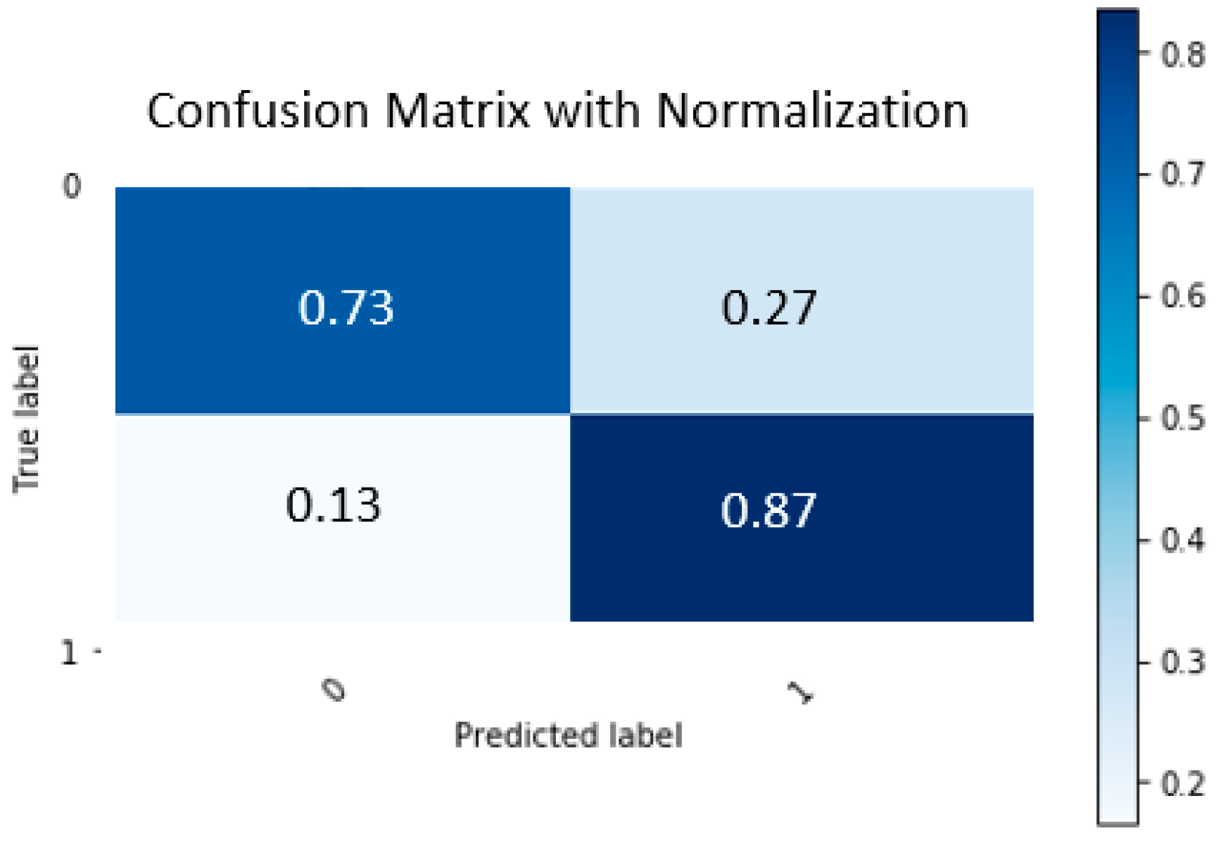
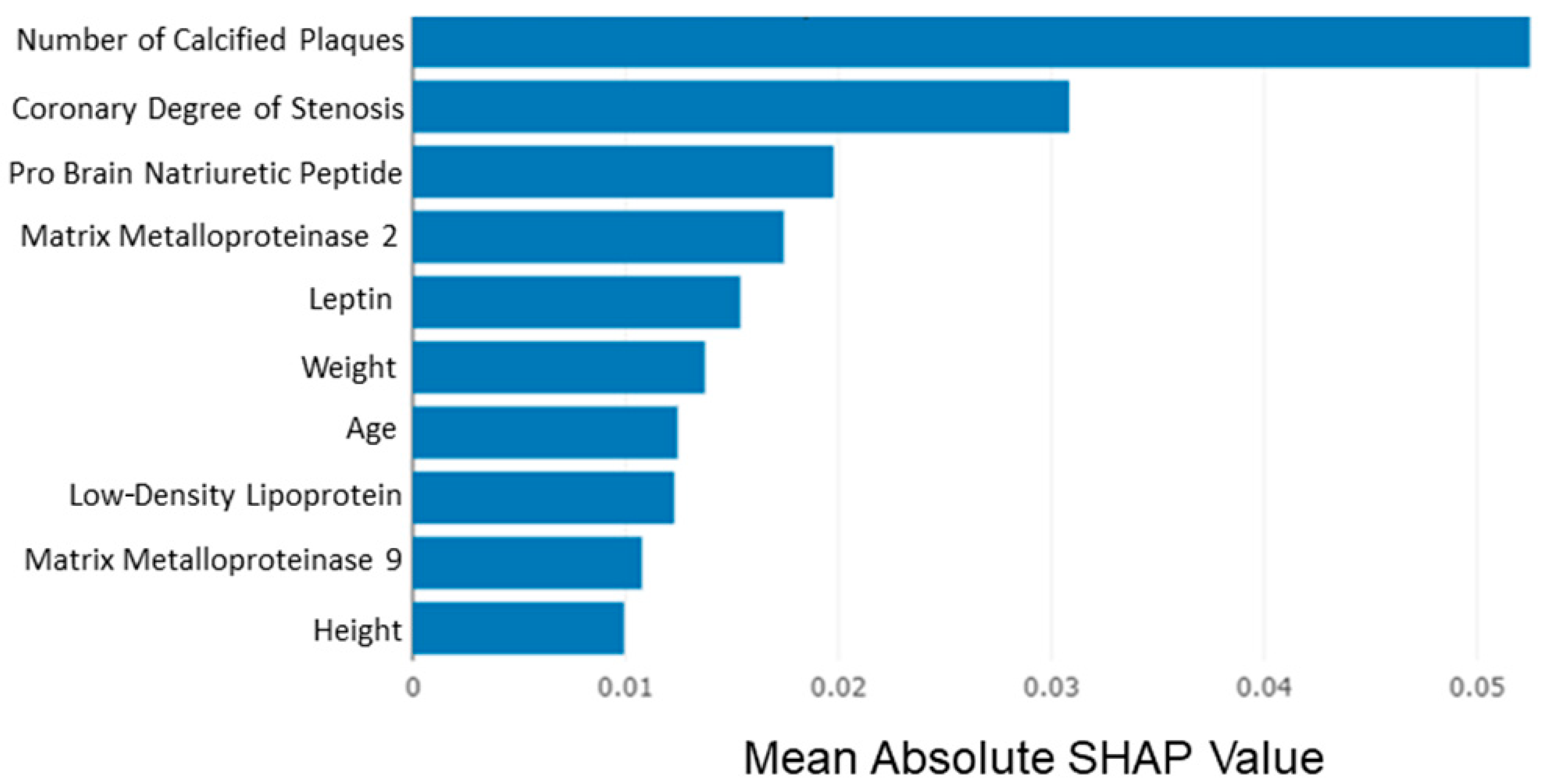
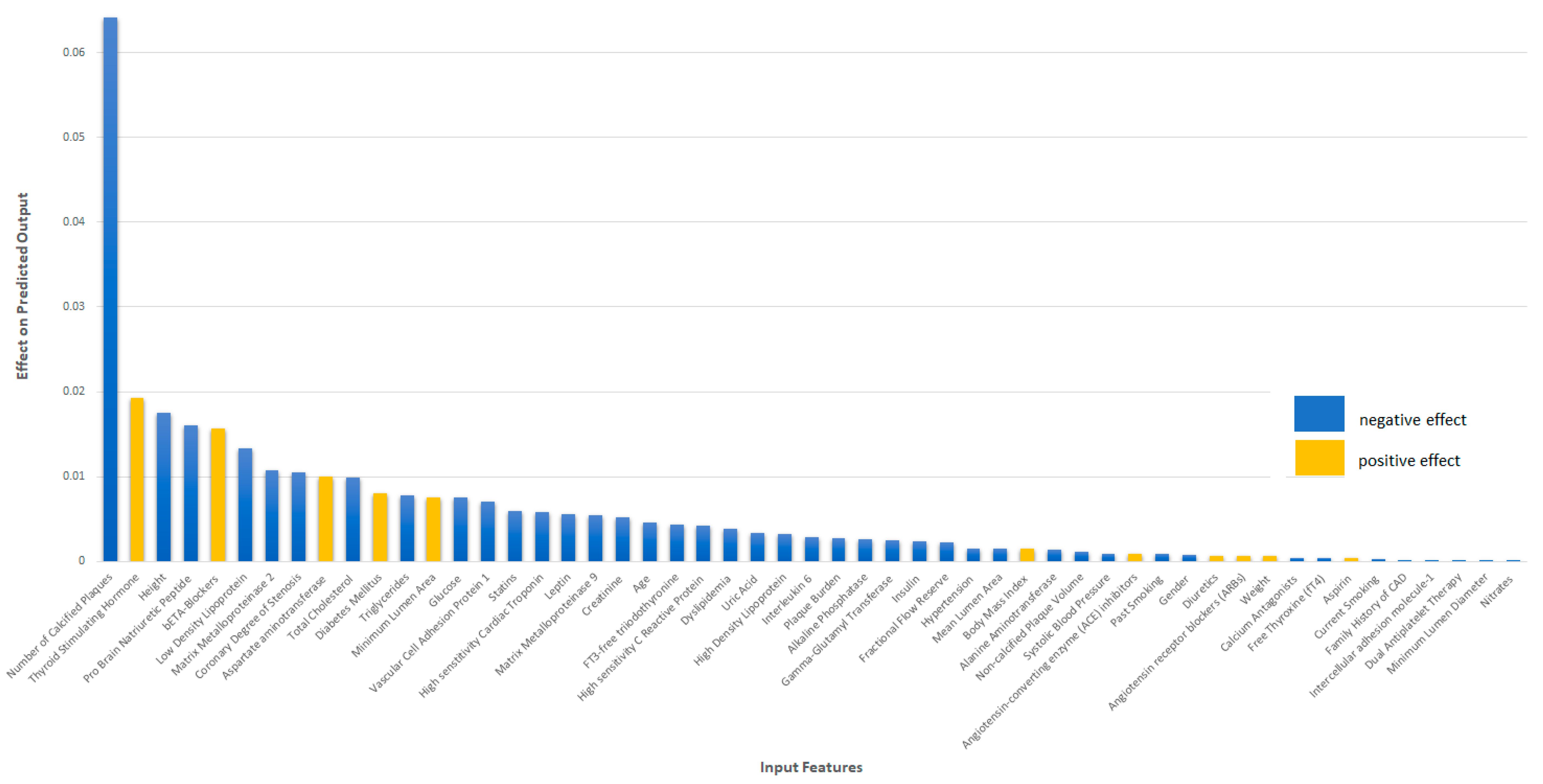
CAD Risk-Prediction Model Performance Evaluation
4. Discussion
5. Conclusions
Supplementary Materials
Author Contributions
Funding
Institutional Review Board Statement
Informed Consent Statement
Conflicts of Interest
References
- Subcommittee on Arteriosclerosis; Andrus, E.C.; Allen, E.V.; Merritt, H.H.; Duff, G.L.; Moore, R.A.; Kendall, F.E.; Shumacker, J.H.B.; Levy, R.L.; Wright, I.S. The pathogenesis of arteriosclerosis 1. Int. J. Epidemiol. 2015, 44, 1791–1793. [Google Scholar] [CrossRef] [PubMed] [Green Version]
- Wong, N.D. Epidemiological studies of CHD and the evolution of preventive cardiology. Nat. Rev. Cardiol. 2014, 11, 276. [Google Scholar] [CrossRef] [PubMed]
- Wexler, L.; Brundage, B.; Crouse, J.; Detrano, R.; Fuster, V.; Maddahi, J.; Rumberger, J.; Stanford, W.; White, R.; Taubert, K. Coronary artery calcification: Pathophysiology, epidemiology, imaging methods, and clinical implications: A statement for health professionals from the American Heart Association. Circulation 1996, 94, 1175–1192. [Google Scholar] [CrossRef] [PubMed]
- Papafaklis, M.; Mavrogiannis, M.; Stone, P. Identifying the progression of coronary artery disease: Prediction of cardiac events. Contin. Cardiol. Educ. 2016, 2, 105–114. [Google Scholar] [CrossRef]
- Roeters van Lennep, J.E.; Westerveld, H.T.; Erkelens, D.W.; van der Wall, E.E. Risk factors for coronary heart disease: Implications of gender. Cardiovasc. Res. 2002, 53, 538–549. [Google Scholar] [CrossRef]
- D’agostino, R.B.; Vasan, R.S.; Pencina, M.J.; Wolf, P.A.; Cobain, M.; Massaro, J.M.; Kannel, W.B. General cardiovascular risk profile for use in primary care: The Framingham Heart Study. Circulation 2008, 117, 743–753. [Google Scholar] [CrossRef] [Green Version]
- Conroy, R.M.; Pyörälä, K.; Fitzgerald, A.E.; Sans, S.; Menotti, A.; De Backer, G.; De Bacquer, D.; Ducimetiere, P.; Jousilahti, P.; Keil, U.; et al. Estimation of ten-year risk of fatal cardiovascular disease in Europe: The SCORE project. Eur. Heart J. 2003, 24, 987–1003. [Google Scholar] [CrossRef]
- Miller, R.J.; Huang, C.; Liang, J.X.; Slomka, P.J. Artificial intelligence for disease diagnosis and risk prediction in nuclear cardiology. J. Nucl. Cardiol. 2022, 1–9. [Google Scholar] [CrossRef]
- Goldstein, B.A.; Navar, A.M.; Carter, R.E. Moving beyond regression techniques in cardiovascular risk prediction: Applying machine learning to address analytic challenges. Eur. Heart J. 2017, 38, 1805–1814. [Google Scholar] [CrossRef] [Green Version]
- Exarchos, K.P.; Carpegianni, C.; Rigas, G.; Exarchos, T.P.; Vozzi, F.; Sakellarios, A.; Marraccini, P.; Naka, K.; Michalis, L.; Parodi, O. A multiscale approach for modeling atherosclerosis progression. IEEE J. Biomed. Health Inform. 2015, 19, 709–719. [Google Scholar] [CrossRef]
- Alizadehsani, R.; Zangooei, M.H.; Hosseini, M.J.; Habibi, J.; Khosravi, A.; Roshanzamir, M.; Khozeimeh, F.; Sarrafzadegan, N.; Nahavandi, S. Coronary artery disease detection using computational intelligence methods. Knowl. Based Syst. 2016, 109, 187–197. [Google Scholar] [CrossRef]
- Ambale-Venkatesh, B.; Yang, X.; Wu, C.O.; Liu, K.; Hundley, W.G.; McClelland, R.; Gomes, A.S.; Folsom, A.R.; Shea, S.; Guallar, E. Cardiovascular event prediction by machine learning: The multi-ethnic study of atherosclerosis. Circ. Res. 2017, 121, 1092–1101. [Google Scholar] [CrossRef] [PubMed]
- Motwani, M.; Dey, D.; Berman, D.S.; Germano, G.; Achenbach, S.; Al-Mallah, M.H.; Andreini, D.; Budoff, M.J.; Cademartiri, F.; Callister, T.Q. Machine learning for prediction of all-cause mortality in patients with suspected coronary artery disease: A 5-year multicentre prospective registry analysis. Eur. Heart J. 2017, 38, 500–507. [Google Scholar] [CrossRef] [PubMed]
- van Rosendael, A.R.; Maliakal, G.; Kolli, K.K.; Beecy, A.; Al’Aref, S.J.; Dwivedi, A.; Singh, G.; Panday, M.; Kumar, A.; Ma, X. Maximization of the usage of coronary CTA derived plaque information using a machine learning based algorithm to improve risk stratification; insights from the CONFIRM registry. J. Cardiovasc. Comput. Tomogr. 2018, 12, 204–209. [Google Scholar] [CrossRef]
- Sakellarios, A.I.; Tsompou, P.; Kigka, V.; Siogkas, P.; Kyriakidis, S.; Tachos, N.; Karanasiou, G.; Scholte, A.; Clemente, A.; Neglia, D. Non-invasive prediction of site-specific coronary atherosclerotic plaque progression using lipidomics, blood flow, and LDL transport modeling. Appl. Sci. 2021, 11, 1976. [Google Scholar] [CrossRef]
- Heo, J.; Yoo, J.; Lee, H.; Lee, I.H.; Kim, J.-S.; Park, E.; Kim, Y.D.; Nam, H.S. Prediction of Hidden Coronary Artery Disease Using Machine Learning in Patients with Acute Ischemic Stroke. Neurology 2022. [Google Scholar] [CrossRef]
- Liga, R.; Vontobel, J.; Rovai, D.; Marinelli, M.; Caselli, C.; Pietila, M.; Teresinska, A.; Aguadé-Bruix, S.; Pizzi, M.N.; Todiere, G. Multicentre multi-device hybrid imaging study of coronary artery disease: Results from the EValuation of INtegrated Cardiac Imaging for the Detection and Characterization of Ischaemic Heart Disease (EVINCI) hybrid imaging population. Eur. Heart J. Cardiovasc. Imaging 2016, 17, 951–960. [Google Scholar] [CrossRef]
- Sakellarios, A.I.; Rigas, G.; Kigka, V.; Siogkas, P.; Tsompou, P.; Karanasiou, G.; Exarchos, T.; Andrikos, I.; Tachos, N.; Pelosi, G. SMARTool: A tool for clinical decision support for the management of patients with coronary artery disease based on modeling of atherosclerotic plaque process. In Proceedings of the 2017 39th Annual International Conference of the IEEE Engineering in Medicine and Biology Society (EMBC), Jeju, Korea, 11–15 July 2017; pp. 96–99. [Google Scholar]
- Kigka, V.I.; Rigas, G.; Sakellarios, A.; Siogkas, P.; Andrikos, I.O.; Exarchos, T.P.; Loggitsi, D.; Anagnostopoulos, C.D.; Michalis, L.K.; Neglia, D.; et al. 3D reconstruction of coronary arteries and atherosclerotic plaques based on computed tomography angiography images. Biomed. Signal Process. Control 2018, 40, 286–294. [Google Scholar] [CrossRef] [Green Version]
- Kigka, V.I.; Sakellarios, A.; Kyriakidis, S.; Rigas, G.; Athanasiou, L.; Siogkas, P.; Tsompou, P.; Loggitsi, D.; Benz, D.C.; Buechel, R. A three-dimensional quantification of calcified and non-calcified plaques in coronary arteries based on computed tomography coronary angiography images: Comparison with expert’s annotations and virtual histology intravascular ultrasound. Comput. Biol. Med. 2019, 113, 103409. [Google Scholar] [CrossRef]
- Siogkas, P.K.; Anagnostopoulos, C.D.; Liga, R.; Exarchos, T.P.; Sakellarios, A.I.; Rigas, G.; Scholte, A.J.H.A.; Papafaklis, M.I.; Loggitsi, D.; Pelosi, G.; et al. Noninvasive CT-based hemodynamic assessment of coronary lesions derived from fast computational analysis: A comparison against fractional flow reserve. Eur. Radiol. 2019, 29, 2117–2126. [Google Scholar] [CrossRef] [Green Version]
- Cury, R.C.; Abbara, S.; Achenbach, S.; Agatston, A.; Berman, D.S.; Budoff, M.J.; Dill, K.E.; Jacobs, J.E.; Maroules, C.D.; Rubin, G.D. CAD-RADSTM coronary artery disease–reporting and data system. An expert consensus document of the Society of Cardiovascular Computed Tomography (SCCT), the American College of Radiology (ACR) and the North American Society for Cardiovascular Imaging (NASCI). Endorsed by the American College of Cardiology. J. Cardiovasc. Comput. Tomogr. 2016, 10, 269–281. [Google Scholar] [PubMed] [Green Version]
- Raff, G.L.; Abidov, A.; Achenbach, S.; Berman, D.S.; Boxt, L.M.; Budoff, M.J.; Cheng, V.; DeFrance, T.; Hellinger, J.C.; Karlsberg, R.P. SCCT guidelines for the interpretation and reporting of coronary computed tomographic angiography. J. Cardiovasc. Comput. Tomogr. 2009, 3, 122–136. [Google Scholar] [CrossRef] [PubMed]
- Robert, C. Machine Learning, A Probabilistic Perspective; Taylor & Francis: Abingdon-on-Thames, UK, 2014. [Google Scholar]
- Liu, X.Y.; Wu, J.; Zhou, Z.H. Exploratory undersampling for class-imbalance learning. IEEE Trans. Syst. Man Cybern. Part B 2009, 39, 539–550. [Google Scholar] [CrossRef]
- Guyon, I.; Weston, J.; Barnhill, S.; Vapnik, V. Gene selection for cancer classification using support vector machines. Mach. Learn. 2002, 46, 389–422. [Google Scholar] [CrossRef]
- Friedman, J.H. Greedy Function Approximation: A Gradient Boosting Machine. Ann. Stat. 2001, 29, 1189–1232. [Google Scholar] [CrossRef]
- Lundberg, S.M.; Lee, S.-I. A unified approach to interpreting model predictions. Adv. Neural Inf. Process. Syst. 2017, 30, 4768–4777. [Google Scholar]
- Cappola, A.R.; Desai, A.S.; Medici, M.; Cooper, L.S.; Egan, D.; Sopko, G.; Fishman, G.I.; Goldman, S.; Cooper, D.S.; Mora, S. Thyroid and cardiovascular disease: Research agenda for enhancing knowledge, prevention, and treatment. Circulation 2019, 139, 2892–2909. [Google Scholar] [CrossRef]
- Inoue, K.; Ritz, B.; Brent, G.A.; Ebrahimi, R.; Rhee, C.M.; Leung, A.M. Association of subclinical hypothyroidism and cardiovascular disease with mortality. JAMA Netw. Open 2020, 3, e1920745. [Google Scholar] [CrossRef] [Green Version]
- Klein, I.; Danzi, S. Thyroid disease and the heart. Circulation 2007, 116, 1725–1735. [Google Scholar] [CrossRef] [Green Version]
- Galli, E.; Pingitore, A.; Iervasi, G. The role of thyroid hormone in the pathophysiology of heart failure: Clinical evidence. Heart Fail. Rev. 2010, 15, 155–169. [Google Scholar] [CrossRef]
- Jin, H.-Y.; Weir-McCall, J.R.; Leipsic, J.A.; Son, J.-W.; Sellers, S.L.; Shao, M.; Blanke, P.; Ahmadi, A.; Hadamitzky, M.; Kim, Y.-J. The relationship between coronary calcification and the natural history of coronary artery disease. Cardiovasc. Imaging 2021, 14, 233–242. [Google Scholar] [CrossRef] [PubMed]
- Virmani, R.; Burke, A.P.; Farb, A.; Kolodgie, F.D. Pathology of the vulnerable plaque. J. Am. Coll. Cardiol. 2006, 47, C13–C18. [Google Scholar] [CrossRef] [PubMed] [Green Version]
- Nelson, C.P.; Hamby, S.E.; Saleheen, D.; Hopewell, J.C.; Zeng, L.; Assimes, T.L.; Kanoni, S.; Willenborg, C.; Burgess, S.; Amouyel, P. Genetically determined height and coronary artery disease. N. Engl. J. Med. 2015, 372, 1608–1618. [Google Scholar] [CrossRef] [PubMed] [Green Version]
- Moon, J.; Hwang, I.C. The link between height and cardiovascular disease: To be deciphered. Cardiology 2019, 143, 114–115. [Google Scholar] [CrossRef]
- Stone, P.H.; Saito, S.; Takahashi, S.; Makita, Y.; Nakamura, S.; Kawasaki, T.; Takahashi, A.; Katsuki, T.; Nakamura, S.; Namiki, A. Prediction of progression of coronary artery disease and clinical outcomes using vascular profiling of endothelial shear stress and arterial plaque characteristics: The PREDICTION Study. Circulation 2013, 127, e489–e490. [Google Scholar] [CrossRef] [Green Version]
- Liu, X.; Wu, G.; Xu, C.; He, Y.; Shu, L.; Liu, Y.; Zhang, N.; Lin, C. Prediction of coronary plaque progression using biomechanical factors and vascular characteristics based on computed tomography angiography. Comput. Assist. Surg. 2017, 22, 286–294. [Google Scholar] [CrossRef]
- Weng, S.F.; Reps, J.; Kai, J.; Garibaldi, J.M.; Qureshi, N. Can machine-learning improve cardiovascular risk prediction using routine clinical data? PLoS ONE 2017, 12, e0174944. [Google Scholar] [CrossRef] [Green Version]
- Bittencourt, M.S.; Hulten, E.; Ghoshhajra, B.; O’leary, D.; Christman, M.P.; Montana, P.; Truong, Q.A.; Steigner, M.; Murthy, V.L.; Rybicki, F.J. Prognostic value of nonobstructive and obstructive coronary artery disease detected by coronary computed tomography angiography to identify cardiovascular events. Circ. Cardiovasc. Imaging 2014, 7, 282–291. [Google Scholar] [CrossRef] [Green Version]
- Dey, D.; Achenbach, S.; Schuhbaeck, A.; Pflederer, T.; Nakazato, R.; Slomka, P.J.; Berman, D.S.; Marwan, M. Comparison of quantitative atherosclerotic plaque burden from coronary CT angiography in patients with first acute coronary syndrome and stable coronary artery disease. J. Cardiovasc. Comput. Tomogr. 2014, 8, 368–374. [Google Scholar] [CrossRef]
- Papafaklis, M.I.; Muramatsu, T.; Ishibashi, Y.; Lakkas, L.S.; Nakatani, S.; Bourantas, C.V.; Ligthart, J.; Onuma, Y.; Echavarria-Pinto, M.; Tsirka, G. Fast virtual functional assessment of intermediate coronary lesions using routine angiographic data and blood flow simulation in humans: Comparison with pressure wire-fractional flow reserve. EuroIntervention 2014, 10, 574–583. [Google Scholar] [CrossRef]


| Type | Features | |
|---|---|---|
| Imaging data * | Geometrical vasculature | Degree of Stenosis, Minimal Lumen Area, Minimal Lumen Diameter, Plaque Burden, Calcified Plaque Volume, Noncalcified Plaque Volume, SmartFFR Index, Number of Calcified Plaques, Number of Non-calcified Plaques |
| Non-imaging data | Demographics | Age, Gender |
| Risk factors | Family History of CAD, Hypertension, Diabetes, Dyslipidemia, Smoking, Obesity, Metabolic Syndrome, Past Smokers | |
| Biohumoral Markers | Creatinine, Uric Acid, Glucose, Total Cholesterol, HDL, LDL, Triglycerides, Insulin, Aspartate Aminotransferase, Alanine Aminotransferase, Alkaline Phosphatase, Gamma-glutamyl Transferase, Hs-C Reactive Protein, Interleukin-6, TSH, fT3, fT4, Leptin, MMP2 Protein Plasma, MMP9 Protein Plasma, hs-cardiac Troponin T, N terminal Fragment of Pro-brain Natriuretic Peptide, Lipidomics, Metabolomics | |
| Proposed Classes | Recommended Stenosis Grading Scale of CAD | Quantitative Stenosis |
|---|---|---|
| 0: Normal | No luminal stenosis | |
| 1: Minimal | Plaque with <25% stenosis | |
| 2: Mild | 25–49% stenosis | |
| 3: Moderate | 50–69% stenosis | |
| 4: Severe | 70–99% stenosis | |
| 5: Occluded | 100% stenosis |
| Folds | Balanced Accuracy | Negative Predictive Value | Positive Predictive Value | ROC AUC | Sensitivity | Specificity |
|---|---|---|---|---|---|---|
| Fold #0 | 0.73 | 0.78 | 0.67 | 0.60 | 0.67 | 0.78 |
| Fold #1 | 0.75 | 0.86 | 0.63 | 0.82 | 0.84 | 0.67 |
| Fold #2 | 0.89 | 1 | 0.72 | 0.92 | 1 | 0.78 |
| Fold #3 | 0.69 | 0.78 | 0.6 | 0.72 | 0.6 | 0.78 |
| Fold #4 | 0.84 | 1 | 0.63 | 0.83 | 1 | 0.67 |
| Fold #5 | 0.84 | 1 | 0.63 | 0.89 | 1 | 0.67 |
| Fold #6 | 0.95 | 1 | 0.84 | 1 | 1 | 0.89 |
| Fold #7 | 0.78 | 1 | 0.56 | 0.78 | 1 | 0.56 |
| Fold #8 | 0.8 | 0.86 | 0.72 | 0.88 | 0.84 | 0.75 |
| Fold #9 | 0.8 | 0.86 | 0.72 | 0.75 | 0.84 | 0.75 |
| Mean ± std | 0.81 ± 0.08 | 0.92 ± 0.1 | 0.68 ± 0.08 | 0.82 ± 0.11 | 0.88 ± 0.15 | 0.73 ± 0.09 |
| Folds | Balanced Accuracy | Negative Predictive Value | Positive Predictive Value | ROC AUC | Sensitivity | Specificity |
|---|---|---|---|---|---|---|
| Fold #0 | 0.5 | 0.6 | 0.4 | 0.33 | 0.33 | 0.67 |
| Fold #1 | 0.56 | 0.64 | 0.5 | 0.65 | 0.33 | 0.78 |
| Fold #2 | 0.72 | 1 | 0.5 | 0.89 | 1 | 0.44 |
| Fold #3 | 0.69 | 0.78 | 0.6 | 0.69 | 0.6 | 0.78 |
| Fold #4 | 0.79 | 0.88 | 0.67 | 0.87 | 0.8 | 0.78 |
| Fold #5 | 0.78 | 1 | 0.56 | 0.78 | 1 | 0.56 |
| Fold #6 | 0.79 | 0.88 | 0.67 | 0.8 | 0.8 | 0.78 |
| Fold #7 | 0.72 | 1 | 0.5 | 0.76 | 1 | 0.44 |
| Fold #8 | 0.6 | 0.64 | 0.67 | 0.79 | 0.33 | 0.88 |
| Fold #9 | 0.71 | 0.75 | 0.67 | 0.83 | 0.67 | 0.75 |
| Mean ± std | 0.69 ± 0.1 | 0.82 ± 0.16 | 0.57 ± 0.1 | 0.74 ± 0.16 | 0.69 ± 0.28 | 0.69 ± 0.15 |
Publisher’s Note: MDPI stays neutral with regard to jurisdictional claims in published maps and institutional affiliations. |
© 2022 by the authors. Licensee MDPI, Basel, Switzerland. This article is an open access article distributed under the terms and conditions of the Creative Commons Attribution (CC BY) license (https://creativecommons.org/licenses/by/4.0/).
Share and Cite
Kigka, V.I.; Georga, E.; Tsakanikas, V.; Kyriakidis, S.; Tsompou, P.; Siogkas, P.; Michalis, L.K.; Naka, K.K.; Neglia, D.; Rocchiccioli, S.; et al. Machine Learning Coronary Artery Disease Prediction Based on Imaging and Non-Imaging Data. Diagnostics 2022, 12, 1466. https://doi.org/10.3390/diagnostics12061466
Kigka VI, Georga E, Tsakanikas V, Kyriakidis S, Tsompou P, Siogkas P, Michalis LK, Naka KK, Neglia D, Rocchiccioli S, et al. Machine Learning Coronary Artery Disease Prediction Based on Imaging and Non-Imaging Data. Diagnostics. 2022; 12(6):1466. https://doi.org/10.3390/diagnostics12061466
Chicago/Turabian StyleKigka, Vassiliki I., Eleni Georga, Vassilis Tsakanikas, Savvas Kyriakidis, Panagiota Tsompou, Panagiotis Siogkas, Lampros K. Michalis, Katerina K. Naka, Danilo Neglia, Silvia Rocchiccioli, and et al. 2022. "Machine Learning Coronary Artery Disease Prediction Based on Imaging and Non-Imaging Data" Diagnostics 12, no. 6: 1466. https://doi.org/10.3390/diagnostics12061466
APA StyleKigka, V. I., Georga, E., Tsakanikas, V., Kyriakidis, S., Tsompou, P., Siogkas, P., Michalis, L. K., Naka, K. K., Neglia, D., Rocchiccioli, S., Pelosi, G., Fotiadis, D. I., & Sakellarios, A. (2022). Machine Learning Coronary Artery Disease Prediction Based on Imaging and Non-Imaging Data. Diagnostics, 12(6), 1466. https://doi.org/10.3390/diagnostics12061466