Next Article in Journal
Toxoplasmosis and Pregnancy: Current Approaches for Favourable Fetal Outcome
Previous Article in Journal
The National Screening Program for Colorectal Cancer
 
 
Romanian Journal of Preventive Medicine is published by MDPI from Volume 3 Issue 1 (2025). Previous articles were published by another publisher in Open Access under a CC-BY (or CC-BY-NC-ND) licence, and they are hosted by MDPI on mdpi.com as a courtesy and upon agreement with the previous journal publisher.
Review

Screening for Congenital Hip Dislocation—An Overview

by
Rodica-Iulia Chirilă
1,
Andreea Calomfirescu-Avramescu
2 and
Vlad Dima
2
1
“Grigore Alexandrescu” Emergency Children’s Hospital, Bucharest, Romania
2
Filantropia Clinical Hospital, Bucharest, Romania
Rom. J. Prev. Med. 2023, 2(3), 7-11; https://doi.org/10.3390/rjpm2030007
Published: 1 September 2023

Abstract

Developmental dysplasia of the hip, previously known as congenital dislocation of the hip, is the most common congenital disease of the musculoskeletal system in newborns. It corresponds to a complete or partial displacement of the femoral head out of the acetabulum. The incidence is around 1:1000 births and it depends on multiple factors (region, family history, gender, mechanical factors, or some specific neonatal conditions). Screening may be by universal neonatal clinical examination (Ortolani or Barlow maneuvers) with the addition of sonographic imaging of the hip (selecting the patients who present abnormalities detected by physical examination or risk factors, and universal screening in the neonate). This screening is recommended at the discharge of newborns in most maternity hospitals in Romania.

History of neonatal screening

The term congenital dislocation of the hip dates back to the time of Hippocrates: “There are persons, who, from birth or from disease, have dislocations outward of both the thighs; in them, then, the bones are affected in like manner, but the fleshy parts in their case lose their strength less; . . . They have the equal use of both their legs, for in walking they totter equally to this side and that.” [1].
There is general agreement among health professionals that starting the treatment as early as possible leads to the best prognosis. Considerable advances have been made since the implementation of Ortolani's technique for diagnosing congenital hip dislocation in the newborn period. This method was not widely disseminated from the beginning, but Von Rosen reported his results in applying Ortolani's technique. This and Barlow's description of a maneuver that provokes dislocation in unstable hips has greatly increased the number of infants who present for early treatment [2,3].
In 1983, Graf introduced his method to detect hip instability by using ultrasonography. This method proved to be powerful in detecting developmental dysplastic hips at a very early age and is therefore associated with significantly reduced rates of severe dysplasia in adults in these populations [4]. This avoids radiological diagnostic procedures that are not sensitive in the diagnosis of hip dysplasia in newborns and reveals features undetectable with the Ortolani-Barlow maneuvers.

Epidemiology and etiology

Developmental dysplasia of the hip, previously known as congenital dislocation of the hip, is the most common congenital disease of the musculoskeletal system in newborns. It corresponds to a complete or partial displacement of the femoral head out of the acetabulum. The etiology of this pathology is not yet fully known, but certain risk factors have proven to be associated with it (e.g., female sex, breech position in the third trimester) [2,5].
The incidence is around 1:1000 births and it depends on multiple factors: region (high frequencies being reported in the Italian population, in Israel, among certain American Indian tribes, and the Japanese), family history (the recurrence risk among siblings of affected persons is about 40 times as great as the incidence expected in the general population), gender (80% of the cases are females), mechanical factors (breech delivery, oligohydramnios, macrosomia or tight lower extremity swaddling and some conditions of the neonate (torticollis, clubfoot and metatarsus varus) [2,6,7,8,9].

Screening techniques

There are considerable discrepancies in newborn hip screening programs among different countries and regions. Most of them have adopted universal or selective ultrasound screening strategies, and both types of strategies are used in combination with neonatal clinical examination (Ortolani or Barlow maneuvers) [5,10].
Ortolani's and Barlow's tests can be performed in the first few weeks of life, while the hip can still be reduced easily; however, once the head of the femur has migrated laterally and proximally onto the lateral surface of the ilium and the adductors have become relatively shortened and tight, other signs must be sought [2].
To perform Barlow's maneuver the examiner grasps the infant's thigh near the hip and with gentle posterior and lateral pressure, attempts to dislocate the femoral head from the acetabulum. Normally, there is no motion in this direction. If the hip is dislocatable, a distinct "clunk" may be felt as the femoral head pops out of the joint (Figure 1a).
After provocation of dislocation by Barlow's maneuver, the hip should be abducted to about 80 degrees while the proximal femur is lifted anteriorly with the fingers placed along the lateral thigh. A positive Ortolani's maneuver is a sensation of a jerk or snap with a reduction in the socket. A click is not necessarily heard and a click without a sensation of abnormal motion is probably not significant (Figure 1b).
In 70–90% of the cases with clinically positive Ortolani and or Barlow tests, the hip instability will spontaneously resolve within 2–4 weeks post-natally. One major problem regarding clinical examination is that these maneuvers failed to identify 66.7% of those hip joints that subsequently required surgical intervention [11,12,13].
One efficient evaluation method for the coxofemoral joint is hip ultrasound according to the Graf method, allowing the early detection of developmental dysplasia and the follow-up of the applied treatment.
Selective ultrasound screening is performed on infants with hip abnormalities detected by physical examination and infants who have risk factors for developmental dysplasia of the hip. In the first case, ultrasound scanning is performed in the first two weeks of life, and infants at risk but exhibiting no clinical abnormalities undergo ultrasound within the first six weeks of life [5].
Universal ultrasound screening involves performing hip ultrasound examinations on all newborns and many studies have reported good results. This type of screening is recommended at the discharge of newborns in most maternity hospitals in Romania. However, the timing of the first ultrasound examination varies among countries, ultrasonography being valuable in the neonatal period. Once the ossifying nucleus of the femoral head appears, ultrasound is less valuable, and a radiograph should be used. If ultrasound is performed too early, some babies with transient immature and physiologically unstable hip joints may be diagnosed as positive cases [5].
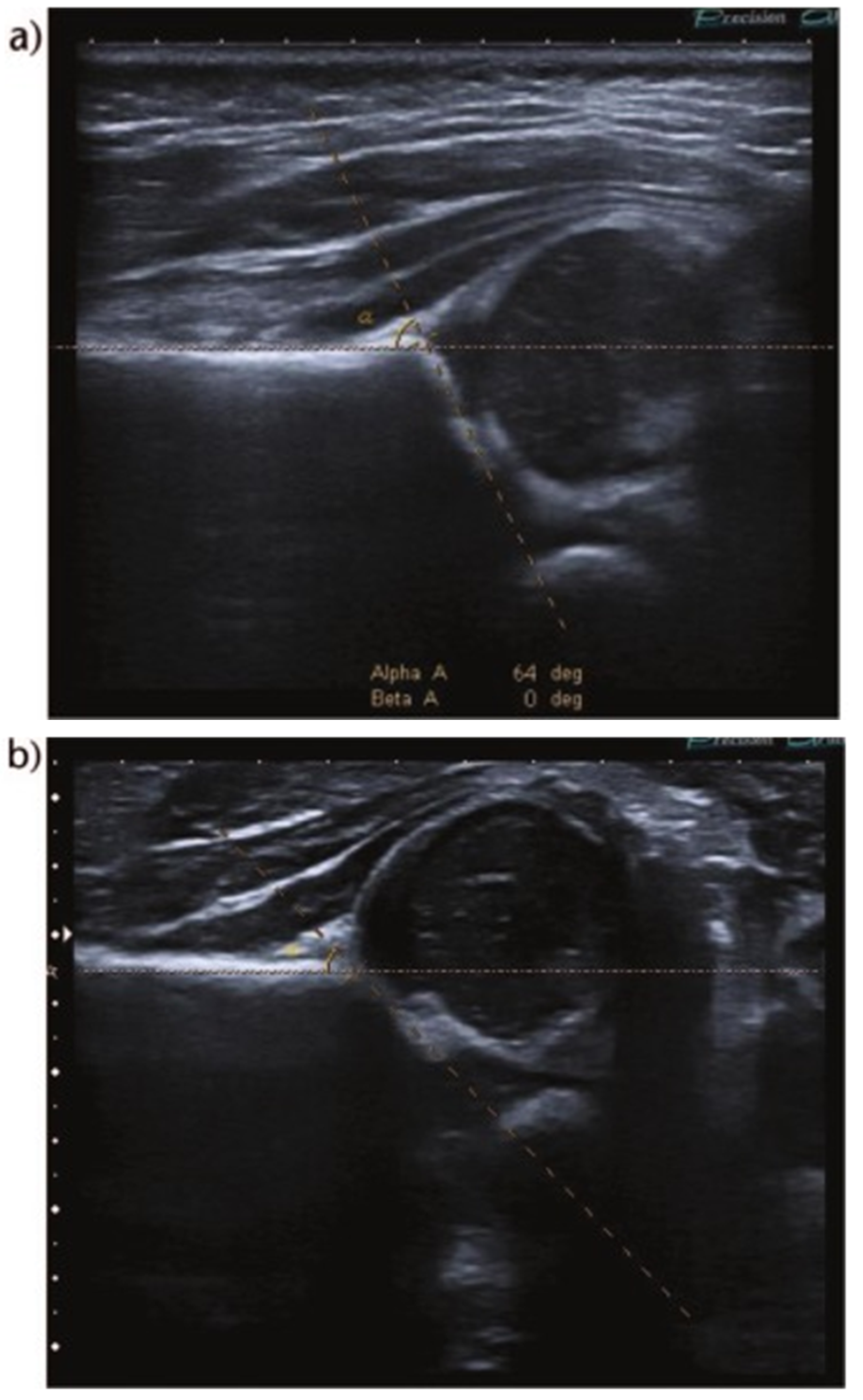
Hip ultrasound according to the Graf method can be used to evaluate development of the hip joint by analyzing the ultrasound images of the hip joint and quantifying the shape of the acetabulum with α and β angles. α angle is formed between the ilion and the osseous wall of the acetabulum; and β angle is formed between the ilion and the cartilaginous labrum (Figure 2a,b). As a general rule, the α angle determines the type, and the β angle determines the subtype:
  • type I
    centered hip
    alpha angle >60° (normal)
    type Ia: beta angle <55°
    type Ib: beta angle >55°
  • type II
    centered hip
    type IIa (physiologically immature): alpha angle 50-59° (<3 months)
    type IIb: alpha angle 50-59° (>3 months)
    type IIc
    alpha angle 43-49°
    beta angle <77°
  • type D (“about to decentre”)
    alpha angle 43-49°
    beta angle >77°
  • type III
    decentred hip
    alpha angle: not measured in a decentred hip
    cartilage roof pushed partly upwards (cephalad), partly downwards (caudal)
    perichondrium goes upward (ultrasound image set as right hip AP)
    type IIIa: no echos in cartilaginous roof
    type IIIb: echos in cartilaginous roof due to structural alteration and damage
  • type IV
    decentred hip
    alpha angle: not measured in a decentred hip
    cartilage roof pushed entirely downwards (cephalad)
    perichondrium goes horizontal (ultrasound image set as right hip AP)
    the hyaline cartilage of the lateral edge of the acetabulum along with the fibrocartilaginous labrum is interposed between the femoral head and the acetabulum [7,14,15,16].
Figure 2. a) Normal sonography. b) In contrast with (a), the α angle is <60° and the hip is subluxated [7].
Rjpm 02 00016 g002
An unresolved issue is that sonographic diagnosis has a higher prevalence of abnormality than clinical diagnosis, raising the possibility of an overdiagnosis of the condition which may lead to over-treatment [11]. Some studies report that 90% of Graf Type II hip dysplasias, <25% of Graf type III hip dysplasias and <90% of Graf Type IV dysplasias may resolve spontaneously [16,17,18,19]. However, one recent large study showed that there was a significant difference in α and β angles between the cases with congenital acetabular dysplasia and those with normal hip joints by ultrasound Graf method. This aspect suggests that the method can be used as one of the methods to screen congenital acetabular abnormalities, with strong operability and high standardization [20].
Opinions are divided regarding the usefulness of universal screening in reducing the rates of late-detected cases (age 12 weeks). The incidences of late-detected Developmental dysplasia of the hip and operatively treated cases were similar among all screening strategies [21].

Conclusions

Screening for congenital hip dislocation represents a controversial subject because there is no international consensus. However, because there is general agreement that starting the treatment as early as possible leads to the best prognosis, screening must be performed at least in the case of newborns considered at risk. Hip ultrasonography in the method according to Graf is currently the most accurate diagnostic tool for developmental dysplasia of the hip in early infancy and optimally timing of the procedure can improve the results.

References

  1. Adams, F. The Genuine Works of Hippocrates. New York, 1886; Volume 2, pp. 131–132. [Google Scholar]
  2. Specht, E.E. Congenital dislocation of the hip. West J Med. 1976, 124, 18–28. [Google Scholar] [PubMed] [PubMed Central]
  3. Wenger, D.R.; Bomar, J.D. Historical Aspects of DDH. Indian J Orthop. 2021, 55, 1360–1371. [Google Scholar] [CrossRef] [PubMed]
  4. Walter, S.G.; Ossendorff, R.; Yagdiran, A.; Hockmann, J.; Bornemann, R.; Placzek, S. Four decades of developmental dysplastic hip screening according to Graf: What have we learned? Front Pediatr. 2022, 10, 990806. [Google Scholar] [CrossRef] [PubMed]
  5. Han, J.; Li, Y. Progress in screening strategies for neonatal developmental dysplasia of the hip. Front. Surg. 2022. [Google Scholar] [CrossRef] [PubMed]
  6. Dunn, P.M. Perinatal observations on the etiology of congenital dislocation of the hip. Clin Orthop Relat Res 1976, 119, 11–22. [Google Scholar] [CrossRef]
  7. Vaquero-Picado, A.; González-Morán, G.; Garay, E.G.; Moraleda, L. Developmental dysplasia of the hip: update of management. EFORT Open Rev. 2019, 4, 548–556. [Google Scholar] [CrossRef] [PubMed]
  8. Ramsey, P.L.; Rosenblum, H. Early detection and treatment of congenital hip dislocation. J Bone Joint Surg 1973, 55A, 1312. [Google Scholar]
  9. Salter, R.B. Etiology, pathogenesis and possible prevention of congenital dislocation of the hip. Can Med Assoc J 1968, 98, 933–945. [Google Scholar] [PubMed]
  10. Paton, R.W. Screening in Developmental Dysplasia of the Hip (DDH). Surgeon 2017, 15, 290–296. [Google Scholar] [CrossRef] [PubMed]
  11. Paton, R.W. Screening in Developmental Dysplasia of the Hip (DDH). The Surgeon 2017. [CrossRef] [PubMed]
  12. Barlow, T.G. Early diagnosis and treatment of congenital dislocation of the hip. J Bone Joint Surg 1962, 44-B, 292e301. [Google Scholar] [CrossRef]
  13. Gardiner, H.M.; Dunn, P.M. Controlled trial of immediate splinting versus ultrasonographic surveillance in congenitally dislocatable hips. Lancet 1990, 336, 1553e6. [Google Scholar] [CrossRef] [PubMed]
  14. Gaillard, F.; Murphy, A.; Ranchod, A.; et al. Graf method for ultrasound classification of developmental dysplasia of the hip. Reference article, Radiopaedia.org. (accessed on 29 October 2023).
  15. Andersson, J.E.; Funnemark, P.O. Neonatal hip instability: screening with anterior-dynamic ultrasound method. J Pediatr Orthop 1995, 15, 322e4. [Google Scholar] [CrossRef] [PubMed]
  16. Castelein, R.M.; Sauter, A.J.; de Viieger, M.; et al. Natural history of ultrasound hip abnormalities in clinically normal newborns. J Pediatr Orthop 1992, 12, 423e7. [Google Scholar] [CrossRef] [PubMed]
  17. Sampath, J.S.; Deakin, S.; Paton, R.W. Splintage in developmental dysplasia of the hip. How low can we go? J Pediatr Orthop 2003, 23, 352e5. [Google Scholar] [CrossRef]
  18. Rosendahl, K.; Toma, P. Ultrasound in the diagnosis of developmental dysplasia of the hip in newborns. The European approach. A review of methods, accuracy and clinical validity. Eur Radiol 2007, 17, 1960e7. [Google Scholar] [CrossRef] [PubMed]
  19. Wood, M.K.; Conboy, V. , Benson, M.K.D. Does early treatment by abduction splintage improve the development of dysplastic but stable neonatal hips. J Pediatr Orthop 2000, 20, 302e5. [Google Scholar] [CrossRef]
  20. Liu, D.; Mou, X.; Yu, G.; Liang, W.; Cai, C.; Li, X.; Zhang, G. The feasibility of ultrasound Graf method in screening infants and young children with congenital hip dysplasia and follow-up of treatment effect. Translational Pediatrics 2021, 10, 1333–1339. [Google Scholar] [CrossRef] [PubMed]
  21. Ziegler, C.M.; Ertl, K.M.; Delius, M.; et al. Clinical examination and patients’ history are not suitable for neonatal hip screening. Journal of Children’s Orthopaedics. 2022, 16, 19–26. [Google Scholar] [CrossRef] [PubMed]
Figure 1. a) Barlow Manuever and b) Ortolani Manuever (https://med.stanford.edu).
Rjpm 02 00016 g001