Article

Environmental Heavy Metal Contamination in Southern Brazilian Mangroves: Biomonitoring Using Crassostrea rhizophorae and Laguncularia racemosa as Green Health Indicators

by
João Carlos Ferreira de Melo Júnior
*,
Celso Voos Vieira
,
Luciano Lorenzi
,
Therezinha Maria Novais de Oliveira
,
Alessandra Betina Gastaldi
,
Aline Krein Moletta
,
Ana Paula de Mello
,
Ana Paula Marcelino de Aquino
,
Daiane Dalmarco
,
Deivid Rodrigo Corrêa
,
Gustavo Borba de Oliveira
,
Laila Cristina Mady
,
Letiane Steinhorst
,
Magda Carrion Bartz
,
Marcelo Lemos Ineu
,
Nara Texeira Barbosa
,
Natalia Cavichioli
,
Ricardo Larroyed de Oliveira
,
Sarah Caroline Lopes
and
Paula Roberta Perondi Furtado
Programa de Pós-Graduação em Saúde e Meio Ambiente, Universidade da Região de Joinville, Joinville 89219-710, Brazil
*
Author to whom correspondence should be addressed.
Green Health 2025, 1(3), 19; https://doi.org/10.3390/greenhealth1030019
Submission received: 10 September 2025 / Revised: 21 October 2025 / Accepted: 24 October 2025 / Published: 3 November 2025

Abstract

Mangrove forests provide critical ecosystem services, including carbon sequestration, shoreline protection, and serving as a food resource for coastal communities. However, these ecosystems face increasing environmental risks due to industrial and urban pollution, particularly contamination by heavy metals. This study assessed environmental quality in mangrove areas of Babitonga Bay, southern Brazil, using biomonitoring with the oyster Crassostrea rhizophorae and the mangrove tree Laguncularia racemosa. Sediment analyses revealed significantly elevated concentrations of copper, nickel, aluminum, and iron in Vila da Glória compared to Espinheiros, exceeding Brazilian environmental guidelines for copper and zinc. Biomonitoring results indicated high accumulation of arsenic and zinc in L. racemosa leaves, while oysters from Espinheiros exhibited higher concentrations of multiple heavy metals and smaller anatomical dimensions compared to those from Vila da Glória. Strong negative correlations were found between metal concentrations in oyster tissues and sediments, suggesting complex bioavailability dynamics. The study demonstrates the applicability of C. rhizophorae and L. racemosa as possible bioindicators of metal contamination in mangrove ecosystems. These findings underscore the importance of integrating biomonitoring approaches into coastal environmental health assessments to inform public health policies and conservation strategies aimed at promoting balanced ecosystem and human health.

1. Introduction

Mangroves are found in the coastal areas of tropical and subtropical climates worldwide, being most common in regions such as the Brazilian coast, Southeast Asia, the eastern coast of Africa, and Australia [1,2]. This ecosystem occurs at river mouths and estuaries, where freshwater mixes with saltwater [3], creating a unique and critical ecosystem [1] capable of harboring specific biodiversity and performing essential ecological functions [2]. Mangroves are characterized by evergreen forests that are tolerant of constant fluctuations in salinity [4]. These fluctuations, combined with wind conditions, low sediment oxygenation, and tidal amplitude, create limiting environments for species survival, making mangroves fragile from a conservation perspective [3].
Mangroves are coastal ecosystems that provide numerous ecological, economic, and social benefits in intertidal zones, where they play critical roles in maintaining the health and productivity of coastal environments [4]. Among the ecosystem functions attributed to mangroves are the provision of habitat and breeding areas for various marine species, nutrient cycling, coastal protection against storms and erosion, and carbon sequestration [4], the latter recently attributing to them the role of a natural climate solution for sustainable development [5]. Mangroves provide essential habitats for a wide range of terrestrial and marine species [5]. Their complex root systems stabilize the environment and provide substrate for various plants and animals, including birds, insects, crustaceans, mammals, reptiles, tunicates, sponges, algae, and bivalves. These habitats support diverse food webs and serve as nurseries for commercially important species such as crabs, shrimp, and fish [6].
Mangroves are highly productive ecosystems that contribute substantial amounts of organic matter to adjacent coastal waters, thereby sustaining complex food webs that involve commercially important species [6,7]. Microbial communities present in these ecosystems play a crucial role in nutrient cycling, transforming dead vegetation into nutrients usable by plants and other organisms. This microorganism–nutrient–plant relationship is fundamental for mangrove productivity and conservation [7]. Mangroves are important carbon sinks, capturing and storing large amounts of carbon dioxide from the atmosphere in various forms across different biogeographic regions and forest conditions, thereby contributing to climate change mitigation; however, the extent of their carbon sequestration capabilities [8].
Despite their importance, mangroves are susceptible to anthropogenic pressures, with their dynamics having been altered exponentially in recent decades [9]. The growing advancement of urbanization and industrialization in Brazilian coastal and shoreline regions has resulted in negative and irreversible impacts on mangroves [10], which are intensified by pollution of water bodies that feed the estuarine system, consequently affecting both environmental and human health [11]. On the southern Brazilian coast, the estuarine region known as Babitonga Bay houses one of the largest estuarine systems of the South Atlantic [12], being considered one of the last remaining mangrove areas in the Southern Hemisphere [13] and thus of high conservation priority [14]. The Babitonga mangroves provide food resources, notably fish and mollusks, for human communities living in their immediate surroundings or those commercially supplied by fishing or mollusk farming in this region. However, population densification, metal-mechanical industries, port activities, and other industrial and urban sources are potential contributors of environmental contaminants in the bay, such as potentially toxic elements and domestic sewage [15].
The assessment of environmental quality in mangrove areas typically involves studies on the accumulation of heavy metals in water and sediments [14,16,17]. The sources and spatial distribution of heavy metals in mangrove sediments vary based on regional activities and natural processes [14,16,18]. The evaluation of ecological risk and pollution status of heavy metals in mangrove sediments can be performed using different indices, such as the geoaccumulation index and the contamination factor [19,20]. In the Klang estuary, Malaysia, these indices indicated minimal risk for most heavy metals, except for Zn, which presented high environmental risk [21]. In the Gulf of Kachchh, India, contamination indices revealed levels ranging from minimal to extremely high, with sediments showing extreme contamination by Sb and As [22]. In Shenzhen, China, a predominance of tungsten and cobalt was identified in mangrove sediments, related to industrial effluent disposal, highlighting the unique contamination profile of this region [23].
This quality assessment is also performed through the analysis of local fauna and flora species, due to the high risk of toxicity from bioaccumulation and biomagnification, intensified by anthropogenic activities, especially in regions of intense urban occupation and industrial hubs [16]. Thus, biological monitoring, also known as biomonitoring, involves the use of living organisms to assess environmental conditions, including the detection of pollutants in ecosystems [17]. These studies are based on the understanding that elements in the environment, whether natural and/or resulting from human action, can unbalance an ecosystem, provoking biological reactions that are quantifiable and analysable [18]. Biomonitoring studies, together with conventional analyses of the physical and chemical nature of the abiotic environment, can provide a more detailed understanding of pollution effects [19]. The passive biomonitoring method utilizes autochthonous organisms that are exposed to pollutants over time, thereby reflecting the real and continuous environmental conditions of the ecosystem, providing a holistic and long-term perspective [20].
Different mangrove plant species have been utilized to indicate the presence of inorganic and organic contaminants in these environments, in addition to serving as potential tools for the removal or immobilization of these pollutants [24]. Species Avicennia marina, a tree found in the mangroves of Saudi Arabia, whose sediments had high levels of Cd, Cu, Pb, and Zn, is a potential bioaccumulator of heavy metals, with Cu and Cr being highly bioaccumulated in different parts of the plant, suggesting potential for phytoextraction [25]. The species Laguncularia racemosa (Combretaceae), widely distributed in Brazilian mangroves, stands out as a potential bioindicator of pollutant presence, especially heavy metals. Its capacity to accumulate toxic elements in structures such as leaves makes it promising for environmental monitoring and mangrove quality assessment [26].
Regarding mangrove biomonitor fauna, bivalve mollusks are used as indicators of variations in aquatic environments because they are sedentary and filter feeders, allowing them to accumulate metals and other substances in their tissues [27]. Bivalves of the genus Crassostrea are recognized as pollution indicators due to their capacity to accumulate heavy metals in varying concentrations, depending on the location of their habitat relative to pollution sources. This reflects their ability to indicate the spatial variation in pollutant concentrations as a function of their biomass production [28].
Therefore, this study combines the analysis of sediment composition with the evaluation of metal bioaccumulation in Crassostrea rhizophorae and Laguncularia racemosa, aiming to identify spatial differences in contamination and assess the ecological status of mangrove areas in Babitonga Bay (southern Brazil). This integrative approach allows the detection of potential injuries or adaptive responses of these organisms to chronic exposure to environmental contaminants, providing a broader understanding of the ecological risks associated with metal enrichment in coastal ecosystems.
The Babitonga Bay estuarine system supports a population of approximately 800,000 inhabitants distributed across six municipalities, whose domestic, port, and industrial activities exert direct influence on local environmental quality [12,13].
Area A1 (Espinheiros Mangrove) was previously affected by a massive herbivory event caused by the moth Hyblaea puera (Lepidoptera: Hyblaeidae), resulting in high mortality of mangrove trees [29].

2. Materials and Methods

2.1. Study Area

The Babitonga Bay water complex comprises an estuarine region located on the northern coast of the State of Santa Catarina, with a territorial extension of approximately 160 km2, encompassing several municipalities around the city of Joinville, such as Araquari, Barra do Sul, Garuva, Itapoá, and São Francisco do Sul [15,30,31]. The Babitonga Bay (26°14′–26°29′ S; 48°32′–48°50′ W) is influenced by a humid subtropical climate (Cfa), with mean annual precipitation exceeding 2000 mm and mean temperature around 21 °C. During the sampling period (austral summer, January 2023), tidal amplitude averaged 0.8 m and salinity ranged from 18 to 28 psu [30,32].
Babitonga Bay is under a microtidal regime (amplitude < 2 m), mixed with a predominantly semidiurnal regime with an average amplitude of 0.84 m, a maximum of 1.9 m, and a minimum of 0.2 m [31,32,33]. The bay is bordered on its northwest portion by the Serra do Mar and to the southeast by the island of São Francisco do Sul [31,34]. It harbors the most significant extensions of mangrove ecosystems in southern Brazil, in addition to relic areas of ombrophilous forests, transitional forests between ombrophilous formation and restinga forests, and small extensions of restinga stricto sensu [29,35,36].
The study was conducted in two mangrove forests within Babitonga Bay (Figure 1). Area A1 (Espinheiros Mangrove) is characterized by its proximity to sources of contamination, including industrial waste and urban densification. Between 2016 and 2017, this forest was severely affected by a massive herbivory event caused by Hyblaea purea, resulting in high mortality of mangrove trees [29,36], which was associated with abiotic factors. Area A2 (Vila da Glória Mangrove) is characterized by its greater distance from industrial and residential areas compared to Forest A. Both sites exhibit similar geomorphological features, but differ markedly in anthropogenic influence, providing a gradient of contamination intensity suitable for comparative analyses.
Joinville is considered the main industrial hub of the state of Santa Catarina, home to more than 1400 industries and nearly 3000 companies, including steel and iron foundries, compressor manufacturers, home appliances manufacturers, and plastic pipes for the construction industry. This should influence Area 1. Vida da Glória, in Area 2, is devoid of industry and has only fishing activities.

2.2. Methodology

2.2.1. Sediment Collection and Analysis

Sediment samples were collected using a PVC sampler with a diameter of 10 cm and a height of 15 cm and stored in plastic bags. Three sediment samples were collected in each of the mangrove forest areas, A1 and A2. Sampling was carried out at low tide from the upper 0–5 cm sediment layer to minimize resuspension. All materials (corers, spatulas, and containers) were sanitized and rinsed with ultrapure water to prevent cross-contamination. Samples were oven-dried at 60 °C for 48 h, homogenized, and gently disaggregated in an agate mortar. Subsequently, the samples were dried to determine the percentages of Organic Matter (%OM) and Calcium Carbonate (%CaCO3) [37].
Grain-size analyses were performed using sieving [37] for sand fractions and the pipette method [38] for silt and clay fractions. The Folk and Ward [39] statistical approach was applied to compute mean grain diameter, sorting, skewness, and kurtosis, as well as to classify the sediment texture based on gravel, sand, silt, and clay percentages. All granulometric and statistical treatments, including graphical visualization of sediment classes, were conducted in SYSGRAN software (3.0, UFPE, Pernambuco, Brazil) [40], which allowed a standardized interpretation of the sedimentological data.

2.2.2. Collection and Preparation of Crassostrea rhizophorae Samples

The species chosen for biomonitoring must meet specific requirements, such as easy identification, high abundance and availability throughout the year, a sedentary habit, sufficient size for analysis, resistance to physical-chemical variations in water, and the ability to accumulate pollutant substance. Therefore, the mangrove oyster (Crassostrea rhizophorae) was chosen as a pollution indicator, as it is distributed in coastal ecosystems that are more susceptible to pollution due to its tolerance of environmental variations, in addition to being abundant and easy to collect [41]. Furthermore, it holds great economic interest, being utilized as a food source [42]. Sampling of C. rhizophorae was carried out in three replicates per site, totaling 36 oysters at each chosen sampling point. Subsequently, oysters were stored in properly labeled plastic bags and sent to the laboratory, where they were kept refrigerated until processing. The dimensions of the oysters (height, length, and width) were measured using a caliper, with results presented in millimeters (mm). For oxidative stress analyses, 50 g of oyster tissue was separated, stored in containers with ice and saline buffer solution, aiming to determine Thio-barbituric acid reactive Substances (TBA-RS, in nmol of MDA/mg protein) and total protein carbonyl content (ProtCarb, in nmol of MDA/mg protein). TBA-RS levels were determined following the method described by Ohkawa et al. [43], which quantifies malondialdehyde (MDA), a lipid peroxidation product generated mainly by hydroxyl free radicals. TBA-RS concentration was measured by absorbance at 535 nm. A calibration curve was obtained using 1,1,3,3-tetramethoxypropane as MDA precursor, and each point of the curve underwent the same treatment as the samples. Results were expressed in nmol of MDA per mg protein. Carbonyl content was determined using the method proposed by Reznick and Packer [44], which is based on the reaction of protein carbonylation with dinitrophenylhydrazine, forming a dinitrophenylhydrazone compound that is measured spectrophotometrically at 370 nm. Results were expressed as total protein carbonyl content (nmol/mg protein).

2.2.3. Collection and Preparation of Laguncularia racemosa Samples

This species has a tree or shrub growth form, characterized by erect trunks with adventitious roots and often pneumatophores. The cylindrical branches turn black at maturity. Leaves are opposite, long-petioled, with two glands in the distal portion. Inflorescences occur in axillary or terminal spikes, with actinomorphic and bisexual flowers. The fruit is nuciform, obovate, striated, with persistent calyx lobes [45]. Although the distribution of L. racemosa occurs along the coast, its representativeness is greater in southern mangroves [46]. In Babitonga Bay, L. racemosa is the tree species with the highest importance value, absolute and relative densities, and relative frequency [47]. Despite variations in soil conditions and salinity within mangroves, L. racemosa can be found in high densities both at edge areas and in transition zones between mangrove and restinga forest [48], as well as in transition areas between restinga and lowland forests [49]. This species occurs in areas with contrasting salinities, ranging from near freshwater to seawater [49]. Ten individuals were selected at each sampling point, from which 20 sun leaves were collected, fully expanded, located between the 3rd and 4th stem nodes, and free of mechanical or herbivore damage. Ten leaves from each specimen were kept refrigerated and in the dark for subsequent analysis of leaf area (LA), fresh mass (FM), specific leaf area (SLA), leaf thickness (LT), chlorophyll a (Chl a), chlorophyll b (Chl b), and total chlorophyll (Chl t = Chl a + Chl b). The remaining leaves were stored in paper bags and placed in a drying oven (SSD 280, SolidSteel, Piracicaba, Brazil) at 70 °C until constant mass was obtained, for subsequent grinding and determination of dry mass (DM) and heavy metals. To determine LA, the Sigma ScanPro 5.0 software (Jandel Scientific Software in California, USA) was used, with results expressed in cm2. For determining DM and FM, an analytical balance with four-decimal precision (AUY 220, Shimadzu Corporation, Kyoto, Japan) was used, and results were expressed in g. The ratio between dry mass and leaf area generates specific leaf area (SLA). For the LT evaluation, a Mitutoyo digital caliper (500-144 B, Mitutoyo Sul Americana, Jundiaí, Brazil) was used, with results presented in millimeters (mm). Chlorophyll a, b, and total contents were measured in 10 leaves per individual (N = 40). Manual maceration was performed in a ceramic crucible (Chiarotti Technical Porcelain, Mauá, Brazil) using 0.5 g from the middle third of each leaf blade, with 5 mL of 80% acetone (Alphatec, Blumenau, Brazil) added. The obtained content was then transferred to test tubes that were pre-covered with aluminum foil to protect it from light. Samples were centrifuged in a Sigma 3K12 centrifuge (Kasvi, Pinhais, Brazil) for 10 min at 2000 rpm. After centrifugation, 0.5 mL of the supernatant was diluted in 5 mL of 80% acetone and transferred to cuvettes (Kasvi, Pinhais, Brazil). Readings were performed in a Biospectro SP 22 spectrophotometer (Kasvi, Pinhais, Brazil) at wavelengths of 645 and 663 nm to measure chlorophyll b and a content, respectively. For chlorophyll a calculation, Equation (12. 7 × A663 − 2.69 × A645) × 1.119 was used; for chlorophyll b, (22.9 × A645 − 4.68 × A663) × 1.102, where A = absorbance. Total chlorophyll was given by the sum of chlorophyll a and b [50].

2.2.4. Heavy Metal Analysis in Biological Samples and Sediments

Elemental chemical analysis of heavy metals was performed by Inductively Coupled Plasma Optical Emission Spectrometry (ICP-OES) (Avio 200, PerkinElmer, MA, USA). Biological samples of Laguncularia racemosa and Crassostrea rhizophorae were separately ground by sampling individual and sampling point, obtaining a fine powder from which 0.2 g of dry mass (DM) was weighed for chemical digestion. Digestions were performed in acid-cleaned glass beakers (Boro 3.3, Uniglas, Itajaí, Brazil) with the addition of 3.0 mL of concentrated nitric acid (HNO3, 65%, analytical grade, Merck, Darmstadt, Germany) per sample. Each beaker was covered with a watch glass and heated on a hot plate at 90 °C for 6 h under a fume hood (YD-B, Yidi Guangdong New Energy Technology Co., Jinhua, China) to ensure complete oxidation of organic matter. After cooling, the resulting extract was centrifuged at 3000 rpm for 10 min and subsequently filtered through black ribbon filter paper (Quantitative black 20 microns, Modern Chemistry, Barueri, Brazil) to remove solid residues. The filtrate was transferred to a volumetric tube (Cral, Cotia, Brazil) and brought to 15 mL with ultrapure water (resistivity ≥ 18.2 MΩ·cm, Millipore SAS, Molsheim, France) and then sent for ICP-OES analysis [51]. Chemical digestion of sediment followed the same procedure, using 3.0 mL of nitric acid per 3 g of homogenized dry sediment, in triplicate for each site. All concentrations were expressed on a dry weight basis (mg kg−1). The elements quantified were copper (Cu), nickel (Ni), chromium (Cr), lead (Pb), zinc (Zn), aluminum (Al), iron (Fe), and manganese (Mn). Analyses were carried out using a Thermo Scientific Avio 200 Series ICP-OES, equipped with a radial plasma torch, argon flow rate of 15 L·min−1, and wavelength calibration for each element using Merck Certipur® (Darmstadt, Germany) multi-element standards. Calibration curves were linear (R2 > 0.995) for all analytes. Method detection limits ranged from 0.001 to 0.01 mg·kg−1 depending on the element. Quality assurance and quality control (QA/QC) were verified through procedural blanks and analytical duplicates, with one blank and one replicate every ten samples. Analytical accuracy was assessed using certified reference material (MESS-4, National Research Council of Canada), with recoveries between 92% and 108% glassware and containers were soaked in 10% HNO3 for 24 h and rinsed with ultrapure water prior to use. The analytical procedures and conditions were adapted from Sanches Filho et al. [52] and Uysal et al. [53].

2.2.5. Statistical Analyses

Data were subjected to statistical analysis using Statistica software, version 14 (TIBCO, California, USA). Initially, data normality was verified, followed by the application of the t-test for independent samples, considering environmental and biological attributes of the two mangrove areas (A1 and A2). A Pearson Correlation Analysis was performed to view the relations between oyster biometry attributes, tissue heavy metal concentrations, TBA-RS and total protein carbonyl content and for L. racemosa morphological attributes, chlorophyll (a and b) and metal concentrations. Results were considered significant when p < 0.05 [54,55]. Additionally, a multivariate PCA (Principal Component Analysis) was performed to investigate sample groupings by sampling area (areas A1 and A2) and their relationships with environmental variables, biological monitors attributes, and metals concentrations in organisms [54,56].
Prior to the inferential tests, data normality was evaluated using the Shapiro–Wilk test, and homogeneity of variances was assessed through Levene’s test. Variables that did not meet parametric assumptions were log10-transformed to stabilize variance and approximate normal distribution. Pearson’s correlation coefficients (r) were interpreted according to Cohen (1988) [57], considering |r| < 0.30 as weak, 0.30 ≤ |r| < 0.50 as moderate, and |r| ≥ 0.50 as strong associations.
To explore overall patterns and co-variation between environmental (sediment) and biological (oyster and plant) parameters, a Principal Component Analysis (PCA) was applied. Prior to PCA, all variables were standardized by z-score (mean = 0, SD = 1) to eliminate unit effects. Eigenvalues > 1 (Kaiser’s criterion) and scree-plot inflection were used to select significant components. The ordination was performed using the correlation matrix to minimize scale bias among variables, and loadings > 0.60 were considered ecologically meaningful.
All statistical analyses followed a confidence level of 95% (p < 0.05). Graphical outputs (biplots and correlation matrices) were generated within the Statistica environment, ensuring consistency across analyses.

3. Results

3.1. Sediments

The sedimentological analyses conducted in areas A1 (Espinheiros) and A2 (Vila da Glória) reveal significant differences in grain size composition and the concentration of organic matter (OM) and calcium carbonate (CaCO3), reflecting distinct depositional environments and sedimentary processes (Table 1). For statistical analysis, sediment data were averaged per area (A1 and A2) using three replicates (A, B, and C) to represent mean environmental conditions at each site. This approach preserves the representativeness of local variability while providing a more consistent basis for comparison between areas.
In general, the sediments from A1 were characterized by a predominance of fine fractions, mainly silt, while A2 exhibited more heterogeneous textures, varying from very fine sand to fine silt, indicating different hydrodynamic regimes between the areas.
In general, the sediments from A1 were characterized by a predominance of fine fractions, mainly silt, while A2 exhibited more heterogeneous textures, varying from very fine sand to fine silt, indicating different hydrodynamic regimes between both areas (Table 1). Sampling points in area A1 are predominantly classified as medium silt, with sand percentages ranging between 16.14% and 27.72%. Silt values are high (69.13–75.03%), while the clay fraction shows lower values (3.15–8.83%). The absence of gravel in all samples indicates a low-energy depositional environment. Such fine-grained, organic-rich sediments are typical of inner mangrove zones, where tidal circulation is reduced and sedimentation rates favor organic matter retention.
Area A2 shows greater variability in sediment classification, with a higher proportion of sand (mean 24.02 ± 8.7%) and occasional gravel fragments, suggesting more energetic depositional conditions compared to the other points (Table 1). This variability indicates that A2, located closer to tidal channels, is subjected to greater hydrodynamic influence and fluvial contribution, contrasting with the more protected environment of A1.
Organic matter values are notably high in all samples from area A1 (mean 50.87 ± 5.3%), reflecting a typical mangrove environment with high organic production and preservation. In contrast, A2 exhibited significantly lower OM (17.08 ± 0.6%, p < 0.05) (Table 1). Similarly, CaCO3 concentration was higher in A1 (8.85 ± 0.7%) compared to A2 (8.14 ± 2.5%), possibly related to biogenic carbonates or calcareous input.
The analysis of heavy metal concentrations in areas A1 (Espinheiros) and A2 (Vila da Glória) reveals marked differences in contamination levels of copper (Cu), aluminum (Al), iron (Fe), zinc (Zn), and nickel (Ni) (Table 2). Although both sites showed metal enrichment typical of estuarine sediments, the mean concentrations were consistently higher in A2, especially for Al (13,003–18,331 mg·kg−1) and Fe (8755–12,102 mg·kg−1), suggesting external input from fluvial or anthropogenic sources.
A detailed comparison of heavy metal concentrations between these two areas highlights significant variations in the analyzed metal levels, reflecting different contamination sources and geological processes. Cu concentrations in area A1 range from 1.90 mg/kg (A1C) to 2.61 mg/kg (A1B), while in area A2, values are significantly higher, ranging from 4.89 mg/kg (A2C) to 6.95 mg/kg (A2B). Al concentrations in area A1 vary from 3436.40 mg/kg (A1C) to 5395.06 mg/kg (A1B). In contrast, area A2 shows significantly higher Al concentrations, ranging from 13,003.09 mg/kg (A2C) to 18,331.64 mg/kg (A2B). These values indicate a substantial difference in geology and depositional processes between the two areas, with A2 showing significant Al accumulation. In area A1, Fe concentrations range from 1606.97 mg/kg (A1C) to 2537.42 mg/kg (A1B). In area A2, values are significantly higher, ranging from 8755.74 mg/kg (A2C) to 12,102.57 mg/kg (A2B). The high Fe concentrations in A2 may be associated with more intense oxidation processes or a greater availability of ferrous minerals in the region. Zn concentrations in area A1 exhibit significant variation, ranging from 32.92 mg/kg (A1C) to 1091.23 mg/kg (A1B). In A2, values are more consistent, ranging from 66.74 mg/kg (A2C) to 103.06 mg/kg (A2A). Zn variation in A1 suggests point sources of contamination, while in A2, sources may be more distributed. In area A1, Ni concentrations range between 1.61 mg/kg (A1C) and 2.95 mg/kg (A1B). In area A2, values are significantly higher, ranging from 6.19 mg/kg (A2C) to 8.67 mg/kg (A2B). The high Ni concentrations in A2 indicate a possible distinct anthropogenic or geological source contributing to elevated contamination levels. The comparison of heavy metal concentrations between areas A1 (Espinheiros) and A2 (Vila da Glória) reveals that A2 has significantly higher concentrations of all analyzed metals (Cu, Al, Fe, Zn, and Ni). These results suggest that area A2 is more susceptible to intense contamination sources or has geological characteristics that favor the accumulation of heavy metals. Although area A1 exhibits variations in the levels of some metals, it generally presents lower concentrations of these metals. These differences highlight the importance of continuous monitoring and targeted environmental management strategies for each area, essential for protecting local ecosystems and public health.
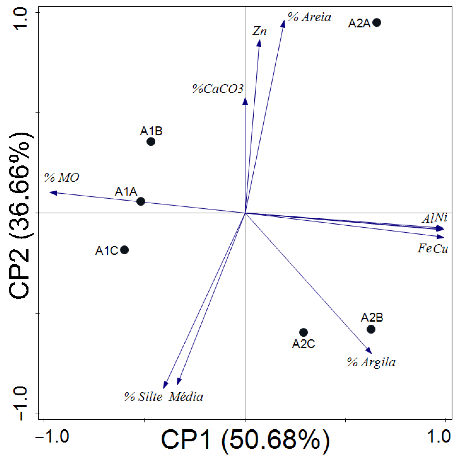
The principal component analysis (PCA) supported these patterns, with PC1 explaining 50.68% and PC2 36.66% of total variance. The A1 cluster was associated with higher organic matter and lower concentrations of metals (Al, Fe, Cu, Ni), while the A2 cluster was linked to higher clay content and metal enrichment. These results indicate a spatial gradient in sediment quality within Babitonga Bay, where A2 is under greater influence of fluvial discharge from the Cubatão River basin and adjacent industrial zones (Figure 2).
Therefore, the sedimentological and geochemical data reveal two contrasting depositional environments: A1 (Espinheiros), dominated by fine, organic-rich sediments with low metal input, and A2 (Vila da Glória), characterized by greater granulometric variability and higher metal concentrations derived from anthropogenic and fluvial sources.

3.2. Crassostrea rhizophorae

Area A1 (Espinheiros) showed significantly higher concentrations of the metals copper, nickel, zinc, aluminum, iron, arsenic, and cadmium compared to area A2 (Vila da Glória) (Table 3). No significant differences were observed in lead concentrations between the areas. These results indicate a higher degree of metal bioaccumulation in oysters from A1, suggesting chronic exposure to contamination sources near this mangrove, which may be associated with local industrial and urban discharges.
Additionally, oysters from area A2 exhibited larger dimensions (width, height, and thickness), as well as higher concentrations of lipid oxidation markers (TBA-RS and Carbonyl). Such contrasting patterns between metal content and biometric traits reflect opposite physiological responses: inhibition of growth under contaminant stress (A1) versus metabolic activation under lower but variable exposure (A2).
The Pearson correlation coefficient revealed strong positive correlations among the concentrations of metals (Cu, Ni, Zn, Al, Fe, As, and Cd) in the C. rhizophorae. However, iron concentrations did not show significant correlations with the other metals detected in the C. rhizophorae. Strong negative correlations were observed between metal concentrations in the fauna and sediments: copper (−0.923, p = 0.009), nickel (−0.921, p = 0.009), aluminum (−0.858, p = 0.029), and iron (−0.941, p = 0.005). Zinc concentrations in fauna and sediments showed no significant correlation (−0.16; p = 0.976). These negative correlations suggest that the bioavailability and accumulation dynamics of metals in oysters are not directly proportional to their sediment concentrations, possibly due to differences in metal speciation, physiological regulation or feeding behavior.
The same statistical test indicated a strong negative correlation between copper concentrations and oyster thickness (−0.812, p = 0.050), with no significant correlations found for other metals detected in C. rhizophorae and sediments. Additionally, no significant correlations were found between other oyster anatomical attributes (height and width) and metals detected in fauna and sediments, nor between oyster anatomical attributes (height, width, and thickness) and sediment characteristics.
TBARS concentrations showed strong negative correlations with metal concentrations in oysters: copper (−0.967, p = 0.002), nickel (−0.989, p < 0.001), zinc (−0.973, p = 0.001), aluminum (−0.963, p = 0.002), iron (−0.993, p < 0.001), and cadmium (−0.872, p = 0.023). Similarly, a strong negative correlation was found between carbonyl content and metals in oysters: copper (−0.954, p = 0.003), nickel (−0.922, p = 0.009), zinc (−0.968, p = 0.002), aluminum (−0.861, p = 0.023), iron (−0.935, p = 0.006), arsenic (−0.914, p = 0.011), and cadmium (−0.954, p = 0.003). However, no significant correlation was found between oxidative stress markers (TBARS and carbonyl) and oyster anatomical attributes (height, width, and thickness). These strong negative associations indicate that individuals under higher metal exposure (A1) exhibit lower oxidative marker levels, suggesting an adaptive physiological adjustment through antioxidant enzyme modulation or metallothionein synthesis, which may reduce oxidative damage under chronic contamination.
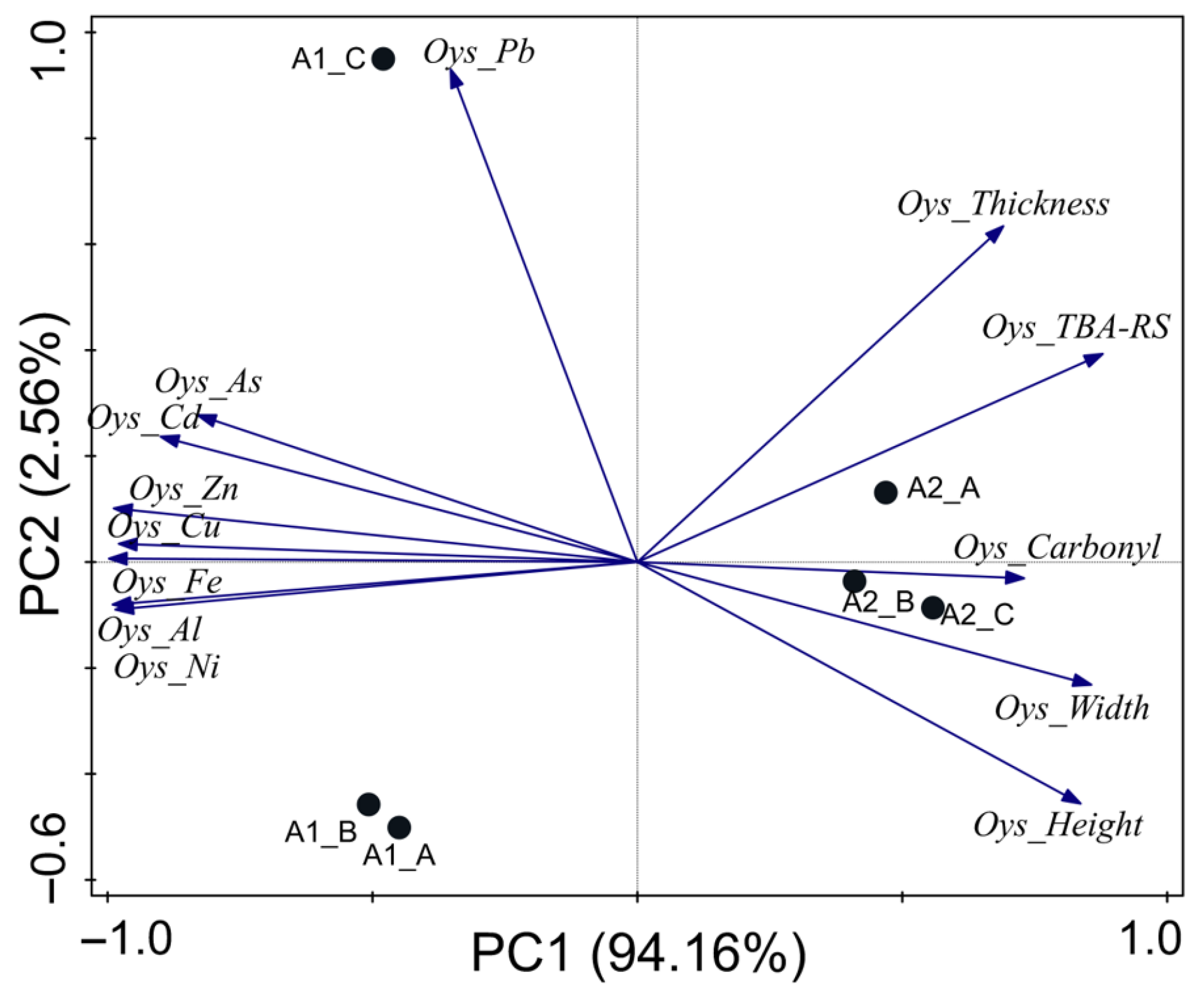
PC1 explained 96.72% of the data variance (Figure 3), and the oysters and sediments from area A2 (Vila da Glória) formed a cluster directly related to higher values of height, width, thickness, TBARS, and carbonyl content in oysters. At the opposite end, a cluster was observed comprising oysters and sediments from area A1 (Espinheiros). Oysters in this area exhibited higher metal concentrations and smaller dimensions in terms of height, width, and thickness. The PCA spatial separation thus supports a dual environmental gradient: A1 reflects chronic exposure with bioaccumulation and physiological suppression, whereas A2 reflects reduced contamination and enhanced metabolic turnover. This distinction confirms that C. rhizophorae is a sensitive bioindicator for both pollutant accumulation and oxidative stress responses in mangrove ecosystems.

3.3. Laguncularia racemosa

Significant differences were observed between the stations in the morphological attributes fresh mass (MFF) and specific leaf area (AEF); in the ecophysiological attributes chlorophyll a (CLa), chlorophyll b (CLb), and total chlorophyll (CLt); and the chemical attributes, specifically arsenic (As) and zinc (Zn) concentrations. All these attributes were found in higher quantities at the Vila da Glória site (Table 4). These variations indicate that plants from A2 presented greater photosynthetic efficiency and metal uptake capacity, suggesting that environmental conditions in this mangrove are more favorable for leaf development and physiological activity, even in the presence of trace contaminants.
Pearson correlation analysis revealed significant correlations between foliar attributes (fresh mass and thickness) and chlorophyll concentrations. Similarly, significant correlations were found between the concentrations of metals detected in the flora (zinc and arsenic). These associations point to the simultaneous influence of environmental factors, such as nutrient content and metal bioavailability, on both structural and functional plant responses.
A significant difference was observed in the zinc (Zn) concentration in leaf samples of Laguncularia racemosa between the different sampling sites, specifically between areas designated as A1 (Espinheiros) and A2 (Vila da Glória) (Table 4). The analyses revealed that the average zinc concentration found in area A2 was 83.64 mg kg−1, significantly higher than that found in area A1, which had an average of 74.34 mg kg−1. This difference was statistically significant, indicating that samples collected in A2 (Vila da Glória) exhibited a significantly greater accumulation of zinc compared to samples collected in A1 (Espinheiros). This result suggests that local environmental characteristics—such as sediment type, hydrodynamics, and exposure to tidal renewal—may influence the bioaccumulation processes of heavy metals in L. racemosa leaves.
Furthermore, the Pearson Correlation demonstrated a significant relationship between zinc content and other foliar attributes, suggesting a possible interaction or co-occurrence of these environmental factors in the two study areas. Table 4 also highlights significant differences in arsenic (As) concentrations in the leaves of L. racemosa collected from two distinct areas (A1 and A2). In the A2 area, the average arsenic concentration recorded was 0.28 mg kg−1, whereas in the A1 area, the average observed was 0.22 mg kg−1. This difference was significant, indicating a higher bioaccumulation of arsenic in samples originating from Vila da Glória. Although both As and Zn are essential or quasi-essential elements at trace levels, their elevated concentrations may indicate anthropogenic influence from distant fluvial inputs or differential retention in sediments and porewaters.
This result suggests the presence of specific environmental factors or contamination sources in the Vila da Glória area that contribute to the greater accumulation of arsenic. Additionally, the Pearson correlation analysis revealed a significant relationship between arsenic concentrations and other attributes such as fresh mass and leaf thickness, as well as chlorophyll concentrations. This correlation suggests possible interactions or co-occurrences among these environmental factors across the two study areas, underscoring the complexity of environmental influences on the bioaccumulation of heavy metals in L. racemosa.
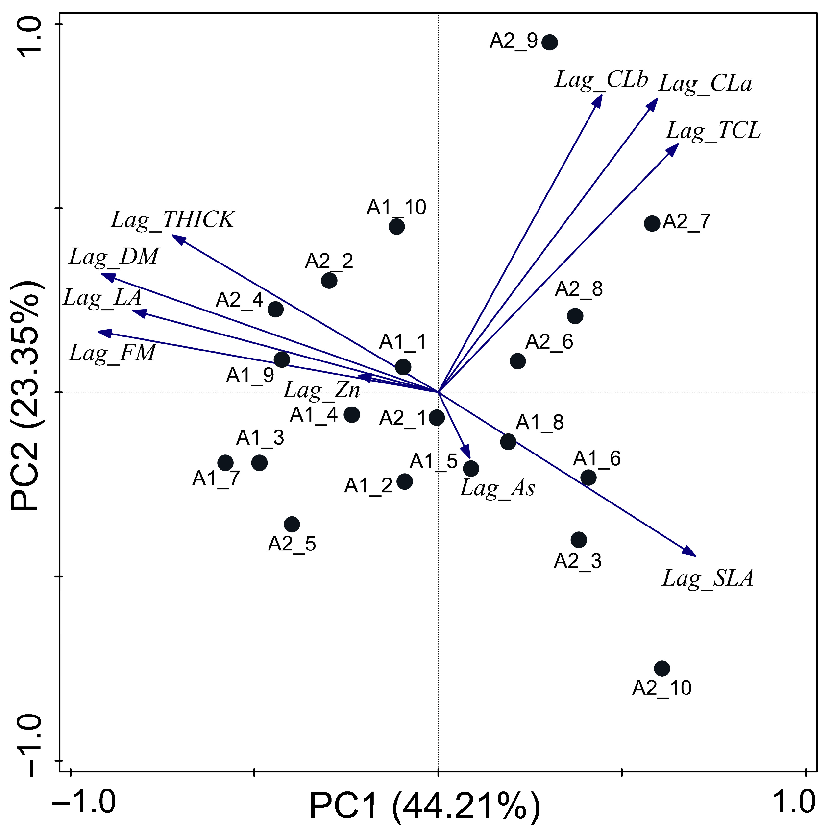
The PC1 represented 44.21% of the total variance, relating the leaves of Espinheiros’s individuals 5, 6 and 8, and Vila da Gloria’s individuals 10, 3, 6, 8, 7, and 9 to the elevation of concentrations in total Chlorophyll and Chlorophyll a and b, arsenic concentration and specific leaf area increasing and tended to be related to low leaf thickness, dry mass, leaf area, fresh mass, and zinc concentration (Figure 4). The grouping of Espinheiro’s individuals 1, 2, 3, 4, 7, 9, and 10, and Vila da Gloria’s individuals 1, 2, 4, and 5, where related to the increasing of leaf thickness, dry mass, leaf area, fresh mass, and zinc concentration, and the decreasing of concentrations in total Chlorophyll and Chlorophyll a and b, arsenic concentration, and specific leaf area (Figure 4).
Overall, the morphological and biochemical variations observed between A1 and A2 confirm that L. racemosa effectively reflects local environmental quality, acting as a reliable bioindicator species for assessing metal bioavailability and physiological responses in mangrove ecosystems.

4. Discussion

4.1. Sediments

Heavy metal contamination in sediments is a recurrent issue in both aquatic and continental environments, resulting from both natural and anthropogenic sources [14,15,16,18]. Due to their high density and specific toxicity, heavy metals are of considerable importance in environmental studies [58]. Assessing the quality of water, sediments, and soils is crucial given the significant role these metals play in the removal and availability of toxic substances across different geomorphological compartments. This study revealed elevated concentrations of metals such as copper (Cu), nickel (Ni), aluminum (Al), and iron (Fe) in samples collected from region A2, Vila da Glória, compared to area A1, Espinheiros.
The levels of heavy metals in sediments (Cu, Ni, and Zn) were compared with the standards established by the Brazilian National Environment Council Resolution No. 454/2012. This regulation defines guidelines and procedures for dredging management in waters under national jurisdiction, establishing two concentration levels: level 1, below which adverse effects on biota are less likely, and level 2, above which such effects are more likely, applicable to freshwater, brackish, and saline waters. In this study, copper (Cu) and zinc (Zn) levels exceeded level 1, indicating potential adverse impacts on local biota, while nickel (Ni) levels remained within the limits established by the legislation.
According to Tureck et al. [14], in Baía Babitonga, heavy metal levels varied considerably and were compared with other Brazilian port regions: Santos and São Vicente Estuary—SP [59], Paranaguá Bay—PR [60], Araçá Bay—SP [61], and Mucuripe Bay—CE [62].
Tureck et al. [14] reported that arsenic (As) concentrations in Baía Babitonga ranged from 1.2 to 43.9 mg kg−1, with maximum values exceeding those found in other port regions such as the Santos and São Vicente Estuary and Paranaguá Bay. Chromium (Cr) levels ranged from 1.2 to 61.1 mg kg−1, with the maximum value being the highest among the compared port areas. Copper (Cu) concentrations ranged from 0.42 to 30.5 mg kg−1, comparable to other regions but with higher maximum concentrations than those in Paranaguá Bay—PR [60], Araçá Bay—SP [61], and Baixada Santista—SP [62]. Nickel (Ni) ranged from less than 0.89 to 21.6 mg kg−1, with the maximum value surpassing all other compared port areas. Lead (Pb) concentrations ranged from 0.89 to 22.8 mg kg−1, slightly higher than those in Paranaguá Bay—PR, Araçá Bay—SP, and Baixada Santista—SP. Zinc (Zn) ranged from 2.0 to 154.9 mg kg−1, with levels higher than those in Paranaguá Bay—PR, Araçá Bay—SP, and the Santos and São Vicente Estuary—SP.
When compared with the limits set by Resolution Conama No. 454/2012, most metals in Baía Babitonga fall within acceptable limits; however, the maximum values for arsenic and zinc may approach or exceed critical levels. This resolution establishes uniform thresholds for all regions, disregarding ecosystem-specific sensitivity, which may increase the risk for fragile environments such as mangroves. This suggests the need for ongoing monitoring and mitigation measures to prevent adverse environmental impacts. It is noteworthy that Baía Babitonga exhibited higher values for all metals when compared to Paranaguá Bay—PR and Araçá Bay—SP.
Metals and metalloids are natural components of the environment, with multiple sources of input, including weathering and soil erosion, resulting in variable concentrations across different geographic regions [63]. Metal speciation in the environment is influenced by physicochemical parameters such as pH, cation exchange capacity, and redox potential [64]. In sediments, geochemical characteristics play a crucial role in the distribution and mobility of these elements. The chemical form in which metals occur, determined by the biogeochemical conditions of the system, defines their toxic potential and their ability to be re-mobilized [65].
The study region exhibits high levels of total organic carbon, carbonates, nutrients (N and P), silt, clay, and organic matter, characteristics that enhance its capacity to adsorb chemical substances. Sediment grain size is directly related to particle cohesion and sorption capacity, being more significant in finer grains. Larger contact surfaces promote the sorption of organic and inorganic elements, whether natural or anthropogenic, resulting in higher concentrations of potentially contaminating elements [66,67,68]. The low hydrodynamics in area A1 favor the deposition of fine sediments, creating ideal conditions for chemical adsorption and transforming it into an accumulation zone with increased contaminant levels.
Among the analysed elements, various anthropogenic sources were identified, primarily related to the industrial sector, which contains Cu and Zn [69,70], as well as electroplating, which uses Ni and Pb [71,72]. The Espinheiros area (A1) is located near a significant industrial hub, the oldest in the municipality of Joinville/SC, which motivated the choice of sampling point A1 for the study. However, the highest metal concentrations were observed at the control point A2, located in the Vila da Glória region, municipality of São Francisco do Sul/SC.
Another relevant aspect influencing the disparity between the studied sites is the sedimentological composition. In this study, area A1 (Espinheiros) was characterized by a predominance of silt (≈72%) and higher organic matter content (≈50.9%), conditions that enhance the adsorption and retention of metals in the sediment matrix. In contrast, area A2 (Vila da Glória) presented higher sand (≈24%) and clay fractions (≈11.6%), combined with a markedly lower organic matter content (≈17%), features typically associated with greater hydrodynamic energy and reduced capacity for metal retention. These textural and compositional differences are consistent with the higher concentrations of Cu, Ni, Zn, and Fe recorded in A1, supporting the interpretation that finer, organic-rich sediments [73] act as effective sinks for heavy metals [14,15,16,17,18,19,20,21,22,56,59,60] within the Babitonga Bay mangrove system.

4.2. Crassostrea rhizophorae

The comparison between the study areas A1 (Espinheiros) and A2 (Vila da Glória) revealed significant differences in heavy metal concentrations in C. rhizophorae and their associated anatomical and biochemical attributes. Oysters from area A1 exhibited significantly higher concentrations of copper, nickel, zinc, aluminum, iron, arsenic, and cadmium compared to area A2. At the same time, no significant difference was observed in lead concentrations between the areas. The higher heavy metal contamination in area A1 can be attributed to its proximity to industrial and urban pollution sources, as described in previous studies on heavy metal bioaccumulation in oysters [74,75]. These metals are frequently found in higher concentrations in areas subject to anthropogenic pollution [76,77]
Oysters from area A2 displayed larger dimensions (width, height, and thickness) and higher concentrations of lipid oxidation markers (TBARS and carbonyl groups). The lower heavy metal contamination in this area may contribute to better growth conditions and reduced oxidative stress, as indicated by studies linking metal bioaccumulation to oxidative stress in bivalves [78,79].
Pearson correlation analysis revealed strong negative correlations between metal concentrations in the fauna and the sediment, suggesting a complex dynamic of metal availability and uptake by oysters [80,81]. Furthermore, the significant negative correlation between oyster thickness and copper concentration highlights the adverse influence of this metal on bivalve growth [82]. TBARS and carbonyl concentrations showed strong negative correlations with metal concentrations in the oysters, indicating that the presence of heavy metals may be associated with increased oxidative stress, as reported in other studies [83,84]. The absence of significant correlations between oxidative stress markers and oyster anatomical attributes suggests that the impact of heavy metals may be more directly related to increased cellular stress than to morphological changes [85].
The clustering patterns of oysters and sediments between the two study areas in the PCA results showed the clustering of oysters from area A2, characterized by larger body dimensions and lower metal contamination, contrasted with the grouping of oysters from area A1, which presented higher metal concentrations and smaller body dimensions. This pattern reinforces the significant influence of environmental contamination on the biological and biochemical attributes of oysters [86,87].

4.3. Laguncularia racemosa

The results obtained from the morphological, ecophysiological, and chemical analyses of L. racemosa in areas A1 (Espinheiros) and A2 (Vila da Glória) reveal significant differences between the two locations, as presented in Table 4. Comparing these data with the findings of Bartz et al. [47], some similarities and disparities can be observed.
The fresh leaf mass of L. racemosa was significantly higher in A2 (2.01 ± 0.43 g) than in A1 (1.82 ± 0.47 g), indicating that environmental conditions in A2 favor greater water retention in the leaves. On the other hand, dry mass showed no significant difference between the two areas (0.53 ± 0.15 g in A1 and 0.55 ± 0.12 g in A2), and the observed values are slightly lower than those reported by Bartz et al. [47], who found 0.62 ± 0.17 g.
Mean leaf area was similar in both studied areas (24.32 ± 5.93 cm2 in A1 and 24.44 ± 4.31 cm2 in A2) with no significant difference. These values are slightly higher than those observed by Bartz et al. [47], who reported an average leaf area of 22.12 ± 4.12 cm2. However, leaf thickness was significantly greater in A2 (0.44 ± 0.04 mm) compared to A1 (0.42 ± 0.03 mm). These values are substantially lower than those reported by Bartz et al. [47], who observed leaf thickness of 0.73 ± 0.11 mm.
Specific leaf area (SLA), an indicator of light-use efficiency and photosynthetic capacity, did not show a significant difference between the areas, and its higher values, compared to other mangroves in the region [47], indicate a greater potential for leaf development in the plants.
Levels of chlorophyll a, chlorophyll b, and total chlorophyll were all significantly higher in A2 than in A1, indicating that plants in A2 may have a higher photosynthetic capacity and thus a potentially more robust growth. These results suggest that environmental conditions in A2 are more favorable for photosynthesis.
Heavy metals such as aluminum, cadmium, lead, copper, and iron were not detected in either area. However, arsenic levels were significantly higher in A2 (0.28 ± 0.06 mg/kg) than in A1 (0.22 ± 0.17 mg/kg), as was zinc (83.64 ± 8.70 mg/kg in A2 and 74.34 ± 13.10 mg/kg in A1). These results suggest that A2 may be susceptible to various pollution sources or biogeochemical processes, resulting in a greater accumulation of these elements.
According to Arrivabene et al. [86], Laguncularia racemosa and Avicennia schaueriana are considered excellent bioindicators due to their anatomical and morphological characteristics, which are closely related to the specific environmental conditions where they develop. Variations in photosynthetic pigment contents indicate changes in photosynthetic activity, which can be influenced by various environmental factors such as salinity, irradiance, flooding, heavy metals, nutritional status, and pollutants, making them potential indicators of adverse environmental conditions [88,89,90].
Mangrove conditions are directly related to pigment content, both chlorophyll a and chlorophyll b, and consequently to total chlorophyll content [91]. For example, more saline environments tend to reduce chlorophyll concentrations, resulting in lower light absorption and chronic photoinhibition [92]. However, in the present study, no differences in sediment salinity were observed between the studied areas, making it impossible to explain the differences in chlorophyll through this mechanism.
In Cavalcante et al. [90], higher concentrations of chlorophyll a and b were observed in sites with greater pollutant loads, both in Rhizophora mangle and L. racemosa. A study using a portable chlorophyll meter to detect differences in chlorophyll a, b, and total indices between control points and two points located in a port region used L. racemosa as a bioindicator of environmental pollution. The results showed significant differences between control areas and port-impacted areas, with higher chlorophyll levels in the port-impacted region. Principal component analysis (PCA) statistical analysis indicated that chlorophyll vectors showed greater pigment activity in the more anthropized segments [93].
Inoue [91] evaluated the effect of dust emissions from a cement plant on chlorophyll levels in six tree species: Araucaria angustifolia, Mimosa scabrella, Ocotea puberula, Schinus terebinthifolius, Pinus taeda, and Matayba elaeagnoides. Increased chlorophyll concentrations were observed in colder months, particularly in trees influenced by cement plant dust, due to reduced radiation.
These studies contrast with the results observed in areas A1 and A2 of Babitonga Bay, where area A1, which is more influenced by pollutants due to greater anthropization, shows lower chlorophyll levels compared to area A2. Another hypothesis to consider is that in the more polluted area, chlorophyll content is reduced, as suggested by other authors [94,95,96]. Thus, it is recommended that physiological parameters, depending on the species studied, may act as bioindicators of atmospheric pollution but with specific behaviors [94].
In the study by Gonçalves et al. [95], most Laguncularia racemosa samples did not present elevated trace metal levels except for copper. It was inferred that trace metal concentrations were higher in sediments than in plants, suggesting that these metals were not in bioavailable form, which aligns with the results of this study.
Sandy sediment characteristics generally contribute to metal availability [86]. Additionally, higher pH values make metals less bioavailable for plant uptake [86]. However, no significant differences were found between the sediments of the studied areas, making it impossible to justify the differences in arsenic and zinc presence in these locations.
Campos [90] reported that Rhizophora mangle was more influenced by salinity in its traits than L. racemosa, which may explain the lack of correlation in Babitonga Bay. Regarding arsenic concentration (As), L. racemosa was more sensitive and presented higher As absorption under more saline conditions. However, as previously mentioned, no salinity differences were observed between the studied areas, so it is not possible to explain the higher foliar As concentration.
Santos et al. [26] found zinc concentrations ranging from 8.42 to 13.33 mg kg−1 in green leaves of L. racemosa in southern Bahia, values lower than those found in the present study. Generally, higher zinc concentrations are found near contamination sources because zinc tends to accumulate in soil and sediments, showing higher phytotoxicity, especially in acidic and silty soils [26].
According to [97], standard or sufficient zinc levels range between 8 and 400 mg kg−1, while levels above 400 mg kg−1 are considered toxic or excessive. Kabata-Pendias [97] defines normal zinc levels between 27 and 150 mg kg−1, with concentrations between 100 and 400 mg kg−1 considered excessive. Furthermore, the World Health Organization (WHO) and the Food and Agriculture Organization (FAO) established a maximum recommended limit of 60 mg kg−1 for zinc. In comparison, the Canadian Council of Ministers of the Environment (CCME) recommended a maximum limit of 124 mg kg−1.
These reference values are essential for interpreting the results obtained in this study, as they provide a context for evaluating the potential toxicity and adequacy of zinc levels observed in L. racemosa samples. Comparing the Babitonga Bay results with literature-established limits, it is possible to infer that, despite the elevated zinc levels found, they do not exceed toxic thresholds. However, the observed values are significantly higher than those reported in other studies, indicating a potential environmental impact and highlighting the potential of L. racemosa as a bioindicator of heavy metal contamination in mangrove ecosystems.
In a study using L. racemosa leaves as indicators of contamination in an estuarine environment [97], quantified the presence of heavy metals, finding zinc concentrations ranging from 6.62 ± 0.16 to 16.93 ± 0.36 mg kg−1. The study concluded that higher values are concerning, pointing to the high potential of L. racemosa as a bioindicator of metal contamination. Another survey of the chemical composition of L. racemosa leaves in a mangrove impacted by industrial residues from the Mucuri River in Bahia found an average zinc content of 10.1 mg kg−1. Both contrast with our results, which evidenced zinc concentrations up to eight times higher in the leaves of this plant.
It is essential to note that high zinc levels in mangrove ecosystems can lead to restricted plant germination, reduced root development, and accelerated plant aging. Considering the results of our samples, the mangrove environment of Babitonga Bay exhibits zinc levels above the average reported in other studies, which suggests environmental stress and reinforces the potential of L. racemosa as an ecological biomonitor.

4.4. Human Health Implications

Consumption of contaminated mollusks and other aquatic organisms represents a potential pathway of human exposure to zinc, arsenic, copper, aluminum, iron, and nickel. The health effects depend on the degree of bioaccumulation, the chemical form of the elements, and the frequency and quantity of intake. The literature demonstrates that bivalves can accumulate these elements to levels of concern in polluted environments [98,99,100,101,102,103].
Arsenic is of particular concern due to its carcinogenic potential. Chronic dietary exposure to inorganic arsenic from contaminated seafood is associated with skin lesions and increased risks of bladder, lung, and skin cancers [98,99,104,105]. Co-exposure with other elements, such as mercury and selenium, may further enhance adverse health outcomes [104]. Nickel, although rarely reaching systemic toxicity at typical dietary levels, may cause allergic contact dermatitis in sensitive individuals [101,102]. Experimental studies also demonstrate combined toxic effects when nickel occurs together with copper and zinc, highlighting that single-element thresholds may underestimate risk [106].
Copper and iron are essential trace elements, but excessive intake may be harmful. High copper exposure can cause hepatic injury, particularly in genetically predisposed individuals, while iron overload may result in oxidative stress and organ damage [101,104,107]. Chronic exposure to mixtures of these metals has also been implicated in endocrine disruption and liver injury [108]. Aluminum, a non-essential element, has been associated with neurotoxicity and bone disorders under chronic exposure, especially in individuals with impaired renal function [103,109]. Reviews indicate that aluminum contamination in aquatic organisms can affect the nervous system, producing oxidative stress and cognitive deficits [110]. Zinc is generally safe at dietary levels, though excessive intake can interfere with copper absorption and cause gastrointestinal effects [101,102,104,109]. Experimental data suggest that zinc supplementation may alleviate arsenic-induced renal toxicity in fish, indicating complex interactions between essential and toxic elements [111].
Risk assessments consistently indicate that arsenic, and to a lesser extent nickel, represent the most significant health risks from mollusk consumption in contaminated areas [101,103,105,112]. For copper, iron, aluminum, and zinc, risks are typically low for the general population but may be elevated in specific subgroups or under chronic high intake [101,102,104,106,107,108,112]. Global evidence further shows that seafood consumption can be a major source of toxic metal exposure, especially in high-consumption regions [113,114,115]. Moreover, contamination of fish and crustaceans demonstrates that metal mixtures can impair reproduction, survival, and biochemical balance in aquatic organisms, reinforcing their potential to indirectly affect human health through the food chain [113,114,115,116].

5. Conclusions

The results indicate significant heavy metal contamination in Vila da Glória (A2) compared to Espinheiros (A1). Levels of Cu and Zn exceed the standards established by CONAMA Resolution No. 454/2012, suggesting possible adverse impacts on the local biota. The higher metal concentrations found in area A2 (Vila da Glória) may be related to fluvial inputs from rivers that drain the industrial district of Joinville and discharge into the estuarine system. Babitonga Bay exhibits elevated heavy metal concentrations compared to other Brazilian port regions, with arsenic (As) and zinc (Zn) levels approaching or exceeding critical thresholds.
The findings demonstrate that Laguncularia racemosa is an excellent bioindicator of environmental conditions due to its anatomical and morphological characteristics. Fresh leaf mass, leaf thickness, chlorophyll levels, and concentrations of zinc and arsenic in L. racemosa leaves were significantly higher in A2 (Vila da Glória) than in A1 (Espinheiros). Samples of Crassostrea rhizophorae from area A1 (Espinheiros) showed significantly higher concentrations of various metals compared to area A2 (Vila da Glória). Oysters from area A2 (Vila da Glória) exhibited larger dimensions (width, height, and thickness) and higher concentrations of lipid oxidation markers.
The bioindicators analyzed in this study (L. racemosa and C. rhizophorae) exhibited distinct responses that reflect differences in environmental conditions across the mangrove areas, particularly related to metal enrichment and biological stress patterns.

Author Contributions

Conceptualization, J.C.F.d.M.J., C.V.V., L.L. and T.M.N.d.O.; methodology, J.C.F.d.M.J., C.V.V. and L.L.; validation, A.B.G., A.K.M., A.P.d.M., A.P.M.d.A., D.D., D.R.C., G.B.d.O., L.C.M., L.S., M.C.B., M.L.I., N.T.B., N.C., R.L.d.O., S.C.L. and P.R.P.F.; formal analysis, R.L.d.O., A.B.G., A.K.M. and A.P.d.M.; investigation, A.B.G., A.K.M., A.P.d.M., A.P.M.d.A., D.D., D.R.C., G.B.d.O., L.C.M., L.S., M.C.B., M.L.I., N.T.B., N.C., R.L.d.O., S.C.L. and P.R.P.F.; resources, J.C.F.d.M.J. and C.V.V.; data curation, R.L.d.O., A.B.G., A.P.d.M. and P.R.P.F.; writing—original draft preparation, all authors; writing—review and editing, all authors; supervision, J.C.F.d.M.J., C.V.V., L.L. and T.M.N.d.O.; project administration, J.C.F.d.M.J., C.V.V. and L.L.; funding acquisition, J.C.F.d.M.J. and C.V.V. All authors have read and agreed to the published version of the manuscript.

Funding

This research was funded by the National Council for Scientific and Technological Development (CNPq), grant number 308777/2025-5; the Santa Catarina Research and Innovation Support Foundation (FAPESC), grant number 2024TR001918; and the Research Support Fund of Univille (FAP), grant number 1282.

Institutional Review Board Statement

Not applicable.

Informed Consent Statement

Not applicable.

Data Availability Statement

The original data presented in the study are openly available in [Zenodo] at https://doi.org/10.5281/zenodo.17106592.

Acknowledgments

During the preparation of this manuscript, the authors used ChatGPT (GPT-5, OpenAI) for the purpose of assisting in the translation of sections of the text from Portuguese to English. The authors have reviewed and edited the output and take full responsibility for the content of this publication.

Conflicts of Interest

The authors declare no conflicts of interest.

References

  1. Giri, C.; Ochieng, E.; Tieszen, L.L.; Zhu, Z.; Singh, A.; Loveland, T.; Masek, J.; Duke, N. Status and distribution of mangrove forests of the world using earth observation satellite data. Glob. Ecol. Biogeogr. 2011, 20, 154–159. [Google Scholar] [CrossRef]
  2. Duke, N.; Nagelkerken, I.; Agardy, T.; Wells, S.; Van Lavieren, H.; Huxham, M. The Importance of Mangroves to People: A Call to Action; United Nations Environment Programme (UNEP): Nairobi, Kenya, 2014. [Google Scholar] [CrossRef]
  3. Madi, A.P.L.M.; Boeger, M.R.T.; Larcher, L.; Pelozo, A.; Sereneski, C.; Reissmann, C.B.; Padial, A.A. Estrutura do componente de regeneração natural e arbóreo de dois manguezais no estado do paraná. Cienc. Florest. 2016, 26, 159–170. [Google Scholar] [CrossRef]
  4. Lee, S.Y.; Primavera, J.H.; Dahdouh-Guebas, F.; McKee, K.; Bosire, J.O.; Cannicci, S.; Diele, K.; Fromard, F.; Koedam, N.; Marchand, C.; et al. Ecological role and services of tropical mangrove ecosystems: A reassessment. Glob. Ecol. Biogeogr. 2014, 23, 726–743. [Google Scholar] [CrossRef]
  5. Gomes, M.T.U.; Lorenzen, P.; Wandscheer, C.B. Carbono azul: Manguezais, um potencial sumidouro de carbono para a América Latina e Caribe. Rev. Estud. Av. 2024, 40, 149–182. [Google Scholar] [CrossRef]
  6. Akram, H.; Hussain, S.; Mazumdar, P.; Chua, K.O.; Butt, T.E.; Harikrishna, J.A. Mangrove Health: A Review of Functions, Threats, and Challenges Associated with Mangrove Management Practices. Forests 2023, 14, 1698. [Google Scholar] [CrossRef]
  7. Holguin, G.; Vazquez, P.; Bashan, Y. The role of sediment microorganisms in the productivity, conservation, and rehabilitation of mangrove ecosystems: An overview. Biol. Fertil. Soils 2001, 33, 265–278. [Google Scholar] [CrossRef]
  8. Godoy, M.D.; de Lacerda, L.D. Mangroves Response to Climate Change: A Review of Recent Findings on Mangrove Extension and Distribution. An. Acad. Bras. Cienc. 2015, 87, 651–667. [Google Scholar] [CrossRef]
  9. Santos, T.O.; Andrade, K.D.; Santos, H.V.S.; Castaneda, D.A.F.G.; Santana, M.B.S.; Holanda, F.S.R.; Santos, M.J.C. Caracterização estrutural de bosques de mangue: Estuário do São Francisco. Sci. Plena 2012, 84b, 1–7. Available online: https://www.scientiaplena.org.br/sp/article/view/1010 (accessed on 28 August 2025).
  10. Amaral, É.B.M.; Santos, C.V. Poluição e invasão de mangues em zonas litorâneas. Unisanta Biosci. 2022, 11, 117–123. [Google Scholar]
  11. Alves, R.J.M.; Pinheiro, D.d.S.; Teixeira, J.M.J.; Alves, R.J.M.; Tavares-Martins, A.C.C. Expansão urbana e percepção ambiental: Uma análise sobre a relação homem-manguezal na cidade de Marapanim, Pará. Mundo Amaz. 2023, 14, 154–179. [Google Scholar] [CrossRef]
  12. Vieira, C.V.; Filho, N.O.H.; Bonetti, C.V.D.H.C.; Bonetti, J. Caracterização morfosedimentar e setorização do complexo estuarino da baía da babitonga/sc. Bol. Parana. Geociencias 2008, 62, 12783. [Google Scholar] [CrossRef]
  13. Herbst, D.F.; Gerhardinger, L.C.; Vila-Nova, D.A.; de Carvalho, F.G.; Hanazaki, N. Integrated and deliberative multidimensional assessment of a subtropical coastal-marine ecosystem (Babitonga bay, Brazil). Ocean Coast. Manag. 2020, 196, 105279. [Google Scholar] [CrossRef]
  14. Tureck, C.R.; Haak, L.; da Cunha, S.M.B.; Destefani, A.; Vieira, C.V.; Kim, B.S.M.; Trevizani, T.H.; Ferreira, P.A.d.L.; Figueira, R. Qualidade de sedimentos na Baía Babitonga e sua implicação na reabertura do Canal do Linguado (SC), Brasil. Acta Biológica Catarin. 2024, 11, 46–61. [Google Scholar] [CrossRef]
  15. Kilca, R.V.; Mel, J.C.F., Jr.; Esemann-Quadros, K.; Larcher, L.; Pfuetzenreuter, A. Os manguezais e marismas da Baía Babitonga: Uma síntese. Rev. CEPSUL-Biodiversidade Conserv. Mar. 2019, 8, eb2019002. [Google Scholar] [CrossRef]
  16. Matias, L.; Silva, M.D. Monitoramento e análise da vegetação de manguezal no litoral sul de Alagoas. J. Environ. Anal. Prog. 2017, 2, 312–319. [Google Scholar] [CrossRef]
  17. Fedorova, D.G.; Karpova, G.V.; Ukenov, B.S. The Accumulation of Heavy Metals in the Leaves of Crataegus Sanguinea Pall. (Redhaw Hawthorn) in the Urban Environment (On the Example of Orenburg). IOP Conf. Ser. Earth Environ. Sci. 2021, 670, 012030. [Google Scholar] [CrossRef]
  18. Acosta-Dacal, A.; Hernández-Marrero, M.E.; Rial-Berriel, C.; Díaz-Díaz, R.; Bernal-Suárez, M.d.M.; Zumbado, M.; Henríquez-Hernández, L.A.; Boada, L.D.; Luzardo, O.P. Comparative study of organic contaminants in agricultural soils at the archipelagos of the Macaronesia. Environ. Pollut. 2022, 301, 118979. [Google Scholar] [CrossRef]
  19. Badamasi, H. Biomonitoring of Air Pollution Using Plants. J. Environ. Sci. 2017, 2, 27–39. [Google Scholar]
  20. Mulgrew, A.; Williams, P. Biomonitoring of Air Quality Using Plants; Monitoring and Assessment Research Centre: London, UK, 2000. [Google Scholar]
  21. Elturk, M.; Abdullah, R.; Zakaria, R.M.; Abu Bakar, N.K. Heavy metal contamination in mangrove sediments in Klang estuary, Malaysia: Implication of risk assessment. Estuarine. Coast. Shelf Sci. 2019, 226, 106266. [Google Scholar] [CrossRef]
  22. Maurya, P.; Kumari, R. Toxic metals distribution, seasonal variations and environmental risk assessment in surficial sediment and mangrove plants (A. marina), Gulf of Kachchh (India). J. Hazard. Mater. 2021, 413, 125345. [Google Scholar] [CrossRef]
  23. Xu, S.; Lin, C.; Qiu, P.; Song, Y.; Yang, W.; Xu, G.; Feng, X.; Yang, Q.; Yang, X.; Niu, A. Tungsten- and cobalt-dominated heavy metal contamination of mangrove sediments in Shenzhen, China. Mar. Pollut. Bull. 2015, 100, 562–566. [Google Scholar] [CrossRef] [PubMed]
  24. Robin, S.L.; Alfaro, A.C.; Gututuauva, K.; Marchand, C. Dynamics of major and trace elements during leaf litter decomposition in semi-arid mangrove subject to urban runoff. Mar. Pollut. Bull. 2025, 211, 117475. [Google Scholar] [CrossRef]
  25. Usman, A.R.; Alkredaa, R.S.; Al-Wabel, M. Heavy metal contamination in sediments and mangroves from the coast of Red Sea: Avicennia marina as potential metal bioaccumulator. Ecotoxicol. Environ. Saf. 2013, 97, 263–270. [Google Scholar] [CrossRef]
  26. Santos, T.A.; Bonfim, T.M.; Silva, F.S.; Silva, A.G.; de Ferreira Bandeira, M.L.S.; de Jesus, R.M.; Nascimento, L.D. Determinação de metais em um sistema estuarino: Laguncularia Racemosa como um potencial indicador de contaminação. Rev. Bras. De Ciências Ambient. 2018, 49, 51–65. [Google Scholar] [CrossRef]
  27. Silva, C.A.R.; Rainbow, P.S.; Smith, B.D. Biomonitoring of trace metal contamination in mangrove-lined Brazilian coastal systems using the oyster Crassostrea rhizophorae: Comparative study of regions affected by oil, salt pond and shrimp farming activities. Hydrobiologia 2003, 501, 199–206. [Google Scholar] [CrossRef]
  28. Senez-Mello, T.M.; Crapez, M.A.C.; e Silva, C.A.R.; Silva, E.T.; Fonseca, E.M. Heavy metals bioconcentration in Crassostrea rhizophorae: A site-to-site transplant experiment at the Potengi estuary, Rio Grande do Norte, Brazil. Sci. Rep. 2020, 10, 246. [Google Scholar] [CrossRef]
  29. Ruthes, A.M.; Silva, M.M.; Melo Júnior, J.C.F. Leaf changes in Avicennia schaueriana following a massive herbivory event by Hyblaea puera (Lepidoptera) in South Brazil. Braz. J. Dev. 2021, 7, 47275–47286. [Google Scholar]
  30. CATARINA, Fundação do Meio Ambiente de Santa. Atlas Ambiental da Região de Joinville: Complexo Hídrico da Baía da Babitonga; FATMA: Florianópolis, Brazil, 2003. [Google Scholar]
  31. Vieira, C.V.; Filho, N.O.H. Paisagem marinha da Baía da Babitonga, nordeste do estado de Santa Catarina. Rev. Bras. Geogr. Física 2017, 10, 1017–1034. [Google Scholar]
  32. Truccolo, E.C.; Schettini, C.A.F. Marés astronômicas na baía Babitonga, SC. Notas Técnicas Facimar 1999, 3, 57–66. [Google Scholar] [CrossRef]
  33. Silva, L.F. Alterações morfodinâmicas no Canal do Linguado pela remoção do dique que o separa da Baía da Babitonga (SC). Ph.D. thesis, Universidade Federal do Rio Grande do Sul, Alegre, Brazil, 2011. [Google Scholar]
  34. Cremer, M.J. O estuário da Baía da Babitonga. In Diagnóstico Ambiental da baía da Babitonga; UNIVILLE: Joinville, France, 2006; pp. 15–19. [Google Scholar]
  35. Kilca, R.V.; Alberti, L.F.; Souza, A.M.; Wolf, L. Estrutura de uma floresta de mangue na Baía da Babitonga, São Francisco do Sul, SC. Ciência E Nat. 2011, 33, 57–72. [Google Scholar]
  36. Júnior, J.C.F.M.; Silva, M.M.; Boeger, M.R.T.; Souza, T.F.; Vieira, C.V. Patrimônio natural das restingas da baía Babitonga, Santa Catarina, Brasil. Rev. CEPSUL-Biodiversidade Conserv. Mar. 2018, 7, eb2018002. [Google Scholar] [CrossRef]
  37. Suguio, K. Introdução a Sedimentologia; EDUSP: São Paulo, Brazil, 1973. [Google Scholar]
  38. Galehouse, J.S. Sedimentation analysis. In Procedures in Sedimentary Petrology; Wiley-Interscience: New York, NY, USA, 1971. [Google Scholar]
  39. Folk, R.L.; Ward, W.C. Brazos river bar: A Study in the Significance of Grain-Size Parameters. J. Sediment. Res. 1957, 27, 3–26. [Google Scholar] [CrossRef]
  40. Camargo, M.G. SysGran: Um sistema de código aberto para análises granulométricas do sedimento. Rev. Bras. Geo-Ciências 2006, 36, 371–378. [Google Scholar] [CrossRef]
  41. Aslam, S.; Chan, M.W.H.; Siddiqui, G.; Boczkaj, G.; Kazmi, S.J.H.; Kazmi, M.R. A comprehensive assessment of environmental pollution by means of heavy metal analysis for oysters’ reefs at Hab River Delta, Balochistan, Pakistan. Mar. Pollut. Bull. 2020, 153, 110970. [Google Scholar] [CrossRef] [PubMed]
  42. Azevedo, J.A.M.; Barros, A.B.; de Miranda, P.R.B.; da Costa, J.G.; Nascimento, V.X. Biomonitoring of Heavy Metals (Fe, Zn, Cu, Mn, Cd and Cr) in Oysters: Crassostrea rhizophorae of Mangrove Areas of Alagoas (Brazil). Braz. Arch. Biol. Technol. 2019, 62, e19180211. [Google Scholar] [CrossRef]
  43. Ohkawa, H.; Ohishi, N.; Yagi, K. Assay for lipid peroxides in animal tissues by thiobarbituric acid reaction. Anal. Biochem. 1979, 95, 351–358. [Google Scholar] [CrossRef] [PubMed]
  44. Reznick, A.Z.; Packer, L. Oxidative damage to proteins: Spectrophotometric method for carbonyl assay. Methods Enzymol. 1994, 233, 357–363. [Google Scholar] [CrossRef]
  45. Filardi, F.L.R.; Barros, F.D.; Baumgratz, J.F.A.; Bicudo, C.E.; Cavalcanti, T.B.; Coelho, M.A.N.; Costa, A.F.; Goldenberg, R.; Labiak, P.H.; Lanna, J.M. Brazil Flora Group, Brazilian Flora 2020: Innovation and collaboration to meet Target 1 of the Global Strategy for Plant Con-servation (GSPC). Rodriguésia 2018, 69, 1513–1527. [Google Scholar]
  46. Schaeffer-Novelli, Y.; Adaime, R.R.; Cintron-Molero, G.; de Camargo, T.M. Variability of Mangrove Ecosystems along the Brazilian Coast. Estuaries 1990, 13, 204–218. [Google Scholar] [CrossRef]
  47. Bartz, M.C.; Júnior, J.C.F.d.M.; Larcher, L. Variação morfológica de Laguncularia racemosa (L.) C. F. Gaertn. (Combretaceae) em áreas de manguezal e de transição entre manguezal e floresta de restinga. Biotemas 2014, 28, 21–29. [Google Scholar] [CrossRef]
  48. Bernini, E.; Rezende, C.E. Litterfall in a mangrove in Southeast Brazil. Pan-Am. J. Aquat. Sci. 2010, 5, 508–519. [Google Scholar]
  49. Sobrado, M.A. Leaf characteristics and gas exchange of the mangrove Laguncularia racemosa as affected by salinity. Photosynthetica 2005, 43, 217–221. [Google Scholar] [CrossRef]
  50. Lee, J.A.; Hendry, G.A.F.; Grime, J.P. Methods in Comparative Plant Ecology: A Laboratory Manual. J. Ecol. 1993, 81, 832. [Google Scholar] [CrossRef]
  51. Martin, D.; Kopp, J. Method 200.7 Determination of Metals and Trace Elements in Water and Wastes by Inductively Coupled Plasma-atomic Emission Spectrometry Revision 4.4. USEPA-ICP Users Group; U.S. Environmental Protection Agency: Cincinnati, OH, USA, 1994. [Google Scholar]
  52. Filho, P.J.S.; Böhm, E.M.; Böhm, G.M.; Montenegro, G.O.; Silveira, L.A.; Betemps, G.R. Determination of hydrocarbons transported by urban runoff in sediments of São Gonçalo Channel (Pelotas—RS, Brazil). Mar. Pollut. Bull. 2017, 114, 1088–1095. [Google Scholar] [CrossRef]
  53. Uysal, K.; Köse, E.; Bülbül, M.; Dönmez, M.; Erdoğan, Y.; Koyun, M.; Ömeroğlu, Ç.; Özmal, F. The comparison of heavy metal accumulation ratios of some fish species in Enne Dame Lake (Kütahya/Turkey). Environ. Monit. Assess. 2008, 157, 355–362. [Google Scholar] [CrossRef]
  54. Gotelli, N.J.; Ellison, A.M. A Primer of Ecological Statistics; Sinauer Associates, Inc.: Sunderland, MA, USA, 2004. [Google Scholar]
  55. Vieira, S. Bioestatística: Tópicos Avançados, 5th ed.; Editora Guanabara Koogan: Rio de Janeiro, Brazil, 2023. [Google Scholar]
  56. Bourgeron, P.; Legendre, P.; Legendre, L. Numerical Ecology. Arctic, Antarct. Alp. Res. 2000, 32, 218. [Google Scholar] [CrossRef]
  57. Cohen, J. Statistical Power Analysis for the Behavioral Sciences, 2nd ed.; Lawrence Erlbaum Associates: Hillsdale, NJ, USA, 1988. [Google Scholar]
  58. El-Sharkawy, M.; Alotaibi, M.O.; Li, J.; Du, D.; Mahmoud, E. Heavy Metal Pollution in Coastal Environments: Ecological Implications and Management Strategies: A Review. Sustainability 2025, 17, 701. [Google Scholar] [CrossRef]
  59. Kim, B.S.M.; Angeli, J.L.F.; Ferreira, P.A.L.; De Mahiques, M.M.; Figueira, R.C.L. A multivariate approach and sediment quality index evaluation applied to Baixada Santista, Southeastern Brazil. Mar. Pollut. Bull. 2019, 143, 72–80. [Google Scholar] [CrossRef]
  60. Angeli, J.L.F.; Kim, B.S.M.; Paladino, Í.M.; Nagai, R.H.; Martins, C.C.; de Mahiques, M.M.; Figueira, R.C.L. Statistical assessment of background levels for metal contamination from a subtropical estuarine system in the SW Atlantic (Paranaguá Estuarine System, Brazil). J. Sediment. Environ. 2020, 5, 137–150. [Google Scholar] [CrossRef]
  61. Kim, B.S.M.; Bícego, M.C.; Taniguchi, S.; Siegle, E.; Oliveira, R.; Alcántara-Carrió, J.; Figueira, R.C.L. Organic and inorganic contamination in sediments from Araçá Bay, São Sebastião, Brazil. Ocean Coast. Manag. 2018, 164, 42–51. [Google Scholar] [CrossRef]
  62. Kim, B.S.M.; Angeli, J.L.F.; Ferreira, P.A.L.; de Mahiques, M.M.; Figueira, R.C.L. Critical evaluation of different methods to calculate the Geoaccumulation Index for environmental studies: A new approach for Baixada Santista—Southeastern Brazil. Mar. Pollut. Bull. 2018, 127, 548–552. [Google Scholar] [CrossRef]
  63. Jeng, A.S.; Bergseth, H. Chemical and Mineralogical Properties of Norwegian Alum Shale Soils, with Special Emphasis on Heavy Metal Content and Availability. Acta Agric. Scand. Sect. B-Soil Plant Sci. 1992, 42, 88–93. [Google Scholar] [CrossRef]
  64. Tessier, A.; Campbell, P.G.C.; Bisson, M. Sequential extraction procedure for the speciation of particulate trace metals. Anal. Chem. 1979, 51, 844–851. [Google Scholar] [CrossRef]
  65. Du Laing, G.; Rinklebe, J.; Vandecasteele, B.; Meers, E.; Tack, F. Trace metal behaviour in estuarine and riverine floodplain soils and sediments: A review. Sci. Total. Environ. 2009, 407, 3972–3985. [Google Scholar] [CrossRef] [PubMed]
  66. Förstner, U. Sediment criteria development. In Sediments and Environmental Geochemistry; Springer: Berlin/Heidelberg, Germany, 1990. [Google Scholar]
  67. Luoma, S.N.; Rainbow, P.S. Metal Contamination in Aquatic Environments: Science and Lateral Management; Cambridge University Press: Cambridge, UK, 2008. [Google Scholar]
  68. Bai, J.; Cui, B.; Chen, B.; Zhang, K.; Deng, W.; Gao, H.; Xiao, R. Spatial distribution and ecological risk assessment of heavy metals in surface sediments from a typical plateau lake wetland, China. Ecol. Model. 2011, 222, 301–306. [Google Scholar] [CrossRef]
  69. Oladimeji, T.; Oyedemi, M.; Emetere, M.; Agboola, O.; Adeoye, J.; Odunlami, O. Review on the impact of heavy metals from industrial wastewater effluent and removal technologies. Heliyon 2024, 10, e40370. [Google Scholar] [CrossRef]
  70. Yap, C.K.; Saleem, M.; Tan, W.S.; Syazwan, W.M.; Azrizal-Wahid, N.; Nulit, R.; Ibrahim, M.H.; Mustafa, M.; Rahman, M.A.A.; Edward, F.B.; et al. Ecological–Health Risk Assessments of Copper in the Sediments: A Review and Synthesis. Pollutants 2022, 2, 269–288. [Google Scholar] [CrossRef]
  71. Che, K.; Zhu, X.; Tang, G.; Zhao, M.; Pan, J. In Situ Electroplating of Ir@Carbon Cloth as High-Performance Selective Oxygen Evolution Reaction Catalyst for Direct Electrolytic Recovery of Lead. Catalysts 2023, 13, 322. [Google Scholar] [CrossRef]
  72. Bari, G.A. Electrodeposition of Nickel. In Modern Electroplating; John Wiley & Sons: New York, NY, USA, 2010; pp. 79–111. [Google Scholar]
  73. Iglesias, I.; Venâncio, S.; Pinho, J.L.; Avilez-Valente, P.; Vieira, J.M.P. Two Models Solutions for the Douro Estuary: Flood Risk Assessment and Breakwater Effects. Estuaries Coasts 2019, 42, 348–364. [Google Scholar] [CrossRef]
  74. Ke, Y.; Ou, C.; Guo, X.; Liu, S.; Yao, C.; Shi, B.; Que, H. Heavy Metal Accumulation in Oysters from an Aquaculture Area in the Luoyangjiang River Estuary. Toxics 2024, 12, 645. [Google Scholar] [CrossRef]
  75. Kanhai, L.D.K.; Gobin, J.F.; Beckles, D.M.; Lauckner, B.; Mohammed, A. Metals in sediments and mangrove oysters (Crassostrea rhizophorae) from the Caroni Swamp, Trinidad. Environ. Monit. Assess. 2013, 186, 1961–1976. [Google Scholar] [CrossRef] [PubMed]
  76. Rebelo, M.d.F.; Amaral, M.C.R.D.; Pfeiffer, W.C. High Zn and Cd accumulation in the oyster Crassostrea rhizophorae, and its relevance as a sentinel species. Mar. Pollut. Bull. 2003, 46, 1354–1358. [Google Scholar] [CrossRef] [PubMed]
  77. Kandziora-Ciupa, M.; Nadgórska-Socha, A.; Barczyk, G.; Ciepał, R. Bioaccumulation of heavy metals and ecophysiological responses to heavy metal stress in selected populations of Vaccinium myrtillus L. and Vaccinium vitis-idaea L. Ecotoxicology 2017, 26, 966–980. [Google Scholar] [CrossRef]
  78. Dang, T.T.; Vo, T.A.; Duong, M.T.; Pham, T.M.; Van Nguyen, Q.; Nguyen, T.Q.; Bui, M.Q.; Syrbu, N.N.; Van Do, M. Heavy metals in cultured oysters (Saccostrea glomerata) and clams (Meretrix lyrata) from the northern coastal area of Vietnam. Mar. Pollut. Bull. 2022, 184, 114140. [Google Scholar] [CrossRef]
  79. Alfonso, J.A.; Handt, H.; Mora, A.; Vásquez, Y.; Azocar, J.; Marcano, E. Temporal distribution of heavy metal concentrations in oysters Crassostrea rhizophorae from the central Venezuelan coast. Mar. Pollut. Bull. 2013, 73, 394–398. [Google Scholar] [CrossRef]
  80. Phillips, D.J.H.; Rainbow, P.S. Biomonitoring of trace aquatic contaminants. J. Mar. Biol. Assoc. UK 1994, 74, 263. [Google Scholar] [CrossRef]
  81. Wallner-Kersanach, M.; Theede, H.; Eversberg, U.; Lobo, S. Accumulation and Elimination of Trace Metals in a Transplantation Experiment with Crassostrea rhizophorae. Arch. Environ. Contam. Toxicol. 2000, 38, 40–45. [Google Scholar] [CrossRef]
  82. Rabaoui, L.; Cusack, M.; Saderne, V.; Krishnakumar, P.K.; Lin, Y.-J.; Shemsi, A.M.; El Zrelli, R.; Arias-Ortiz, A.; Masqué, P.; Duarte, C.M.; et al. Anthropogenic-induced acceleration of elemental burial rates in blue carbon repositories of the Arabian Gulf. Sci. Total. Environ. 2020, 719, 135177. [Google Scholar] [CrossRef]
  83. Defew, L.H.; Mair, J.M.; Guzman, H.M. An assessment of metal contamination in mangrove sediments and leaves from Punta Mala Bay, Pacific Panama. Mar. Pollut. Bull. 2005, 50, 547–552. [Google Scholar] [CrossRef]
  84. Kulkarni, V.; Awad, J.; van Leeuwen, J.; Drikas, M.; Chow, C.; Cook, D.; Medlock, A.; Trolio, R.; Amal, R. Impact of zinc on biologically mediated monochloramine decay in waters from a field based pilot scale drinking water distribution system. Chem. Eng. J. 2018, 339, 240–248. [Google Scholar] [CrossRef]
  85. Senez-Mello, T.M.; Crapez, M.A.C.; e Silva, C.A.R.; da Silva, E.T.; da Fonseca, E.M. Spatial Variability of Heavy Metals in Native Oysters From an Anthropogenically Impacted Estuary in Northeastern Brazil. Front. Mar. Sci. 2020, 7, 412. [Google Scholar] [CrossRef]
  86. Arrivabene, H.P.; Souza, I.; Có, W.L.O.; Rodella, R.A.; Wunderlin, D.A.; Milanez, C.R. Functional traits of selected mangrove species in Brazil as biological indicators of different environmental conditions. Sci. Total. Environ. 2014, 476–477, 496–504. [Google Scholar] [CrossRef]
  87. Esteban, R.; Barrutia, O.; Artetxe, U.; Fernández-Marín, B.; Hernández, A.; García-Plazaola, J.I. Internal and external factors affecting photosynthetic pigment composition in plants: A meta-analytical approach. New Phytol. 2015, 206, 268–280. [Google Scholar] [CrossRef]
  88. Silva, B.P.; Saballo, H.M.; Lobo, A.K.M.; Neto, M.C.L. The plasticity of the photosynthetic apparatus and antioxidant responses are critical for the dispersion of Rhizophora mangle along a salinity gradient. Aquat. Bot. 2022, 185, 103609. [Google Scholar] [CrossRef]
  89. Pascoalini, S.S.; Lopes, D.M.d.S.; Falqueto, A.R.; Tognella, M.M.P. Abordagem ecofisiológica dos manguezais: Uma revisão. Biotemas 2014, 27, 1–11. [Google Scholar] [CrossRef]
  90. Cavalcante, E.d.R.; Ribeiro, V.V.; Taddei, R.R.; Castro, Í.B.; Alves, M.J. High levels of anthropogenic litter trapped in a mangrove area under the influence of different uses. Mar. Pollut. Bull. 2024, 200, 116045. [Google Scholar] [CrossRef] [PubMed]
  91. Inoue, M.T. Teor de clorofila de seis espécies arbóreas sob influência da poeira de cimento. Floresta 2010, 40, 17841. [Google Scholar] [CrossRef]
  92. Thawale, P.R.; Babu, S.S.; Wakode, R.R.; Singh, S.K.; Kumar, S.; Juwarkar, A.A. Biochemical changes in plant leaves as a biomarker of pollution due to anthropogenic activity. Environ. Monit. Assess. 2011, 177, 527–535. [Google Scholar] [CrossRef]
  93. Mondal, I.; De, A.; Nandi, S.; Thakur, S.; Raman, M.; Jose, F.; De, T.K. Estimation of Chlorophyll-a, TSM and salinity in mangrove dominated tropical estuarine areas of Hooghly River, North East Coast of Bay of Bengal, India using sentinel-3 data. Acta Geophys. 2023, 72, 303–322. [Google Scholar] [CrossRef]
  94. Wei, Z.; Yu, Y.; Yi, Y. Prediction of Future Chlorophyll-a Concentrations in Large Eutrophic Shallow Lakes Under Multiple Stressors. Earth’s Futur. 2025, 13, e2024EF005293. [Google Scholar] [CrossRef]
  95. Gonçalves, M.V.P.; Alva, J.C.R.; Silva, I.I.; Cruz, M.J.M.; Silva, A.C.M.; Junior, A.B.d.S.R. Biogeoquímica e conservação dos bosques de mangue da APA Tinharé-Boipeba, Baixo Sul da Bahia (Brasil): Metais-traços em sedimentos e folhas da Laguncularia racemosa (L.) C. F. Gaertn (Combretaceae). Rev. Concil. 2022, 22, 419–522. [Google Scholar] [CrossRef]
  96. Souza, A.K.R.; Morassuti, C.Y.; de Deus, W.B. Poluição do ambiente por metais pesados e utilização de vegetais como bioindicadores. Acta Biomed. Bras. 2018, 9, 95–106. [Google Scholar] [CrossRef]
  97. Kabata-Pendias, A.; Pendias, H. Trace Elements in Soils and Plants; CRC: Boca Raton, FL, USA, 1985. [Google Scholar]
  98. Ahmadi, A.; Moore, F.; Keshavarzi, B.; Shahimi, H.; Hooda, P.S. Bioaccumulation of selected trace elements in some aquatic organisms from the proximity of Qeshm Island ecosystems: Human health perspective. Mar. Pollut. Bull. 2022, 182, 113966. [Google Scholar] [CrossRef]
  99. Ozuni, E.; Andoni, E.; Castrica, M.; Balzaretti, C.M.; Brecchia, G.; Agradi, S.; Curone, G.; Di Cesare, F.; Fehri, N.E.; Luke, B.; et al. Human exposure to heavy metals and possible public health risks via consumption of mussels M. galloprovincialis from the Albanian sea cost. Chemosphere 2024, 368, 143689. [Google Scholar] [CrossRef] [PubMed]
  100. Vieira, K.; Delgado, J.; Lima, L.; Souza, P.; Crapez, M.; Correa, T.; Aguiar, V.; Neto, J.B.; Fonseca, E. Human health risk assessment associated with the consumption of mussels (Perna perna) and oysters (Crassostrea rhizophorae) contaminated with metals and arsenic in the estuarine channel of Vitória Bay (ES), Southeast Brazil. Mar. Pollut. Bull. 2021, 172, 112877. [Google Scholar] [CrossRef]
  101. Fu, L.; Lu, X.; Niu, K.; Tan, J.; Chen, J. Bioaccumulation and human health implications of essential and toxic metals in freshwater products of Northeast China. Sci. Total. Environ. 2019, 673, 768–776. [Google Scholar] [CrossRef] [PubMed]
  102. Chiesa, L.; Ceriani, F.; Caligara, M.; Di Candia, D.; Malandra, R.; Panseri, S.; Arioli, F. Mussels and clams from the italian fish market. is there a human exposition risk to metals and arsenic? Chemosphere 2018, 194, 644–649. [Google Scholar] [CrossRef] [PubMed]
  103. Liu, D.; Shi, Q.; Liu, C.; Sun, Q.; Zeng, X. Effects of Endocrine-Disrupting Heavy Metals on Human Health. Toxics 2023, 11, 322. [Google Scholar] [CrossRef]
  104. Guo, C.; Hu, L.; Jiang, L.; Feng, H.; Hu, B.; Zeng, T.; Song, S.; Zhang, H. Toxic arsenic in marketed aquatic products from coastal cities in China: Occurrence, human dietary exposure risk, and coexposure risk with mercury and selenium. Environ. Pollut. 2021, 295, 118683. [Google Scholar] [CrossRef]
  105. Duarte, L.F.d.A.; Ortega, A.d.S.B.; Paço, M.d.S.; Nobre, C.R.; Sadauskas-Henrique, H.; Gusso-Choueri, P.K.; Souza, I.C.; Carvalho, M.V.; Monferrán, M.V.; Wunderlin, D.A.; et al. Coastal fallout of settleable particulate matter: Metal bioaccumulation and sublethal toxicity in estuarine bivalves with implications for human health. Mar. Pollut. Bull. 2025, 220, 118441. [Google Scholar] [CrossRef]
  106. Angon, P.B.; Islam, S.; Kc, S.; Das, A.; Anjum, N.; Poudel, A.; Suchi, S.A. Sources, effects and present perspectives of heavy metals contamination: Soil, plants and human food chain. Heliyon 2024, 10, e28357. [Google Scholar] [CrossRef] [PubMed]
  107. Teschke, R. Aluminum, Arsenic, Beryllium, Cadmium, Chromium, Cobalt, Copper, Iron, Lead, Mercury, Molybdenum, Nickel, Platinum, Thallium, Titanium, Vanadium, and Zinc: Molecular Aspects in Experimental Liver Injury. Int. J. Mol. Sci. 2022, 23, 12213. [Google Scholar] [CrossRef] [PubMed]
  108. Zhuzzhassarova, G.; Azarbayjani, F.; Zamaratskaia, G. Fish and Seafood Safety: Human Exposure to Toxic Metals from the Aquatic Environment and Fish in Central Asia. Int. J. Mol. Sci. 2024, 25, 1590. [Google Scholar] [CrossRef] [PubMed]
  109. El-Shorbagy, M.A.; Abdel-Moniem, S.M.; Ghanem, M.H.; Embaby, M.A.; Kourany, M.S.; El-Kady, A.A.; Abbas, M.M.M. Elucidating the Environmental and Health Risks of Trace Element Pollution in Red Sea Fish from Nuweiba City, Aqaba Gulf, Egypt. Biol. Trace Element Res. 2025, 203, 1618–1636. [Google Scholar] [CrossRef]
  110. Closset, M.; Cailliau, K.; Slaby, S.; Marin, M. Effects of Aluminium Contamination on the Nervous System of Freshwater Aquatic Vertebrates: A Review. Int. J. Mol. Sci. 2021, 23, 31. [Google Scholar] [CrossRef]
  111. Sohrabi, N.; Kalantari, N.; Amiri, V.; Saha, N.; Berndtsson, R.; Bhattacharya, P.; Ahmad, A. A probabilistic-deterministic analysis of human health risk related to the exposure to potentially toxic elements in groundwater of Urmia coastal aquifer (NW of Iran) with a special focus on arsenic speciation and temporal variation. Stoch. Environ. Res. Risk Assess. 2020, 35, 1509–1528. [Google Scholar] [CrossRef]
  112. Banaee, M.; Zeidi, A.; Mikušková, N.; Faggio, C. Assessing Metal Toxicity on Crustaceans in Aquatic Ecosystems: A Comprehensive Review. Biol. Trace Element Res. 2024, 202, 5743–5761. [Google Scholar] [CrossRef]
  113. Lu, H.; Zhang, Y.; Zhang, X.; Wang, R.; Guo, T.; Wang, Q.; Zhao, H.; Xing, M. New insights into zinc alleviating renal toxicity of arsenic-exposed carp (Cyprinus carpio) through YAP-TFR/ROS signaling pathway. Pestic. Biochem. Physiol. 2024, 196, 106153. [Google Scholar] [CrossRef]
  114. Teschke, R. Copper, Iron, Cadmium, and Arsenic, All Generated in the Universe: Elucidating Their Environmental Impact Risk on Human Health Including Clinical Liver Injury. Int. J. Mol. Sci. 2024, 25, 6662. [Google Scholar] [CrossRef]
  115. Singh, G.; Sharma, S. Heavy metal contamination in fish: Sources, mechanisms and consequences. Aquat. Sci. 2024, 86, 107. [Google Scholar] [CrossRef]
  116. Timpano, A.J.; Jones, J.W.; Beaty, B.; Hull, M.; Soucek, D.J.; Zipper, C.E. Combined effects of copper, nickel, and zinc on growth of a freshwater mussel (Villosa iris) in an environmentally relevant context. Aquat. Toxicol. 2022, 242, 106038. [Google Scholar] [CrossRef]
Figure 1. Map of study area showing the areas A1: Espinheiros and A2: Vila da Glória in Babitonga Bay, Santa Catarina, Brazil, where sediment, Crassostrea rhizophorae and Laguncularia racemosa samples were collected.
Greenhealth 01 00019 g001
Figure 2. Graphical representation of Principal Component Analysis (PCA) results of sediment variables mean diameter and percentages of sand, silt, clay, organic matter (OM), and calcium carbonate (CaCO3), and the concentrations of heavy metals copper (Cu), aluminum (Al), iron (Fe), zinc (Zn) and nickel (Ni) from the areas of Babitonga Bay, A1: Espinheiros, and A2: Vila da Glória and respective points A1, A2, and A3. PC1: Principal Component 1, and PC2: Principal Component 2 and the percentages of variance in the parentheses.
Greenhealth 01 00019 g002
Figure 3. Graphical representation of Principal Component Analysis (PCA) results of sediment variables metals (mg/kg), anatomical measurements (mm), and lipid peroxidation markers (TBA-RS and Carbonyl in nmol MDA/mg protein) in Crassostrea rhizophorae (Oys), collected from the areas of Babitonga Bay, A1: Espinheiros, and A2: Vila da Glória and respective replicates A, B, and C. PC1: Principal Component 1, and PC2: Principal Component 2 and the percentages of variance in the parentheses.
Greenhealth 01 00019 g003
Figure 4. Graphical representation of Principal Component Analysis (PCA) results of morphological, ecophysiological, and chemical attributes of Laguncularia racemosa (Lag) and respective individuals from 1 to 10, which leaves were collected from the areas of Babitonga Bay, A1: Espinheiros, and A2: Vila da Glória. Variables: fresh mass (FM g), leaf thickness (THICK mm), dry mass (DM g), leaf area (LA cm2), specific leaf area (SLA cm2/g), Chlorophyll a (CLa µmol·m−2), Chlorophyll b (CLb µmol·m−2), Total Chlorophyll (TCL µmol·m−2), zinc (Zn mg/kg), arsenic (As mg/kg) PC1: Principal Component 1, and PC2: Principal Component 2 and the percentages of variance in the parentheses.
Greenhealth 01 00019 g004
Table 1. Mean sediment classification and composition in Babitonga Bay (mean ± SD).
AreaSediment TypeGravel (%)Sand
(%)
Silt
(%)
Clay
(%)
OM
(%)
CaCO3 (%)
A1 (Espinheiros)Medium silt021.37 ± 5.972.19 ± 2.96.43 ± 2.850.87 ± 5.38.85 ± 0.7
A2 (Vila da Glória)Fine to medium silt1.33 ± 2.324.02 ± 8.763.05 ± 7.611.60 ± 3.117.08 ± 0.68.14 ± 2.5
Note: Values represent mean ± standard deviation (n = 3). OM: organic matter; CaCO3: calcium carbonate.
Table 2. Mean concentrations (mg·kg−1) of heavy metals in sediments from Babitonga Bay (mean ± SD).
AreaCu (mg·kg−1)Al (mg·kg−1)Fe (mg·kg−1)Zn (mg·kg−1)Ni (mg·kg−1)
A1 (Espinheiros)2.17 ± 0.384149 ± 10462090 ± 472452 ± 5802.14 ± 0.70
A2 (Vila da Glória)5.96 ± 1.0514,478 ± 272710,407 ± 175889.8 ± 18.77.48 ± 1.26
Note: Values represent mean ± standard deviation (n = 3). Significant differences between areas for all metals (Student’s t-test, p < 0.05).
Table 3. Comparisons between Espinheiros area (A1) and Vila da Glória area (A2) means and standard deviation (s.d.) of concentrations of metals (mg/kg); anatomical measurements: height, width, and thickness (mm); and lipid peroxidation markers: TBA-RS and carbonyl in nmol MDA/mg protein in oysters of the species Crassostrea rhizophorae collected from Babitonga Bay. TBA-RS: Thiobarbituric acid reactive substances; t: Student’s t-test result.
A1A2
VariablesMean (s.d.)Mean (s.d.)tp-Value
Cu85.76 (±8.60)37.10 (±4.38)8.720.0009 *
Ni0.47 (±0.60)0.00 (±0.00)12.660.0002 *
Zn13,735.96 (±1713.27)3555.33 (±193.45)10.220.0005 *
Al279.57 (±57.92)77.26 (±15.25)5.850.0042 *
Fe421.68 (±44.36)98.26 (±17.23)11.760.0002 *
Cd0.38 (±0.03)0.26 (±0.04)3.740.0199 *
Pb0.06 (±0.09)0.01 (±0.02)0.760.4859 ns
As1.39 (±0.19)0.91 (±0.22)2.790.0490 ns
Height43.17 (±5.95)58.57 (±4.34)−3.620.0223 *
Width35.67 (±2.15)45.71 (±4.68)−3.370.0279 *
Thickness16.57 (±4.57)22.76 (±2.14)−2.120.1009
TBA-RS1.86 (±0.46)3.13 (±0.13)−4.600.0099 *
Carbonyl5.15 (±1.04)6.86 (±1.00)−2.040.1097 ns
* Significant differences in p-values < 0.05; ns: non-significant differences.
Table 4. Comparisons between Espinheiros area (A1) and Vila da Glória area (A2) means and standard deviation (s.d.) of Laguncularia racemosa morphological, ecophysiological, and chemical attributes. Variables: metal concentrations (mg/kg) and total Chlorophyll, and Chlorophyll a and b (μmol·m−2).
A1A2
VariablesMean (s.d.)Mean (s.d.)tp-Value
Fresh Mass (g)1.82 (±0.47)2.01 (±0.43)−3.000.00 *
Leaf Thickness (mm)0.42 (±0.03)0.44 (±0.04)−4.690.00 *
Dry Mass (g)0.53 (±0.15)0.55 (±0.12)−0.930.35 ns
Leaf Area (cm2)24.32 (±5.93)24.44 (±4.31)−0.1530.89 ns
Specific Leaf Area (cm2/g)45.80 (±3.78)44.98 (±3.33)1.650.10 ns
Chlorophyll a1.55 (±0.38)1.79 (±0.37)−4.480.00 *
Chlorophyll b1.71 (±0.28)1.94 (±0.49)−4.160.00 *
Total Chlorophyll3.25 (±0.60)3.73 (±0.82)−4.670.00 *
Zn74.34 (±13.10)83.64 (±8.70)−5.910.00 *
As0.22 (±0.17)0.28 (±0.06)−3.830.00 *
Cdndnd--
Pbndnd--
Cundnd--
Fendnd--
Nindnd--
Alndnd--
* Significant values with p-value < 0.05; ns: non-significant values; nd: metals not detected by ICP-OES.
Disclaimer/Publisher’s Note: The statements, opinions and data contained in all publications are solely those of the individual author(s) and contributor(s) and not of MDPI and/or the editor(s). MDPI and/or the editor(s) disclaim responsibility for any injury to people or property resulting from any ideas, methods, instructions or products referred to in the content.