Article

Determination of Gamma-Glutamylcysteine Ethyl Ester Efficacy via Enzymatic Analysis in Moderate Traumatic Brain Injury

1
Department of Chemistry, Eastern Kentucky University, Richmond, KY 40475, USA
2
Spinal Cord and Brain Injury Research Center, University of Kentucky, Lexington, KY 40506, USA
*
Author to whom correspondence should be addressed.
Clin. Bioenerg. 2025, 1(1), 3; https://doi.org/10.3390/clinbioenerg1010003
Submission received: 31 March 2025 / Revised: 1 May 2025 / Accepted: 16 May 2025 / Published: 21 May 2025

Abstract

:
Background/Objectives: Traumatic brain injury (TBI) affects millions of people worldwide, with approximately 2.8 million cases occurring in the United States each year. These injuries may be mild, moderate, or severe based on intensity of impact. The damage caused by TBI results not only from the initial injury, but also from secondary damage due to oxidative stress. Oxidative stress is the increase in reactive oxygen and nitrogen species and the decrease in overall antioxidant capacity, which can lead to a loss of protein function. There is currently no treatment for TBI, only alleviation of symptoms. Glutathione, the most potent antioxidant in the brain, is capable of reducing oxidative damage. Methods: This study investigates the efficacy of gamma-glutamylcysteine ethyl ester (GCEE), a glutathione analog, as a post-therapeutic treatment option in moderate TBI using enzymatic analysis. Enzymatic analysis indicates that key metabolic enzymes of TBI samples treated with GCEE significantly increase in activity relative to traumatically brain injured rats treated with a saline treatment. Protein and gene expression of TBI samples treated with GCEE was also analyzed and compared to that of control and saline-treated samples. Results: Glutathione-related enzymes were found to be increased in GCEE-treated animals compared to saline, thereby showing an increase in antioxidant defense from gamma-glutamylcysteine ethyl ester. Conclusions: Results demonstrate GCEE as a promising post-therapeutic treatment for moderate TBI.

Graphical Abstract

1. Introduction

There are approximately 2.8 million cases of traumatic brain injury (TBI) reported annually in the United States alone. TBI can be mild, moderate, or severe depending on intensity of impact [1]. Life-long deficiencies can stem from TBI, including several long-term neurodegenerative effects [2,3,4]. Traumatic brain injury is caused by primary damage from an initial trauma, while secondary damage can be a result of inflammation, blood–brain barrier disruption, mitochondrial dysfunction, calcium and sodium influx into neurons, and oxidative stress [5,6]. There are currently no treatments for TBI, only an alleviation of symptoms. Various biochemical alterations following TBI include altered protein trafficking, complement activation, modifications of cytoskeletal organization, and other changes [7].
In moderate TBI, primary damage caused by initial insult is present as well as secondary damage such as oxidative stress, caused by reactive oxygen and nitrogen species (ROS and RNS, respectively). This will lead to a decrease in overall antioxidant capacity [8,9] and cause many of the long-term neurodegenerative effects associated with TBI [10]. ROS/RNS are capable of damaging complex cellular biomolecules including proteins, DNA, and lipids [11]. In addition to initial trauma, these subsequent effects can directly affect further cell death and loss of protein function post-injury [12,13].
One way the body combats oxidative stress is with antioxidants, which aid in the elimination of ROS and RNS. However, an excessive increase in ROS/RNS formation can cause reduced activity of antioxidants [1]. Glutathione (GSH), as shown in Figure 1, is the most potent antioxidant in the brain, playing an essential role to many metabolic processes, including several antioxidant enzyme mechanisms.
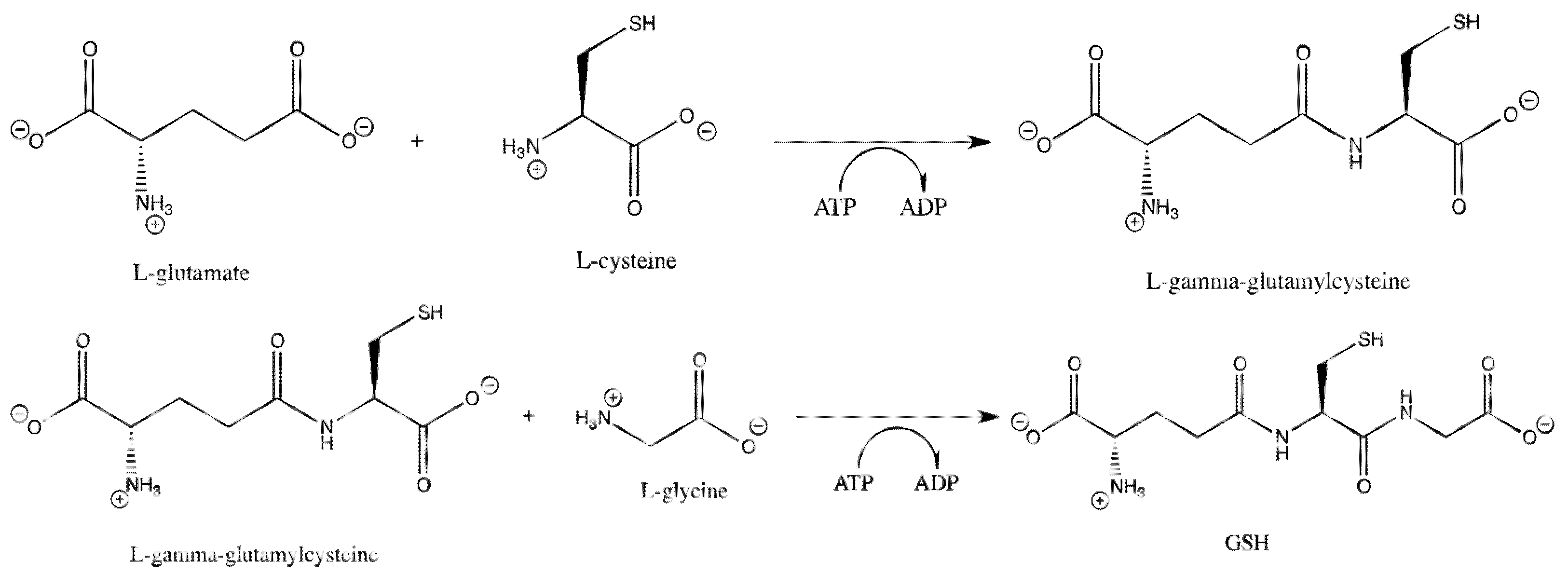
Capable of conjugating with toxic species to mark for removal, GSH is also capable of reversibly conjugating directly with amino acids in proteins to prevent irreversible damage caused by oxidative stress [14]. This tripeptide has been found to be a promising solution to cell death in the event of injury [15]. Glutathione is produced in a two-step process (Figure 2), where the first step is rate-limiting.
Once glutathione has reacted with a harmful species, it is then converted to its oxidized form (GSSG), which requires reduction before becoming available for neutralization. Once the interaction between glutathione and peroxide takes place, glutathione peroxidase will neutralize the peroxide (Figure 3).
Once the peroxide is neutralized, the remaining glutathione is oxidized and requires reduction by glutathione reductase (Figure 4).
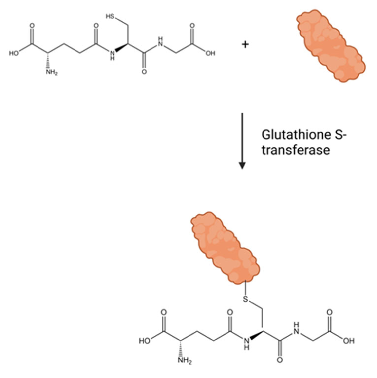
Oxidized glutathione combined with NADPH+ in a catalyzed reaction via glutathione reductase results in reduced glutathione, which is readily available for further protec-tion. Research conducted with the chemotherapeutic drug adriamycin has shown that glutathione peroxidase (GPx) activity increased, while the activity of glutathione reductase (GR) decreased when oxidants were introduced to the brain [16]. The antioxidants that normally function to neutralize these damaging species become oxidized and in turn lose functionality, contributing to increased secondary damage to the brain after a traumatic brain injury. Glutathione S-transferase uses glutathione in conjugation reactions to protect proteins from further oxidation, as illustrated in Figure 5.
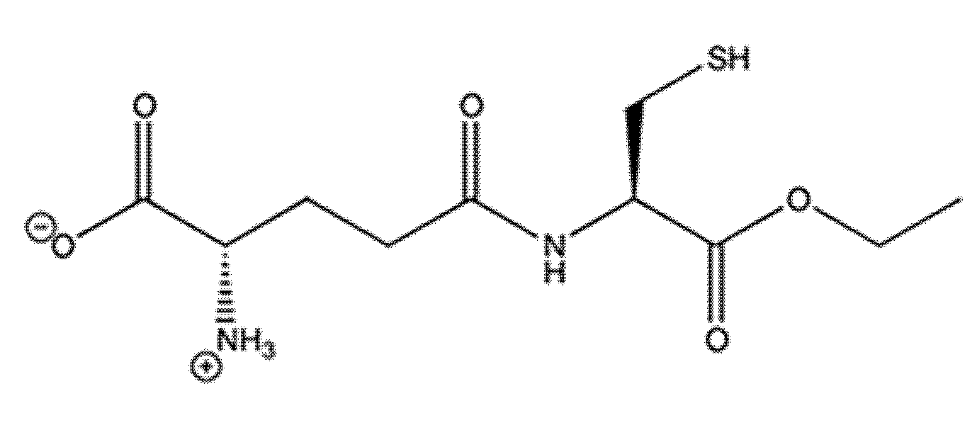
The glutathione mimetic, gamma-glutamylcysteine ethyl ester (GCEE), as depicted in Figure 6, is the amino acid sequence gamma-glutamylcysteine and will produce gluta-thione through the addition of glycine by glutathione synthetase (Figure 7).
The movement of gamma-glutamylcysteine across the blood–brain barrier requires the use of a polar moiety, provided by an ethyl ester, thereby making it a plausible treatment in brain-related injuries [17]. This allows uptake into the brain, promoting the formation of glutathione. When this intermediate molecule, GCEE, is present, there will be an upregulation of glutathione in the brain. This upregulation will then provide increased antioxidant capacity and relief from increased oxidative stress. Cellular cysteine is in minute quantities inadequate for the formation of glutathione. When GCEE is administered, the rate-limiting step in the formation of glutathione is bypassed. Various free radical scavengers including edaravone and tempol are neuroprotective in several TBI animal models [18,19]. Many of these agents are administered pre TBI, in which a drug is administered before an injury is given. In contrast, we investigated the use of GCEE 30 and 60 min post injury to investigate their protection against oxidative stress. Antioxidant treatments including L-theanine, curcumin, and quercetin [20,21,22] and current medications including nicorandil, oridonin, imipramine, valproate, and flumazenil have shown cognitive benefit when administered post injury, making this study essential in the investigation of post-TBI therapeutic strategies [23,24,25,26,27]. The focus of this research effort is to investigate the enzymatic activity of glutathione reductase (GR) and glutathione peroxidase (GPx), two enzymes responsible for protecting the brain in the event of a moderate TBI. These enzymes work synergistically to allow glutathione to continuously protect the brain when injury is present. We hypothesize that rats given GCEE after a moderate traumatic brain injury would demonstrate an increase in activity of these glutathione-related neuroprotective enzymes, thereby allowing the system to better combat damaging effects caused by TBI such as oxidative stress and protein dysfunction.

2. Materials and Methods

All chemicals, unless indicated, were obtained from Sigma Aldrich (St. Louis, MO, USA) and were of the highest purity. GCEE was purchased from Bachem (Torrance, CA, USA). All water used in this procedure was distilled and deionized. Brain tissue samples were stored at −80 °C until used. Once homogenized in a buffer solution, samples were stored at −20 °C until used.

2.1. Surgical Methods

The surgical procedures and drug administration protocols described below are in compliance with and have been approved by the University of Kentucky Institutional Animal Care and Use Committee and are consistent with all animal care procedures established by the U.S. Public Health and Service Policy on Humane Care and Use of Laboratory Animals.
In this study, 30 Wistar adult male rats were used, each weighing 300–350 g. Isoflurane (3.0%) was used to anesthetize the animals. The rats were shaved and then placed in a stereotaxic frame (David Kopf Instruments, Tujunga, CA, USA). A craniotomy and a 1.5 mm contusion was introduced for the moderate traumatic brain as described by Sullivan [28]. Sham animals were excluded from the injury but received a craniotomy (n = 6). Post craniotomy, each Wistar rat had a 4 mm dental cement seal placed on the craniotomy site. The rats were then placed on warming mats to prevent hypothermia until they regained consciousness. The rats were given 150 mg/kg body weight of GCEE 30 or 60 min post injury (n = 6). Injections were given intraperitoneally based on prior research in which improvement with post-neurosteroidal treatment was observed [17,29]. Another group was given saline at 30 or 60 min post injury, with a concentration of 150 mg/kg body weight (n = 6). Concentrations for GCEE and saline were determined from previous studies [30]. All rats were then sacrificed 24 h post injury with immediate removal and freezing of the whole brain at −80 °C. The time period of 24 h was chosen due to having maximum decrease in glutathione levels as well as enzymatic activity [31]. Brain samples were then sonicated and placed in 10 mM HEPES buffer (pH 7.4) containing 137 mM NaCl, 4.6 mM KCl, 1.1 mM KH2PO4, 0.1 mM EDTA, and 0.6 mM MgSO4 along with protease inhibitors: leupeptin (0.5 mg/mL), pepstatin (0.7 µg/mL), type II S soybean trypsin inhibitor (0.5 µg/mL), and phenylmethylsulfonyl fluoride (PMSF; 40 µg/mL). After homogenation, samples were then centrifuged at 14,000× g for 10 min to remove all cellular debris that could potentially cause matrix effects within the sample. Protein concentration in the supernatant was determined by the BCA protein assay (Pierce, Rockford, IL, USA). Supernatant was then decanted from the centrifuged samples and stored for future determination of enzymatic activity.

2.2. Analysis of Glutathione Peroxidase Activity

All samples were analyzed using a modified procedure from Sigma Aldrich (St. Louis, MO, USA). Reagents were combined and analyzed at room temperature for five minutes. Each sample (200 µL) was analyzed in triplicate with final concentrations in solution being 48 mM sodium phosphate, 0.38 mM ethylenediaminetetraacetic acid, 0.12 mM β-nicotinamide adenine dinucleotide phosphate (NADPH) reduced form, 0.95 mM sodium azide, 3.2 units of glutathione reductase, 1 mM glutathione, 0.02 mM DL-dithiothreitol, and 0.0007% (w/w) hydrogen peroxide. Each sample was analyzed at 340 nm for indirect evaluation of glutathione peroxidase activity by direct measurement of NADPH concentration. This absorbance represents the ability of glutathione peroxidase to detoxify H2O2.

2.3. Analysis of Glutathione Reductase Activity

All samples were analyzed using a modified procedure from Sigma Aldrich (St. Louis, MO, USA). Reagents were combined and analyzed for five minutes at room temperature. Each sample (200 µL) was analyzed in triplicate with final concentrations in solution being 75 mM potassium phosphate, 2.6 mM ethylenediaminetetraacetic acid, 1 mM glutathione, 0.09 mM β-nicotinamide adenine dinucleotide phosphate (NADPH), reduced form, and 0.13% (w/v) bovine serum albumin. Each sample was analyzed at 340 nm for indirect evaluation of glutathione reductase activity by direct measurement of NADPH concentration. This absorbance represents the ability of glutathione reductase to produce reduced glutathione.

2.4. Gene Expression Analysis Sample Preparation

All materials/reagents used were RNAse/nuclease free. Fresh brain tissue was stored at −80 °C and weighed approximately 100 mg per sample and was soaked in RNAlater solution (Life technologies, Carlsbad, CA, USA) for at least 30 min on ice. Brain tissue was then placed in 1.5 mL tubes with lysing matrix D and 1 mL of trizol per 100 mg of sample. The tubes were then processed in a FastPrep-24 instrument for 45 s at a high setting for six cycles and placed on ice for one minute between cycles to prevent sample overheating. Homogenate was then transferred to a new tube by pipette, avoiding the transfer of any matrix beads. This was shaken for five minutes at room temperature. Tubes were then centrifuged at 12,000× g for 10 min at 4 °C to begin liquid-liquid extraction. The upper aqueous layer (approximately 750 μL, without any cell debris) was removed and transferred to a new tube. Chloroform (200 μL) was added and shaken for 15 s then incubated at room temperature for 2–3 min. Tubes were centrifuged at 12,000× g for 15 min at 4 °C. The upper aqueous layer was transferred to a new tube. Cold isopropanol (300 µL) was added and mixed by inversion five times. This solution was then stored at −20 °C for 30 min. Tubes were then centrifuged at 12,000× g for 10 min at 4 °C. Supernatant was removed, and pellets were washed with 1 mL of cold 75% ethanol. The pellets were vortexed back into solution and centrifuged at 7500× g for 10 min at 4 °C. Ethanol was removed and the pellet was air dried for five minutes at room temperature. It was ensured that pellet did not completely dry. The pellet was then resuspended in 100 μL of water and stored at −80 °C until further use.

2.5. RNA Purity and Concentration Determination

RNA samples were vortexed, and it was ensured that no precipitate was present. If precipitate was present, it was removed using centrifugation. A NanoDrop spectrophotometer was used to determine the concentration (ng/μL) and purity (260/280 abs) of samples. A ratio of 2 for the 260/280 abs represents a purer sample.

2.6. Converting RNA to cDNA

A high-capacity cDNA kit (Applied Biosystems, Foster City, CA, USA) was used. Reaction for the conversion of RNA to cDNA was performed in 0.2 mL PCR tubes with supplies from the kit (10% 10X RT buffer, 4% 25X dNTP mix (100 mM), 10% 10X RT random primers, 5% multiscribe reverse transcriptase, 5% RNAse inhibitor), as well as 66% RNA sample and water (containing 1 μg of RNA) for a total of 20 μL in each PCR tube. The conversion of RNA to cDNA allows for a more stable sample to be used during TaqMan PCR. Each PCR tube was mixed by plunging with a pipette and centrifuged a few seconds to remove any air bubbles. A PCR protocol was then performed (annealing at 25 °C for 10 min, elongation at 37 °C for 120 min, final elongation at 85 °C for five seconds, hold at 4 °C).

2.7. TaqMan PCR (qRT-PCR)

TaqMan wells were prepared using 1 μL of 20X TaqMan gene expression assay solution, 10 μL of 2X gene expression master mix, and 8 μL of RNAse free water. TaqMan gene expression assay solutions used included glutathione peroxidase 1 (GPX1, 77 amplicon length), glutathione reductase (GSR, 64 amplicon length), glutathione S-transferase theta 1 (GSTT1, 83 amplicon length), and glyceraldehyde-3-phosphate dehydrogenase (GAPDH, 174 amplicon length) used as a control (Life technologies, Carlsbad, CA, USA). This was mixed pipetting and centrifugation. Lastly, 1 μL of cDNA sample was added to each designated well. A standard TaqMan PCR program was immediately performed.

2.8. Statistical Analysis

All statistical analyses were performed using ANOVA testing. Alpha was set to 0.05. Grubb’s test was used to identify outliers. Error bars were calculated as SEM. Significance of each result was confirmed by calculation of the p-value by two-tailed Student’s t-test (p-values < 0.05).

3. Results

3.1. Enzymatic Analysis

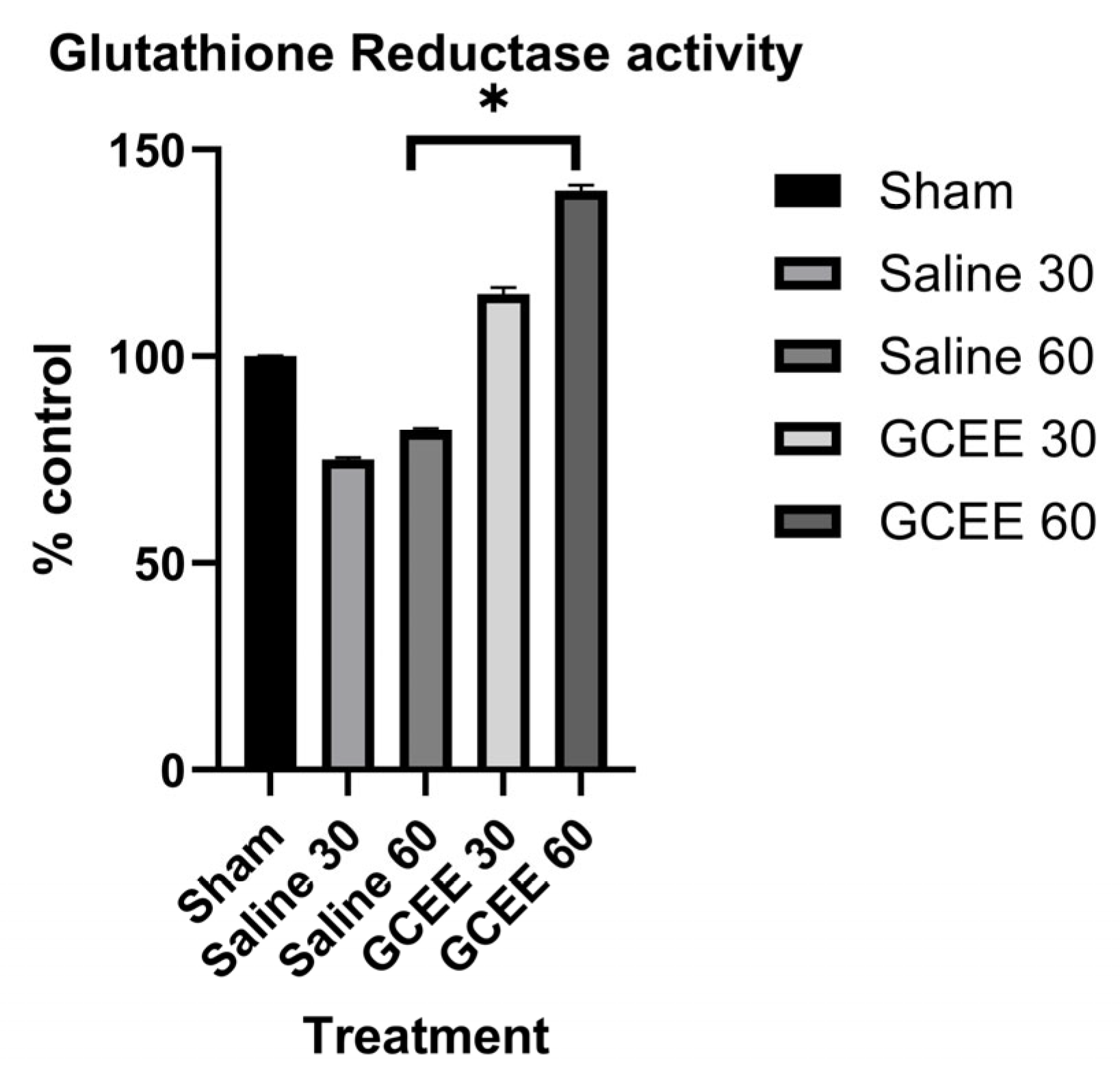
Upon evaluation of data for glutathione reductase, the activity of glutathione reductase (Figure 8) increased in the GCEE samples when compared to saline samples at 30 min by 15%; however, this increase was not deemed statistically significant by ANOVA or Student’s t-test. A significant increase (40%) is shown at the 60 min time interval (p-value < 0.05) for glutathione reductase activity in GCEE-treated samples when compared to those treated with saline.
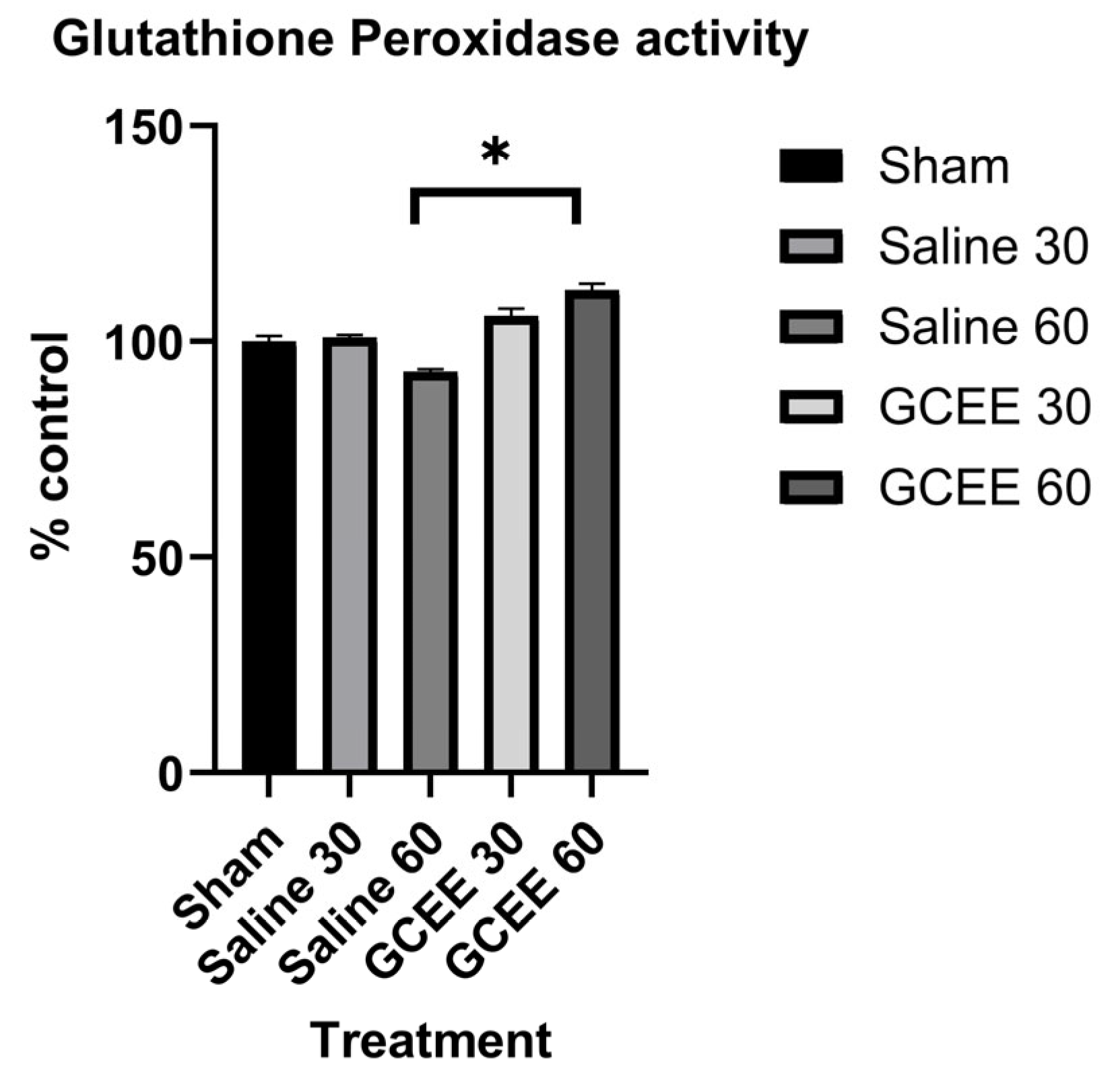
Data for glutathione peroxidase activity (Figure 9) show a small increase in activity (6%) in GCEE-treated samples when compared to those treated with saline at 30 min post traumatic brain injury; however, this was not found to be statistically significant. Enzymatic activity at 60 min shows a statistically significant 12% increase in activity in GCEE-treated samples compared to saline-treated samples (p-value < 0.001).

3.2. Gene Expression Analysis

To further bolster the importance of glutathione in relation to traumatic brain injury, gene expressions of glutathione peroxidase 1 (GPX1), glutathione S-transferase theta 1 (GSTT1), and glutathione reductase (GR) were analyzed. Only the sham and 30 min time post treatment samples were used for this evaluation. These are key enzymes involved in glutathione metabolism. GPX1 is capable of reducing radical species by oxidizing glutathione to form glutathione disulfide (GSSG). GSTT1 conjugates glutathione to proteins, a reversible reaction that can protect proteins from permanent oxidative stress. Glutathione reductase reduces oxidized glutathione back to a usable glutathione form. This gene expression analysis was performed to determine how mRNA production of these enzymes is altered following brain injury and GCEE treatments. The gene expressions for the glutathione-related genes GPX1, GSTT1, and GR have not be evaluated using a moderate traumatic injury model and treatment with GCEE, making these findings novel (Figure 10). Results from this analysis (Table 1) demonstrate that mRNA expression of GPX1 and GR is significantly greater in saline-treated samples compared to sham and GCEE-treated samples. Previous studies have found glutathione peroxidase to have a twofold increase following a cortical contusion model [32]. Other glutathione peroxidases such as GPX3 have been recently shown as pivotal hub genes in traumatic brain injury as their expression is significantly altered [33], while glutathione peroxidase 4 (GPX4) protects cells from ferroptosis, programmed iron-dependent cell death facilitated by lipid peroxidation. The mechanisms of ferroptosis in traumatic brain injury are not well understood, but they could explain other findings regarding lipid peroxidation and moderate traumatic brain injury [34,35]. This upregulation of GPX1 and GR suggests that these enzymes are essential to brain-injured samples, as these enzymes are key to combating oxidative stress. GCEE-treated samples also showed significant increase in GPX1 and GR mRNA expression when compared to sham samples, further supporting the importance of these enzymes. If glutathione-metabolizing enzymes are being upregulated, it is likely that an increase in glutathione production would be beneficial, as glutathione is essential for these enzymes to function. This suggests that GCEE may be a viable post-therapeutic treatment for TBI against ROS/RNS that cause oxidative stress.

4. Discussion

There are approximately 60 million people affected worldwide with traumatic brain injury annually with the estimated treatment costs in the billion [36,37]. These numbers do not include the high incidence of those serving in the armed forces, whose risk of suffering a TBI is high [38,39]. Males between the ages of 15 and 24 years have the highest incidence of TBI [40]. Sudden brain trauma is described as TBI. There is no known cure, but immediate medical care after injury is most advantageous for patient recovery. Unfortunately, the number of Level I and Level II trauma centers that contain suitable neurotrauma units that can adequately accept, treat, and rehabilitate those with moderate to severe traumatic brain injuries is limited globally [41]; therefore, it is imperative that an appropriate pharmacological intervention to moderate TBI be developed. ROS production occurs in TBI, as can be observed by increased protein nitration and protein carbonyls [42,43]. Ansari et al. demonstrated that oxidative stress occurs within 3 hr post-injury [31]. Currently, several drugs show promise in treating TBI, including scriptaid, a histone deacetylase inhibitor [44]; dexmedetomidine, an α2- adrenoreceptor agonist [45]; pycnogenol, a novel bioflavonoid [46]; and edaravone, a free radical scavenger [47]. Imiprine, a tricyclic antidepressant [24]; valproate, an antiepileptic [23]; sulforaphane, a detoxification agent and free radical scavenger [48]; flumazenil, a benzodiazepine antagonist [25]; nicorandil, an ATP-sensitive potassium channel opener with anti-inflammatory and neuroprotective activities [26]; and oridonin, a NLRP3-selective inhibitor with anti-inflammatory properties [27], have shown cognitive improvement when administered post-injury. These treatments do not have any mechanistic commonalities, but they all work to reduce post-TBI cognitive deficits. GCEE, an ester moiety of the dipeptide gamma-glutamylcysteine, is a vital antioxidant that can easily cross the plasma membrane and up-regulate GSH in the brain [49]. None of the current post therapeutic treatment strategies involve increasing antioxidant capacity. GCEE prevents oxidative stress induced by amyloid β peptide and other moieties by scavenging and free radicals [30].
This work indicates that GCEE is a viable treatment for moderate traumatic brain injury. The synergistic effects of the antioxidant enzymes are of utmost importance in maintaining a healthy antioxidant-oxidant ratio. These enzymes work concomitantly to relieve the oxidative stress and possibly halt the cycle of its formation. When GCEE is administered, it is quickly converted to the protective antioxidant glutathione by way of glutathione synthetase [17]. Once glutathione is created in its reduced form, it is readily available to alleviate reactive species in the event of traumatic brain injury. Glutathione peroxidase accomplishes this mechanism of alleviation. This enzyme can conjugate reduced glutathione to cause the neutralization of reactive peroxide species and renders glutathione in the inactive oxidized form [12]. The recycling of this oxidized glutathione is accomplished by glutathione reductase. The beta-oxidation pathway from the reaction catalyzed by glucose-6-phosphate dehydrogenase provides the electron donor, NADPH [12]. Once NADPH is available, glutathione reductase can readily reduce the oxidized form of glutathione to allow further negation of reactive species. The enzyme glutathione S-transferase is directly responsible for the neutralization of ROS and RNS in a similar mechanism to that of glutathione peroxidase, which also forms the oxidized glutathione. GPx has been shown to have a significant decreased activity in TBI in the ipsilateral hippocampus of rats at 24 h post injury [31]. Therefore, as GPx plays a role in peroxide neutralization, it is clear why there is no significant change in the activity of glutathione peroxidase between saline- and GCEE-treated samples at the 30 min timepoint but a more significant increase at a longer time point. The increase in glutathione reductase levels in GCEE-treated samples supports the reduction of oxidative stress via protein carbonyls and 3-nitrotyrosine by increasing antioxidant defense, as shown by Reed et al. [34]. This study suggests that GCEE administration 30 and 60 min post-TBI demonstrates an increase in the antioxidant enzyme glutathione reductase, which plays a role in antioxidant defense, thereby decreasing levels of oxidative stress. Due to the significant roles that glutathione peroxidase and glutathione reductase play in detoxifying reactive oxygen species, increased activity could lead to decreased oxidative stress, thus decreasing the likelihood of debilitating secondary effects. Increased gene expression of glutathione metabolizing enzymes following TBI shows that glutathione plays an essential role in brain injury. These results illustrate that expression of these enzymes maintains a significant role with GCEE treatments, meaning that components of a native antioxidant mechanism involving glutathione are being produced, possibly to take advantage of an increase in glutathione.

5. Conclusions

Previous research to increase glutathione levels shows that it can be ineffective if it is not altered to allow crossing of the blood–brain barrier [50,51]. The ethyl ester moiety that is present in GCEE allows it to passively cross the blood–brain barrier due to the increased polarity of the molecule, thus increasing permeability [52]. When GCEE is readily available, the rate-limiting step is bypassed; therefore, glutathione levels increase, which could provide a potentially viable treatment for TBI. The time period available for treatment is vital and possibly limited to a specific time frame. The research presented focuses on the time periods of 30 and 60 min. Shorter time frames have been assessed in previous literature [35]. Determination of an optimal time frame at which the treatment is most advantageous is crucial for development of a possible therapeutic for TBI. This study shows that GCEE administration at times of 30 min and 60 min post-injury can increase enzyme activity of metabolic enzymes, suggesting that GCEE can lead to improved protection against ROS and RNS that cause cellular damage. Therefore, GCEE offers promise as a potential post-injury therapeutic for TBI.

Author Contributions

Conceptualization, T.T.R.; methodology, T.T.R., A.S. and P.G.S.; software, T.T.R., J.O. and J.T.J.; validation, M.H., A.K., J.T.J., J.O., Z.P.S. and R.W.; formal analysis, A.K., J.T.J., J.O. and Z.P.S.; investigation, A.K., J.T.J., J.O., T.T.R., P.G.S. and Z.P.S.; resources, T.T.R. and P.G.S.; data curation, A.K., J.T.J., J.O. and Z.P.S.; writing—original draft preparation, A.K., J.T.J. and J.O.; writing—review and editing, T.T.R.; visualization, A.K., J.T.J., J.O. and T.T.R.; supervision, T.T.R.; project administration, T.T.R.; funding acquisition, T.T.R. All authors have read and agreed to the published version of the manuscript.

Funding

This work was supported by National Institutes of Health grant (1R15NS072870-01A1).

Institutional Review Board Statement

The surgical procedures and drug administration proto-cols described below are in compliance with and have been approved by the University of Kentucky Institutional Animal Care and Use Committee and are consistent with all animal care procedures established by the U.S. Public Health and Service Policy on Humane Care and Use of Laboratory Animals.

Informed Consent Statement

Not applicable.

Data Availability Statement

The raw data supporting the conclusions of this article will be made available by the authors on request.

Acknowledgments

This work was supported by National Institutes of Health and the Department of Chemistry, Eastern Kentucky University. All surgical methods were performed at the University of Kentucky Spinal Cord & Brain Injury Research Center in Patrick Sullivan’s lab with assistance from Andrea Sebastian.

Conflicts of Interest

The authors declare no conflicts of interest.

Abbreviations

The following abbreviations are used in this manuscript:
GAPDHglyceraldehyde-3-phosphate dehydrogenase
GCEEgamma-glutamylcysteine ethyl ester
GPxglutathione peroxidase
GPX1glutathione peroxidase 1
GPX4glutathione peroxidase 4
GR/GSRglutathione reductase
GSHglutathione
GSSGglutathione disulfide
GSTT1glutathione S-transferase 1
ROSreactive oxygen species
RNSreactive nitrogen species
TBItraumatic brain injury

References

  1. Sellers, Z.P.; Williams, R.A.; Overbay, J.W.; Cho, J.; Henderson, M.; Reed, T.T. Current Therapeutic Modalities, Enzyme Kinetics, and Redox Proteomics in Traumatic Brain Injury. In Traumatic Brain Injury; InTechOpen: London, UK, 2014; pp. 39–77. [Google Scholar]
  2. Stelfa, G.; Svalbe, B.; Vavers, E.; Duritis, I.; Dambrova, M.; Zvejniece, L. Moderate traumatic brain injury triggers long-term risks for the development of peripheral pain sensitivity and depressive-like behavior in mice. Front. Neurol. 2022, 13, 985895. [Google Scholar] [CrossRef] [PubMed]
  3. Lannsjo, M.; Borg, J.; Lewen, A.; von Seth, C.; Enblad, P.; Abu Hamdeh, S. Long-Term Outcomes of Moderate to Severe Diffuse Axonal Traumatic Brain Injury: A Prospective Study. J. Rehabil. Med. Clin. Commun. 2025, 8, 42299. [Google Scholar] [CrossRef] [PubMed]
  4. Haynes, A.; Johnson, L.; Ashpole, R.; Mamo, A.; Chagpar, S.; Williams, G.; Clanchy, K.; Waters, N.; Vassallo, G.; Scheinberg, A.; et al. The long road back to physical activity: The experience of people with moderate-to-severe traumatic brain injury. Disabil. Rehabil. 2025, 1–12. [Google Scholar] [CrossRef] [PubMed]
  5. Bigler, E.D. Distinguished Neuropsychologist Award Lecture 1999. The lesion(s) in traumatic brain injury: Implications for clinical neuropsychology. Arch. Clin. Neuropsychol. 2001, 16, 95–131. [Google Scholar] [CrossRef]
  6. Weber, J.T. Altered calcium signaling following traumatic brain injury. Front. Pharmacol. 2012, 3, 60. [Google Scholar] [CrossRef]
  7. Surgucheva, I.; He, S.; Rich, M.C.; Sharma, R.; Ninkina, N.N.; Stahel, P.F.; Surguchov, A. Role of synucleins in traumatic brain injury—An experimental in vitro and in vivo study in mice. Mol. Cell Neurosci. 2014, 63, 114–123. [Google Scholar] [CrossRef]
  8. Butterfield, D.A.; Sultana, R. Identification of 3-Nitrotyosine Modified Brain Proteins byRedox Proteomics. In Methods in Enzymology; Elsevier: Amsterdam, The Netherlands, 2008; Volume 440, pp. 295–308. [Google Scholar]
  9. Dai, W.; Cheng, H.L.; Huang, R.Q.; Zhuang, Z.; Shi, J.X. Quantitative detection of the expression of mitochondrial cytochrome c oxidase subunits mRNA in the cerebral cortex after experimental traumatic brain injury. Brain Res. 2009, 1251, 287–295. [Google Scholar] [CrossRef]
  10. Hall, E.D.; Vaishnav, R.A.; Mustafa, A.G. Antioxidant therapies for traumatic brain injury. Neurotherapeutics 2010, 7, 51–61. [Google Scholar] [CrossRef]
  11. Wu, D.; Cederbaum, A.I. Alcohol, oxidative stress, and free radical damage. Alcohol. Res. Health 2003, 27, 277–284. [Google Scholar]
  12. Ansari, M.A.; Roberts, K.N.; Scheff, S.W. Oxidative stress and modification of synaptic proteins in hippocampus after traumatic brain injury. Free. Radic. Biol. Med. 2008, 45, 443–452. [Google Scholar] [CrossRef]
  13. Karve, I.P.; Taylor, J.M.; Crack, P.J. The contribution of astrocytes and microglia to traumatic brain injury. Br. J. Pharmacol. 2016, 173, 692–702. [Google Scholar] [CrossRef] [PubMed]
  14. Pompella, A.; Visvikis, A.; Paolicchi, A.; De Tata, V.; Casini, A.F. The changing faces of glutathione, a cellular protagonist. Biochem. Pharmacol. 2003, 66, 1499–1503. [Google Scholar] [CrossRef] [PubMed]
  15. Pocernich, C.B.; Cardin, A.L.; Racine, C.L.; Lauderback, C.M.; Butterfield, D.A. Glutathione elevation and its protective role in acrolein-induced protein damage in synaptosomal membranes: Relevance to brain lipid peroxidation in neurodegenerative disease. Neurochem. Int. 2001, 39, 141–149. [Google Scholar] [CrossRef]
  16. Joshi, G.; Aluise, C.D.; Cole, M.P.; Sultana, R.; Pierce, W.M.; Vore, M.; St Clair, D.K.; Butterfield, D.A. Alterations in brain antioxidant enzymes and redox proteomic identification of oxidized brain proteins induced by the anti-cancer drug adriamycin: Implications for oxidative stress-mediated chemobrain. Neuroscience 2010, 166, 796–807. [Google Scholar] [CrossRef]
  17. Lok, J.; Leung, W.; Zhao, S.; Pallast, S.; van Leyen, K.; Guo, S.; Wang, X.; Yalcin, A.; Lo, E.H. gamma-glutamylcysteine ethyl ester protects cerebral endothelial cells during injury and decreases blood-brain barrier permeability after experimental brain trauma. J. Neurochem. 2011, 118, 248–255. [Google Scholar] [CrossRef] [PubMed]
  18. Itoh, T.; Satou, T.; Nishida, S.; Tsubaki, M.; Hashimoto, S.; Ito, H. The novel free radical scavenger, edaravone, increases neural stem cell number around the area of damage following rat traumatic brain injury. Neurotox. Res. 2009, 16, 378–389. [Google Scholar] [CrossRef]
  19. Singh, I.N.; Sullivan, P.G.; Hall, E.D. Peroxynitrite-mediated oxidative damage to brain mitochondria: Protective effects of peroxynitrite scavengers. J. Neurosci. Res. 2007, 85, 2216–2223. [Google Scholar] [CrossRef]
  20. Mohan, M.C.; Anjana, A.S.; Hilmi Jaufer, T.A.; Deepti, A.; Krishnakumar, I.M.; Baby Chakrapani, P.S. Co-delivery of curcumin-resveratrol-carnosic acid complex promotes neurogenesis and cognitive recovery in a rodent model of repeated mild traumatic brain injury. Biomed. Pharmacother. 2025, 183, 117818. [Google Scholar] [CrossRef]
  21. Park, M.K.; Choi, B.Y.; Kho, A.R.; Lee, S.H.; Hong, D.K.; Kang, B.S.; Lee, C.J.; Yang, H.W.; Woo, S.Y.; Park, S.W.; et al. L-theanine ameliorates traumatic-brain-injury-induced hippocampal neuronal death in rats. Phytomedicine 2025, 139, 156457. [Google Scholar] [CrossRef]
  22. Tanhai, G.; Chahardehi, A.M.; Sohrabi, M.A.; Afshoon, M.; Saberian, P.; Pourshams, M.; Ghasemi, D.; Motaghi, S.M.; Arefnezhad, R.; Niknam, Z. Ameliorative properties of quercetin in the treatment of traumatic brain injury: A mechanistic review based on underlying mechanisms. Mol. Biol. Rep. 2024, 51, 695. [Google Scholar] [CrossRef]
  23. Dash, P.K.; Orsi, S.A.; Zhang, M.; Grill, R.J.; Pati, S.; Zhao, J.; Moore, A.N. Valproate administered after traumatic brain injury provides neuroprotection and improves cognitive function in rats. PLoS ONE 2010, 5, e11383. [Google Scholar] [CrossRef] [PubMed]
  24. Han, X.; Tong, J.; Zhang, J.; Farahvar, A.; Wang, E.; Yang, J.; Samadani, U.; Smith, D.H.; Huang, J.H. Imipramine treatment improves cognitive outcome associated with enhanced hippocampal neurogenesis after traumatic brain injury in mice. J. Neurotrauma 2011, 28, 995–1007. [Google Scholar] [CrossRef]
  25. Ochalski, P.G.; Fellows-Mayle, W.; Hsieh, L.B.; Srinivas, R.; Okonkwo, D.O.; Dixon, C.E.; Adelson, P.D. Flumazenil administration attenuates cognitive impairment in immature rats after controlled cortical impact. J. Neurotrauma 2010, 27, 647–651. [Google Scholar] [CrossRef]
  26. Tu, Y.; Han, D.; Liu, Y.; Hong, D.; Chen, R. Nicorandil attenuates cognitive impairment after traumatic brain injury via inhibiting oxidative stress and inflammation: Involvement of BDNF and NGF. Brain Behav. 2024, 14, e3356. [Google Scholar] [CrossRef]
  27. Zhao, X.J.; Zhu, H.Y.; Wang, X.L.; Lu, X.W.; Pan, C.L.; Xu, L.; Liu, X.; Xu, N.; Zhang, Z.Y. Oridonin Ameliorates Traumatic Brain Injury-Induced Neurological Damage by Improving Mitochondrial Function and Antioxidant Capacity and Suppressing Neuroinflammation through the Nrf2 Pathway. J. Neurotrauma 2022, 39, 530–543. [Google Scholar] [CrossRef] [PubMed]
  28. Sullivan, P.G.; Keller, J.N.; Bussen, W.L.; Scheff, S.W. Cytochrome c release and caspase activation after traumatic brain injury. Brain Res. 2002, 949, 88–96. [Google Scholar] [CrossRef] [PubMed]
  29. Djebaili, M.; Guo, Q.; Pettus, E.H.; Hoffman, S.W.; Stein, D.G. The neurosteroids progesterone and allopregnanolone reduce cell death, gliosis, and functional deficits after traumatic brain injury in rats. J. Neurotrauma 2005, 22, 106–118. [Google Scholar] [CrossRef]
  30. Joshi, G.; Hardas, S.; Sultana, R.; St Clair, D.K.; Vore, M.; Butterfield, D.A. Glutathione elevation by gamma-glutamyl cysteine ethyl ester as a potential therapeutic strategy for preventing oxidative stress in brain mediated by in vivo administration of adriamycin: Implication for chemobrain. J. Neurosci. Res. 2007, 85, 497–503. [Google Scholar] [CrossRef]
  31. Ansari, M.A.; Roberts, K.N.; Scheff, S.W. A time course of contusion-induced oxidative stress and synaptic proteins in cortex in a rat model of TBI. J. Neurotrauma 2008, 25, 513–526. [Google Scholar] [CrossRef]
  32. Goss, J.R.; Taffe, K.M.; Kochanek, P.M.; DeKosky, S.T. The antioxidant enzymes glutathione peroxidase and catalase increase following traumatic brain injury in the rat. Exp. Neurol. 1997, 146, 291–294. [Google Scholar] [CrossRef]
  33. Wang, Y.; Fang, J.; Yuan, Q.; Yu, J.; Hu, J. GPX3 as a Novel and Potential Therapeutic Target in the Shared Molecular Mechanisms of Traumatic Brain Injury and Parkinson’s Disease. J. Inflamm. Res. 2025, 18, 1911–1928. [Google Scholar] [CrossRef] [PubMed]
  34. Henderson, M.; Rice, B.; Sebastian, A.; Sullivan, P.G.; King, C.; Robinson, R.A.; Reed, T.T. Neuroproteomic study of nitrated proteins in moderate traumatic brain injured rats treated with gamma glutamyl cysteine ethyl ester administration post injury: Insight into the role of glutathione elevation in nitrosative stress. Proteom. Clin. Appl. 2016, 10, 1218–1224. [Google Scholar] [CrossRef]
  35. Reed, T.T.; Owen, J.; Pierce, W.M.; Sebastian, A.; Sullivan, P.G.; Butterfield, D.A. Proteomic identification of nitrated brain proteins in traumatic brain-injured rats treated postinjury with gamma-glutamylcysteine ethyl ester: Insights into the role of elevation of glutathione as a potential therapeutic strategy for traumatic brain injury. J. Neurosci. Res. 2009, 87, 408–417. [Google Scholar] [CrossRef] [PubMed]
  36. Matney, C.; Bowman, K.; Berwick, D. (Eds.) Traumatic Brain Injury: A Roadmap for Accelerating Progress; The National Academies Press: Washington, DC, USA, 2022. [Google Scholar]
  37. Dismuke-Greer, C.E.; Esmaeili, A.; Pugh, M.J.; Pogoda, T.K.; Amuan, M.; Cifu, D.X. Association of Clinical TBI Severity and Military Factors With Veteran TBI Service-Connected Disability Ratings and Total Compensation: A Long-Term Impact of Military Brain Injury Consortium (LIMBIC) Study. J. Head Trauma Rehabil. 2025; online ahead of print. [Google Scholar] [CrossRef]
  38. Clark, D.; Joannides, A.; Adeleye, A.O.; Bajamal, A.H.; Bashford, T.; Biluts, H.; Budohoski, K.; Ercole, A.; Fernandez-Mendez, R.; Figaji, A.; et al. Casemix, management, and mortality of patients rreseceiving emergency neurosurgery for traumatic brain injury in the Global Neurotrauma Outcomes Study: A prospective observational cohort study. Lancet Neurol. 2022, 21, 438–449. [Google Scholar] [CrossRef]
  39. Orman, J.A.; Geyer, D.; Jones, J.; Schneider, E.B.; Grafman, J.; Pugh, M.J.; Dubose, J. Epidemiology of moderate-to-severe penetrating versus closed traumatic brain injury in the Iraq and Afghanistan wars. J. Trauma Acute Care Surg. 2012, 73, S496–S502. [Google Scholar] [CrossRef]
  40. Rutland-Brown, W.; Langlois, J.A.; Thomas, K.E.; Xi, Y.L. Incidence of traumatic brain injury in the United States, 2003. J. Head Trauma Rehabil. 2006, 21, 544–548. [Google Scholar] [CrossRef]
  41. Dasic, D.; Morgan, L.; Panezai, A.; Syrmos, N.; Ligarotti, G.K.I.; Zaed, I.; Chibbaro, S.; Khan, T.; Prisco, L.; Ganau, M. A scoping review on the challenges, improvement programs, and relevant output metrics for neurotrauma services in major trauma centers. Surg. Neurol. Int. 2022, 13, 171. [Google Scholar] [CrossRef] [PubMed]
  42. Hall, E.D.; Detloff, M.R.; Johnson, K.; Kupina, N.C. Peroxynitrite-mediated protein nitration and lipid peroxidation in a mouse model of traumatic brain injury. J. Neurotrauma 2004, 21, 9–20. [Google Scholar] [CrossRef]
  43. Opii, W.O.; Nukala, V.N.; Sultana, R.; Pandya, J.D.; Day, K.M.; Merchant, M.L.; Klein, J.B.; Sullivan, P.G.; Butterfield, D.A. Proteomic identification of oxidized mitochondrial proteins following experimental traumatic brain injury. J. Neurotrauma 2007, 24, 772–789. [Google Scholar] [CrossRef]
  44. Wang, G.; Jiang, X.; Pu, H.; Zhang, W.; An, C.; Hu, X.; Liou, A.K.; Leak, R.K.; Gao, Y.; Chen, J. Scriptaid, a Novel Histone Deacetylase Inhibitor, Protects Against Traumatic Brain Injury via Modulation of PTEN and AKT Pathway: Scriptaid Protects Against TBI via AKT. Neurotherapeutics 2013, 10, 124–142. [Google Scholar] [CrossRef] [PubMed]
  45. Schoeler, M.; Loetscher, P.D.; Rossaint, R.; Fahlenkamp, A.V.; Eberhardt, G.; Rex, S.; Weis, J.; Coburn, M. Dexmedetomidine is neuroprotective in an in vitro model for traumatic brain injury. BMC Neurol. 2012, 12, 20. [Google Scholar] [CrossRef]
  46. Scheff, S.W.; Ansari, M.A.; Roberts, K.N. Neuroprotective effect of Pycnogenol(R) following traumatic brain injury. Exp. Neurol. 2013, 239, 183–191. [Google Scholar] [CrossRef] [PubMed]
  47. Wang, G.H.; Jiang, Z.L.; Li, Y.C.; Li, X.; Shi, H.; Gao, Y.Q.; Vosler, P.S.; Chen, J. Free-radical scavenger edaravone treatment confers neuroprotection against traumatic brain injury in rats. J. Neurotrauma 2011, 28, 2123–2134. [Google Scholar] [CrossRef] [PubMed]
  48. Dash, P.K.; Zhao, J.; Orsi, S.A.; Zhang, M.; Moore, A.N. Sulforaphane improves cognitive function administered following traumatic brain injury. Neurosci. Lett. 2009, 460, 103–107. [Google Scholar] [CrossRef]
  49. Drake, J.; Kanski, J.; Varadarajan, S.; Tsoras, M.; Butterfield, D.A. Elevation of brain glutathione by gamma-glutamylcysteine ethyl ester protects against peroxynitrite-induced oxidative stress. J. Neurosci. Res. 2002, 68, 776–784. [Google Scholar] [CrossRef]
  50. Aoyama, K. Glutathione in the Brain. Int. J. Mol. Sci. 2021, 22, 5010. [Google Scholar] [CrossRef]
  51. Cornford, E.M.; Braun, L.D.; Crane, P.D.; Oldendorf, W.H. Blood-brain barrier restriction of peptides and the low uptake of enkephalins. Endocrinology 1978, 103, 1297–1303. [Google Scholar] [CrossRef]
  52. Itoh, T.; Tabuchi, M.; Mizuguchi, N.; Imano, M.; Tsubaki, M.; Nishida, S.; Hashimoto, S.; Matsuo, K.; Nakayama, T.; Ito, A.; et al. Neuroprotective effect of (-)-epigallocatechin-3-gallate in rats when administered pre- or post-traumatic brain injury. J. Neural Transm. 2012, 120, 767–783. [Google Scholar] [CrossRef]
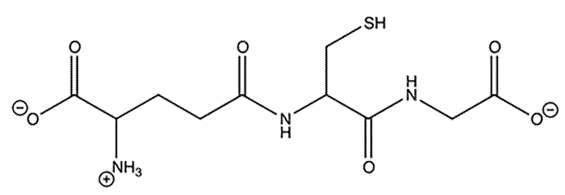
Figure 1. Structure of glutathione (pH 7.0), a tripeptide consisting of glutamate, cysteine, and glycine with a gamma linkage between the glutamate and cysteine and an alpha linkage between the cysteine and glycine.
Clinbioenerg 01 00003 g001
Figure 2. The two-step process of glutathione synthesis, with the first step being the rate-limiting step due to relatively low concentrations of cysteine available for the reaction. All amino acids are at pH 7.0.
Clinbioenerg 01 00003 g002
Figure 3. Catalytic reaction of glutathione peroxidase.
Clinbioenerg 01 00003 g003
Figure 4. Catalytic reaction of glutathione reductase.
Clinbioenerg 01 00003 g004
Figure 5. Catalytic reaction of glutathione S-transferase. Created in https://BioRender.com.
Clinbioenerg 01 00003 g005
Figure 6. Structure of γ-glutamylcysteine ethyl ester (pH 7.0), a glutathione mimetic.
Clinbioenerg 01 00003 g006
Figure 7. Catalytic reaction of glutathione synthetase.
Clinbioenerg 01 00003 g007
Figure 8. Activity of glutathione reductase at 30 and 60 min post traumatic brain injury. n = 6. Error is reported in SEM (* p-value = 0.0359).
Clinbioenerg 01 00003 g008
Figure 9. Activity of glutathione peroxidase at 30 and 60 min post traumatic brain injury. n = 6. Error is reported in SEM (* p-value <0.0001).
Clinbioenerg 01 00003 g009
Figure 10. Gene expression analysis for sham, saline at 30 minute and saline at 60 minute samples. (* p < 0.039, ** p < 0.07, n = 6).
Clinbioenerg 01 00003 g010
Table 1. Delta-Delta CT (fold increase).
TreatmentGPX1GSTT1GR
Saline to Sham 3034.968 *43.0087.198 *
GCEE to Sham 3012.943 *21.2793.479
Saline to GCEE 30 2.702 *2.021 *2.069 **
Comparison of gene expressions of various samples including sham and TBI induced with saline and GCEE treatments at 30 min post-injury in terms of GPX1, GSTT1, and GR (expressed as fold increase). * p < 0.039, ** p < 0.07, n = 6.
Disclaimer/Publisher’s Note: The statements, opinions and data contained in all publications are solely those of the individual author(s) and contributor(s) and not of MDPI and/or the editor(s). MDPI and/or the editor(s) disclaim responsibility for any injury to people or property resulting from any ideas, methods, instructions or products referred to in the content.