Article

The Trend of Long Pentraxin 3 and Other Inflammatory Serum Markers in the 30 Days After Total Hip Arthroplasty

Department of Life, Health & Environmental Sciences, University of L’Aquila, 67100 L’Aquila, Italy
*
Author to whom correspondence should be addressed.
Acta Microbiol. Hell. 2025, 70(1), 7; https://doi.org/10.3390/amh70010007
Submission received: 30 December 2024 / Revised: 28 January 2025 / Accepted: 5 February 2025 / Published: 7 February 2025
(This article belongs to the Special Issue Feature Papers in Medical Microbiology in 2024)

Abstract

:
One of the most dangerous and difficult side effects to treat after total hip arthroplasty (THA) is periprosthetic or superficial site infection. Blood and synovial fluid biomarkers have recently come into focus in addition to conventional systemic indicators of inflammation in order to assess their potential utility in the diagnosis of infections. Long pentraxin 3 (PTX3) appears to be a sensitive biomarker of acute-phase inflammation. The purpose of this study is to determine plasma PTX3 in patients undergoing THA and compare its trend with other common serum markers, such as CRP, D-dimer, procalcitonin, and ESR up to 30 days post-operatively. Patients with hip arthritis or avascular necrosis of the femoral head were consecutively enrolled in a single-center study. Each patient underwent blood testing for ESR, CRP, procalcitonin, D-dimer, and PTX3 levels before surgery and at 1, 3, 5, 15, and 30 days after THA. PTX3 was measured using the ELISA method. Other markers’ values and trends were compared with PTX3’s. A total of 50 patients met our inclusion criteria. When different trends were evaluated, PTX3 was found to have a trajectory and sensitivity comparable to other inflammatory markers. Notably, PTX3 changed more quickly than the other markers, with a sharp increase immediately post-operatively, followed by normalization at the 5-, 15-, and 30-day follow-ups, corresponding to the resolution of the inflammatory condition. However, 30 days post surgery, no patients exhibited signs or symptoms of early prosthetic infection. PTX3 is confirmed as a reliable and promising serum biomarker for tracking the level of inflammation in patients undergoing total hip replacements. Blood PTX3 values rise even more rapidly than CRP and procalcitonin and then quickly return to normal values when the inflammatory process resolves. One of the primary barriers to PTX3’s inclusion in routine studies on early periprosthetic infections is the waiting period for PTX3 sample analysis.

1. Introduction

The frequency of symptomatic hip osteoarthritis (OA) is constantly rising in a culture where life expectancy is higher and joint replacement is considered one of the most successful surgical procedures in orthopedic surgery for most patients [1].
Total hip arthroplasty (THA), although a safe procedure, is not free from complications. According to numerous published registries and large cohort studies, the onset of periprosthetic joint infections (PJIs) is the primary reason for THA revision [2], and the long-term social cost of an infected THA can range from USD 389,307 for a patient aged 65 to USD 474,004 for those aged 55 [3]. Furthermore, a recent systematic review identified PJI mortality rates after THA of 4.22% at 1 year and 21.12% at 5 years [4].
This highlights the need to prevent or at least identify early post-operative complications. Acute infections occur within 3 months of the index surgery and are characterized by joint pain, fever, redness, and heat at the surgical site [5,6]. Several guidelines for diagnosing PJI have been developed, including those from the Musculoskeletal Infection Society [7], the International Consensus Meeting on Musculoskeletal Infection [8], and the European Bone and Joint Infection Society [9]. Various tests have been identified as useful, including serum markers (CRP, ESR, D-dimer, procalcitonin, and IL-6), synovial markers (leukocytes, polymorphonuclear cells, alpha-defensin, leukocyte esterase, CRP, and IL-6), and microbiological and histological examination of periprosthetic samples [10].
Recently, a new acute-phase inflammation marker has emerged: long pentraxin 3 (PTX3) [11]. It belongs to the family of evolutionary conserved proteins known as pentraxins, which are divided into short (25 kDa) and long (40–50 kDa) proteins. Among the short pentraxins, we find C-reactive protein (CRP), while PTX3 is classified as a long pentraxin. While CRP is synthesized in the liver, PTX3 is produced at the side of infection or inflammation and can be defined as sensitive but not specific for infectious processes [12,13,14].
A small number of studies have already examined the use of PTX3 in orthopedics, assessing this marker in synovial and plasmatic samples of patients with unquestionably infected prostheses of the hip and knee and comparing them with the most extensively researched but still non-specific markers. In fact, no serum markers have been demonstrated to be sufficiently accurate in diagnosing PJIs in the hip and knee [11,13,15].
This study’s main goal is to evaluate the biomarker PTX3 plasma levels in patients undergoing primary and uncomplicated THA. Secondly, the authors wish to examine the trend of PTX3 up to 30 days post-operatively, in comparison with other common serum markers, such as CRP, D-dimer, procalcitonin, and erythrocyte sedimentation rate (ESR).

2. Materials and Methods
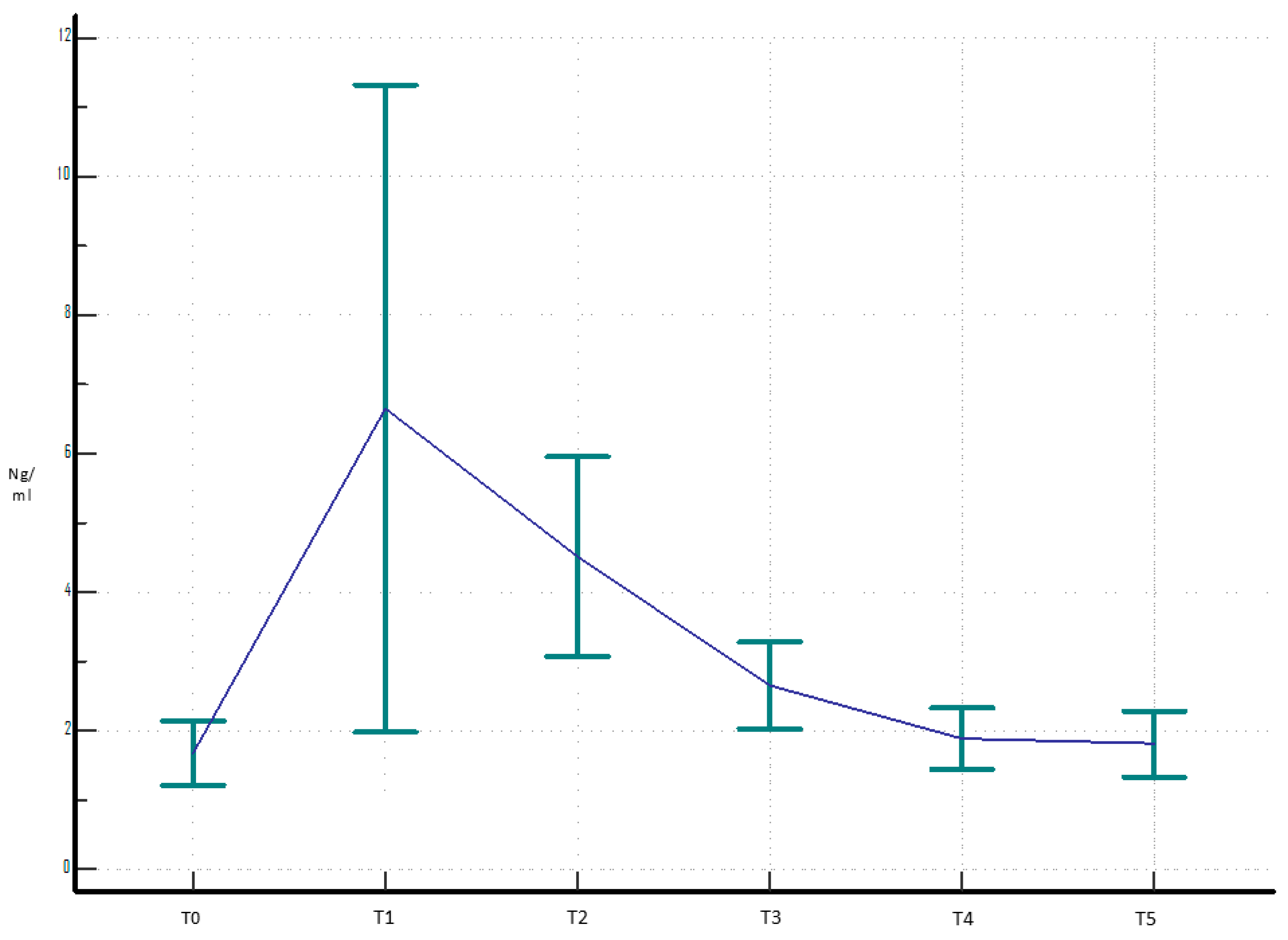
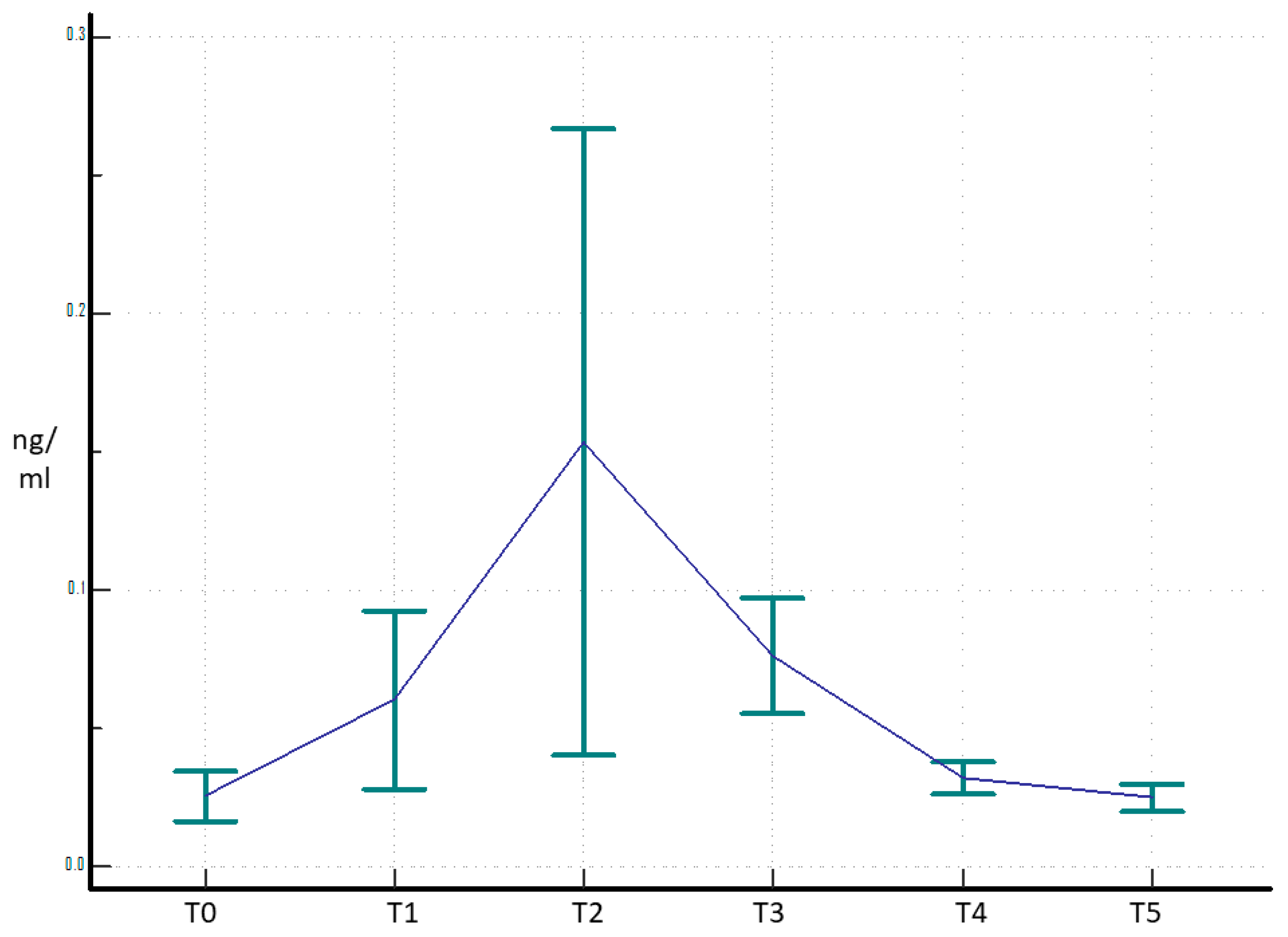
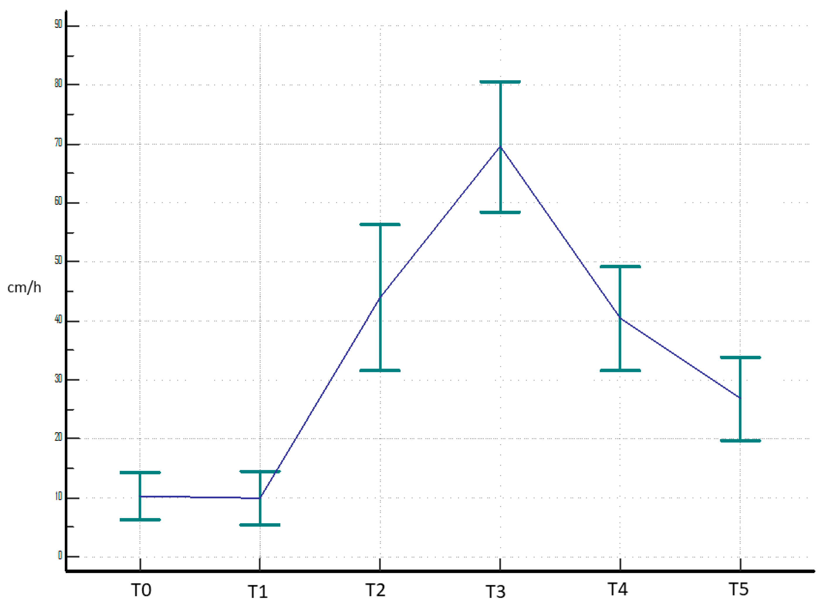
Patients aged 40 to 90 years, with a maximum body mass index (BMI) of 35, undergoing THA for primary hip OA or affected by avascular necrosis of the femoral head were consecutively recruited in a single-center study. All patients were treated according to the ethical standards of the Declaration of Helsinki and were asked to read, understand, and sign the informed consent form to publish data for scientific purposes. This study was approved by the Internal Review Board (authorization number 55/2021–2022, approved date: 25 January 2022).
Exclusion criteria were as follows: inflammatory diseases, previous surgical infections, active infections, intraoperative complications promoting inflammation, cemented prostheses, existing contralateral hip prostheses, revision surgeries, invasive surgical procedures within the previous 90 days, liver disease, coagulopathies, previous thrombosis or embolisms, BMI ≥36, diabetes, and all other pathologies that increase the risk of post-operative infection [5].
Serial blood samples of ESR, CRP, D-dimer, procalcitonin, and PTX3 were taken at specific intervals: pre-surgery (T0); on the day of intervention (T1); and on days 3, 5, 15, and 30 post surgery (T2, T3, T4, and T5). ESR, CRP, D-dimer, and procalcitonin samples were analyzed in a standard manner on the collection day. Physiological values were considered <3 cm/h for ESR, <5 ng/dL for CRP, and <0.8 for D-dimer. PTX3 plasma levels were measured by an ELISA kit (Human Pentraxin 3, HK347, HycultBiotech, Uden, The Netherlands). The kit is specific for human pentraxin 3. The sensitivity and specificity of this kit were previously assessed to be 63.4 and 84.5, respectively, whereas the lowest detectable concentration of a sample is 0.16 ng/mL [16]. As per manufacturer instructions, EDTA plasma was used: the blood, collected in EDTA tubes, was maintained on ice and centrifuged (1500× g, 4 °C, 15 min, twice) within 20 min from venipuncture; then, it was stored at −80 °C.
To perform the analysis, the samples were thawed, diluted 1:4 with the supplied dilution buffer, and analyzed in duplicate. The samples, along with the provided properly diluted PTX3 standard, were subjected to several steps, each one followed, when required, by washings: incubation on the plate and addition of the biotinylated antibody, streptavidin-peroxidase conjugate, and TMB solution. The reaction was stopped by adding a stop solution, and the optical density was read using a plate reader set at 450 nm. The PTX3 concentration was calculated as ng/mL using the standard curve.
To improve the readings, the analysis was duplicated and the average of the two obtained PTX3’s values was reported. The ELISA methodology enables highly sensitive quantitative and qualitative measurements of antigens (proteins, peptides, and nucleic acids) [17].
Additionally, the patients were assessed for localized inflammation at the surgery side and the occurrence of fever. Other considered parameters included the presence of urinary catheters or additional venous access.
Lastly, the collected values of the various biomarkers were analyzed using the Friedman test. This is a non-parametric test used as an alternative to the ANOVA test to assess whether there is a statistically significant difference between several values from the same subjects obtained at various time points.
Finally, the PTX3 trend was discussed in comparison with the trend of other inflammatory markers.
For the analyses, a statistical confidence level of 95% was selected. A p-value < 0.05 determined significance. All statistical analyses were performed with SAS System version 9.4 (SAS, Cary, NC, USA) and MedCalc version 19.1.3 (MedCalc Software Ltd., Ostend, Belgium) software.

3. Results

A total of 50 consecutive patients (25 W and 25 M) met our inclusion criteria, with an average age of 66 years (min 43 and max 88) and an average BMI of 27.
Demographic characteristics of local signs of inflammation, the use of catheters, additional venous access, and the occurrence of fever are summarized in Table 1. Only one patient showed a surgical wound suffering, which resolved autonomously within stitch removal. The bladder catheter, if used, was usually removed on the first post-operative day. The average hospital stay was 5 days.
The corresponding graphs were created by calculating the trends of several serum markers. A curve that depicts the trend of each variable over time can be seen in each graph, and the interval that the gathered data falls within has been added.
The values of PTX3 (ng/mL) for each time ranged as follows: T0 (0.41–3.51); T1 (0.77–27.6); T2 (0.48–9.00), T3 (0.35–4.40); T4 (0.73–3.65); and T5 (0.60–3.75). The mean and median were calculated, allowing us to draw Figure 1, which shows the trend of PTX3. The comparison between each mean resulted in similar values in T0, T4, and T5 and differed from those in T1, T2, and T3 (p < 0.00001).
Figure 2 shows the trend for D-dimer. Range of values (ng/mL): T0 (0.27–4.20), T1 (0.30–3.95), T2 (0.57–5.30), T3 (1.11–7.30), T4 (0.70–6.00), and T5 (0.16–3.50). The comparison between the means showed a closeness in the results at T0, T1, and T2 and a difference compared to T3, T4, and T5 (p < 0.00001).
Figure 3 shows the trend of CRP. Range of values (ng/mL): T0 (0.04–0.8), T1 (0.08–7.43), T2 (0.41–19.38), T3 (2.14–26.40), T4 (0.11–6.30), and T5 (0.04–1.77). The comparison between means highlighted that values in T0 and T5 are similar to each other, while they differ from those of T1, T2, T3, and T4 (p < 0.00001).
The values of procalcitonin (ng/mL) ranged as follows: T0 (0.01–0.09), T1 (0.01–0.27), T2 (0.03–0.97), T3 (0.02–0.14), T4 (0.02–0.06), and T5 (0.02–0.05). The trend of this marker is shown in Figure 4. There is a close relationship between the means of the variables at T0, T4, and T5, but they are different at T1, T2, and T3, which are instead related to each other (p < 0.00001).
Finally, Figure 5 shows the trend of ESR (cm/h). The range of each sampling are as follows: T0 (2.5–36.0), T1 (2.0–36.0), T2 (8.0–103.0), T3 (19.0–101.0), T4 (10.0–75.0), and T5 (9.0–58.0). The comparison of means shows a correlation with the variables at T2, T3, T4, and T5, that are different from T0 and T1 (p < 0.00001).

4. Discussion

The main finding of this study is that PTX3 is a reliable and promising serum biomarker for monitoring inflammatory status and thus the risk of early infection in patients undergoing total hip arthroplasty.
Osteoarthritis of the hip and knee is the eleventh largest contributor to global disability [18], and as the population ages, this burden will rise. Hip replacements are one of the most successful surgical procedures performed, with a reported 10-year failure rate of only 4.56%, and PJI is the most feared complication [19].
Many authors have defined PJI as a true health emergency from an economic viewpoint, regardless of whether it is backed by public or private welfare systems. This is due to the fact that the total cost of treatment is impacted by longer in-house physical therapy sessions, prolonged antibiotic therapy, rehabilitation, hospital readmissions, and revision surgeries [20]. Although there are currently no serum markers that are sufficiently accurate for diagnosing PJIs in THA patients, it is crucial to monitor blood markers that can detect the potential onset of PJI as soon as possible in order to make an early diagnosis [21].
According to the literature, PTX3 is mostly used to assess respiratory and cardiac diseases, such as myocardial infarction and atherosclerosis. In all these cases analyzed, it appears to be a reliable marker, as an aseptic inflammatory condition develops [12,13]. This protein, belonging to the family of long pentraxins, is synthesized in response to pro-inflammatory cytokines (TNF-α and IL-1), Toll-like receptor agonists, microbial fragments, and certain intact microorganisms. In contrast to short pentraxins like CRP, which is produced in the liver and widely used as a systemic, albeit generic, marker of inflammation, PTX3 is locally induced at sites of infection and inflammation by proinflammatory mediators and is anticipated to be an earlier and local marker of disease [11]. As described by Granata et al., high levels of PTX3 have been correlated, with a specificity of 93%, with prosthetic revision surgeries, both for hip and knee replacements [22]. Several studies have documented increased levels of PTX3, both local and systemic, in infections brought on by a variety of bacteria, including Pseudomonas; Aspergillus fumigatus; S. aureus; and more recently, SARS-CoV-2. According to these investigations, individuals with infections had raised PTX3 levels linked to higher IL-1β levels, which was in contrast to individuals without infections [14,15,23]. IL-10 also appears to play a key role by acting in conjunction with IL-1β in regulating the expression of genes leading to PTX3 production in infected patients. This finding could support the evaluation of these markers (IL-10 and IL-1β) alongside PTX3 during the diagnostic phase of patients with periprosthetic infections [22].
However, as highlighted by other studies as well, PTX3 plays a fundamental role in the regulation of innate immunity. It acts as a precursor to antibodies, activating the complement system and the process of pathogen opsonization. Acting as an opsonin for bacteria and fungi and neutralizing virus infectivity, PTX3 has a protective role against pathogens [15]. In addition to the classical form, neutrophils also include a stored version of PTX3 that is available for use. This form, which is generated in reaction to inflammatory stimuli, prevents an early decline in neutrophil numbers and ensures protection against sepsis by delaying neutrophil death [12]. As a result, the production and release of PTX3 are reflected by an increase in its serum levels.
Recent reports of septic patients have shown a correlation between circulating levels of PTX3 and other indicators of bacterial infections, including procalcitonin and D-dimer [24,25]. Procalcitonin procalcitonin and D-dimer both showed low diagnostic accuracy as PJI markers [19], and further study is required to properly appreciate their potential in this condition. Because PTX3 lies at the intersection of infection immunology and bone biology, it is therefore a great choice for research into novel PJI biomarkers [15].
Intraoperative fractures are one of the most dreaded surgical consequences associated with periprosthetic infections and revision prosthesis procedures: it is fundamental to note that PTX3 plays a crucial role in bone homeostasis. In fact, as demonstrated by both in vivo and in vitro studies, PTX3 is expressed in human osteoblastic cells [26]. Specific stages of the inflammatory cascade, such as angiogenesis, mesenchymal progenitor cell recruitment, cartilage and bone production, the creation of a local microenvironment, and callus remodeling, are what define bone fracture healing. PTX3 gene expression is specifically detected in non-hematopoietic compartment cells, including CD51+ preosteoblasts and α-smooth muscle actin (α-SMA), which are responsible for populating the soft callus in the center of the first bone healing process. Additionally, under physiological conditions of bone turnover, PTX3 is expressed by osteoblasts and osteocytes. PTX3 expression promotes osteoblastic differentiation by activating a pro-inflammatory cascade that includes IL-6, IL-1β, and TNF-α. By activating RANK-L, PTX3 aids in bone remodeling during the last phases of fracture healing. Therefore, the identification and post-operative assessment of complications, including periprosthetic fractures, may be greatly aided by the analysis of PTX3 [26].
The current report’s findings further corroborate the recent literature and could provide more proof of the significance and dependability of this biomarker in the study of infections. In fact, this marker appears to be extremely sensitive, as unlike the others, it shows a significant peak immediately after surgery. This peak is related to surgical stress, as the body perceives the procedure as a trauma, triggering an inflammatory response that promotes cell recruitment, vascularization, and tissue healing. Subsequently, in the absence of infections, there is a rapid normalization of the values, which return to levels similar to the pre-operative range within 15 days after the surgery.
The biomarkers whose trends are closest to PTX3 are CRP and procalcitonin, where a peak in values was observed a few days after surgery, with normalization at 15 and 30 days: in-fact, PCR expressed a peak 3 days after surgery and further increased 5 days after surgery, while procalcitonin manifested a peak 3 days after surgery. Similarly, the post-operative values of the ESR showed the same increase as PTX3, but there was no rapid normalization, and thus, high values were still present even at 30 days. In contrast, the D-dimer showed a much delayed increase, resulting in late normalization of values.
Due to the lack of acute failures in the current cohort, no statistically valid association between PTX3 levels and PJI could be established. However, considering that an inflammatory insult (such as surgery alone) immediately increases PTX3, and the latter returns to normal values rapidly and only when the inflammation has subsided, then it is licit to speculate that in the presence of PJI-related symptoms, PTX3 assessment may be of fundamental help for an immediate diagnosis.
Nevertheless, despite the promising results, this study has limitations. Since PTX3 must be tested using the ELISA method with specialized plates and kits, which are costly and not available in all institutions, the first problem is with the analysis process itself. In contrast, traditional serum markers offer immediate findings using standard procedures. Additionally, the kits that are offered are big and have the capacity to hold around 40 samples. Because of the high expense, it is thus recommended to freeze the samples while awaiting the completion of the kits in order to conduct research prior to making each test. This will result in lengthy wait periods for the PTX3 readings.
A further limitation of the present study is the relatively small number of patients recruited. However, to the best of our knowledge, this is the first report to prospectively assess this issue by studying consecutive patients in order to prevent selection bias. The large proportion of the cohort is consistent with other publications and with the research conducted by other researchers into PTX3 in orthopedics.

5. Conclusions

PTX3 appears to be a reliable and valid marker of acute inflammatory diseases. Blood values increase even more rapidly than CRP and procalcitonin and then return quickly to normal ranges when the inflammatory process resolves.
It is advisable that the waiting time for sample analysis, which is one of the primary barriers for PTX3 to be included in standard diagnostic examinations on PJI, should be shortened in the future, in order to obtain more comprehensive data for comparison with other reports and a clinical application.

Author Contributions

This manuscript is the result of a collaborative effort. A.F., V.M., L.V. and G.L.—conception, data collection, and drafting. I.G. and G.P.—data collection, drafting, laboratory analysis. S.N.—data collection, drafting, and statistical analysis. A.F. and G.L.—editing and supervision. All authors have read and agreed to the published version of the manuscript.

Funding

This research received no external funding.

Institutional Review Board Statement

This study was conducted in accordance with the Declaration of Helsinki and approved by the Institutional Review Board (or Ethics Committee) of University of L’Aquila, Italy (authorization number 55/2021–2022) on 25 January 2022.

Informed Consent Statement

All patients were treated according to the ethical standards of the Declaration of Helsinki and were asked to read, understand, and sign the informed consent form to publish data for scientific purposes.

Data Availability Statement

The datasets used and/or analyzed during the current study are available from the corresponding author on reasonable request.

Conflicts of Interest

The authors declare no conflicts of interest.

Abbreviations

The following abbreviations are used in this manuscript:
PTX3Long Pentraxin 3
THATotal Hip Arthroplasty
CRPC-Reactive Protein
ESRErythrocyte Sedimentation Rate
OAOsteoarthritis
PJIPeriprosthetic Joint Infection
IL-6Interleukin-6
BMIBody Mass Index
EDTAEthylenediaminetetraacetic acid
ANOVAAnalysis of Variance
TNF-αTumor Necrosis Factor

References

  1. Saracco, M.; Fidanza, A.; Necozione, S.; Maccauro, G.; Logroscino, G. Could Short Stems THA Be a Good Bone-Saving Option Even in Obese Patients? J. Clin. Med. 2022, 11, 7114. [Google Scholar] [CrossRef] [PubMed]
  2. Kanwar, S.; Al-Mansoori, A.A.; Chand, M.R.; Villa, J.M.; Suarez, J.C.; Patel, P.D. What Is the Optimal Criteria to Use for Detecting Periprosthetic Joint Infections Before Total Joint Arthroplasty? J. Arthroplast. 2018, 33, S201–S204. [Google Scholar] [CrossRef] [PubMed]
  3. Ghirardelli, S.; Touloupakis, G.; Antonini, G.; Violante, B.; Fidanza, A.; Indelli, P.F. Debridement, antibiotic, pearls, irrigation and retention of the implant and other local strategies on hip periprosthetic joint infections. Minerva Orthop 2022, 73, 4. [Google Scholar] [CrossRef]
  4. Kunutsor, S.K.; Beswick, A.D.; Peters, T.J.; Gooberman-Hill, R.; Whitehouse, M.R.; Blom, A.W.; Moore, A.J.; Hills, R.K. Health Care Needs and Support for Patients Undergoing Treatment for Prosthetic Joint Infection following Hip or Knee Arthroplasty: A Systematic Review. PLoS ONE 2017, 12, e0169068. [Google Scholar] [CrossRef]
  5. Iannotti, F.; Prati, P.; Fidanza, A.; Iorio, R.; Ferretti, A.; Pèrez Prieto, D.; Kort, N.; Violante, B.; Pipino, G.; Schiavone Panni, A.; et al. Prevention of Periprosthetic Joint Infection (PJI): A Clinical Practice Protocol in High-Risk Patients. Trop. Med. Infect. Dis. 2020, 5, 186. [Google Scholar] [CrossRef]
  6. Nelson, S.B.; Pinkney, J.A.; Chen, A.F.; Tande, A.J. Periprosthetic Joint Infection: Current Clinical Challenges. Clin. Infect. Dis. 2023, 77, e34–e45. [Google Scholar] [CrossRef]
  7. Parvizi, J.; Zmistowski, B.; Berbari, E.F.; Bauer, T.W.; Springer, B.D.; Della Valle, C.J.; Garvin, K.L.; Mont, M.A.; Wongworawat, M.D.; Zalavras, C.G. New Definition for Periprosthetic Joint Infection: From the Workgroup of the Musculoskeletal Infection Society. Clin. Orthop. 2011, 469, 2992–2994. [Google Scholar] [CrossRef]
  8. Shohat, N.; Bauer, T.; Buttaro, M.; Budhiparama, N.; Cashman, J.; Della Valle, C.J.; Drago, L.; Gehrke, T.; Marcelino Gomes, L.S.; Goswami, K.; et al. Hip and Knee Section, What is the Definition of a Periprosthetic Joint Infection (PJI) of the Knee and the Hip? Can the Same Criteria be Used for Both Joints?: Proceedings of International Consensus on Orthopedic Infections. J. Arthroplast. 2019, 34, S325–S327. [Google Scholar] [CrossRef]
  9. McNally, M.; Sousa, R.; WouthuyzenBakker, M.; Chen, A.F.; Soriano, A.; Vogely, H.C.; Clauss, M.; Higuera, C.A.; Trebše, R. The EBJIS definition of periprosthetic joint infection: A practical guide for clinicians. Bone Jt. J. 2021, 103-B, 18–25. [Google Scholar] [CrossRef]
  10. Luppi, V.; Regis, D.; Sandri, A.; Magnan, B. Diagnosis of periprosthetic hip infection: A clinical update. Acta Biomed. Atenei Parm. 2023, 94, e2023095. [Google Scholar] [CrossRef]
  11. Logroscino, G.; Fidanza, A.; Giusti, I.; Poppa, G.; Troianello, L.; Calafiore, F.; Saracco, M.; Avigni, R.; Leone, R.; Mantovani, A.; et al. Pentraxin 3, a new biomarker for the diagnosis and management of PJI in primary and revision hip arthroplasty. Acta Biomed. Atenei Parm. 2023, 94, e2023100. [Google Scholar] [CrossRef]
  12. Kunes, P.; Holubcova, Z.; Kolackova, M.; Krejsek, J. Pentraxin 3(PTX 3): An Endogenous Modulator of the Inflammatory Response. Mediat. Inflamm. 2012, 2012, 920517. [Google Scholar] [CrossRef] [PubMed]
  13. Erenler, A.K.; Yücel, O.; Baydın, A. Clinical Use of Pentraxin 3: A Review. Direct Res. J. Health Pharmacol. (DRJHP) 2017, 5, 7–13. [Google Scholar]
  14. Garlanda, C.; Jaillon, S.; Doni, A.; Bottazzi, B.; Mantovani, A. PTX3, a humoral pattern recognition molecule at the interface between microbe and matrix recognition. Curr. Opin. Immunol. 2016, 38, 39–44. [Google Scholar] [CrossRef]
  15. Loppini, M.; Di Maio, M.; Avigni, R.; Leone, R.; Inforzato, A.; Grappiolo, G.; Mantovani, A.; Bottazzi, B. Long Pentraxin 3 as a New Biomarker for Diagnosis of Hip and Knee Periprosthetic Joint Infections. J. Clin. Med. 2023, 12, 1055. [Google Scholar] [CrossRef]
  16. Hurler, L.; Mescia, F.; Bergamaschi, L.; Cambridge Institute of Therapeutic Immunology and Infectious Disease-National Institute of Health Research (CITIID-NIHR) COVID BioResource Collaboration; Kajdácsi, E.; Sinkovits, G.; Cervenak, L.; Prohászka, Z.; Lyons, P.A.; Toonen, E.J.M. sMR and PTX3 levels associate with COVID-19 outcome and survival but not with Long COVID. iScience 2024, 27, 110162. [Google Scholar] [CrossRef]
  17. Sakamoto, S.; Putalun, W.; Vimolmangkang, S.; Phoolcharoen, W.; Shoyama, Y.; Tanaka, H.; Morimoto, S. Enzyme-linked immunosorbent assay for the quantitative/qualitative analysis of plant secondary metabolites. J. Nat. Med. 2018, 72, 32–42. [Google Scholar] [CrossRef]
  18. Cross, M.; Smith, E.; Hoy, D.; Nolte, S.; Ackerman, I.; Fransen, M.; Bridgett, L.; Williams, S.; Guillemin, F.; Hill, C.L.; et al. The global burden of hip and knee osteoarthritis: Estimates from the Global Burden of Disease 2010 study. Ann. Rheum. Dis. 2014, 73, 1323–1330. [Google Scholar] [CrossRef]
  19. Evans, J.T.; Evans, J.P.; Walker, R.W.; Blom, A.W.; Whitehouse, M.R.; Sayers, A. How long does a hip replacement last? A systematic review and meta-analysis of case series and national registry reports with more than 15 years of follow-up. Lancet 2019, 393, 647–654. [Google Scholar] [CrossRef]
  20. Parisi, T.J.; Konopka, J.F.; Bedair, H.S. What is the Long-term Economic Societal Effect of Periprosthetic Infections After THA? A Markov Analysis. Clin. Orthop 2017, 475, 1891–1900. [Google Scholar] [CrossRef]
  21. Sigmund, I.K.; Puchner, S.E.; Windhager, R. Serum Inflammatory Biomarkers in the Diagnosis of Periprosthetic Joint Infections. Biomedicines 2021, 9, 1128. [Google Scholar] [CrossRef] [PubMed]
  22. Granata, V.; Strina, D.; Possetti, V.; Leone, R.; Valentino, S.; Chiappetta, K.; Loppini, M.; Mantovani, A.; Bottazzi, B.; Asselta, R. Inforzato Interleukin-1β Polymorphisms Are Genetic Markers of Susceptibility to Periprosthetic Joint Infection in Total Hip and Knee Arthroplasty. Genes 2024, 15, 596. [Google Scholar] [CrossRef] [PubMed]
  23. Doni, A.; Michela, M.; Bottazzi, B.; Peri, G.; Valentino, S.; Polentarutti, N.; Garlanda, C.; Mantovani, A. Mantovani; Regulation of PTX3, a key component of humoral innate immunity in human dendritic cells: Stimulation by IL-10 and inhibition by IFN-γ. J. Leukoc. Biol. 2006, 79, 797. [Google Scholar] [CrossRef]
  24. Chen, H.; Li, T.; Yan, S.; Liu, M.; Liu, K.; Zhang, H.; Gao, M.; Xiao, X. Pentraxin-3 Is a Strong Biomarker of Sepsis Severity Identification and Predictor of 90-Day Mortality in Intensive Care Units via Sepsis 3.0 Definitions. Diagnostics 2021, 11, 1906. [Google Scholar] [CrossRef] [PubMed]
  25. Davoudian, S.; Piovani, D.; Desai, A.; Mapelli, S.N.; Leone, R.; Sironi, M.; Valentino, S.; Silva-Gomes, R.; Stravalaci, M.; Asgari, F.; et al. A cytokine/PTX3 prognostic index as a predictor of mortality in sepsis. Front. Immunol. 2022, 13, 979232. [Google Scholar] [CrossRef]
  26. Parente, R.; Sobacchi, C.; Bottazzi, B.; Mantovani, A.; Grčevic, D.; Inforzato, A. The Long Pentraxin PTX3 in Bone Homeostasis and Pathology. Front. Immunol. 2019, 10, 2628. [Google Scholar] [CrossRef]
Figure 1. PTX3 trend from pre-operative to 30 days post THA shows an immediate post-operative peak (T1) and then returns to normal values already at 15 days of follow-up (T4).
Amh 70 00007 g001
Figure 2. D-dimer trend from pre-operative to 30 days post THA shows a delayed increase with high values still at 30 days of follow-up.
Amh 70 00007 g002
Figure 3. CRP trend from pre-operative to 30 days post THA shows a rapid increase after surgery and a normalization of values 15 days after surgery.
Amh 70 00007 g003
Figure 4. Procalcitonin trend from pre-operative to 30 days post THA shows a peak 3 days after surgery (T2) and a return to normal values 15 days after surgery (T4).
Amh 70 00007 g004
Figure 5. ESR trend from pre-operative to 30 days post THA shows an increase in values a few days after surgery, but there is no rapid normalization, maintaining high values even at 30 days of follow-up.
Amh 70 00007 g005
Table 1. Frequency of fever, local signs of superficial inflammation at the surgical wound, use of urinary catheters, or additional venous access in the 50 patients included in the study.
FeverLocal Sign of InfectionCatheterAdditional Venous AccessInfection
No 45 (90%)No 48 (96%)No 38 (76%)No 47 (94%)No 50 (100%)
Yes 5 (10,%)Yes 2(4%)Yes 12 (24%)Yes 3 (6%)Yes 0
Disclaimer/Publisher’s Note: The statements, opinions and data contained in all publications are solely those of the individual author(s) and contributor(s) and not of MDPI and/or the editor(s). MDPI and/or the editor(s) disclaim responsibility for any injury to people or property resulting from any ideas, methods, instructions or products referred to in the content.

