Communication

Development of a 3D Microfluidic Analytical Device for the Detection of Pathogenic Bacteria in Commercial Food Samples with Loop-Mediated Isothermal Amplification

by
Spyridon-Andreas Papatheodorou
1,
Dimitra Houhoula
1,*,
Sotirios Magoulas
1,
Andreas G. Tsantes
2,
Efstathia Tsakali
1,3,
Simen Akkermans
3,
Jan Van Impe
3,
Panagiotis Halvatsiotis
4 and
Argyrios E. Tsantes
5
1
Department of Food Science and Technology, University of West Attica, 28 Agiou Spiridonos Str., 12243 Egaleo, Greece
2
Department of Microbiology, ‘Saint Savvas’ Oncology Hospital, 11522 Athens, Greece
3
BioTeC+—Biochemical Process and Control, Department of Chemical Engineering, KU Leuven Technology Campus Gent, Gebroeders de Smetstraat 1, 9000 Gent, Belgium
4
2nd Propaedeutic Department of Internal Medicine, Medical School, National and Kapodistrian University of Athens, “ATTIKON” University Hospital, 1 Rimini Str., 12462 Chaidari, Greece
5
Laboratory of Haematology, Blood Bank Unit, “Attiko” Hospital, School of Medicine, National and Kapodistrian University of Athens, 12462 Athens, Greece
*
Author to whom correspondence should be addressed.
Acta Microbiol. Hell. 2024, 69(1), 41-49; https://doi.org/10.3390/amh69010006
Submission received: 6 January 2024 / Revised: 25 February 2024 / Accepted: 4 March 2024 / Published: 14 March 2024
(This article belongs to the Special Issue Feature Papers in Medical Microbiology in 2024)

Abstract

:
Traditional methods of detecting foodborne pathogens take several days to produce the required results. Furthermore, various molecular techniques (e.g., PCR) that also produce reliable results in the detection of pathogenic bacteria have been introduced, but the cost–time ratio required does not allow them to be considered a substantial solution to this specific problem. Three-dimensional (3D) printing technology provides the ability to design and manufacture microfluidic analytical devices using conventional 3D printers, which, in combination with colorimetric loop-mediated isothermal amplification (LAMP), may further simplify the process. The overall reduction in time and cost may provide the opportunity to upscale this diagnostic modality. Moreover, unlike most microfluidic analytical devices, this technique is simpler and more user-friendly, as it does not require any expertise or additional equipment apart from a conventional oven. A 3D-printed microfluidic analytical device in combination with LAMP was developed and tested for the simultaneous detection of foodborne pathogens in food samples. A total of 150 commercial food specimens (50 milk, 50 chicken, 50 lettuce samples) were analyzed for possible contamination with Salmonella typhimurium, Listeria monocytogenes and Escherichia coli. The 3D-printed microfluidic device was 100% precise for both negative (80 samples) and positive samples (7 samples were positive for S. typhimurium, 28 for L. monocytogenes, and 35 for E. coli) for all pathogens. Overall, the amount of data analyzed led to a high level of confidence in the precision of this device. As such, this new 3D device in combination with LAMP provides a precise detection method for food pathogens with a low detection limit.

1. Introduction

Foodborne diseases remain a current issue with consequences for public health but also the economy. They are brought on by consuming tainted food or drink. The great majority of the estimated more than 250 different foodborne diseases are bacterial infections [1]. Reports from the EFSA (European Food Safety Authority) indicate that Salmonella spp., Escherichia coli, and Listeria monocytogenes are among the top ten foodborne pathogens regarding both the number of cases and the severity of the diseases [2]. Epidemiological data from recent years show that cases of salmonellosis that occurred in Europe decreased slightly from 94,477 in 2015 to 65,208 cases in 2023; however, they still maintained high levels. In the case of L. monocytogenes, which reached 2183 cases in 2015, and Shiga toxin-producing E. coli (STEC), of which there were 5035 incidents in 2015, their numbers increased to 2738 and 6084, respectively [3]. From the above data, it can be concluded that incidents of foodborne infections do not seem to have decreased over the years. Based on this, the detection of pathogenic bacteria in the food industry is still a current issue and needs to be addressed.
However, the traditional methods of detecting foodborne pathogens take several days to produce the required results and although they are reliable, they are considered insufficient, since they cannot keep up with increases in production in the food industry. Furthermore, various molecular techniques (e.g., PCR) that also give reliable results in the detection of pathogenic bacteria have been developed, but the cost–time ratio they require does not allow them to act as a substantial solution to this specific problem. Some studies on innovative (pneumatic) techniques for the detection and identification of food contaminants, such as microfluidics (a new technological trend that is developing rapidly in the food sector), have had very promising results as they are used to optimize already existing techniques. In these techniques, the several steps of sample preparation and analysis are combined and controlled by the movement of fluids within a miniaturized device called a microchip [4]. Microfluid chips can be further combined with loop-mediated isothermal amplification (LAMP), which is a well-established, rather rapid and dependable tool for the detection of DNA [5]. Its greatest advantages include constant temperature for the amplification of the target sequence instead of thermal cycles and the greater amount of DNA received when compared to PCR.
Three-dimensional printing technology provides the ability to design and manufacture microfluidic analytical devices using conventional 3D printers, which in combination with LAMP will further simplify the process. The overall reduction in time and cost will give the opportunity to apply this diagnostic modality at a large scale. Moreover, unlike most microfluidic analytical devices, this technique is simpler and more user-friendly, as it does not require any expertise or additional equipment beyond a conventional oven.
The aim of the present study was the development and testing of a 3D-printed microfluidic analytical device in combination with LAMP for the simultaneous detection of foodborne pathogens in food samples, in order to demonstrate its suitability for the rapid, sensitive, and simultaneous processing of several samples. This device can serve as a standardized, easy to use, and inexpensive tool for routine analysis.

2. Materials and Methods

2.1. Food Sample Collection

During the study period, from November 2022 to March 2023, a total of 150 commercial food specimens (50 pasteurized milk, 50 raw chicken, 50 lettuce samples) were collected from local markets in order to be analyzed for possible contamination with S. Typhimurium, L. monocytogenes, and E. coli.

2.2. Microbiological Tests

Each sample was microbiologically tested on selective agar plates. A sample quantity of 25 g along with 225 mL of buffered peptone water (BPW) was homogenized for 30 s in a stomacher (Laboratory Blender Stomacher 400, Seward Medical, West Sussex, England). A total of 0.1 mL of the homogenized sample was then moved to 9.9 mL BPW and 0.1 mL of this diluted sample was placed on selective CHROMagar™ LISTERIA (Bioprepare Microbiology, Athens, Greece) for L. monocytogenes testing, on selective CHROMagar™ ORIENTATION (Bioprepare Microbiology, Athens, Greece) for E. coli testing, and, after 24 h of pre-enrichment in BPW, was transferred to selective CHROMagar™ SALMONELLA PLUS (Bioprepare Microbiology, Athens, Greece) for S. Typhimurium.

2.3. DNA Extraction

Initially, 25 g of the food samples was placed in sterile bags with the addition of 225 g of diluent solution (10% dilution) and homogenized in a stomacher. From the homogenized solution, 1500 µL of sample was taken and centrifuged for 3 min at 12,000 rpm. The supernatant was then discarded to collect its lower phase, which consisted of a concentrate of the sample cells. After the collection was made, the DNA of these cells was isolated following a methodology developed in our laboratory [6], based on the classic phenol–chloroform method with some modifications to improve its performance. In particular, genomic DNA (gDNA) extraction relied on cell wall disruption by boiling for 10 min and ultrasonic bathing (Elma TI-H 10, Elma Schmidbauer GmbH, Singen, Germany) for additional 10 min with a power of 200 w. Eventually, each sample was centrifuged at 11,000 rpm for 2 min and the aqueous phase was collected. When compared to other commercial kits for DNA extraction, this particular almost instrument-free protocol (which only requires a centrifuge, freezer, heating block, and ultrasonic bath) presents significantly similar limits of detection while requiring almost the same amount of time but is much less costly for each processed sample [6].

2.4. Fabrication of the Microfluidic Analytical Device

The microfluidic device was designed with Autodesk Fusion 360 software, with final dimensions of 24 × 22 × 9 mm and with 1.5 mm diameter channels for better and easy transfer of fluid. Then, the design was further processed in CHITUBOX software (v1.8.1, CBD Technology Co., Ltd., Shenzhen, China) with the following settings: exposure time: 2 s, lift distance: 6 mm, lift speed: 65 mm/min, bottom exposure time: 12 s, layer height: 0.040 mm, retract speed: 120 mm/min. Finally, printing was carried out using a Phrozen sonic mini 4 k 3D printer, with transparent liquid resin PRIMA Value UV Resin STANDARD CLEAR, density 1.1 g/mL, as the printing material.
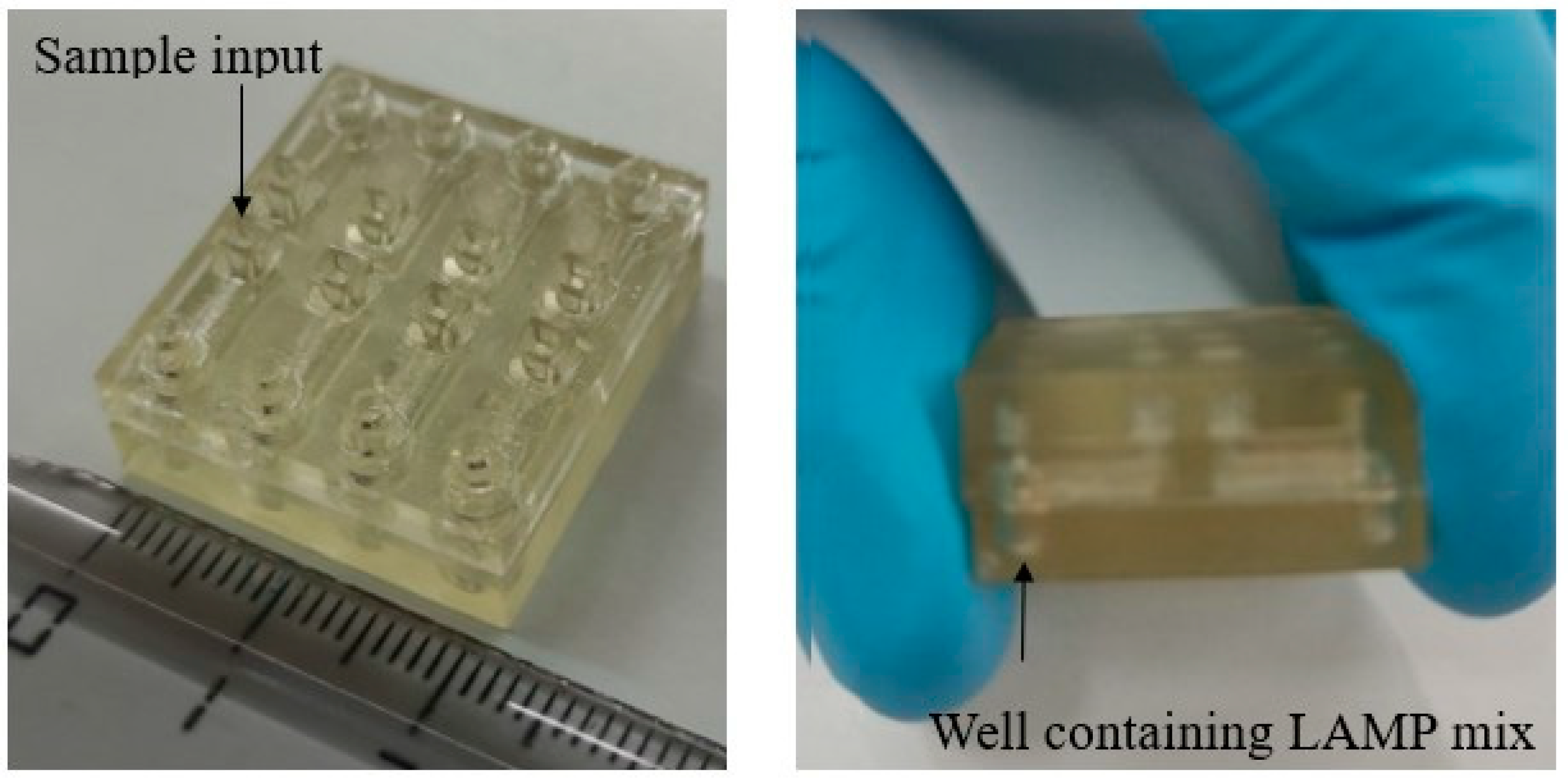
The device was divided into 3 different parts, namely a lid, upper part, and bottom part, each of which would be complementary to the other and would be combined after printing (Figure 1). The reason this device was not designed as a single piece was to avoid other problems that could occur after printing. For example, during curing with UV radiation, a small amount of liquid resin remained inside the channels, which then solidified, causing them to clog, and the fluid could not pass through them. Each device contains 8 different channels, so it can analyze 8 samples simultaneously, as shown in Figure 1 and Figure 2.

2.5. Loop-Mediated Isothermal Amplification (LAMP) Method

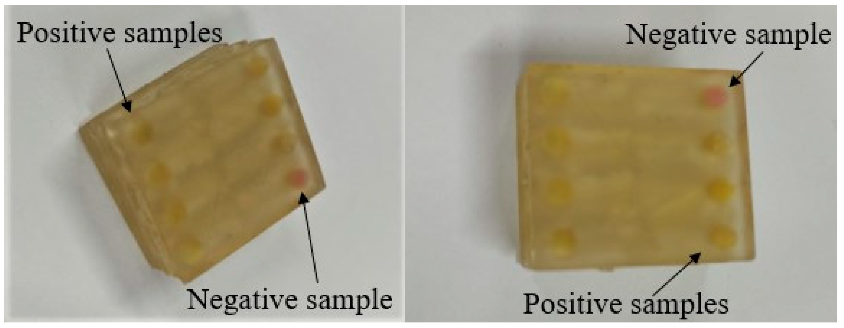
The technique used to detect the DNA of food pathogens was the colorimetric loop-mediated isothermal amplification (LAMP) method. The final assay solution prepared amounted to 25 μL and consisted of 15 μL WarmStart® Colorimetric LAMP 2X Master Mix, 2 μL primer solution, 5 μL molecular-grade water, and 3 μL bacterial DNA isolated from the samples. Each solution contained separate primers for S. typhimurium, L. monocytogenes, and E. coli, which were selected according to Srisawat and Panbangred, 2015 [7], Tang et al., 2011 [8], and Ramezani et al., 2018 [9], respectively. The prepared LAMP mixture was placed in each of the 8 small wells contained in the bottom part of the above-mentioned device, closed with the lid to protect the mixture, and stored in frozen conditions (−20 °C). On the day of analysis, microbial DNA was added and placed into an oven at 65 °C for 40 min. Any color change from purple to yellow (the color of the pH indicator dyes) indicated that the sample was positive for the particular pathogen according to the LAMP principle [10]. The color change occurs due to the production of protons during the amplification of Bst DNA polymerase in combination with the low concentration of the buffer in the colorimetric LAMP mixture.

2.6. Method Standardization

Briefly, 925 g of the studied food (milk, chicken, lettuce) was divided into two almost equal amounts. An initial amount of 475 g was further contaminated with an overnight culture of (S. Typhimurium ATCC 14028, L. monocytogenes ATCC 35152, and E. coli ATCC 25922). The known microbial pathogens’ concentrations in the foods started from 107 cfu/mL and then sub-tenfold serial dilutions were made until the final concentration reached 101 CFU/mL (positive controls). The remaining amount of 450 g was used for the preparation of negative controls and it was contaminated with an overnight culture of Staphylococcus aureus ATCC 25923 to obtain a final concentration of 104 CFU/g (negative controls) (Figure 3). The cultural microbiological method and the colorimetric LAMP assay methodologies were compared for the detection of the food pathogens.

2.7. Statistical Analyses

The 95% confidence intervals of the positive predictive value (PPV) and negative predictive value (NPV) were calculated based on the Wilson Score interval [11]. Data from all food products were pooled for each pathogen to calculate these statistics. The PPV and NPV were defined as follows:
P P V = T r u e   p o s i t i v e s T r u e   p o s i t i v e s + F a l s e   p o s i t i v e s
N P V = T r u e   n e g a t i v e s T r u e   n e g a t i v e s + F a l s e   n e g a t i v e s
The 95% confidence interval ( C I ) of an estimated positive or negative predictive value p ¯ was calculated based on the following equation:
C I = p ¯ + Z 2 2 n ± Z p ¯ ( 1 p ¯ ) n + Z 2 4 n 2 1 + Z 2 n
with the Z -score equal to 1.96 for the 95% confidence intervals and n being the total number of positive or negative samples. The following aggregate gold standard was used: positive cultured and real-time PCR isolates expressed a positive result while negative cultured and real-time PCR isolates expressed a negative result.

3. Results and Discussion

The European Commission has highly prioritized food monitoring due to the food safety crises that occurred in recent years. The European Commission’s White Paper on Food Safety suggests controls from “from farm to fork” and includes official controls, the implementation of advanced food safety standards as per Codex Alimentarius’ microbiological criteria, improved methods of detection, and extensive quality control at laboratory level [12]. The detection thresholds of the developed device are listed in Table 1. The sensitivity of the technique shows similar results with previous studies [13] in which the same variant of the present DNA isolation method (based on the classical phenol–chloroform method) was used and followed by identification using colorimetric LAMP. As shown in Table 1, the limit of detection for L. monocytogenes is 102 CFU/mL for all the food samples compared to 101 CFU/mL for S. Typhimurium and E. coli.
Regarding the results of the analyzed commercial food samples, of the 50 milk samples, 0 (0%) were positive for S. Typhimurium, 2 (4%) were positive for L. monocytogenes, and 4 (8%) were positive for E. coli. Of the 50 chicken samples, 5 (10%) were positive for S. Typhimurium, 11 (22%) were positive for L. monocytogenes, and 17 (34%) were positive for E. coli. Finally, regarding the 50 lettuce samples, 2 (4%) were positive for S. Typhimurium, 15 (30%) were positive for L. monocytogenes, and 14 (28%) were positive for E. coli (Table 2 and Figure 4).
As shown in Table 2, all samples were correctly identified as positive or negative with both the LAMP 3D microfluidic device and the reference methods. Therefore, the sensitivities and specificities for all pathogens were 100%. As such, the estimated values of the PPV and NPV were 1, or 100%, for each pathogen. Based on the available data, the statistical analysis in Table 3 demonstrates narrow confidence intervals (Cis) for the PPVs of L. monocytogenes and E. coli, of 12 and 10%, respectively. The PPV of S. Typhimurium had a wider CI of 35% due to the low quantity of commercial food samples that tested positive for this pathogen (7 samples). As such, more data are needed to statistically prove the method’s precision with respect to positive results for S. Typhimurium, but based on the results for L. monocytogenes and E. coli, it is expected to be precise. The CIs of the NPV were just 3% for each pathogen, demonstrating that the method is certain to provide precise negative results.
The 3D microfluidic analytical device developed in the present study has the potential to provide rapid analyses of food samples, with an analysis time of approximately 70 min (including extraction) and with high precision at a low cost. Its applicability was tested in three different food matrices (milk, chicken, and lettuce) for the detection of three common food pathogens, L. monocytogenes, S. Typhimurium, and E. coli. Multiple uses of integrated microfluidic LAMP systems have been identified in recent years [14,15,16]. Qi et al., 2021 [17], reported the development of a biosensor which uses a single microchip for the detection of S. Typhimurium in chicken meat with a limit of detection (LOD) of 14 CFU/mL and an analysis time of 1 h. A similar application was described by Man et al., 2021 [18], who developed a glass microfluidic colorimetric biosensor for the detection of S. Typhimurium in freshly made salad with a LOD of 6.1 × 101 CFU/mL in 45 min. The detection of two pathogens in milk, namely E. coli and S. Typhimurium, at a level of 103 CFU/mL and 102 CFU/mL, respectively, by a microfluidic paper-based aptasensor device was reported by Somvanshi et al., 2022 [19]. A microdevice for the detection of three food pathogens (E. coli, Salmonella, and Vibrio cholerae) in chicken meat was described by Sayad et al., 2018 [20], but with a higher LOD of 2.7 × 10⁴ CFU/mL and an analysis time of 65 min. The same three pathogens as in the present study (E. coli, Salmonella, and L. monocytogenes) were detected in ready-to-eat meals by a paper-based analytical device for colorimetric detection developed by Jokerst et al., 2012 [21], with a similarly low LOD of 101 CFU/mL but with a much longer analysis time of 8–12 h.
Due to the impact of foodborne pathogens on human health, governments and the food industry have been developing strategies to reduce the levels of foodborne pathogen contaminations. As the incidence of foodborne infections in Europe is not decreasing, but on the contrary, is increasing for most pathogens, the need to develop new rapid and simplified detection methods has become critical in the food industry. Microfluidic analytical devices are a technology that has been developing rapidly in recent years and is very promising for the evolution of analyses to the point where they can keep up with the continuous increase in production. Based on their flexibility in terms of integration, multiplexing, automation, and miniaturization, “micro total analysis systems (μTAS)” or “labs-on-a-chip” have become increasingly well known [22]. The use of microfluidic chip-based detection methods has certain clear benefits, such as high throughput, portability, relatively low reagent volumes and sample consumption, high speed, and the capacity to combine numerous components on a single chip [23]. The loop-mediated isothermal amplification (LAMP) technique was introduced by Notomi et al. in 2000 [24] as a unique DNA amplification technique that amplifies DNA under isothermal conditions with excellent specificity, efficiency, and speed. LAMP has been proven to be faster and more stable, sensitive, and specific for DNA identification when compared to conventional molecular detection techniques [25].
These microdevices can be manufactured using a number of materials like polymers, glass, silicon, and paper [26]. A number of applications have been developed for the detection of microorganisms including bacteria and viruses [5,26] in different matrices like clinical and food samples. In the case of food, applications for the detection of pathogens such as Campylobacter, E. coli, Salmonella, Vibrio cholera, L. monocytogenes, and Clostridium sporogenes use different detection methodologies like colometry, surface-enhanced Raman spectroscopy, fluorescence, electrochemistry, and turbidimetry. In order to minimize or eliminate the threat of possible infections, reduce the expensive storage of food until distribution, or prevent product recalls, the use of easy, non-time-consuming but sensitive detection methods for trace levels of bacterial pathogens is crucial. New technologies can expand the current possibilities by a drastically shortened detection time and a significantly lowered detection limit, with the additional benefit of being operable by non-experts. The design of innovative devices could also support the profitability of the European food industry.

4. Conclusions

This paper described the development of a 3D-printed microfluidic analytical device for the detection of pathogenic bacteria in commercial food samples using loop-mediated isothermal amplification. The detection limits were kept low while at the same time requiring minimal analysis time compared to existing methods. Each device, combined with a simplified DNA isolation methodology, is capable of analyzing eight different food samples for possible contamination with S. Typhimurium, L. monocytogenes, or E. coli using colorimetric LAMP. The above-mentioned device is easy to use, cheap, and fast, thereby showing potential to fill a huge gap in food safety practices in the future. The assay comprises a standalone technology that could provide a quantitative detection method when combined with UV-Vis spectroscopic analysis for low levels of pathogens (less than 10 CFU/sample). Finally, in the long term, this technology represents a platform for the development of testing devices in other areas besides food safety, such as in the case of the intentional release of pathogenic microorganisms, infectious disease diagnosis, environmental protection, and others. It should be emphasized that the goal of this innovative method is to be used as an alternative method to culture with equal performance and certain advantages such as shorter turn-around time, and not to achieve the maximum sensitivity and specificity seen with molecular methods.

Author Contributions

Conceptualization, D.H. and S.-A.P.; methodology, D.H., P.H., A.G.T., A.E.T. and S.-A.P.; validation, E.T. and S.A.; formal analysis, S.-A.P. and S.M.; investigation, S.-A.P., D.H. and E.T.; resources, D.H., A.G.T., A.E.T. and P.H.; data curation, S.-A.P., S.A. and S.M.; writing—original draft preparation, D.H., E.T., S.-A.P. and J.V.I.; writing—review and editing D.H., E.T., S.A. and J.V.I.; visualization, S.-A.P. and E.T.; supervision, D.H.; funding acquisition, J.V.I. All authors have read and agreed to the published version of the manuscript.

Funding

Author Simen Akkermans was funded by the Research Foundation Flanders (FWO), grant number 1224623N.

Institutional Review Board Statement

Not applicable.

Informed Consent Statement

Not applicable.

Data Availability Statement

The data presented in this study are available on request from the corresponding author (accurately indicate status).

Conflicts of Interest

The authors declare no conflicts of interest.

References

  1. Programme “Global Salm-Surv” de l’OMS “Global Salm-Surv”. Note D’information INFOSAN No. 6/2005. Available online: http://www.who.int/foodsafety/fs_management/No_06_GSS_Oct05_fr.pdf (accessed on 4 January 2024).
  2. European Food Safety Authority (EFSA). The Community Summary Report on Trends and Sources of Zoonoses, Zoonotic Agents, Antimicrobial Resistance and Foodborne Outbreaks in the European Union in 2006. EFSA J. 2007, 5, 130r. [Google Scholar] [CrossRef]
  3. European Food Safety Authority and European Centre for Disease Prevention and Control. The European Union One Health 2019 Zoonoses Report. EFSA J. 2023, 19, e06406. [Google Scholar] [CrossRef]
  4. Zhang, D.; Bi, H.; Liu, B.; Qiao, L. Detection of Pathogenic Microorganisms by Microfluidics Based Analytical Methods. Anal. Chem. 2018, 90, 5512–5520. [Google Scholar] [CrossRef] [PubMed]
  5. Luo, Z.; Ye, C.; Xiao, H.; Yin, J.; Liang, Y.; Ruan, Z.; Luo, D.; Gao, D.; Tan, Q.; Li, Y.; et al. Optimization of loop-mediated isothermal amplification (LAMP) assay for robust visualization in SARS-CoV-2 and emerging variants diagnosis. Chem. Eng. Sci. 2022, 251, 117430. [Google Scholar] [CrossRef] [PubMed]
  6. Papatheodorou, S.A.; Halvatsiotis, P.; Houhoula, D. A comparison of different DNA extraction methods and molecular techniques for the detection and identification of foodborne pathogens. AIMS Microbiol. 2021, 7, 304–319. [Google Scholar] [CrossRef]
  7. Srisawat, M.; Panbangred, W. Efficient and Specific Detection of Salmonella in Food Samples Using a stn-Based Loop-Mediated Isothermal Amplification Method. BioMed Res. Int. 2015, 2015, 356401. [Google Scholar] [CrossRef]
  8. Tang, M.J.; Zhou, S.; Zhang, X.Y.; Pu, J.H.; Ge, Q.L.; Tang, X.J.; Gao, Y.S. Rapid and Sensitive Detection of Listeria monocytogenes by Loop-Mediated Isothermal Amplification. Curr. Microbiol. 2011, 63, 511–516. [Google Scholar] [CrossRef]
  9. Ramezani, R.; Parizi, Z.K.; Ghorbanmehr, N.; Mirshafiee, H. Rapid and Simple Detection of Escherichia coli by Loop-Mediated Isothermal Amplification Assay in Urine Specimens. Avicenna J. Med. Biotechnol. 2018, 10, 269–272. [Google Scholar]
  10. Tanner, N.A.; Zhang, Y.; Evans, T.C. Visual detection of isothermal nucleic acid amplification using pH-sensitive dyes. BioTechniques 2015, 58, 59–68. [Google Scholar] [CrossRef] [PubMed]
  11. Wilson, E.B. Probable Inference, the Law of Succession, and Statistical Inference. J. Am. Stat. Assoc. 1927, 22, 209–212. [Google Scholar] [CrossRef]
  12. Commission of the European Communities. White Paper on Food Safety Com (1999) 719 Final. 12.1.2000. Available online: https://eur-lex.europa.eu/legal-content/EN/TXT/PDF/?uri=CELEX:51999DC0719 (accessed on 15 September 2023).
  13. Papatheodorou, S.A.; Tsironi, T.; Giannakourou, M.; Halvatsiotis, P.; Houhoula, D. Application of microfluidic paperbased analytical devices (μPADs) for food microbial detection. J. Sci. Food Agric. 2023, 103, 2215. [Google Scholar] [CrossRef]
  14. Li, Z.; Bai, Y.; You, M.; Hu, J.; Yao, C.; Cao, L.; Xu, F. Fully integrated microfluidic devices for qualitative, quantitative and digital nucleic acids testing at point of care. Biosens. Bioelectron. 2021, 177, 112952. [Google Scholar] [CrossRef] [PubMed]
  15. Escobar, A.; Chiu, P.; Qu, J.; Zhang, Y.; Xu, C. Integrated Microfluidic-Based Platforms for On-Site Detection and Quantification of Infectious Pathogens: Towards On-Site Medical Translation of SARS-CoV-2 Diagnostic Platforms. Micromachines 2021, 12, 1079. [Google Scholar] [CrossRef]
  16. Donia, A.; Furqan Shahid, M.; Hassan, S.; Shahid, R.; Ahmad, A.; Javed, A.; Nawaz, M.; Yaqub, T.; Bokhari, H. Integration of RT-LAMP and Microfluidic Technology for Detection of SARS-CoV-2 in Wastewater as an Advanced Point-of-Care Platform. Food Environ. Virol. 2022, 14, 364–373. [Google Scholar] [CrossRef]
  17. Qi, W.; Zheng, L.; Wang, S.; Huang, F.; Liu, Y.; Jiang, H.; Lin, J. A microfluidic biosensor for rapid and automatic detection of Salmonella using metal-organic framework and Raspberry Pi. Biosens. Bioelectron. 2021, 178, 113020. [Google Scholar] [CrossRef] [PubMed]
  18. Man, Y.; Ban, M.; Li, A.; Jin, X.; Du, Y.; Pan, L. A microfluidic colorimetric biosensor for in-field detection of Salmonella in fresh-cut vegetables using thiolated polystyrene microspheres, hose-based microvalve and smartphone imaging APP. Food Chem. 2021, 354, 129578. [Google Scholar] [CrossRef] [PubMed]
  19. Somvanshi, S.B.; Ulloa, A.M.; Zhao, M.; Liang, Q.; Barui, A.K.; Lucas, A.; Jadhav, K.M.; Allebach, J.P.; Stanciu, L.A. Microfluidic paper-based aptasensor devices for multiplexed detection of pathogenic bacteria. Biosens. Bioelectron. 2022, 207, 114214. [Google Scholar] [CrossRef]
  20. Sayad, A.; Ibrahim, F.; Mukim Uddin, S.; Cho, J.; Madou, M.; Thong, K.L. A microdevice for rapid, monoplex and colorimetric detection of foodborne pathogens using a centrifugal microfluidic platform. Biosens. Bioelectron. 2018, 100, 96–104. [Google Scholar] [CrossRef]
  21. Jokerst, J.C.; Adkins, J.A.; Bisha, B.; Mentele, M.M.; Goodridge, L.D.; Henry, C.S. Development of a Paper-Based Analytical Device for Colorimetric Detection of Select Foodborne Pathogens. Anal. Chem. 2012, 84, 2900–2907. [Google Scholar] [CrossRef]
  22. Huang, G.; Huang, Q.; Xie, L.; Xiang, G.; Wang, L.; Xu, H.; Ma, L.; Luo, X.; Xin, J.; Zhou, X.; et al. A rapid, low-cost, and microfluidic chip-based system for parallel identification of multiple pathogens related to clinical pneumonia. Sci. Rep. 2017, 7, 6441. [Google Scholar] [CrossRef]
  23. Nguyen, N.T.; Hejazian, M.; Ooi, C.; Kashaninejad, N. Recent Advances and Future Perspectives on Microfluidic Liquid Handling. Micromachines 2017, 8, 186. [Google Scholar] [CrossRef]
  24. Notomi, T.; Okayama, H.; Masubuchi, H.; Yonekawa, T.; Watanabe, K.; Amino, N.; Hase, T. Loop-mediated isothermal amplification of DNA. Nucleic Acids Res. 2000, 28, e63. [Google Scholar] [CrossRef] [PubMed]
  25. Mori, Y.; Notomi, T. Loop-mediated isothermal amplification (LAMP): A rapid, accurate, and cost-effective diagnostic method for infectious diseases. J. Infect. Chemother. 2009, 15, 62–69. [Google Scholar] [CrossRef] [PubMed]
  26. Lonchamps, P.L.; He, Y.; Wang, K.; Lu, X. Detection of pathogens in foods using microfluidic “lab-on-chip”: A mini review. J. Agric. Food Res. 2022, 10, 100430. [Google Scholar] [CrossRef]
Figure 1. Three-dimensional image of the microfluidic analytical device.
Amh 69 00006 g001
Figure 2. Microfluidic analytical device.
Amh 69 00006 g002
Figure 3. Overview of the experimental approach.
Amh 69 00006 g003
Figure 4. Example of results of the analysis of commercial food samples using LAMP and the 3D microfluidic device.
Amh 69 00006 g004
Table 1. Detection limits of the 3D microfluidic analytical device.
Limits of Detection (cfu/mL)
MilkL. monocytogenes102
S. typhimurium101
E. coli101
ChickenL. monocytogenes102
S. typhimurium101
E. coli101
LettuceL. monocytogenes102
S. typhimurium101
E. coli101
Table 2. Analyses of 150 commercial food samples for detection and identification of S. typhimurium, L. monocytogenes, and E. coli.
S. typhimurium
Culture/3D Device
L. monocytogenes
Culture/3D Device
E. coli
Culture/3D Device
Milk0/02/24/4
Chicken5/511/1117/17
Lettuce2/215/1514/14
Total7/15028/15035/150
Table 3. Confidence intervals (95%) of the positive and negative predictive values of the 3D microfluidic device for each food pathogen for all 150 food samples.
S. typhimuriumL. monocytogenesE. coli
Positive predictive value65–100%88–100%90–100%
Negative predictive value97–100%97–100%97–100%
Disclaimer/Publisher’s Note: The statements, opinions and data contained in all publications are solely those of the individual author(s) and contributor(s) and not of MDPI and/or the editor(s). MDPI and/or the editor(s) disclaim responsibility for any injury to people or property resulting from any ideas, methods, instructions or products referred to in the content.