Review

Monoclonal Protein Evaluation in the Diagnostic Algorithm for Cardiac Amyloidosis

Division of Cardiology, Johns Hopkins University, Baltimore, MD 21217, USA
LabMed 2025, 2(3), 13; https://doi.org/10.3390/labmed2030013
Submission received: 21 December 2024 / Revised: 15 May 2025 / Accepted: 25 June 2025 / Published: 28 July 2025

Abstract

Cardiac amyloidosis (CA) results from the deposition of either immunoglobulin light chain (AL) or transthyretin (ATTR) amyloid fibrils in the myocardium, causing restrictive cardiomyopathy and, if left untreated, can lead to early death. Advancements in non-invasive diagnostic modalities have led to an increased recognition of the disease. Monoclonal gammopathy plays a pivotal role in the diagnostic algorithm for CA, particularly in differentiating AL from ATTR. This review highlights the importance of monoclonal protein detection through serum protein electrophoresis, immunofixation electrophoresis, and serum free light chain assays as initial screening tools. However, these tests alone are insufficient for a definitive diagnosis due to the complexities associated with coexisting monoclonal gammopathies and the potential for false negative and positive results. Advanced imaging modalities, such as echocardiography, cardiac magnetic resonance, and nuclear scintigraphy, along with tissue biopsy, are crucial for confirming CA and accurately determining the CA subtype.

1. Introduction

Amyloidosis refers to a collection of diseases characterized by the accumulation of misfolded proteins in various organs, leading to tissue damage and dysfunction [1]. Cardiac amyloidosis (CA), which affects the heart, carries the worst prognosis [2]. The condition primarily results from the deposition of either immunoglobulin light chain (AL) or transthyretin (ATTR) fibrils in the myocardium, causing restrictive cardiomyopathy and, if left untreated, can lead to early death [3]. AL is caused by the misfolding of a monoclonal immunoglobulin light chain fragment produced by plasma cells in the bone marrow and has a more aggressive course of the two subtypes. In contrast, ATTR is caused by the misfolding of TTR, a protein that is primarily synthesized in the liver and transports thyroxine and retinol-binding protein in the blood and could occur sporadically in wild-type ATTR (ATTRwt) or could be inherited as mutation in the genetic sequence (ATTRv) [3].
Recent studies have highlighted evolving trends in the epidemiology of CA, with an increasing prevalence primarily driven by enhanced clinical awareness and advancements in diagnostic methods [4]. These improvements in diagnostic capabilities have facilitated earlier detection of CA, despite challenges posed by the phenotypic overlap with other cardiomyopathies [5,6]. Early diagnosis has significant implications for the timely initiation of treatment and for improving patient outcomes. Laboratory evaluation of mosubtypes andeins plays a crucial role in the diagnostic algorithm for CA, aiding in the differentiation between CA subtypes, and can often provide valuable insights without the need for histological confirmation.
In this review, we will emphasize the role of multimodality imaging in the diagnosis of CA, with a particular focus on the significance of laboratory workup, specifically in relation to monoclonal gammopathy, within the diagnostic algorithm. By integrating advanced imaging techniques with comprehensive laboratory evaluations, we aim to provide a precise and efficient approach to diagnosing CA, enabling differentiation between its subtypes to help institute appropriate treatment strategies.

2. Recognizing the ‘Red Flags’ to Suspect CA

The typical clinical presentation of CA often involves decompensated heart failure, which manifests as progressive shortness of breath on exertion and/or signs of right ventricular failure, including peripheral lower extremity edema and jugular venous distention. In rare instances, cardiogenic shock can be the initial presentation in severe cases [7]. Patients with CA may also present with angina, even in the absence of obstructive epicardial coronary artery disease, due to coronary microvascular dysfunction as a result of deposition of amyloid fibrils in the interstitial tissues, intramyocardial coronary vessels and perivascular regions of the heart [8,9]. Arrythmias are a common clinical finding in CA [10,11]. Atrial fibrillation is the most frequently encountered arrhythmia in CA patients, especially in ATTRwt [12,13]. While bradyarrhythmias, particularly heart blocks, may serve as an early clue for the diagnosis of CA, sudden cardiac arrest caused by ventricular arrhythmias can rarely be the first presentation [14,15].
In AL, amyloid deposits can affect any extra-cardiac tissue, except for the brain. Renal involvement is common, often presenting as nephrotic syndrome and proteinuria due to glomerular amyloid deposition [16]. Hepatomegaly is another common finding, which can result from either direct amyloid infiltration of the liver or secondary congestion due to right-sided heart failure. Autonomic nervous system involvement can cause symptoms such as orthostatic hypotension, gastroparesis, erectile dysfunction, and intestinal dysmotility [17]. Peripheral nerve involvement leads to painful, bilateral, symmetric distal sensory neuropathy that progresses to motor neuropathy. Finally, macroglossia, an enlargement of the tongue, is a hallmark feature of AL.
Several non-cardiac manifestations can raise suspicion for CA. Carpal tunnel syndrome, which occurs due to amyloid fibril deposition in the flexor retinaculum and tenosynovial tissue within the carpal tunnel, is the earliest and most common non-cardiac manifestation. Approximately 50% of ATTRwt patients experience carpal tunnel syndrome, often occurring 5–9 years before cardiac involvement [18]. Studies show that around 10–16% of patients undergoing carpal tunnel surgery have tenosynovial amyloid deposits, while only up to 2% are diagnosed with CA at that time [19]. Spinal stenosis, which occurs due to amyloid deposition in the ligamentum flavum, is exclusive to ATTR, particularly ATTRwt. Amyloid deposits are detected in more than a third of older adults undergoing surgery for lumbar spinal stenosis, and the incidence increases with age [20]. The spontaneous rupture of the distal biceps tendon has been reported in upto 1/3rd of patients with ATTRwt [21]. Furthermore, ATTRwt patients have a higher prevalence of total hip and knee arthroplasties compared to the general population [22].

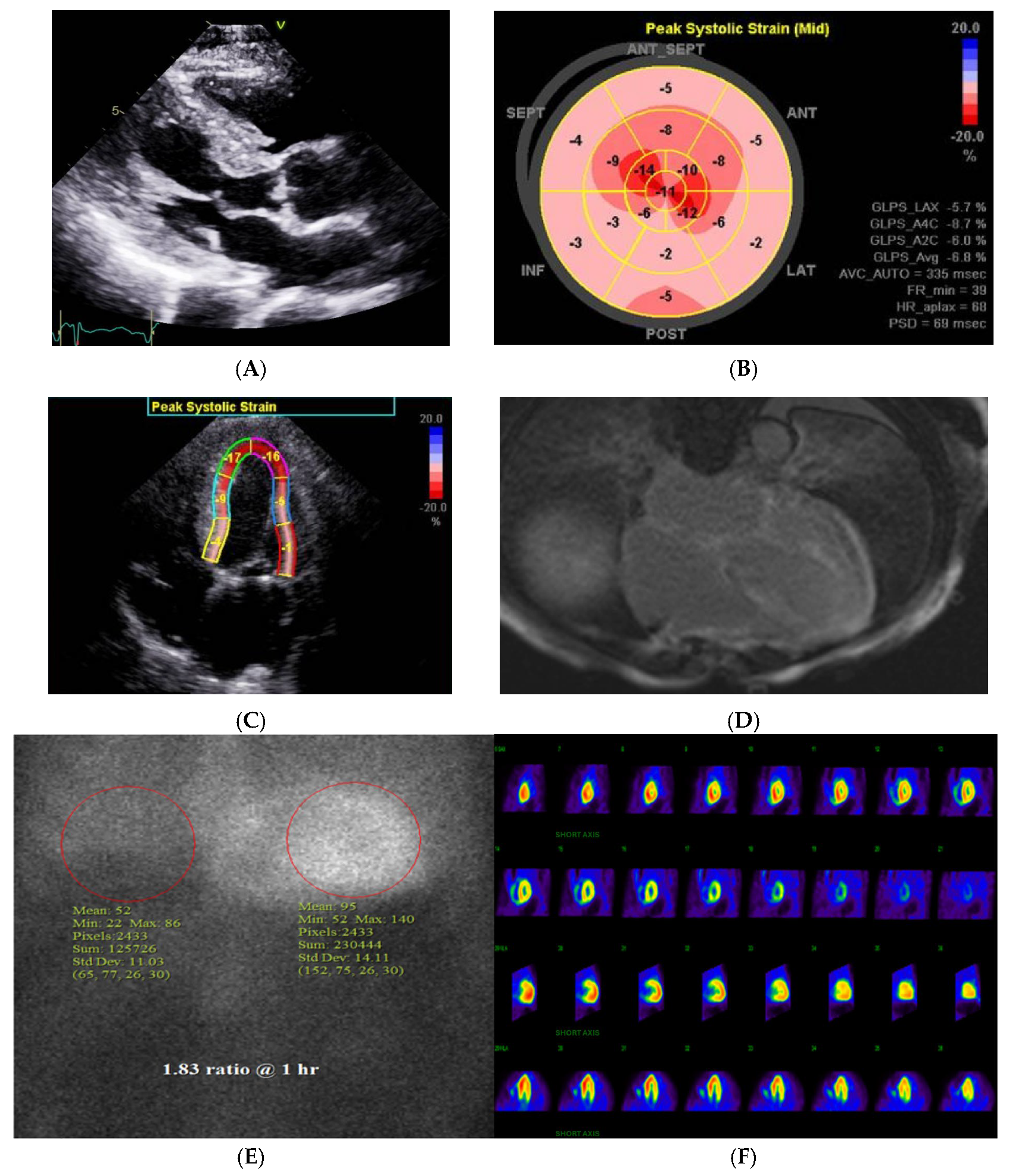
3. Diagnostic Imaging in CA

Echocardiography is the first-line imaging modality for suspected CA and can provide some clues to differentiate it from other conditions such as hypertensive cardiomyopathy, hypertrophic cardiomyopathy, aortic stenosis, and Fabry’s disease (Figure 1 Central illustration). Key findings in CA include significant left ventricular (LV) thickening, often concentric and exceeding 15 mm, with asymmetric thickening observed in 23% of ATTRwt cases [23]. LV outflow obstruction can also mimic hypertrophic obstructive cardiomyopathy. Additional echocardiographic features include small LV cavity, atrial enlargement, and a granular myocardial appearance, though the latter is nonspecific for CA [24,25]. Diastolic dysfunction, particularly elevated E/e ratio and low tissue Doppler velocity, is consistently present. Tissue Doppler imaging and speckle-tracking strain imaging can help detect subclinical LV dysfunction, with CA showing characteristic patterns, such as apical sparing of longitudinal strain, which can differentiate it from other etiologies of LV hypertrophy.
Cardiac magnetic resonance (CMR) is invaluable in distinguishing CA from other hypertrophic conditions, providing high-resolution structural and functional imaging, though it cannot reliably differentiate between ATTR and AL. CMR detects amyloid infiltration via late gadolinium enhancement (LGE), a pattern that is pathognomonic for CA [26]. T1 mapping is particularly useful, with elevated native T1 values serving as an early indicator of CA, even before ventricular thickening is evident [27]. This technique also aids in tracking disease progression and prognosis [28]. In addition, T2 mapping can visualize myocardial edema, providing further insight, especially in AL cases [28]. Similarly, extracellular volume (ECV) measurement can also provide important prognostic information, which will be described in more detail below [29].
Nuclear scintigraphy using bone-avid tracers, such as 99mTc-PYP, is a non-invasive method for diagnosing ATTR [30,31]. However, this test is always performed in conjunction with serum and urine studies to rule out paraproteinemia, and hence AL (Figure 2). The visual grading of myocardial tracer uptake can help confirm the diagnosis of ATTR with a high specificity in patients who do not demonstrate monoclonal gammopathy. A high heart-to-lung ratio on scintigraphy, coupled with diffuse myocardial uptake on SPECT, can confirm a diagnosis of ATTR without the need for biopsy [32,33]. On the other hand, if monoclonal gammopathy is present, it always necessitates further workup for AL. If a monoclonal protein is present, tissue confirmation of amyloid type is mandatory because nuclear scintigraphy may be mildly positive in AL [34].

4. Monoclonal Gammopathy Testing in Suspected CA

The diagnosis of AL involves detecting a clonal plasma cell disorder, beginning with the evaluation of monoclonal proteins. These proteins can include intact immunoglobulins, immunoglobulin fragments such as free light chains (FLC), or, less commonly, free heavy chains. The primary laboratory method used to identify monoclonal proteins is serum protein electrophoresis (SPEP). In this process, serum is applied to a medium, and an electric current is passed through it, causing the proteins to separate into five distinct regions based on their size and electrical charge: albumin, α1, α2, β, and γ. Since most antibodies migrate into the γ region, monoclonal proteins typically appear as a sharp peak, known as the “M spike”. However, SPEP alone cannot determine the subtype of the monoclonal protein or confirm the presence of monoclonal protein. To do so, immunofixation electrophoresis (IFE) is required, where the serum is exposed to antibodies targeting different heavy and light chain subtypes. Therefore, IFE should be performed in patients with abnormal SPEP.
It is important to note that SPEP/IFE has its own limitations as a screening test. In approximately 25% of AL cases, SPEP/IFE may show normal results. This can occur if the monoclonal light chains responsible for amyloidosis are produced in small amounts, or if they are filtered out by the kidneys, making them undetectable in the serum. This can lead to a missed diagnosis, especially in patients with early-stage disease or renal impairment. In addition, SPEP/IFE may not detect all subtypes of monoclonal proteins associated with AL. For example, in some cases, the monoclonal protein may be composed of free light chains or heavy chains that are not detected by routine SPEP, or the amount produced may be insufficient to form a clear M spike on electrophoresis. There is high dependance on protein quantity, as SPEP/IFE is less effective in detecting AL in patients with low levels of monoclonal proteins or with light chain deposition that does not form distinct bands.
To improve detection, combining SPEP/IFE with a 24 h urine collection for urine protein electrophoresis and IFE should be obtained, which can identify 90% of amyloidosis cases. While a 24 h urine protein electrophoresis and immunofixation (UPEP/IFE) may have limited sensitivity on its own, it remains valuable for detecting clonal light chains, particularly when monoclonal protein is only indicated by a slight abnormality in serum free light chains (SFLC). In these cases, 24 h UPEP/IFE could be the definitive test for identifying monoclonal protein. Additionally, 24 h UPEP/IFE is beneficial in assessing the cause of renal damage in patients who present with new renal insufficiency and monoclonal gammopathy. Therefore, even when a monoclonal protein has already been detected, obtaining a 24 h UPEP/IFE can provide further insights and may influence management decisions.
Finally, it is essential to measure SFLC as part of the diagnostic workup. Both kappa and lambda light chain concentrations, as well as the kappa/lambda ratio (KLR), should be measured. Combining SFLC measurements with SPEP/IFE significantly improves the ability to detect monoclonal protein in patients with AL, reaching a sensitivity of up to 99% [35]. However, abnormal SFLC results do not always confirm the presence of monoclonal protein. For example, elevated kappa and lambda levels with a normal KLR are not indicative of monoclonal protein and are often seen in nonspecific inflammatory conditions. Since light chains are excreted by the kidneys, patients with chronic kidney disease may have impaired clearance of light chains, particularly kappa chains, which are typically cleared more efficiently than lambda chains. As a result, a mildly elevated KLR in these patients may reflect an imbalance between production and clearance, rather than monoclonal gammopathy.
Despite abnormalities in protein studies, a tissue biopsy is essential to confirm the diagnosis of amyloidosis. The sensitivity of diagnostic biopsies varies, with abdominal fat pad aspiration having an accuracy of around 85%, rectal biopsy ranging from 75 to 85%, and bone marrow biopsy showing sensitivity of about 50% [36,37]. If clinical suspicion remains high and non-invasive tests are inconclusive, biopsy of the other affected organs may be considered. However, this decision must be made with caution, given the increased risk of hemorrhagic complications in amyloidosis patients. It is generally advisable to start with the least invasive procedures before progressing to more invasive options.

5. Pitfalls and Challenges with Monoclonal Gammopathy

The identification of monoclonal gammopathy presents several challenges and pitfalls, which can complicate diagnosis. One of the challenges can be subclinical or low-level monoclonal proteins that are difficult to detect through standard testing. Subtle abnormalities, such as minimal SFLC elevation or low-intensity monoclonal spikes, may be missed or misinterpreted, leading to delayed diagnosis or misdiagnosis. Another challenge can be minor elevations in free light chains or isolated monoclonal protein spikes, especially when detected through SPEP, may not always represent a true monoclonal gammopathy. These can be seen in benign conditions, such as chronic inflammation or renal insufficiency, leading to overdiagnosis if not carefully interpreted. Similarly, monoclonal gammopathies and ATTR are often coincident, owing to shared associations with older age and Black race. The presence of monoclonal gammopathy in these patients typically does not suggest a primary cause of amyloidosis; however, distinguishing between the two subtypes requires further diagnostic testing, as it is not possible to do so based solely on clinical presentation or initial screening. Finally, in cases where monoclonal gammopathy is present, clonal plasma cells may not always be detectable in a bone marrow biopsy, especially when the plasma cell burden is low or when disease is located outside the bone marrow (extramedullary). This can result in negative biopsy results despite the presence of a monoclonal gammopathy, leading to diagnostic uncertainty. Addressing these challenges involves a multi-faceted approach, combining advanced diagnostic tests, careful clinical interpretation, and, when necessary, histological confirmation to establish the diagnosis and differentiate between AL and ATTR.

6. Advanced Typing Techniques When Non-Invasive Diagnostics Are Inconclusive

While most cases of CA can be diagnosed using non-invasive methods combined with laboratory tests for monoclonal proteins, a subset of patients presents diagnostic ambiguity that necessitates advanced tissue-based amyloid typing. In such instances, immunoelectron microscopy (IEM) serves as a valuable adjunctive diagnostic technique [38]. IEM combines immunolabeling with ultrastructural imaging, allowing precise visualization of amyloid fibrils tagged with subtype-specific antibodies. This technique is particularly useful when conventional mass spectrometry (MS) is not available or when MS results are inconclusive or conflicting, and can reliably type amyloid with high sensitivity, specificity, and predictive values [39,40].
van den Broek et al. illustrates the utility of IEM and droplet digital PCR (ddPCR) in a challenging case of a 55-year-old patient with an IgG kappa-positive lymphoplasmacytic lymphoma (LPL) and suspected CA [40]. Conventional immunohistochemistry was unable to conclusively type the amyloid deposits due to overlapping staining profiles, but IEM—using gold-labeled antibodies—confirmed AL of kappa light chain type. Furthermore, ddPCR identified the MYD88 L265P mutation in endomyocardial tissue, confirming the presence of LPL in the myocardium itself. This rare co-localization of lymphoma and amyloid in the same cardiac biopsy underscores the diagnostic value of IEM and ddPCR when conventional tools fall short. Both techniques are highly sensitive, can be applied to formalin-fixed paraffin-embedded tissue, and are more accessible in certain settings than mass spectrometry. Their incorporation into the diagnostic workflow can improve accuracy and guide appropriate therapy, particularly in complex or ambiguous cases.

7. Advances in Imaging-Role of PET

Amyloid positron emission tomography (PET) tracers offer a promising alternative to traditional bone-avid SPECT imaging for the diagnosis and management of CA, particularly for detecting AL and early disease [41]. Unlike SPECT tracers, PET tracers such as 11C-PiB, 18F-florbetapir, and 18F-flutemetamol bind directly to the β-sheet structure of amyloid fibrils, allowing detection of amyloid deposits regardless of precursor protein [41,42]. These tracers provide quantitative and semiquantitative metrics, including standardized uptake values (SUV), retention index (RI), and percentage injected dose, which may better reflect amyloid burden than weight-normalized SUV metrics alone [43]. PET imaging can detect early cardiac involvement before structural changes appear and may be especially valuable for identifying AL, where SPECT is ineffective. However, the sensitivity of β-amyloid PET tracers appears lower in ATTR, particularly in patients with certain fibril subtypes. New tracers such as 124I-evuzamitide, which targets glycosaminoglycans on amyloid fibrils, may improve detection of both AL and ATTR [41]. While amyloid PET cannot distinguish between amyloid types and currently lacks FDA-approved tracers for CA, ongoing research and trials may soon establish PET as a key modality for early diagnosis, disease quantification, and monitoring of therapeutic response in CA.

8. Prognostic Assessment in CA

In systemic amyloidosis, cardiac involvement is the primary factor contributing to both morbidity and mortality, making the assessment of CA burden crucial for prognosis. Widely used staging systems, particularly in AL amyloidosis, rely on serum biomarkers. The Mayo classification incorporates NT-proBNP and troponin levels to stratify risk at diagnosis, with the addition of free SFLC difference enhancing prognostic accuracy [44]. Similarly, Boston University developed a staging model using BNP and troponin, which aligns closely with the Mayo system [45]. The UK’s National Amyloidosis Centre risk stratification system includes NT-proBNP and eGFR, applying to both ATTRwt and ATTRv forms [46], while the Mayo Clinic’s staging for ATTRwt also uses NT-proBNP and troponin [47]. The UK’s National Amyloidosis Centre system was subsequently expanded to a four-stage model using a 10,000 ng/L NT-proBNP cutoff to identify high-risk patients [48]. Columbia University further refined staging by integrating NYHA class and diuretic dosage [49].
Although NT-proBNP is a cornerstone of all biomarker-based staging systems in CA, its elevation does not necessarily reflect amyloid infiltration alone. Rather, it often represents a complex interplay of physiological derangements, including impaired kidney function, systemic congestion, and neurohormonal activation. As such, elevated NT-proBNP may be more indicative of downstream systemic consequences rather than the true extent of myocardial amyloid deposition. This limitation highlights the growing importance of cardiac imaging in providing a more direct and nuanced assessment of disease burden.
Advanced echocardiographic parameters offer valuable insights beyond what serum biomarkers can provide. The presence of reduced global longitudinal strain has consistently demonstrated a strong and independent association with mortality in CA [50]. This is likely because amyloid infiltration tends to begin in the subendocardial region, where longitudinal myocardial fibers are concentrated, making this parameter particularly sensitive to early disease. Stroke volume—especially when indexed to body surface area—is another robust prognostic marker, as it reflects both the restrictive filling pattern and impaired systolic output seen in amyloid cardiomyopathy [51].
Additionally, the myocardial contraction index (MCI), defined as the ratio of stroke volume to myocardial volume, provides a volumetric measure of cardiac contractile efficiency. MCI captures subtle changes in myocardial function more effectively than conventional metrics like ejection fraction and has been shown to offer superior prognostic value [52,53]. Together, these imaging-based markers help overcome the limitations of serum biomarkers and allow for a more comprehensive evaluation of disease severity and progression in CA.
The markers of structural and functional assessment also provide important prognostic information. The presence and extent of transmural LGE is a significant predictor of mortality [54], while ECV mapping allows for direct quantification of amyloid burden and has emerged as the most robust CMR-derived prognostic marker [28,29]. Although native T1 mapping also offers prognostic information, its signal reflects a mix of intracellular and extracellular changes, limiting its specificity [55]. ECV, in contrast, correlates directly with amyloid infiltration. Hepatic ECV has also shown independent prognostic value in systemic AL amyloidosis. Nuclear imaging with bone-seeking tracers is sensitive for diagnosing ATTR, but the prognostic value of tracer uptake is less clear. While higher grades of uptake signal worse outcomes initially, differences in survival between moderate and severe uptake grades are not significant in established disease. PET imaging, still investigational, shows promise; increased uptake of specific tracers like Pittsburgh B compound has been linked to worse outcomes in AL [56,57], but larger studies are needed to validate its role in prognostic stratification.

9. Amyloid-Specific Disease-Modifying Therapies

9.1. Management of ATTR

9.1.1. TTR Silencers

Advancements in gene-silencing technology have led to the development of TTR silencers, which reduce TTR production through RNA interference (RNAi) mechanisms [58]. These therapies are particularly effective in ATTRv with neuropathy. Patisiran, the first FDA-approved small interfering RNA (siRNA) therapeutic, demonstrated significant benefits in the APOLLO phase III trial, improving neurological symptoms, quality of life, and autonomic function [59]. Administered intravenously every three weeks, patisiran requires steroid premedication due to infusion-related reactions and carries a risk of vitamin A deficiency, for which supplementation is recommended. Inotersen, an antisense oligonucleotide administered via weekly subcutaneous injection, also stabilizes neuropathy in ATTRv [60]. However, it carries risks of glomerulonephritis, severe thrombocytopenia, and requires intensive monitoring of platelet counts and renal function due to serious adverse events noted in the NEURO-TTR trial [61].

9.1.2. TTR Stabilizers and Degraders

TTR stabilizers function by preventing the dissociation of TTR tetramers into monomers, a critical step in amyloid formation. Tafamidis is the first oral FDA-approved drug for ATTRwt and ATTRv cardiomyopathy. The ATTR-ACT trial confirmed its efficacy in reducing mortality, hospitalizations, and functional decline over a 30-month period, with excellent tolerability [62]. Diflunisal, an NSAID with TTR-stabilizing properties, has shown benefit in slowing neurological and cardiac deterioration in ATTR in multiple studies, though its use is limited by renal, cardiac, and bleeding risks [63,64]. Its cost-effectiveness makes it a viable alternative for select patients. In parallel, investigational therapies targeting TTR degradation, such as monoclonal antibodies like NI006, aim to clear amyloid deposits from tissues, though these remain in clinical trials. In the phase 1 trial of the recombinant human antibody NI006 for the treatment of patients with ATTR cardiomyopathy and heart failure, the use of NI006 was associated with no apparent drug-related serious adverse events [65]. The combination of doxycycline and tauroursodeoxycholic acid (TUDCA) has been examined for TTR degradation, but results have not been promising clinically, and significant gastrointestinal and dermatologic side effects have been witnessed [66,67].

9.2. Management of AL

Treatment of AL amyloidosis is complex and generally managed by hematologists experienced in plasma cell disorders. Therapeutic strategies aim to suppress the underlying plasma cell clone using combinations of proteasome inhibitors (e.g., bortezomib, carfilzomib), immunomodulatory drugs (lenalidomide, pomalidomide), and monoclonal antibodies (notably daratumumab) [68,69]. Selected patients may also undergo autologous stem cell transplantation. Treatment response is assessed through hematologic and cardiac markers. Hematologic responses are categorized as complete (CR), very good partial (VGPR), partial (PR), or no response (NR), with definitions based on changes in free light chain levels and immunofixation results [70]. Cardiac response is primarily monitored using NT-proBNP, with a reduction of more than 30% indicating meaningful clinical improvement. The ultimate goal is to achieve both hematologic and organ-specific remission to improve survival and quality of life.

10. Challenges and Limitations

The management of CA presents several significant challenges, primarily due to delayed diagnosis, disease heterogeneity, and limited access to specialized care. CA often mimics other more common cardiac conditions such as hypertensive heart disease or heart failure with preserved ejection fraction (HFpEF), leading to under-recognition and diagnostic delays that adversely affect prognosis. Furthermore, diagnostic tools like CMR or nuclear scintigraphy, while effective, are not universally available and require expert interpretation. Differentiating between amyloid subtypes (AL vs. ATTR) is critical, as treatment strategies differ drastically, yet this requires access to advanced laboratory testing, tissue biopsy, or genetic evaluation—resources not always readily accessible, particularly in low-resource settings.
Therapeutically, while disease-modifying treatments have advanced significantly, their availability and affordability remain major limitations. FDA-approved therapies such as tafamidis, patisiran, and inotersen are often cost-prohibitive and not uniformly covered by insurance, creating disparities in care. Additionally, AL requires aggressive chemotherapy-like regimens that are not well tolerated by frail or elderly patients and demand close hematologic monitoring. Many emerging therapies are still in clinical trials, and long-term data on efficacy and safety are limited. Moreover, even with treatment, cardiac involvement often progresses due to irreversible amyloid deposition, underscoring the need for earlier detection and therapies that can actively reverse amyloid burden rather than merely halt its progression.
Another key limitation in the management of CA is the lack of standardized treatment pathways and long-term outcome data, especially in patients with overlapping or atypical presentations. Clinical trials often enroll highly selected patient populations, which may not reflect the broader, more diverse group of individuals seen in routine clinical practice—such as those with multiple comorbidities, mixed amyloid subtypes, or advanced heart failure. Additionally, the absence of universally accepted guidelines for monitoring treatment response, particularly in ATTR, complicates longitudinal care. Biomarker-based staging systems, while useful, can be influenced by non-cardiac factors such as renal function or volume status, making it difficult to accurately assess disease progression or treatment efficacy. This variability underscores the need for more robust, real-world data and the integration of imaging and biomarker strategies to guide individualized treatment plans and improve long-term outcomes in CA patients.

11. Conclusions

Monoclonal gammopathy plays a critical role in the diagnostic algorithm for CA, particularly in distinguishing AL from ATTR. While monoclonal protein detection, through serum and urine protein electrophoresis and SFLC assays, can provide valuable insights, it is not definitive on its own. Given the complexities and potential for coexisting monoclonal gammopathies, comprehensive diagnostic strategies—incorporating clinical correlation, advanced imaging techniques, and most importantly tissue biopsy—are essential for accurate diagnosis and subtype differentiation.

Funding

This research received no external funding.

Institutional Review Board Statement

Not applicable.

Informed Consent Statement

Not applicable.

Data Availability Statement

No new data were created or analyzed in this study. Data sharing is not applicable to this article.

Conflicts of Interest

The author declares no conflicts of interest.

References

  1. Merlini, G.; Bellotti, V. Molecular mechanisms of amyloidosis. N. Engl. J. Med. 2003, 349, 583–596. [Google Scholar] [CrossRef]
  2. Masri, A.; Bukhari, S.; Eisele, Y.S.; Soman, P. Molecular Imaging of Cardiac Amyloidosis. J. Nucl. Med. 2020, 61, 965–970. [Google Scholar] [CrossRef] [PubMed]
  3. Bukhari, S. Cardiac amyloidosis: State-of-the-art review. J. Geriatr. Cardiol. 2023, 20, 361–375. [Google Scholar] [CrossRef]
  4. Ravichandran, S.; Lachmann, H.J.; Wechalekar, A.D. Epidemiologic and Survival Trends in Amyloidosis, 1987–2019. N. Engl. J. Med. 2020, 382, 1567–1568. [Google Scholar] [CrossRef] [PubMed]
  5. Garcia-Pavia, P.; Rapezzi, C.; Adler, Y.; Arad, M.; Basso, C.; Brucato, A.; Burazor, I.; Caforio, A.L.P.; Damy, T.; Eriksson, U.; et al. Diagnosis and treatment of cardiac amyloidosis: A position statement of the ESC Working Group on Myocardial and Pericardial Diseases. Eur. Heart J. 2021, 42, 1554–1568. [Google Scholar] [CrossRef]
  6. Masri, A.; Bukhari, S.; Ahmad, S.; Nieves, R.; Eisele, Y.S.; Follansbee, W.; Brownell, A.; Wong, T.C.; Schelbert, E.; Soman, P. Efficient 1-Hour Technetium-99 m Pyrophosphate Imaging Protocol for the Diagnosis of Transthyretin Cardiac Amyloidosis. Circ. Cardiovasc. Imaging 2020, 13, e010249. [Google Scholar] [CrossRef]
  7. Oye, M.; Dhruva, P.; Kandah, F.; Oye, M.; Missov, E. Cardiac amyloid presenting as cardiogenic shock: Case series. Eur. Heart J.-Case Rep. 2021, 5, ytab252. [Google Scholar] [CrossRef] [PubMed]
  8. Mueller, P.S.; Edwards, W.D.; Gertz, M.A. Symptomatic ischemic heart disease resulting from obstructive intramural coronary amyloidosis. Am. J. Med. 2000, 109, 181–188. [Google Scholar] [CrossRef]
  9. Dorbala, S.; Vangala, D.; Bruyere, J.; Quarta, C.; Kruger, J.; Padera, R.; Foster, C.; Hanley, M.; Di Carli, M.F.; Falk, R. Coronary microvascular dysfunction is related to abnormalities in myocardial structure and function in cardiac amyloidosis. JACC Heart Fail. 2014, 2, 358–367. [Google Scholar] [CrossRef]
  10. Bukhari, S.; Khan, S.Z.; Ghoweba, M.; Khan, B.; Bashir, Z. Arrhythmias and Device Therapies in Cardiac Amyloidosis. J. Clin. Med. 2024, 13, 1300. [Google Scholar] [CrossRef]
  11. Bukhari, S.; Khan, S.Z.; Bashir, Z. Atrial Fibrillation, Thromboembolic Risk, and Anticoagulation in Cardiac Amyloidosis: A Review. J. Card. Fail. 2023, 29, 76–86. [Google Scholar] [CrossRef]
  12. Bukhari, S.; Barakat, A.F.; Eisele, Y.S.; Nieves, R.; Jain, S.; Saba, S.; Follansbee, W.P.; Brownell, A.; Soman, P. Prevalence of Atrial Fibrillation and Thromboembolic Risk in Wild-Type Transthyretin Amyloid Cardiomyopathy. Circulation 2021, 143, 1335–1337. [Google Scholar] [CrossRef] [PubMed]
  13. Bukhari, S.; Oliveros, E.; Parekh, H.; Farmakis, D. Epidemiology, Mechanisms, and Management of Atrial Fibrillation in Cardiac Amyloidosis. Curr. Probl. Cardiol. 2023, 48, 101571. [Google Scholar] [CrossRef] [PubMed]
  14. Bukhari, S.; Kasi, A.; Khan, B. Bradyarrhythmias in Cardiac Amyloidosis and Role of Pacemaker. Curr. Probl. Cardiol. 2023, 48, 101912. [Google Scholar] [CrossRef]
  15. Bukhari, S.; Khan, B. Prevalence of ventricular arrhythmias and role of implantable cardioverter-defibrillator in cardiac amyloidosis. J. Cardiol. 2023, 81, 429–433. [Google Scholar] [CrossRef]
  16. Dember, L.M. Amyloidosis-associated kidney disease. J. Am. Soc. Nephrol. 2006, 17, 3458–3471. [Google Scholar] [CrossRef]
  17. Bashir, Z.; Younus, A.; Dhillon, S.; Kasi, A.; Bukhari, S. Epidemiology, diagnosis, and management of cardiac amyloidosis. J. Investig. Med. 2024, 72, 620–632. [Google Scholar] [CrossRef]
  18. Milandri, A.; Farioli, A.; Gagliardi, C.; Longhi, S.; Salvi, F.; Curti, S.; Foffi, S.; Caponetti, A.G.; Lorenzini, M.; Ferlini, A.; et al. Carpal tunnel syndrome in cardiac amyloidosis: Implications for early diagnosis and prognostic role across the spectrum of aetiologies. Eur. J. Heart Fail. 2020, 22, 507–515. [Google Scholar] [CrossRef]
  19. Sperry, B.W.; Reyes, B.A.; Ikram, A.; Donnelly, J.P.; Phelan, D.; Jaber, W.A.; Shapiro, D.; Evans, P.J.; Maschke, S.; Kilpatrick, S.E.; et al. Tenosynovial and Cardiac Amyloidosis in Patients Undergoing Carpal Tunnel Release. J. Am. Coll. Cardiol. 2018, 72, 2040–2050. [Google Scholar] [CrossRef]
  20. Maurer, M.S.; Smiley, D.; Simsolo, E.; Remotti, F.; Bustamante, A.; Teruya, S.; Helmke, S.; Einstein, A.J.; Lehman, R.; Giles, J.T.; et al. Analysis of lumbar spine stenosis specimens for identification of amyloid. J. Am. Geriatr. Soc. 2022, 70, 3538–3548. [Google Scholar] [CrossRef] [PubMed]
  21. Geller, H.I.; Singh, A.; Alexander, K.M.; Mirto, T.M.; Falk, R.H. Association Between Ruptured Distal Biceps Tendon and Wild-Type Transthyretin Cardiac Amyloidosis. JAMA 2017, 318, 962–963. [Google Scholar] [CrossRef]
  22. Rubin, J.; Alvarez, J.; Teruya, S.; Castano, A.; Lehman, R.A.; Weidenbaum, M.; Geller, J.A.; Helmke, S.; Maurer, M.S. Hip and knee arthroplasty are common among patients with transthyretin cardiac amyloidosis, occurring years before cardiac amyloid diagnosis: Can we identify affected patients earlier? Amyloid 2017, 24, 226–230. [Google Scholar] [CrossRef]
  23. Bukhari, S.; Bashir, Z. Diagnostic Modalities in the Detection of Cardiac Amyloidosis. J. Clin. Med. 2024, 13, 4075. [Google Scholar] [CrossRef]
  24. Bashir, Z.; Musharraf, M.; Azam, R.; Bukhari, S. Imaging modalities in cardiac amyloidosis. Curr. Probl. Cardiol. 2024, 49, 102858. [Google Scholar] [CrossRef]
  25. Bukhari, S.M.; Shpilsky, S.; Nieves, D.; Soman, R. Amyloidosis prediction score: A clinical model for diagnosing Transthy-retin Cardiac Amyloidosis. J. Card. Fail. 2020, 26 (Suppl. 10), 33. [Google Scholar] [CrossRef]
  26. Boynton, S.J.; Geske, J.B.; Dispenzieri, A.; Syed, I.S.; Hanson, T.J.; Grogan, M.; Araoz, P.A. LGE Provides Incremental Prognostic Information Over Serum Biomarkers in AL Cardiac Amyloidosis. JACC Cardiovasc. Imaging 2016, 9, 680–686. [Google Scholar] [CrossRef]
  27. Karamitsos, T.D.; Piechnik, S.K.; Banypersad, S.M.; Fontana, M.; Ntusi, N.B.; Ferreira, V.M.; Whelan, C.J.; Myerson, S.G.; Robson, M.D.; Hawkins, P.N.; et al. Noncontrast T1 mapping for the diagnosis of cardiac amyloidosis. JACC Cardiovasc. Imaging 2013, 6, 488–497. [Google Scholar] [CrossRef]
  28. Banypersad, S.M.; Fontana, M.; Maestrini, V.; Sado, D.M.; Captur, G.; Petrie, A.; Piechnik, S.K.; Whelan, C.J.; Herrey, A.S.; Gillmore, J.D.; et al. T1 mapping and survival in systemic light-chain amyloidosis. Eur. Heart J. 2015, 36, 244–251. [Google Scholar] [CrossRef] [PubMed]
  29. Olausson, E.; Wertz, J.; Fridman, Y.; Bering, P.; Maanja, M.; Niklasson, L.; Wong, T.C.; Fukui, M.; Cavalcante, J.L.; Cater, G.; et al. Diffuse myocardial fibrosis associates with incident ventricular arrhythmia in implantable cardioverter defibrillator recipients. medRxiv 2023. Preprint. [Google Scholar] [CrossRef] [PubMed]
  30. Bukhari, S.B.; Nieves, A.; Eisele, R.; Follansbee, Y.; Soman, W.P. Clinical Predictors of positive 99mTc-99m pyrophosphate scan in patients hospitalized for decompensated heart failure. J. Nucl. Med. 2020, 61 (Suppl. 1), 659. [Google Scholar]
  31. Bukhari, S.; Masri, A.; Ahmad, S.; Eisele, Y.S.; Brownell, A.; Soman, P. Discrepant Tc-99m PYP Planar grade and H/CL ratio: Which correlates better with diffuse tracer uptake on SPECT? J. Nucl. Med. 2020, 61 (Suppl. 1), 1633. [Google Scholar]
  32. Bokhari, S.; Morgenstern, R.; Weinberg, R.; Kinkhabwala, M.; Panagiotou, D.; Castano, A.; DeLuca, A.; Jin, Z.; Maurer, M.S. Standardization of 99mTechnetium pyrophosphate imaging methodology to diagnose TTR cardiac amyloidosis. J. Nucl. Cardiol. 2018, 25, 181–190. [Google Scholar] [CrossRef] [PubMed]
  33. Gillmore, J.D.; Maurer, M.S.; Falk, R.H.; Merlini, G.; Damy, T.; Dispenzieri, A.; Wechalekar, A.D.; Berk, J.L.; Quarta, C.C.; Grogan, M.; et al. Nonbiopsy Diagnosis of Cardiac Transthyretin Amyloidosis. Circulation 2016, 133, 2404–2412. [Google Scholar] [CrossRef]
  34. Bokhari, S.; Castaño, A.; Pozniakoff, T.; Deslisle, S.; Latif, F.; Maurer, M.S. (99m)Tc-pyrophosphate scintigraphy for differentiating light-chain cardiac amyloidosis from the transthyretin-related familial and senile cardiac amyloidoses. Circ. Cardiovasc. Imaging 2013, 6, 195–201. [Google Scholar] [CrossRef]
  35. Katzmann, J.A.; Abraham, R.S.; Dispenzieri, A.; Lust, J.A.; Kyle, R.A. Diagnostic performance of quantitative kappa and lamb-da free light chain assays in clinical practice. Clin. Chem. 2005, 51, 878–881. [Google Scholar] [CrossRef]
  36. van Gameren, I.I.; Hazenberg, B.P.; Bijzet, J.; van Rijswijk, M.H. Diagnostic accuracy of subcutaneous abdominal fat tissue aspiration for detecting systemic amyloidosis and its utility in clinical practice. Arthritis Rheum. 2006, 54, 2015–2021. [Google Scholar] [CrossRef] [PubMed]
  37. Swan, N.; Skinner, M.; O’Hara, C.J. Bone marrow core biopsy specimens in AL (primary) amyloidosis. A morphologic and immunohistochemical study of 100 cases. Am. J. Clin. Pathol. 2003, 120, 610–616. [Google Scholar] [CrossRef]
  38. Arbustini, E.; Verga, L.; Concardi, M.; Palladini, G.; Obici, L.; Merlini, G. Electron and immuno-electron microscopy of abdominal fat identifies and characterizes amyloid fibrils in suspected cardiac amyloidosis. Amyloid 2002, 9, 108–114. [Google Scholar] [CrossRef]
  39. Abildgaard, N.; Rojek, A.M.; Møller, H.E.; Palstrøm, N.B.; Nyvold, C.G.; Rasmussen, L.M.; Hansen, C.T.; Beck, H.C.; Marcussen, N. Immunoelectron microscopy and mass spectrometry for classification of amyloid deposits. Amyloid 2020, 27, 59–66. [Google Scholar] [CrossRef]
  40. Leguit, R.J.; Vink, A.; de Jonge, N.; Minnema, M.C.; Oerlemans, M.I. Endomyocardial biopsy with co-localization of a lymphoplasmacytic lymphoma and AL amyloidosis. Cardiovasc. Pathol. 2021, 53, 107348. [Google Scholar] [CrossRef]
  41. Dorbala, S.; Kijewski, M.F. Molecular Imaging of Systemic and Cardiac Amyloidosis: Recent Advances and Focus on the Future. J. Nucl. Med. 2023, 64, 20S–28S. [Google Scholar] [CrossRef]
  42. Rauf, M.U.; Hawkins, P.N.; Cappelli, F.; Perfetto, F.; Zampieri, M.; Argiro, A.; Petrie, A.; Law, S.; Porcari, A.; Razvi, Y.; et al. Tc-99m labelled bone scintigraphy in suspected cardiac amyloidosis. Eur. Heart J. 2023, 44, 2187–2198. [Google Scholar] [CrossRef]
  43. Wu, Z.; Yu, C. Diagnostic performance of CMR, SPECT, and PET imaging for the detection of cardiac amyloidosis: A meta-analysis. BMC Cardiovasc. Disord. 2021, 21, 482. [Google Scholar] [CrossRef]
  44. Kumar, S.; Dispenzieri, A.; Lacy, M.Q.; Hayman, S.R.; Buadi, F.K.; Colby, C.; Laumann, K.; Zeldenrust, S.R.; Leung, N.; Dingli, D.; et al. Revised prognostic staging system for light chain amyloidosis incorporating cardiac biomarkers and serum free light chain measurements. J. Clin. Oncol. 2012, 30, 989–995. [Google Scholar] [CrossRef]
  45. Lilleness, B.; Ruberg, F.L.; Mussinelli, R.; Doros, G.; Sanchorawala, V. Development and validation of a survival staging system incorporating BNP in patients with light chain amyloidosis. Blood 2019, 133, 215–223. [Google Scholar] [CrossRef]
  46. Gillmore, J.D.; Damy, T.; Fontana, M.; Hutchinson, M.; Lachmann, H.J.; Martinez-Naharro, A.; Quarta, C.C.; Rezk, T.; Whelan, C.J.; Gonzalez-Lopez, E.; et al. A newstaging system for cardiac transthyretin amyloidosis. Eur. Heart J. 2018, 39, 2799–2806. [Google Scholar] [CrossRef]
  47. Grogan, M.; Scott, C.G.; Kyle, R.A.; Zeldenrust, S.R.; Gertz, M.A.; Lin, G.; Klarich, K.W.; Miller, W.L.; Maleszewski, J.J.; Dispenzieri, A. Natural History of Wild-Type Transthyretin Cardiac Amyloidosis and Risk Stratification Using a Novel Staging System. J. Am. Coll. Cardiol. 2016, 68, 1014–1020. [Google Scholar] [CrossRef]
  48. Nitsche, C.; Ioannou, A.; Patel, R.K.; Razvi, Y.; Porcari, A.; Rauf, M.U.; Bandera, F.; Aimo, A.; Emdin, M.; Martinez-Naharro, A.; et al. Expansion of the National Amyloidosis Centre staging system to detect early mortality in transthyretin cardiac amyloidosis. Eur. J. Heart Fail. 2024, 26, 2008–2012. [Google Scholar] [CrossRef]
  49. Cheng, R.K.; Levy, W.C.; Vasbinder, A.; Teruya, S.; Santos, J.D.L.; Leedy, D.; Maurer, M.S. Diuretic Dose and NYHA Functional Class are Independent Predictors of Mortality in Patients with Transthyretin Cardiac Amyloidosis. JACC CardioOncol. 2020, 2, 414–424. [Google Scholar] [CrossRef]
  50. Cohen, O.C.; Ismael, A.; Pawarova, B.; Manwani, R.; Ravichandran, S.; Law, S.; Foard, D.; Petrie, A.; Ward, S.; Douglas, B.; et al. Longitudinal strain is an independent predictor of survival and response to therapy in patients with systemic AL amyloidosis. Eur. Heart J. 2022, 43, 333–341. [Google Scholar] [CrossRef]
  51. Milani, P.; Dispenzieri, A.; Scott, C.G.; Gertz, M.A.; Perlini, S.; Mussinelli, R.; Lacy, M.Q.; Buadi, F.K.; Kumar, S.; Maurer, M.S.; et al. Independent Prognostic Value of Stroke Volume Index in Patients With Immunoglobulin Light Chain Amyloidosis. Circ. Cardiovasc. Imaging 2018, 11, e006588. [Google Scholar] [CrossRef]
  52. Rubin, J.; Steidley, D.E.; Carlsson, M.; Ong, M.-L.; Maurer, M.S. Myocardial Contraction Fraction by M-Mode Echocardiography Is Superior to Ejection Fraction in Predicting Mortality in Transthyretin Amyloidosis. J. Card. Fail. 2018, 24, 504–511. [Google Scholar] [CrossRef]
  53. Bukhari, S.; Bashir, Z.; Shpilsky, D.; Eisele, Y.S.; Soman, P. Abstract 16145: Reduced ejection fraction at diagnosis is an independent predictor of mortality in transthyretin amyloid cardiomyopathy. Circulation 2020, 142 (Suppl. 3), A16145. [Google Scholar] [CrossRef]
  54. Fontana, M.; Pica, S.; Reant, P.; Abdel-Gadir, A.; Treibel, T.A.; Banypersad, S.M.; Maestrini, V.; Barcella, W.; Rosmini, S.; Bulluck, H.; et al. Prognostic Value of Late Gadolinium Enhancement Cardiovascular Magnetic Resonance in Cardiac Amyloidosis. Circulation 2015, 132, 1570–1579. [Google Scholar] [CrossRef]
  55. Martinez-Naharro, A.; Kotecha, T.; Norrington, K.; Boldrini, M.; Rezk, T.; Quarta, C.; Treibel, T.A.; Whelan, C.J.; Knight, D.S.; Kellman, P.; et al. Native T1 and Extracellular Volume in Transthyretin Amyloidosis. JACC Cardiovasc. Imaging 2019, 12, 810–819. [Google Scholar] [CrossRef]
  56. Lee, S.-P.; Suh, H.-Y.; Park, S.; Oh, S.; Kwak, S.-G.; Kim, H.-M.; Koh, Y.; Park, J.-B.; Kim, H.-K.; Cho, H.-J.; et al. Pittsburgh B Compound Positron Emission Tomography in Patients With AL Cardiac Amyloidosis. J. Am. Coll. Cardiol. 2020, 75, 380–390. [Google Scholar] [CrossRef]
  57. Bukhari, S.; Fatima, S.; Barakat, A.F.; Fogerty, A.E.; Weinberg, I.; Elgendy, I.Y. Venous thromboembolism during pregnancy and postpartum period. Eur. J. Intern. Med. 2022, 97, 8–17. [Google Scholar] [CrossRef] [PubMed]
  58. Elbashir, S.M.; Harborth, J.; Lendeckel, W.; Yalcin, A.; Weber, K.; Tuschl, T. Duplexes of 21-nucleotide RNAs mediate RNA interference in cultured mammalian cells. Nature 2001, 411, 494–498. [Google Scholar] [CrossRef] [PubMed]
  59. Adams, D.; Gonzalez-Duarte, A.; O’Riordan, W.D.; Yang, C.C.; Ueda, M.; Kristen, A.V.; Tournev, I.; Schmidt, H.H.; Coelho, T.; Berk, J.L.; et al. Patisiran, an RNAi Therapeutic, for Hereditary Transthyretin Amyloidosis. N. Engl. J. Med. 2018, 379, 11–21. [Google Scholar] [CrossRef]
  60. Crooke, S.T.; Wang, S.; Vickers, T.A.; Shen, W.; Liang, X.-H. Cellular uptake and trafficking of antisense oligonucleotides. Nat. Biotechnol. 2017, 35, 230–237. [Google Scholar] [CrossRef] [PubMed]
  61. Benson, M.D.; Waddington-Cruz, M.; Berk, J.L.; Polydefkis, M.; Dyck, P.J.; Wang, A.K.; Planté-Bordeneuve, V.; Barroso, F.A.; Merlini, G.; Obici, L.; et al. Inotersen Treatment for Patients with Hereditary Transthyretin Amyloidosis. N. Engl. J. Med. 2018, 379, 22–31. [Google Scholar] [CrossRef]
  62. Maurer, M.S.; Schwartz, J.H.; Gundapaneni, B.; Elliott, P.M.; Merlini, G.; Waddington-Cruz, M.; Kristen, A.V.; Grogan, M.; Witteles, R.; Damy, T.; et al. Tafamidis Treatment for Patients with Transthyretin Amyloid Cardiomyopathy. N. Engl. J. Med. 2018, 379, 1007–1016. [Google Scholar] [CrossRef]
  63. Berk, J.L.; Suhr, O.B.; Obici, L.; Sekijima, Y.; Zeldenrust, S.R.; Yamashita, T.; Heneghan, M.A.; Gorevic, P.D.; Litchy, W.J.; Wiesman, J.F.; et al. Repurposing diflunisal for familial amyloid polyneuropathy: A randomized clinical trial. JAMA 2013, 310, 2658–2667. [Google Scholar] [CrossRef]
  64. Sekijima, Y.; Tojo, K.; Morita, H.; Koyama, J.; Ikeda, S.-I. Safety and efficacy of long-term diflunisal administration in hereditary transthyretin (ATTR) amyloidosis. Amyloid 2015, 22, 79–83. [Google Scholar] [CrossRef]
  65. Garcia-Pavia, P.; Siepen, F.A.D.; Donal, E.; Lairez, O.; van der Meer, P.; Kristen, A.V.; Mercuri, M.F.; Michalon, A.; Frost, R.J.; Grimm, J.; et al. Phase 1 Trial of Antibody NI006 for Depletion of Cardiac Transthyretin Amyloid. N. Engl. J. Med. 2023, 389, 239–250. [Google Scholar] [CrossRef]
  66. Karlstedt, E.; Jimenez-Zepeda, V.; Howlett, J.G.; White, J.A.; Fine, N.M. Clinical Experience With the Use of Doxycycline and Ursodeoxycholic Acid for the Treatment of Transthyretin Cardiac Amyloidosis. J. Card. Fail. 2019, 25, 147–153. [Google Scholar] [CrossRef]
  67. Wixner, J.; Pilebro, B.; Lundgren, H.-E.; Olsson, M.; Anan, I. Effect of doxycycline and ursodeoxycholic acid on transthyretin amyloidosis. Amyloid 2017, 24, 78–79. [Google Scholar] [CrossRef]
  68. Bukhari, S.; Bashir, Z.; Shah, N.; Patel, Y.; Hulten, E. Investigating Cardiac Amyloidosis: A Primer for Clinicians. Rhode Isl. Med. J. 2025, 108, 49–55. [Google Scholar]
  69. Hasib Sidiqi, M.; Gertz, M.A. Immunoglobulin light chain amyloidosis diagnosis and treatment algorithm 2021. Blood Cancer J. 2021, 11, 90. [Google Scholar] [CrossRef]
  70. Dispenzieri, A.; Buadi, F.; Kumar, S.K.; Reeder, C.B.; Sher, T.; Lacy, M.Q.; Kyle, R.A.; Mikhael, J.R.; Roy, V.; Leung, N.; et al. Treatment of Immunoglobulin Light Chain Amyloidosis: Mayo Stratification of Myeloma and Risk-Adapted Therapy (mSMART) Consensus Statement. Mayo Clin. Proc. 2015, 90, 1054–1081. [Google Scholar] [CrossRef]
Figure 1. Central illustration: Role of Monoclonal Gammopathy in the Diagnostic Pathway for Cardiac Amyloidosis. CA—cardiac amyloidosis; ATTRwt—wild-type transthyretin cardiac amyloidosis; ATTRv—variant transthyretin cardiac amyloidosis; AL—light-chain cardiac amyloidosis; CMR—cardiac magnetic resonance.
Labmed 02 00013 g001
Figure 2. Diagnostic tools for the diagnosis of cardiac amyloidosis. (A) Echocardiography reveals LVH, which is the most common echocardiographic feature in CA; (B,C) speckle-tracking echocardiography reveals apical sparing pattern, and reduced GLS; (D) CMR reveals diffuse transmural LGE; (E) diffuse myocardial tracer uptake (visual grade 3) on planar image of 99mTc-pyrophosphate scintigraphy; (F) diffuse myocardial tracer uptake on SPECT confirms the diagnosis of ATTR. ATTR—transthyretin cardiac amyloidosis; CA—cardiac amyloidosis; CMR—cardiac magnetic resonance; GLS—global longitudinal strain; LGE—late gadolinium enhancement; LVH—left ventricular hypertrophy.
Labmed 02 00013 g002
Disclaimer/Publisher’s Note: The statements, opinions and data contained in all publications are solely those of the individual author(s) and contributor(s) and not of MDPI and/or the editor(s). MDPI and/or the editor(s) disclaim responsibility for any injury to people or property resulting from any ideas, methods, instructions or products referred to in the content.

