Article

Elevated D-Dimer Levels in Older Medical Emergency Department Patients: Real-Life Data on Associations with Severe Acute Medical Problems and Occult Malignancy

by
Beau Elshout
1,2,†,
Noortje Zelis
1,2,*,†,
Jacqueline Buijs
2,
Peter W. de Leeuw
1,3 and
Patricia M. Stassen
1,3
1
Division of General Internal Medicine, Department of Internal Medicine, Maastricht University Medical Centre, Maastricht University, Postbus 5800, 6202 AZ Maastricht, The Netherlands
2
Department of Internal Medicine and Gastroenterology, Zuyderland Medical Centre, 6419 PC Heerlen, The Netherlands
3
CARIM School for Cardiovascular Diseases, Maastricht University Medical Centre, Maastricht University, Postbus 616, 6200 MD Maastricht, The Netherlands
*
Author to whom correspondence should be addressed.
These authors contributed equally to this work.
Emerg. Care Med. 2025, 2(4), 56; https://doi.org/10.3390/ecm2040056
Submission received: 26 July 2025 / Revised: 20 October 2025 / Accepted: 6 November 2025 / Published: 25 November 2025

Abstract

Background: D-dimers are frequently elevated in older Emergency Department (ED) patients and often lead to diagnostic dilemmas as specific underlying causes remain unclear. We aimed to investigate the association of elevated D-dimer levels with serious diseases with special focus on occult malignancy in the following 6 months. Methods: In this Dutch prospective cohort study in older (≥65 years) medical ED patients, D-dimer levels were routinely measured upon ED arrival but blinded to clinicians. Associations with serious medical conditions were evaluated using Cox regression, in a real-life clinical context. Results: Among 407 patients (median age 79 years), 69.8% had elevated age-adjusted D-dimers (AADD). Sepsis, ischemia, and venous thromboembolism (VTE) were all associated with AADD, although VTE was present in only 4.2% of patients. In 336 patients without active malignancy, occult malignancy was diagnosed in 9.2% within 6 months, with a time to diagnosis of 5 days. D-dimer levels ≥2000 µg/L predicted occult malignancy (HR of 2.61) and interval likelihood ratios (LRs) increased with higher D-dimer levels (highest LR 2.88). Low D-dimers (<500 µg/L) had very low LR 0.21. Conclusions: Older ED patients frequently have elevated D-dimer levels, and these levels are often associated with non-thrombotic conditions including sepsis, ischemia, and occult malignancy. While elevated levels are associated with an increased risk of occult malignancy, the absolute risk increment is modest. Consequently, routine screening for occult malignancy solely based on D-dimers is not recommended, as most malignancies were diagnosed within a short timeframe. Interestingly, occult malignancy is extremely unlikely in patients with low D-dimer levels.

1. Introduction

D-dimer levels are frequently elevated in Emergency Department (ED) patients [1,2], a finding often resulting in diagnostic dilemmas since clinicians are inclined to pursue imaging to exclude venous thromboembolism (VTE). Moreover, elevated D-dimers is not unique to VTE and is also seen in serious non-thrombotic conditions, including malignancy, sepsis, ischemia, autoimmune diseases, and mortality [2,3,4,5,6,7]. While several studies demonstrate these associations, the prevalence and implications of elevated D-dimers specifically in older ED patients remain poorly defined, limiting clinicians’ ability to tailor diagnostic work-up appropriately.
Most studies examining conditions associated with elevated D-dimer levels have measured them in patients already suspected of VTE, which limits understanding of how these levels behave in the broader ED population. One study in unselected adult ED patients found that elevated D-dimers were associated with serious conditions including infection and previously diagnosed (i.e., known or active) malignancy [2]. Given that malignancy is often associated with elevated D-dimer levels, it is plausible that undiagnosed (i.e., occult) malignancy may also contribute to these elevations [8]. Furthermore, several studies have shown that D-dimer levels increase with age [9], which not only impacts their diagnostic utility but may also alter their association with underlying disease. While early identification of serious conditions can improve outcomes [10,11], there remains a lack of data on the cause of elevated D-dimers when routinely measured in older ED patients.
We hypothesize that elevated D-dimer levels in older medical ED patients are associated with several severe underlying diseases, including occult malignancy. The aim of this prospective blinded study was therefore to investigate the diseases associated with elevated D-dimer levels, with special focus on occult malignancy, in older medical ED patients, irrespective of their presenting symptoms.

2. Materials and Methods

2.1. Study Design and Patient Selection

This study is a prospective observational single-center study in Zuyderland Medical Center. This study is based on the data of the RISE UP study (Risk Stratification in the ED in Acutely Ill Older Patients study) that aimed to identify predictors of mortality in older medical ED patients [12]. Zuyderland MC is a large teaching hospital providing secondary care with approximately 55,000 ED visits per year. Most ED patients return to the same hospital for follow-up or in case new medical problems occur.
This study was approved by the medical ethics committee of Zuyderland MC (NL55867.096.15) and all participants provided written informed consent. This study was reported in line with the Strengthening the Reporting of Observational Studies in Epidemiology (STROBE) [13].
Patients could be enrolled in the study from July 2016 to February 2017, if they were ≥65 years of age, were assessed and treated by an internist or gastroenterologist, and provided written informed consent. Patients were excluded when D-dimer levels were not available, when they already participated in the study, and/or were unable to speak English, German, or Dutch.

2.2. Data Collection

Clinical data were collected from patients’ medical records, and blood samples for measurements of D-dimer were collected at the time of the ED presentation in all study participants, regardless of their presenting complaint or suspicion of VTE. We collected data on age, sex, and comorbidities according to the Charlson Comorbidity Index (CCI) [14]. In addition, the use of anticoagulants or antiplatelet drugs was obtained. The reason for the ED visit was derived according to the International Statistical Classification of Diseases and Related Health Problems (ICD-10) [15]. Furthermore, information on admission and mortality was retrieved.
We collected the following diagnoses, present during the ED visit, which are known to be associated with elevated D-dimer levels: sepsis, infection, inflammation, and ischemia. In addition, we collected data regarding a new diagnosis of VTE within 7 days of the ED visit and data on active malignancies diagnosed prior to the ED visit. The timespan of 7 days after the ED visit was used, since diagnostic imaging or ultrasounds are often repeated after one week to obtain a definite diagnosis of VTE. The presence of VTE, sepsis, infection, inflammation, and ischemia was recorded as part of the clinical data, but these conditions did not influence whether D-dimer was measured.
Sepsis was diagnosed based on the quick Sepsis-Related Organ Failure Assessment (qSOFA) score or the Systemic Inflammatory Response Syndrome (SIRS) criteria [16,17]. Inflammation was considered present when a patient had a history of chronic inflammatory disease (e.g., inflammatory bowel disease, vasculitis, rheumatic arthritis) or when an acute or chronic inflammatory disease was diagnosed during the ED visit. Ischemia was defined as the occurrence of cerebral, peripheral, myocardial, intestinal, or other organ ischemic events present during the ED visit. Events were verified using medical records, based on documented clinical diagnoses and, when available, supporting imaging (e.g., computed tomographic (CT) scans) or laboratory results (e.g., troponin, lactate). For information regarding VTE, results of CT pulmonary angiography (CTa) and duplex ultrasound were used.
Active malignancy was considered present when a patient had a history of any malignancy (except non-melanoma skin cancer), unless the patient had been treated with curative intent and clinical signs of malignancy had been absent for at least 12 months.

2.3. Occult Malignancy

Occult malignancy was defined as a new diagnosis of malignancy within six months after the ED visit. For this purpose, a six-month follow-up after the ED visit was achieved by checking all medical files. If patients did not return to the hospital (ED, ward, or outpatient clinics) at the end of this follow-up period, or in case of uncertainty about a diagnosis, the GP and/or patient were contacted to obtain further information. No pre-specified work-up was prescribed. A new diagnosis of malignancy was considered present either when confirmed by pathological testing or when highly suspected based on radiology imaging without further pathological testing because of advanced age and/or poor health status.

2.4. D-Dimer Measurement and Classification

In all patients, during their stay in the ED, one extra venous blood sample was taken together with routine blood sampling. This venous blood sample was centrifuged at 1800× g for 10 min and the plasma was stored in a freezer by −20 degrees Celsius. Samples were frozen for a maximum of 4 weeks before analysis to guarantee reliable test results. D-dimer levels were measured using the INNOVANCE® D-dimer assay (Siemens Healthinees, Marburg, Germany) on the Sysmex CS-2100i. Results of the D-dimer analysis were blinded for the professionals involved in patient care and available for the investigators only, except when D-dimer levels were requested for medical reasons (i.e., on indication for exclusion of VTE) by the professionals.

2.5. Analysis

A sample size of 280 patients was required to detect a 10% difference in occult malignancy rates (15% in patients with elevated D-dimers versus 5% in those without), with a significance level of α = 0.05 and a statistical power of 0.8. Therefore, the study population, in whom D-dimer levels were measured (n = 407), was deemed sufficiently large.
We performed descriptive analyses on the observed data of all patients. In case of missing values, valid percentages were used.
In the first part of the analysis, to assess the prevalence of diseases associated with elevated D-dimer levels, we compared three groups of patients (<age adjusted D-dimer (AADD), ≥AADD—2000 µg/L, and ≥2000 µg/L) with normal and elevated D-dimer levels using Pearson’s chi-square or Fisher’s exact test, when appropriate. D-dimer levels were considered elevated when higher than the AADD cut-off value (age × 10 µg/L) [18].
In the second part of the analysis, to assess the association of D-dimer levels with occult malignancy, we excluded all patients with active malignancy at the time of the ED visit. We then performed four analyses. First, we created Kaplan–Meier curves with log-rank tests to analyze differences in malignancy free survival in the three predefined D-dimer groups. Secondly, we analyzed the discriminatory value of D-dimer for occult malignancy by calculating the area under the Receiver Operating Characteristic (ROC) curve (AUC), and an optimum cut-off value was chosen based on the value being closest to the upper left corner of the AUC. Thirdly, the association of D-dimer with occult malignancy was analyzed using the Cox regression method both as categorical and continuous variable using a log-transformed scale. We tested for the proportional hazards assumption by testing the influence of a time-dependent Cox model and by creating a log(−log(survival)) curve. The models were adjusted for age, sex, CCI-score, infection, sepsis, and hospitalization. In addition, we performed a competing risk analysis with death as a competing event using the Fine-Gray method. Hazard ratios (HR) with 95% confidence interval (CI) were calculated, as well as sensitivity and specificity regarding occult malignancy. Fourth, to assess the diagnostic accuracy of D-dimers for occult malignancy, interval likelihood ratios (LRs) were calculated for different D-dimer levels.
p-values below 0.05 were considered statistically significant. Statistical analysis was carried out using IBM SPSS Statistics 26.0, IBM Corp., Armonk, NY, USA, and R version 4.5.1 (R Foundation For Statistical Computing, Vienna, Austria).

3. Results

3.1. Study Population and Patient Characteristics

During the study period, 450 patients were included in the study. D-dimer levels were missing in 43 patients due to technical errors or insufficient material. Therefore, 407 patients (90.4%) were included in the first analysis on disease associations (Figure 1). Follow-up regarding the second analysis on occult malignancy was complete in all patients (n = 336) without active malignancy, assuring a 6-month follow-up.
The patients had a median age of 79 years (IQR 73–85) and 52.8% were male (Table 1). The main reasons for the ED visit were infectious diseases (27.8%) or diseases of the digestive system (27.8%). In total, 79 (19.4%) patients died within the first 90 days and 101 (24.8%) within 6 months.

3.2. Distribution of D-Dimer Levels

D-dimer levels were >500 µg/L in 351 (86.2%) patients and above the age-adjusted value in 284 (69.8%) patients (Table 1, Figure 2). Consequently, the cohort was categorized as follows: 123 patients (30.2%) with D-dimers < AADD, 129 patients (31.7%) with D-dimers ≥ AADD-2000 µg/L, and 155 patients (38.1%) with D-dimer ≥ 2000 µg/L (Table 2). Physicians requested D-dimer levels in 23 patients (5.7%) and the proportion of requested (on indication) D-dimer tests were higher when D-dimer levels exceeded 2000 µg/L (Figure 2; 2.8% vs. 10.3% for <2000 vs. ≥2000 µg/L resp.).

3.3. Prevalence of Diseases Associated with Elevated D-Dimer Levels

In the 407 patients, sepsis (26.0%), infection (23.1%), and active malignancy (17.4%) were the most prevalent diseases at the time of the ED visit (Table 2). VTE was rare, affecting only 4.2% of entire cohort and 11.0% in patients with D-dimer ≥2000 µg/L. Sepsis prevalence increased, corresponding with rising D-dimer levels (17.9% in <AADD vs. 26.4% in ≥AADD-2000 vs. 32.3% in ≥ 2000 µg/L, p = 0.03). Ischemia was only present in a minority (n = 6, 1.5%) of patients and exclusively seen in patients with elevated D-dimer levels. Infection, active malignancy, and inflammatory diseases were equally present in the three groups.

3.4. Occult Malignancy

Of the 336 patients without a diagnosis of active malignancy at the time of the ED visit, 31 patients (9.2%) were diagnosed with a malignancy within 6 months of follow-up (Table 3). Of these, 11 patients (35.5%) had metastasized malignancy at the time of diagnosis. A variety of malignancies were seen with colorectal and prostate carcinoma being most prevalent (Supplemental Table S1). The median time to diagnosis of malignancy was 5 days (IQR 1–23 days). In total, 24 (77.4%) patients were diagnosed within the first month, 4 (12.9%) within 1–3 months, and 3 (9.6%) patients within 3–6 months. Most of the occult malignancies (n = 20, 64.5%) were diagnosed during the subsequent hospital admission after the ED visit.

3.5. Association of D-Dimer Levels with Occult Malignancy

Kaplan–Meier curves showed that the malignancy free survival was lower in patients with D-dimer ≥ 2000 µg/L than in patients with D-dimer < AADD and D-dimer ≥ AADD-2000 µg/L (log-rank test p = 0.02, Figure 3).
The AUC of D-dimer for occult malignancy within 6 months was 0.63 (95% CI 0.53–0.74). The optimum cut-off value of D-dimer for occult malignancy was ≥2000 µg/L, according to the value being closest to the upper left corner of the AUC. In this ≥2000 µg/L group, 18 of 123 (14.6%) were diagnosed with occult malignancy, in contrast to 6.1% in those <2000 µg/L.
Cox regression analysis showed that a D-dimer ≥2000 µg/L was significantly associated with occult malignancy (HR 2.61, 95% CI 1.28–5.33, p < 0.01), yielding a sensitivity of 58.1% and a specificity of 65.6%. When D-dimer was analyzed as a continuous variable using a log-transformed scale, similar results were found (HR 1.57, 95% CI 1.17–2.10, p < 0.01). After adjusting the models for age, sex, CCI score, infection, sepsis, hospitalization, and the competing risk of death, the association remained comparable (Supplemental Table S2). In a sensitivity analysis including patients who were diagnosed with malignancy more than 30 days after the ED visit only, similar results were observed (Supplemental Table S3).
Interval LRs increased with higher D-dimer levels, with a maximum interval LR of 2.88 when D-dimer was ≥8000 µg/L corresponding to a post-test probability of occult malignancy of 22.7% (Table 4). However, this applied only to a limited number of patients (n = 22, 6.5% of all patients).

4. Discussion

In this prospective, blinded real-life study, we found that approximately 70% of older medical ED patients present with elevated D-dimer levels (≥AADD), irrespective of their presenting complaint. Despite this high prevalence, sepsis was far more common in patients with elevated D-dimers than in those with normal levels, while VTE was only found in 6% of patients. Notably, both ischemia and VTE were exclusively observed in patients with elevated D-dimers. Occult malignancy was diagnosed in nearly 10% of patients within six months of their ED visit. In over one-third (35.5%), the malignancy was already metastasized at the time of diagnosis, while the median time until the diagnosis was 5 days. Elevated D-dimer levels were associated with occult malignancy with a HR of 2.61 (95% CI 1.28–5.33) when D-dimer ≥ 2000 µg/L. Although HRs and LRs increased with higher D-dimer levels, the post-test probability of occult malignancy showed only modest increases across cut-off values. In contrast, for patients with D-dimer levels < 500 µg/L, the LR for occult malignancy was low (0.21), resulting in a low post-test probability of occult malignancy of 2.1%.
We routinely measured and blinded D-dimer results to treating physicians, regardless of the reason for the ED visit. Elevated values were found in 70% of patients, yet VTE was confirmed in only 6% of patients with elevated D-dimers. Interestingly, clinicians were more likely to request D-dimer testing when levels were ≥2000 µg/L (10.3% vs. 2.8% for levels < 2000 µg/L). In these high D-dimer groups, sepsis, VTE, and occult malignancy were significantly more prevalent compared to patients in whom no test was ordered by the clinician. Only one other study, a Danish prospective cohort, measured D-dimer levels in unselected adult non-trauma ED patients of all ages, irrespective of presenting complaint [2]. That Danish study found that VTE, infection, malignancy, and anemia were associated with elevated D-dimer levels, which aligns with our findings. Interestingly, the proportion of patients with elevated D-dimer was higher in our study (70%) compared to the Danish cohort (47%). This difference likely reflects our older and sicker study population, which also had higher hospital admission (74% vs. 62%) and 90-day mortality rates (19% vs. 5%) [2,19]. Both older age [18] and disease severity [20] are known to correlate with higher D-dimer levels. The higher prevalence of infection/sepsis (49% vs. 36%) and malignancy (17% vs. 8%) in our cohort further supports this difference in D-dimer elevation [2]. Our findings support that in older ED patients, elevated D-dimers are not specific to VTE and that other conditions including malignancy, ischemia, and sepsis need to be considered when interpreting high D-dimer values.
To the best of our knowledge, this is the first study to evaluate the association between D-dimer levels and occult malignancy in an unselected, older ED population. Previous research typically measured D-dimers in patients with suspected or confirmed VTE, limiting generalizability. Our finding, a nearly 10% incidence of occult malignancy, is consistent with prior VTE-focused studies, which report rates of 8–14% [21,22]. Crucially, the majority of patients in our cohort were not initially suspected of having VTE. Indeed, only one patient with occult malignancy had a D-dimer test ordered by the physician, indicating that VTE was often not the initial diagnostic suspicion. The median time for the diagnosis of occult malignancy was just 5 days, and approximately 65% of cases were diagnosed during the index hospitalization, indicating that many patients likely presented with symptoms of their underlying malignancy. However, sensitivity analysis revealed that D-dimer could still predict occult malignancy in patients diagnosed after 30 days.
Our analysis revealed that D-dimer is significantly associated with occult malignancy, even after adjustment for potential confounders such as age, infection, and sepsis, as well as accounting for the competing risk of death. A D-dimer cutoff of ≥2000 µg/L was most predictive for occult malignancy. However, this threshold offered only moderate diagnostic performance with a sensitivity of 58.1% and specificity of 65.6%. Higher cutoffs produced higher LRs, but their clinical impact remained limited. For instance, a D-dimer > 8000 µg/L increased the post-test probability of occult malignancy from 9.2% to 22.7%, but this applied to just 6.5% of patients. Even the >2000 µg/L cutoff yielded a smaller increase. Therefore, overall diagnostic utility of D-dimer for occult malignancy is low. One meta-analysis found that elevated D-dimer levels are associated with advanced disease and metastasis in breast cancer patients suggesting that it could be a potential tumor marker [23]. However, due to the lack of specificity, D-dimers are unlikely to serve as an ideal tumor marker for cancer screening or follow-up. Nevertheless, in patients with markedly elevated D-dimer levels, the possibility of an underlying malignancy should be considered.
Conversely, a normal D-dimer < 500 µg/L was highly reassuring since the LR for occult malignancy was only 0.21, and the probability of VTE and ischemia were also very low. This aligns with findings from a Danish ED cohort study, which showed that patients with D-dimers < 500 µg/L had markedly lower 30-day mortality compared to those with elevated levels (0.5% vs. 5.8%) [7]. This supports its use as a reliable rule-out threshold for serious conditions in older ED patients.

4.1. Implications for Clinical Practice

Currently, D-dimer is used in the ED to rule out VTE. Routine measurement of D-dimer, outside VTE suspicion, may trigger additional investigations that are not clinically warranted and could lead to overuse of healthcare resources. Since most patients were diagnosed with occult malignancy shortly after their index ED visit, regardless of the knowledge of D-dimer levels and without pre-specified work-up, and only a slight post-test increase in the probability of occult malignancy was found, we do not recommend routine determination of D-dimer levels in older patients at the ED. If D-dimer is measured on indication (i.e., to exclude VTE) and elevated, screening for occult malignancy is not recommended either. Studies evaluating the effectiveness of screening for occult malignancy in VTE patients have shown similar results, with no survival benefit of extensive screening with additional CT scans and mammography [24,25]. Most suggest careful history and physical examination with basic laboratory tests and chest X-ray [24,25]. Nevertheless, in patients with a D-dimer level > 2000 µg/L found during routine ED patient care (e.g., for screening of VTE), the possibility of a malignancy should be considered on the basis of clinical findings [3]. Conversely, low D-dimer levels (<500 µg/L) are strongly reassuring since they correlate with low risk of serious conditions including VTE, ischemia, and occult malignancy. Therefore, a low D-dimer can reliably rule out these conditions in older ED patients.

4.2. Strengths and Limitations

The present study has several strengths. First, it was a prospective blinded study and D-dimer levels were measured routinely, irrespective of the reason for admission. In addition, complete follow-up was obtained for all patients by careful investigation of medical records and by contacting the GP or the patient, if necessary. A limitation of our study is that we did not systematically search for occult malignancies, but this allows for a real-life evaluation of the patients. We achieved complete patient follow-up through systematic review of medical records and direct contact with general practitioners or patients when needed. Nearly all occult malignancies (94%) were diagnosed within three months, suggesting that our follow-up window was sufficient to capture relevant events and link them to initial D-dimer levels. In addition, the study was conducted in older patients presenting to the ED with a range of acute and chronic conditions, which limits generalizability to healthier populations. Nevertheless, D-dimers were measured in all participants regardless of chief complaint or suspicion of VTE, allowing assessment of the association between D-dimer and occult malignancy in a real-world clinical ED setting.

5. Conclusions

D-dimer levels were elevated in 70% of older ED patients and these were not only associated with VTE, but also significantly with sepsis, ischemia, VTE, and occult malignancy. While elevated D-dimers correlated with occult malignancy, we do not recommend routine D-dimer testing or malignancy screening based solely on elevated results. Even extremely high values (>8000 µg/L) only modestly increased malignancy probability and most malignancies were diagnosed soon after ED presentation independent of D-dimer levels. Conversely, a D-dimer <500 µg/L was strongly reassuring since serious conditions such as VTE, ischemia, and occult malignancy were very unlikely. Consequently, low D-dimers serve as a reliable rule-out threshold for serious conditions in elderly ED patients.

Supplementary Materials

The following supporting information can be downloaded at: https://www.mdpi.com/article/10.3390/ecm2040056/s1, Table S1: Overview of occult malignancies; Table S2: Association of log D-dimer with occult malignancy (cox-regression); Table S3: Association of log D-dimer with occult malignancy (cox-regression) in patients diagnosed > 30 days after admission.

Author Contributions

N.Z., J.B., P.W.d.L., and P.M.S. were responsible for developing the research question and study design. N.Z. and P.M.S. were responsible for study management and N.Z., B.E., and P.M.S. for data collection. B.E. and N.Z. performed the statistical analyses and the data were interpreted by all authors. B.E. and N.Z. drafted the first version of the manuscript. N.Z., J.B., P.W.d.L., and P.M.S. critically revised the manuscript. All authors have read and agreed to the published version of the manuscript.

Funding

This research received no external funding.

Institutional Review Board Statement

The study was conducted according to the guidelines of the Declaration of Helsinki and approved by the medical ethics committee of Zuyderland MC (NL55867.096.15, 19 April 2016).

Informed Consent Statement

Informed consent was obtained from all subjects involved in the study.

Data Availability Statement

The data supporting the findings of this study will be made available upon request after the manuscript has been accepted for publication.

Conflicts of Interest

All authors have no conflicts of interest to disclose.

References

  1. Yao, Y.; Cao, J.; Wang, Q.; Shi, Q.; Liu, K.; Luo, Z.; Chen, X.; Chen, S.; Yu, K.; Huang, Z.; et al. D-dimer as a biomarker for disease severity and mortality in COVID-19 patients: A case control study. J. Intensive Care 2020, 8, 49. [Google Scholar] [CrossRef]
  2. Nickel, C.H.; Kellett, J.; Cooksley, T.; Lyngholm, L.E.; Chang, S.; Imfeld, S.; Bingisser, R.; Brabrand, M. The Diagnoses and Outcomes of Emergency Patients with an Elevated D-Dimer Over the Next 90 Days. Am. J. Med. 2021, 134, 260–266.e2. [Google Scholar] [CrossRef] [PubMed]
  3. Schutte, T.; Thijs, A.; Smulders, Y.M. Never ignore extremely elevated D-dimer levels: They are specific for serious illness. Neth. J. Med. 2016, 74, 443–448. [Google Scholar]
  4. Klok, F.A.; Djurabi, R.K.; Nijkeuter, M.; Eikenboom, H.C.; Leebeek, F.W.; Kramer, M.H.; Kaasjager, K.; Kamphuisen, P.W.; Büller, H.R.; Huisman, M.V. High D-dimer level is associated with increased 15-d and 3 months mortality through a more central localization of pulmonary emboli and serious comorbidity. Br. J. Haematol. 2008, 140, 218–222, Erratum in Br. J. Haematol. 2008, 141, 274. [Google Scholar] [CrossRef]
  5. Lippi, G.; Bonfanti, L.; Saccenti, C.; Cervellin, G. Causes of elevated D-dimer in patients admitted to a large urban emergency department. Eur. J. Intern. Med. 2014, 25, 45–48. [Google Scholar] [CrossRef]
  6. Lim, J.; Cardle, C.; Isles, C. Patients with markedly elevated D-dimer who do not have pulmonary embolism. Postgrad. Med. J. 2021, 97, 77–82. [Google Scholar] [CrossRef]
  7. Brabrand, M.; Fløjstrup, M.; Bogh, S.B.; Cooksley, T.; Nickel, C.H. Utilizing D-dimer levels for predicting survival probability in unplanned hospital admissions: Insights from a 5-year nationwide population-based register study. Eur. J. Intern. Med. 2025, 137, 149–151. [Google Scholar] [CrossRef]
  8. Yu, J.; Li, D.; Lei, D.; Yuan, F.; Pei, F.; Zhang, H.; Yu, A.; Wang, K.; Chen, H.; Chen, L.; et al. Tumor-Specific D-Dimer Concentration Ranges and Influencing Factors: A Cross-Sectional Study. PLoS ONE 2016, 11, e0165390. [Google Scholar] [CrossRef] [PubMed]
  9. Righini, M.; Van Es, J.; Den Exter, P.L.; Roy, P.M.; Verschuren, F.; Ghuysen, A.; Rutschmann, O.T.; Sanchez, O.; Jaffrelot, M.; Trinh-Duc, A.; et al. Age-adjusted D-dimer cutoff levels to rule out pulmonary embolism: The ADJUST-PE study. JAMA 2014, 311, 1117–1124, Erratum in JAMA 2014, 311, 1694. [Google Scholar] [CrossRef] [PubMed]
  10. Neal, R.D.; Tharmanathan, P.; France, B.; Din, N.U.; Cotton, S.; Fallon-Ferguson, J.; Hamilton, W.; Hendry, A.; Hendry, M.; Lewis, R.; et al. Is increased time to diagnosis and treatment in symptomatic cancer associated with poorer outcomes? Systematic review. Br. J. Cancer 2015, 112 (Suppl. S1), S92–S107. [Google Scholar] [CrossRef]
  11. Rivers, E.; Nguyen, B.; Havstad, S.; Ressler, J.; Muzzin, A.; Knoblich, B.; Peterson, E.; Tomlanovich, M. Early goal-directed therapy in the treatment of severe sepsis and septic shock. N. Engl. J. Med. 2001, 345, 1368–1377. [Google Scholar] [CrossRef] [PubMed]
  12. Zelis, N.; Buijs, J.; de Leeuw, P.W.; van Kuijk, S.M.J.; Stassen, P.M. Study protocol for a multicentre prospective cohort study to identify predictors of adverse outcome in older medical emergency department patients (the Risk Stratification in the Emergency Department in Acutely Ill Older Patients (RISE UP) study). BMC Geriatr. 2019, 19, 65. [Google Scholar] [CrossRef]
  13. von Elm, E.; Altman, D.G.; Egger, M.; Pocock, S.J.; Gøtzsche, P.C.; Vandenbroucke, J.P. The Strengthening the Reporting of Observational Studies in Epidemiology (STROBE) statement: Guidelines for reporting observational studies. J. Clin. Epidemiol. 2008, 61, 344–349. [Google Scholar] [CrossRef]
  14. Charlson, M.E.; Pompei, P.; Ales, K.L.; MacKenzie, C.R. A new method of classifying prognostic comorbidity in longitudinal studies: Development and validation. J. Chronic Dis. 1987, 40, 373–383. [Google Scholar] [CrossRef] [PubMed]
  15. World Health Organization. International Statistical Classification of Diseases and Related Health Problems, 5th ed.; 10th revision; World Health Organization: Geneva, Switzerland, 2015. [Google Scholar]
  16. Vincent, J.L.; de Mendonça, A.; Cantraine, F.; Moreno, R.; Takala, J.; Suter, P.M.; Sprung, C.L.; Colardyn, F.; Blecher, S. Use of the SOFA score to assess the incidence of organ dysfunction/failure in intensive care units: Results of a multicenter, prospective study. Working group on “sepsis-related problems” of the European Society of Intensive Care Medicine. Crit. Care Med. 1998, 26, 1793–1800. [Google Scholar] [CrossRef]
  17. Bone, R.C.; Balk, R.A.; Cerra, F.B.; Dellinger, R.P.; Fein, A.M.; Knaus, W.A.; Schein, R.M.; Sibbald, W.J. Definitions for sepsis and organ failure and guidelines for the use of innovative therapies in sepsis. The ACCP/SCCM Consensus Conference Committee. American College of Chest Physicians/Society of Critical Care Medicine. Chest 1992, 101, 1644–1655. [Google Scholar] [CrossRef]
  18. Douma, R.A.; le Gal, G.; Söhne, M.; Righini, M.; Kamphuisen, P.W.; Perrier, A.; Kruip, M.J.; Bounameaux, H.; Büller, H.R.; Roy, P.M. Potential of an age adjusted D-dimer cut-off value to improve the exclusion of pulmonary embolism in older patients: A retrospective analysis of three large cohorts. BMJ 2010, 340, c1475. [Google Scholar] [CrossRef]
  19. Zelis, N.; Buijs, J.; de Leeuw, P.W.; van Kuijk, S.M.J.; Stassen, P.M. A new simplified model for predicting 30-day mortality in older medical emergency department patients: The rise up score. Eur. J. Intern. Med. 2020, 77, 36–43. [Google Scholar] [CrossRef] [PubMed]
  20. Halaby, R.; Popma, C.J.; Cohen, A.; Chi, G.; Zacarkim, M.R.; Romero, G.; Goldhaber, S.Z.; Hull, R.; Hernandez, A.; Mentz, R.; et al. D-Dimer elevation and adverse outcomes. J. Thromb. Thrombolysis 2015, 39, 55–59. [Google Scholar] [CrossRef]
  21. Han, D.; Hartaigh, B.ó.; Lee, J.H.; Cho, I.J.; Shim, C.Y.; Chang, H.J.; Hong, G.R.; Ha, J.W.; Chung, N. Impact of D-Dimer for Prediction of Incident Occult Cancer in Patients with Unprovoked Venous Thromboembolism. PLoS ONE 2016, 11, e0153514. [Google Scholar] [CrossRef]
  22. Schutgens, R.E.; Beckers, M.M.; Haas, F.J.; Biesma, D.H. The predictive value of D-dimer measurement for cancer in patients with deep vein thrombosis. Haematologica 2005, 90, 214–219. [Google Scholar]
  23. Lu, Y.; Zhang, L.; Zhang, Q.; Zhang, Y.; Chen, D.; Lou, J.; Jiang, J.; Ren, C. The association of D-dimer with clinicopathological features of breast cancer and its usefulness in differential diagnosis: A systematic review and meta-analysis. PLoS ONE 2019, 14, e0221374. [Google Scholar] [CrossRef] [PubMed]
  24. Van Doormaal, F.F.; Terpstra, W.; Van Der Griend, R.; Prins, M.H.; Nijziel, M.R.; Van De Ree, M.A.; Büller, H.R.; Dutilh, J.C.; ten Cate-Hoek, A.; Van Den Heiligenberg, S.M.; et al. Is extensive screening for cancer in idiopathic venous thromboembolism warranted? J. Thromb. Haemost. 2011, 9, 79–84. [Google Scholar] [CrossRef] [PubMed]
  25. Carrier, M.; Lazo-Langner, A.; Shivakumar, S.; Tagalakis, V.; Zarychanski, R.; Solymoss, S.; Routhier, N.; Douketis, J.; Danovitch, K.; Lee, A.Y.; et al. Screening for Occult Cancer in Unprovoked Venous Thromboembolism. N. Engl. J. Med. 2015, 373, 697–704. [Google Scholar] [CrossRef] [PubMed]
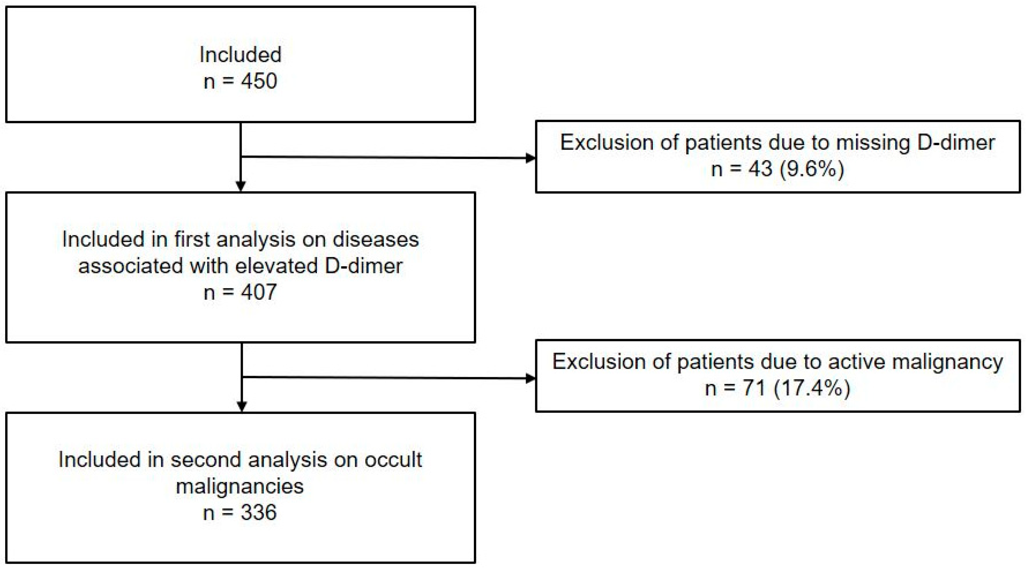
Figure 1. Flowchart of patient selection.
Ecm 02 00056 g001
Figure 2. Distribution of D-dimer levels.
Ecm 02 00056 g002
Figure 3. Kaplan–Meier curve of malignancy free survival. Colors represent different categories: green is D-dimer < age-adjusted value; blue is D-dimer > age-adjusted − 2000 µg/L; and red is D-dimer > 2000 µg/L.
Ecm 02 00056 g003
Table 1. Baseline characteristics of the study participants.
CharacteristicsAll Patients
n = 407
Age, median (IQR), years79 (73–85)
Male sex, n%215 (52.8)
Comorbidity
Median CCI score a (IQR)1 (1–3)
Active malignancy before ED visit, n%71 (17.4)
Medication, n%
Anticoagulants124 (30.5)
Antiplatelet therapy159 (39.1)
Reason for ED visit (ICD-10), n%
Infectious and parasitic diseases113 (27.8)
Diseases of the digestive system113 (27.8)
Neoplasms b40 (9.8)
Diseases of the circulatory system34 (8.4)
Endocrine, nutritional, and metabolic diseases23 (5.7)
Diseases of the respiratory system22 (5.4)
Diseases of blood and blood-forming organs20 (4.9)
Diseases of the genitourinary system17 (4.2)
Miscellaneous25 (6.1)
Laboratory tests
D-dimer, median (IQR), µg/L (normal <500)1364 (683–3314)
AADD cut-off value c, n%284 (69.8)
Admission, n%302 (74.2)
Mortality
90-day mortality, n%79 (19.4)
6-month mortality, n%101 (24.8)
Abbreviations: AADD = age-adjusted D-dimer; CCI = Charlson Comorbidity Index; ED = Emergency Department; ICD-10 = International Statistical Classification of Diseases and Related Health Problems 10th revision; IQR = interquartile range. a According to the Charlson Comorbidity Index [12]. b Diagnosed before the ED visit. c Age-adjusted cut-off value D-dimer: patient’s age in years × 10 µg/L [16].
Table 2. Prevalence of underlying diseases in patients with and without elevated D-dimer levels.
DiagnosisTotalD-Dimer Levels (µg/L)
n = 407<AADD a
n = 123
(30.2%)
≥AADD-2000
n = 129
(31.7%)
≥2000
n = 155
(38.1%)
p-Value
Sepsis106 (26.0)22 (17.9)34 (26.4)50 (32.3)0.03
Infection (excluding sepsis)94 (23.1)22 (17.9)36 (27.9)36 (23.2)0.18
Active malignancy71 (17.4)20 (16.3)19 (14.7)32 (20.6)0.39
VTE b17 (4.2)0017 (11.0)0.02
Acute or chronic inflammatory
diseases
15 (3.7)4 (3.3)6 (4.7)5 (3.2)0.80
Ischemia6 (1.5)02 (1.6)4 (2.6)0.26
Abbreviations: AADD = age-adjusted D-dimer; VTE = venous thromboembolism. a AADD: patient’s age in years × 10 µg/L [18]. b Diagnosed within 7 days of ED visit.
Table 3. Occult malignancies.
Total
n = 336
Occult malignancy, n%31 (9.2)
Metastasized at diagnosis11 (35.5)
Time to diagnosis, median (IQR), days5 (1–23)
Type of malignancy, n%
Colorectal carcinoma5 (16.1)
Prostate carcinoma4 (12.9)
Acute myeloid leukemia3 (9.7)
Unknown primary3 (9.7)
Multiple myeloma2 (6.5)
Lung carcinoma2 (6.5)
Pancreatic carcinoma2 (6.5)
All other sites10 (32.3)
Abbreviations: IQR = interquartile range.
Table 4. Interval likelihood ratios (LRs).
D-Dimer
Levels (µg/L)
n = 336Occult Malignancy
Prevalence Per D-Dimer Group (%)Yes (n%)
n = 31
No (n%)
n = 305
LR (95% CI)
0–499482.11 (3.2)47 (15.4)0.21 (0.03–1.47)
500–999837.26 (19.4)77 (25.2)0.77 (0.37–1.62)
1000–1999827.36 (19.4)76 (24.9)0.78 (0.37–1.64)
2000–39995512.77 (22.6)48 (15.7)1.44 (0.71–2.90)
4000–79994613.06 (19.4)40 (13.1)1.48 (0.68–3.21)
≥80002222.75 (16.1)17 (5.6)2.88 (1.14–7.26)
Abbreviations: LR = likelihood ratio.
Disclaimer/Publisher’s Note: The statements, opinions and data contained in all publications are solely those of the individual author(s) and contributor(s) and not of MDPI and/or the editor(s). MDPI and/or the editor(s) disclaim responsibility for any injury to people or property resulting from any ideas, methods, instructions or products referred to in the content.