Review

Firearm Injuries: A Review of Wound Ballistics and Related Emergency Management Considerations

by
Panagiotis K. Stefanopoulos
1,*,
Gustavo A. Breglia
2,
Christos Bissias
3,
Alexandra S. Nikita
4,
Chrysovalantis Papageorgiou
5,
Nikolaos E. Tsiatis
6,
Efrem Serafetinides
7,
Dimitrios A. Gyftokostas
8,
Stavros Aloizos
9 and
Georgios Mikros
10
1
Independent Researcher, 22100 Tripolis, Greece
2
Orthopaedic Surgeon, National University of Comahue, Cipolletti CP 8324, Rio Negro, Argentina
3
Orthopaedic and Trauma Surgeon, Royal Commission Medical Center, Yanbu 46451, Saudi Arabia
4
Radiological Research and Medical Imaging Unit, Institute of Communication and Computer Systems (ICCS), National Technical University of Athens, 15773 Athens, Greece
5
Department of Pulmonary Medicine, Laikon General Hospital, 11527 Athens, Greece
6
Hellenic Police Forensic Science Division (FSD), 10442 Athens, Greece
7
Urologic Surgeon, Department of Urology, Asklipieion Voulas General Hospital, 16673 Athens, Greece
8
Independent Researcher, 34200 Istiaia, Greece
9
Intensive Care Unit, NIMTS Army Share Fund Hospital, 11521 Athens, Greece
10
Second Department of Surgery, 401 Army General Hospital, 11525 Athens, Greece
*
Author to whom correspondence should be addressed.
Emerg. Care Med. 2025, 2(4), 52; https://doi.org/10.3390/ecm2040052
Submission received: 25 July 2025 / Revised: 8 October 2025 / Accepted: 6 November 2025 / Published: 12 November 2025

Abstract

Gunshot injuries are challenging conditions because of the unique characteristics of the wounding agents producing soft tissue damage that may be compounded by the formation of an expanding temporary cavity (cavitation). Variations in ballistic performance leading to higher energy transfer by the projectile, including bullet tumbling, deformation, and fragmentation, cause increased soft tissue injury and may also lead to more extensive bone comminution compromising local blood supply. Once life-threatening injuries have been excluded or properly addressed, the emergency management of localized trauma from bullets and shotgun pellets may be complicated due to progressive tissue necrosis within the zone of injury. Additionally, the risk of infection should be tackled, especially in high energy bone injuries. War experience suggests a baseline separation between wounds with limited tissue destruction which can routinely be managed as simple penetrating injuries and those resulting from high energy transfer to the tissues involving a substantial amount of necrotic elements surrounding the wound channel which call for a more aggressive surgical approach. A further justification for such a distinction is the need for antibiotic therapy, which varies according to most studies depending on the wounding mechanism, the nature of the wound, and the extent of tissue injury. The emergency physician should also be aware of the possibility of “bizarre” bullet paths resulting in occult injuries of important anatomic structures.

1. Introduction

The greater availability of firearms in modern societies has resulted in increased frequency of gunshot injuries and deaths among civilians, due to suicide attempts, assaults, and unintentional discharge of guns [1]. Whereas most self-inflicted injuries of this type result in death and only few wounded arrive in hospital emergency departments alive, more victims of firearm-related violence, in general, suffer nonfatal injuries than die [2]. Firearm injuries remain challenging because of the unique characteristics of the wounding agents and the resultant trauma which may involve unpredictable wound tracks and additional collateral damage due to the phenomenon of cavitation leading to progressive tissue necrosis, especially with high energy injuries. Such injuries are seen in mass shooting incidents against civilians by lone actors and terrorist groups using military-type rifles [3,4].
Effective management of gunshot injuries in the emergency department (ED) requires an understanding of the behavior of bullets and other types of projectiles (e.g., fragments, shotgun pellets) in the human body to support the diagnostic process and the interpretation of imaging studies [5,6]. Moreover, certain aspects of the initial management of these injuries, such as the benefit of antibiotic prophylaxis and the removal of retained projectiles, have to be addressed according to the currently best available evidence [7].
The aim of this article was to review ballistic aspects of gunshot wounds pertinent to the ED management of these injuries. To identify relevant studies, an online search was performed in the PubMed database from 2000 to 2025, using the MeSH terms “firearms”, “forensic ballistics”, and “gunshot wounds” in various combinations with specific injuries and procedures (e.g., “bone fractures”, “thoracic injuries”, “thoracotomy”, “abdominal injuries”, and “vascular system injuries”) and additional keywords, such as “emergency medical services”, “debridement”, and “antibiotics”. A second search included the Scopus database for articles and book chapters, with the keywords “gunshot injury”, “gunshot wounds”, and “ballistics”, excluding publications about air guns and rubber bullets. All search was limited to the English language. Inclusion criteria were for studies which encompassed ballistics of gunshot injuries, such as high-velocity projectiles; those considered applicable in the context of ED management were selected for full review.

2. Wound Ballistics: The Dynamics and Interaction of Projectiles Penetrating Tissue

Gunshot injuries are generally classified according to the velocity of the penetrating projectile [8] based on the premise that high-velocity projectiles, usually defined as exceeding 600–760 m/s, have the potential for far greater tissue destruction because of the magnitude of the temporary cavity they produce [9,10,11]. Common handgun bullets, which are associated with the vast majority of civilian gunshot injuries, are low-velocity projectiles since they have an initial (muzzle) velocity around the speed of the sound in air (approximately 340 m/s) considered the upper limit for the low-velocity range [12], whereas bullets from rifles (military and hunting) fall into the high-velocity category [11]. Medium velocities between 400 m/s and 450 m/s are typically attained by bullets from Magnum handgun cartridges [8].
The role attributed to the projectile velocity as the single most important wounding factor is theoretically justified by its contribution to the kinetic energy of the projectile, which represents the quantification of its wounding potential [8,10,13]. The formula K = 1/2 mv2 states that the kinetic energy K associated with the motion of an object is proportional to its mass m but the major determinant is velocity v which enters the equation raised to the second power. This energy can be transferred once a decelerating force applies on the moving object [14], which is the case with the retarding force (drag) from the tissue resistance against the projectile motion. A projectile that comes to rest within the body has delivered all of its available energy to the tissues [9].
Nevertheless, the kinetic energy formula is not an accurate depiction of the complex projectile–tissue interaction [15,16]; certainly, it does not reflect the tissue damage from tumbling projectiles and the type of injury due to the temporary cavity and bullet disintegration (fragmentation) [17]. Projectiles with the same kinetic energy at impact but different masses and velocities behave differently in the same type of tissue, creating different wound morphologies [16]. In experimental ballistic tests using gelatin as an ideal skeletal muscle simulant [18], the penetration depth is usually less for the projectile that is smaller and faster compared to the heavier, slower one [19]. The latter causes mainly crushing and disruption of the material it contacts, while the lighter, faster sphere dissipates most of its energy by propelling tissue sideways, resulting in the formation of the temporary cavity (cavitation) [18].
The energy transfer characteristics, namely, the amount of energy transferred to the tissue, the timeframe within which this occurs, and the surface area over which the transferred energy is distributed, determine the degree and extent of tissue destruction [10]. Certain bullet types, such as those designed to expand upon impact, available in semi-jacketed soft-point and hollow-point types, aim at maximizing the tissue damage by increasing the amount and rate of energy transfer and the surface of energy distribution [20,21,22]. The jacket in these bullets leaves the soft lead core exposed at the tip, which expands in the form of mushrooming within 2 to 4 cm after penetration [23]. As a consequence, the bullet diameter may increase by 2.5 times, resulting in a corresponding increase in the surface area of the tissue that sustains direct damage from crushing and tearing approximately 6.25 times [19]. However, this increase in wound diameter becomes significant only with expanding rifle bullets, normally used for hunting [9].
Modern designs of military bullets are full metal-jacketed (FMJ), containing various components of steel with or without a lead core. The jacket completely covers the tip preventing expansion—but usually leaves the base of the bullet open [9] (Figure 1). Despite the ban imposed on expanding (“Dum-Dum”) bullets for military purposes by the Third Declaration of the 1899 Hague Convention, on the grounds of avoiding unnecessary suffering of the wounded [24], currently there are reports of illegal ammunition with modified expanding bullets used in the war of Ukraine [25,26].
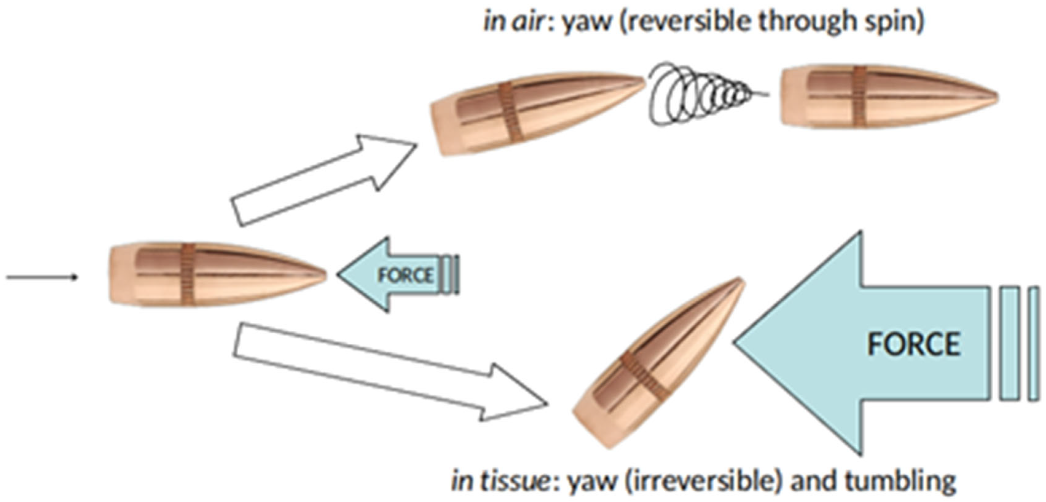
Military rifle bullets penetrate tissue initially with the longitudinal axis of the bullet close to its line of trajectory (i.e., with nose-forward orientation) [27] creating a wound channel with roughly the same diameter as the projectile, termed the narrow channel [23,28]. Beyond the narrow channel the wound diameter increases as the bullet becomes destabilized because of the action of the retarding force. By concentrating on the bullet’s forepart, the retarding force creates an overturning moment which prompts the bullet to yaw [29]. Yaw is the deviation of the bullet’s longitudinal axis from its line of trajectory [9]. Since modern rifle bullets have a streamlined (“spitzer”) contour, any such change in their orientation can only increase their presented area, thereby increasing the magnitude of the retarding force by positive feedback [29,30] (Figure 2). In the case of the 5.56 mm NATO bullet used in the M16 and M4 military rifles, at a yaw angle of 90 degrees its presented area increases approximately 3.5 times (from 25.4 mm2 in its nose-forward orientation to 88.1 mm2) [30].
A tendency to yaw is a feature of military rifle bullets (mushrooming of expanding bullets provides shoulder stabilization preventing yaw [23]). This tendency already exists during the flight in air but is effectively counteracted by the gyroscopic stabilization of the spin (imparted to the bullet by the rifling of the bore of the gun). In tissue, however, stabilization is rapidly lost because of the higher density of the medium and the enormous retarding forces [9,19]. Consequently, tumbling (rapid yaw) of the bullet increases the diameter of the wound channel. Maximum tissue damage occurs once the bullet travels sideways (at 90 degrees of yaw) striking tissue with its whole length [19]. In terms of energy transfer, the energy exchange at this point also reaches its maximum [8,9,10,31].
Bullet fragmentation is common as a result of bone impact while it also occurs with expanding rifle bullets containing a lead core as they penetrate soft tissue [9,32,33]. A different pattern of breakup, also within soft tissue, affects military rifle bullets with a lead core. Above a certain penetration velocity, approximately at 600 m/s, as these bullets tumble moving sideways, the extreme bending stresses encountered due to the retarding force cause flattening of the jacket [23]. At still higher velocities, the bullet may even fracture, usually at the cannelure (the circumferential crimp on the jacket around which the cartridge case holds firmly the bullet in the unused ammunition) releasing metallic fragments within the tissue [19]. This results in a snowstorm appearance of the wound in plain radiographs [9].

2.1. Pathophysiology of Bullet Wounds and Cavitation Injuries

The nature and severity of bullet injuries depend primarily on anatomic factors related to the shot placement and the bullet trajectory [34,35,36] and the functional effects resulting from the anatomic structures and organs disrupted [37,38] (Figure 3). Projectile characteristics, some of them inherent (mass, contour, and construction) and other developing during the passage from the gun barrel (velocity, stability), also play a role as wounding factors [19]. Although the caliber, i.e., the diameter of the bullet (expressed either as fraction of an inch or in millimeters), is not considered an important determinant of the wounding effect [19], larger caliber handguns have been associated with an increased likelihood of death from gunshots following criminal assaults [39]. The most common cause of death in shooting incidents is hemorrhage [40,41]. Gunshot wounds to the head may be compatible with survival depending on the bullet trajectory [16,37], whereas in case of involvement of the brainstem, death ensues instantaneously [41].
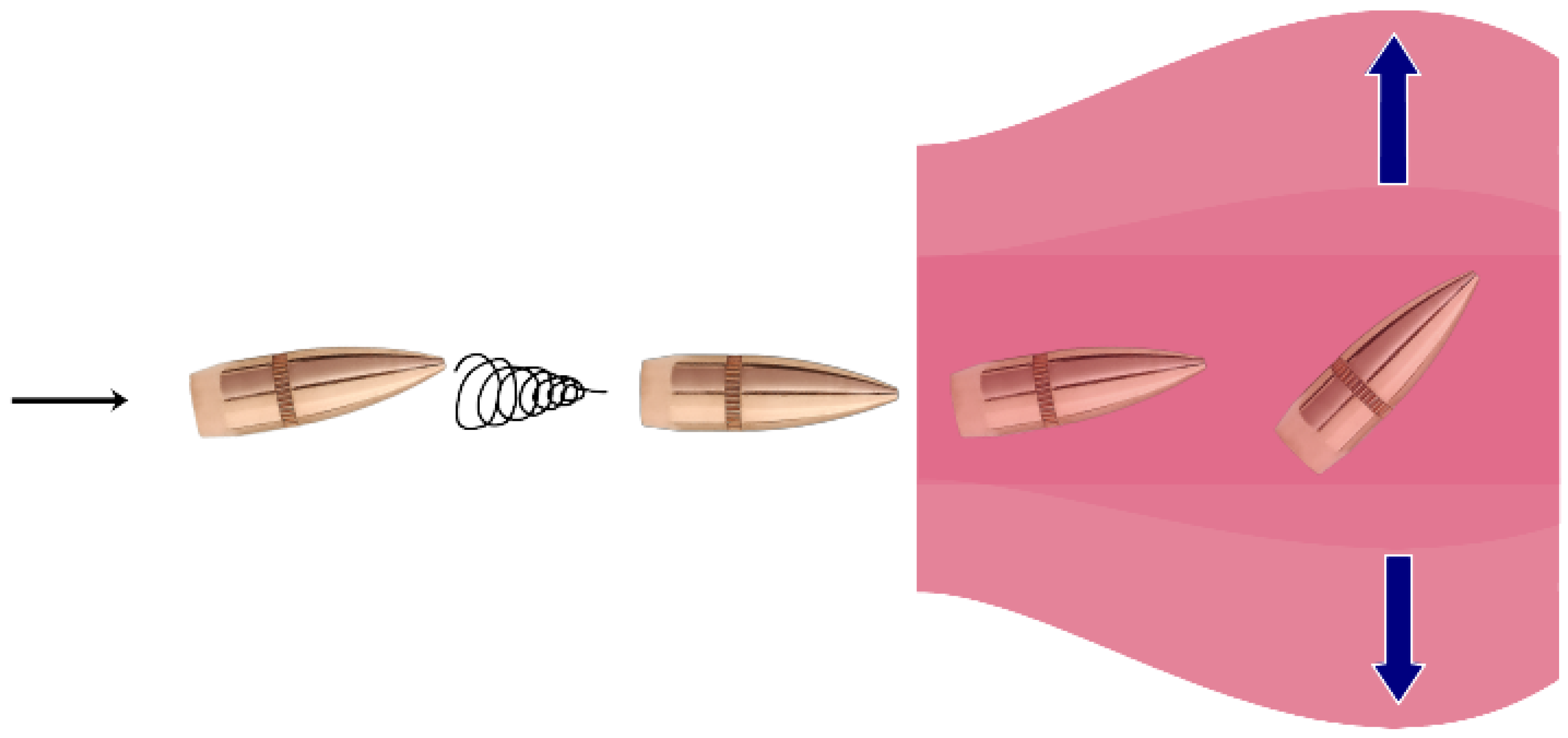
Two basic components of tissue damage have been recognized in bullet wounds [8,19,28,42]: the permanent wound channel which is the area of overt tissue destruction from the passage of the bullet, and the temporary cavity that forms in the wake of the bullet during the very wounding process, invisible to the naked eye. Injury from low-velocity projectiles results from direct tissue damage due to crushing and laceration. As a result, in order to cause significant injury to a structure, a handgun bullet must penetrate that structure (Figure 4). High-velocity bullets possess enough energy to cause additional indirect damage through cavitation and possibly secondary injury due to bullet fragmentation [9,27]. Cavitation occurs as the bullet track expands with its walls thrust outwards, at velocities that reach up to one tenth that of the projectile from the deposition of its kinetic energy (Figure 4). The cavity created by this phenomenon involves massive tissue displacement and it increases in size several times the diameter of the projectile, within milliseconds after the latter has left the body [43,44]. In sufficiently elastic tissue, the cavity after some undulation collapses undergoing a violent implosion, thus demonstrating both its temporary character and the highly dynamic nature of this phenomenon [31,43].
The degree of tissue damage from cavitation depends on the type and properties of the tissue(s) perforated by the bullet [9]. The tissue properties also affect the projectile–tissue interaction which has a central role in the wounding process [43]. Cavity expansion results in tensile injury [43,45], consisting in the so-called stretch mechanism of the temporary cavity [16]. Solid inelastic parenchymatous organs, such as the liver, suffer disruption over the whole extent of the temporary cavity [8,43,46], whereas skeletal muscle, due to its elasticity, is believed to undergo mostly reversible injury from the stretch mechanism [18]. Indirect injury to internal organs, lying at a distance of several cm from the path of the projectile, can occur from the pressure waves associated with cavitation, sometimes referred to as “blast injury” [47,48,49,50].
The lungs are affected to a significantly lower degree from cavitation because of the compliance and low density of the lung parenchyma [8,46,51], although transthoracic injuries may be associated with extensive pulmonary contusion in radiographs [5]. Increased pulmonary injury from cavitation has been reported with tangential thoracic gunshot wounds [52,53,54], probably because of involvement of the high-density tissues of the chest wall [52,53,55].
In head injuries, formation of a temporary cavity is an important cause of the ensuing pathology since even low-velocity FMJ bullets create a significant cavitation effect with pressure waves reaching 644 kPa [56]. The resultant zone of destruction of brain tissue, which extends up to 18 mm, can account for the onset of respiratory arrest leading to death in case of brainstem involvement [57]. Cavitation from rifle bullets penetrating the head is associated with extreme pressures exceeding 2760 kPa, which, amplified due to the unyielding brain case, produce explosive fractures of the calvarium from within [28].

2.2. High Energy Injuries

High energy bullet wounds are characterized by tissue damage extending peripherally to the wound track as a result of the temporary cavity formation following penetration by a high-velocity projectile [58]. The size of the temporary cavity is directly related to the projectile energy transferred to the tissue [9,10]. Clinically significant cavitation is a predominant manifestation of high energy transfer resulting in a “zone of injury”, often extensive, surrounding the wound channel [13]. Due to the extensive rupture of small blood vessels from the stretching effect of the expanding temporary cavity, the zone of injury has the appearance of bruised discolored muscle [29]. Within this zone, a pattern of progressive necrosis is encountered, which is not obvious during the initial examination [59,60,61]. This evolving process has been reported in experimental animals too, sometimes even after formal debridement [62,63,64]. Extensive endothelial trauma affecting the microcirculation beyond the limits of obvious tissue damage, which is proportional to the energy transferred, appears to be a critical event in the underlying pathology [43,65,66].
In the absence of gross infection or vascular impairment, whether muscle necrosis occurs other than at a microscopic level has been a matter of debate [28]. The different response of muscle to cavitation in various experimental studies may be related to the animal species used [58] and the underlying patterns of involvement of vascular territories in the muscle tissue [67]. The temporary cavity also creates a strong suction effect forcing debris to enter the wound from the environment [27]. The wound demonstrating a large amount of dead and ischemic non-viable tissue contaminated with foreign material represents the unique pathology of the high energy gunshot injury [28].
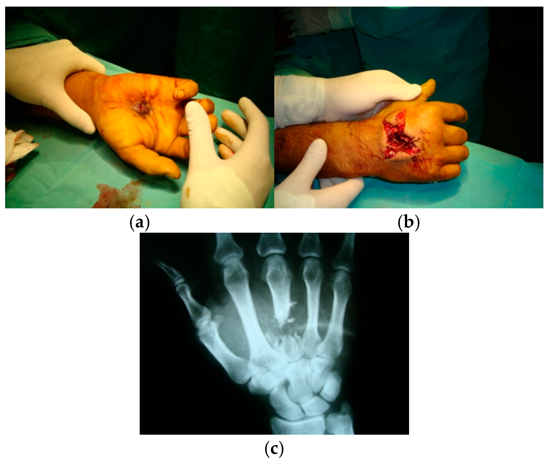
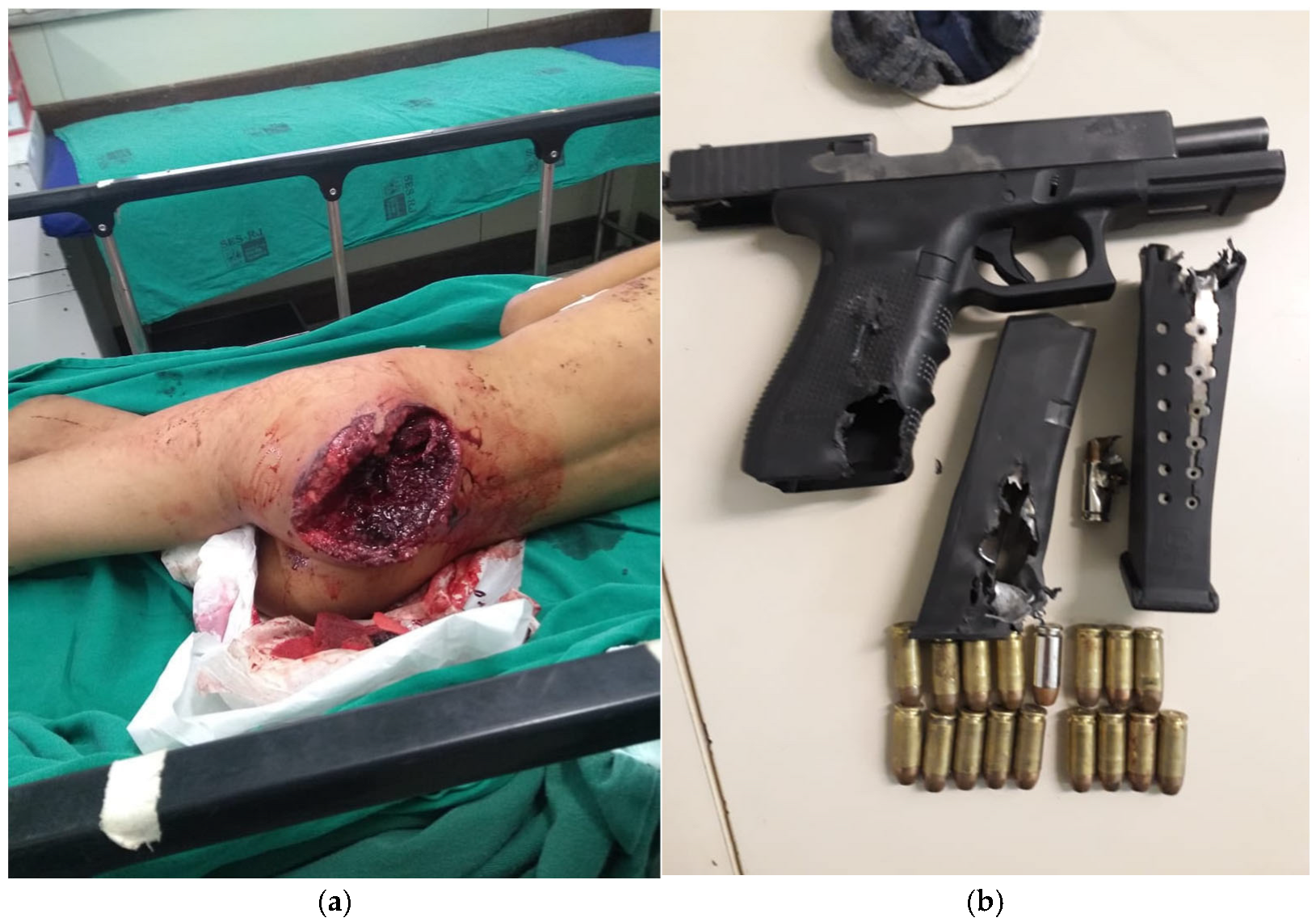
In general, high-velocity projectiles have the capacity of completely perforating the human body [20,22] creating longer wound tracks and potentially penetrating through multiple anatomic parts. They also cause a wider zone of injury around their path compared to wounds produced by lower velocity bullets [68]. A deforming or disintegrating bullet from a powerful handgun is also capable of producing high energy effects to the wound with a large temporary cavity [58,69]; a large handgun bullet may produce a similar effect because of a bone hit (Figure 5).
A large exit wound is always a sign of high energy transfer [20,58]. In the case of a non-deforming military-type rifle bullet, it results from expansion of a large temporary cavity near the point of exit associated with bullet tumbling or breakup, or expulsion of bone fragments acting as secondary projectiles [64,70,71,72]. This is more likely to occur with wounds of the torso involving a longer wound track [73]. A small exit hole from a tumbling bullet that has eventually turned 180 degrees leaving the body base-forward may conceal extensive injury in a deeper layer from high energy transfer [27,28] (Table 1).
Bullet fragmentation in soft tissue can be regarded as the most extreme manifestation of high energy transfer by high-velocity projectiles, consistently associated with greater tissue destruction [55,72] (Figure 6). The fragments created from a high-velocity bullet often have no sufficient energy to exit the target [7,26,74]. The resultant tissue damage involves a synergistic effect with cavitation, as the fragments create secondary wound channels extending radially from the main wound track, and the subsequent stretch mechanism causes more extensive disruption of this damaged tissue than what would be expected by this mechanism alone [32,74].
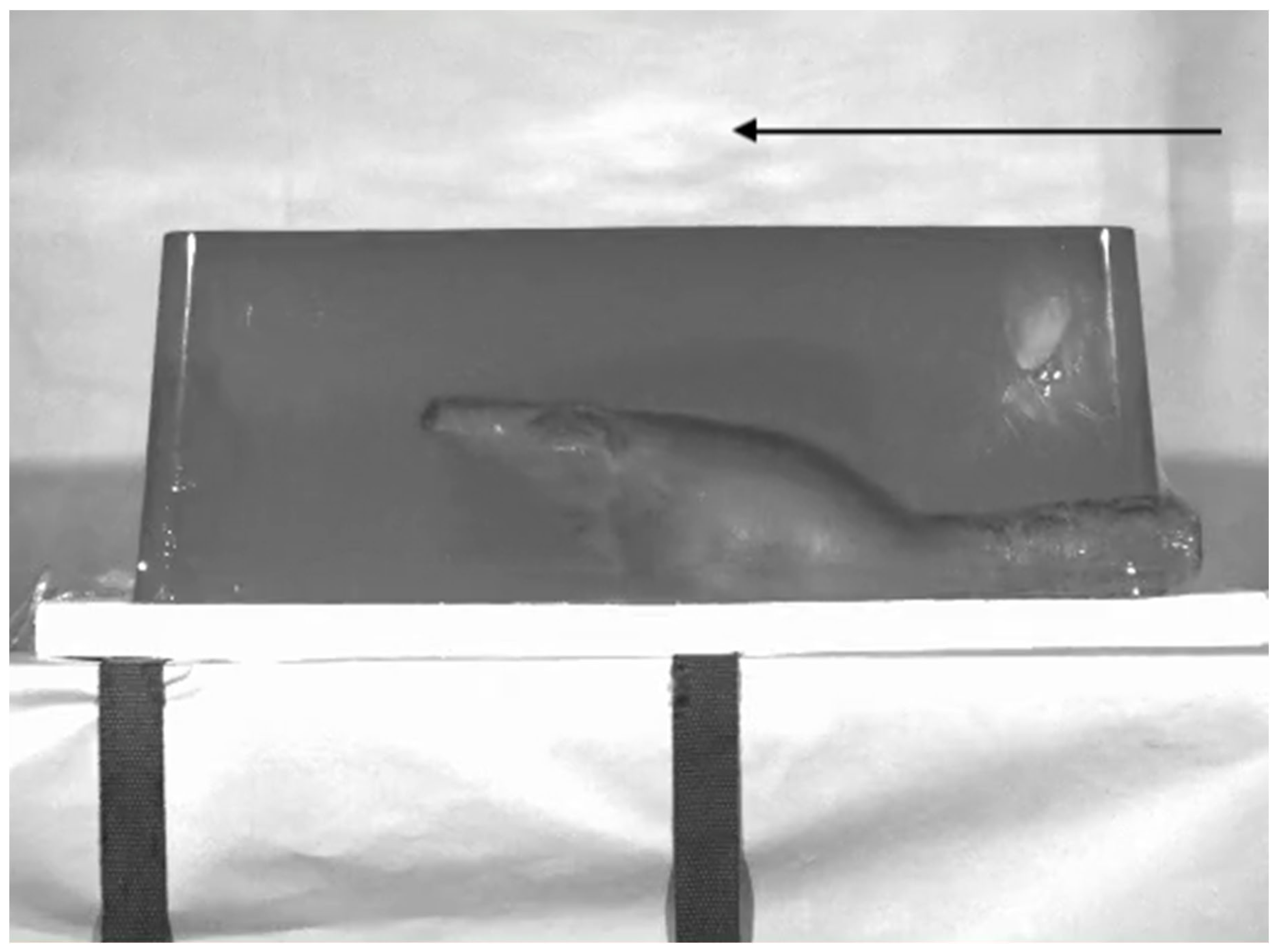
Following mass shootings in the United States, in which the AR-15 rifle (the semi-automatic civilian version of the M16 assault rifle) had been used, press releases falsely attributed the large wounds of the victims to the high velocity of its bullet (exceeding 900 m/s), sometimes exaggerating its effects stating that the bullet blows up in the body [9]. The 5.56 mm NATO lead core bullet used in the M16 rifle and the M4 carbine (the corresponding sporting ammunition for the AR-15 rifle is the 0.223 Remington) is particularly effective in energy transfer, not because of its velocity but because of a tendency for tumbling and possibly fragmentation during soft tissue penetration. The bullet actually relies on this mechanism to create extensive tissue damage [9,19], although it has been shown that fragmentation does not consistently occur with penetration velocities below 760 m/s [71] while it also necessitates a sufficient width of tissue to allow for bullet tumbling (Figure 7). In borderline cases, pre-impact yaw has a critical role in reducing the length of the narrow channel and precipitating early tumbling [23,45].
Gunshot wounds of the extremities are often complicated with vascular trauma, in some cases possibly as an indirect result of a large temporary cavity [75], which can cause compartment syndrome leading to ischemic necrosis and infection [17,76]. High energy gunshot injuries sustained in combat have shown an increased rate of vascular injury [77], amounting to 26–33% [78]. Significantly lower rates (<10%) among civilian cases [60,79] likely suggest a causative role of cavitation in the context of vascular injury associated with high energy ballistic trauma. The mechanism of vascular damage most commonly involves stretching of the vessel wall resulting in intimal tears and thrombosis [80]. Neurologic deficits of different degrees may also be encountered in extremity wounds, either indirectly from the severe stretch produced by cavitation or from direct laceration or contusion by the projectile [81,82].

2.3. Shotgun Injuries

Shotguns are smooth-bored (non-rifled) long-rifle firearms, which fire either multiple pellets collectively known as the shot or a single projectile (slug). Pellets are separated by size into two general categories, birdshot (of smaller diameter used for birds and small game) and buckshot (for large game) [83]. Lead pellets have an upper limit of velocity, approximately 420 m/s, above which collisions between individual pellets cause deformation whereas for steel pellets this is not a limiting factor and higher velocities can be attained [9]. In many areas, including the European Union and the United States, lead shot has been banned for game hunting in wetlands because of lead poisoning of waterbirds [83]. Upon discharge, the pellet spread (“pattern”) attains a conical shape [76] and the wad, usually a plastic insert isolating the shot from the propellant within the shotgun cartridge, is also expelled from the muzzle [19,83]. As a result, the firing range from muzzle to target is the primary determinant of the wound morphology [9,84]; it affects not only the number of pellets that hit the target but also the density of the shot pattern.
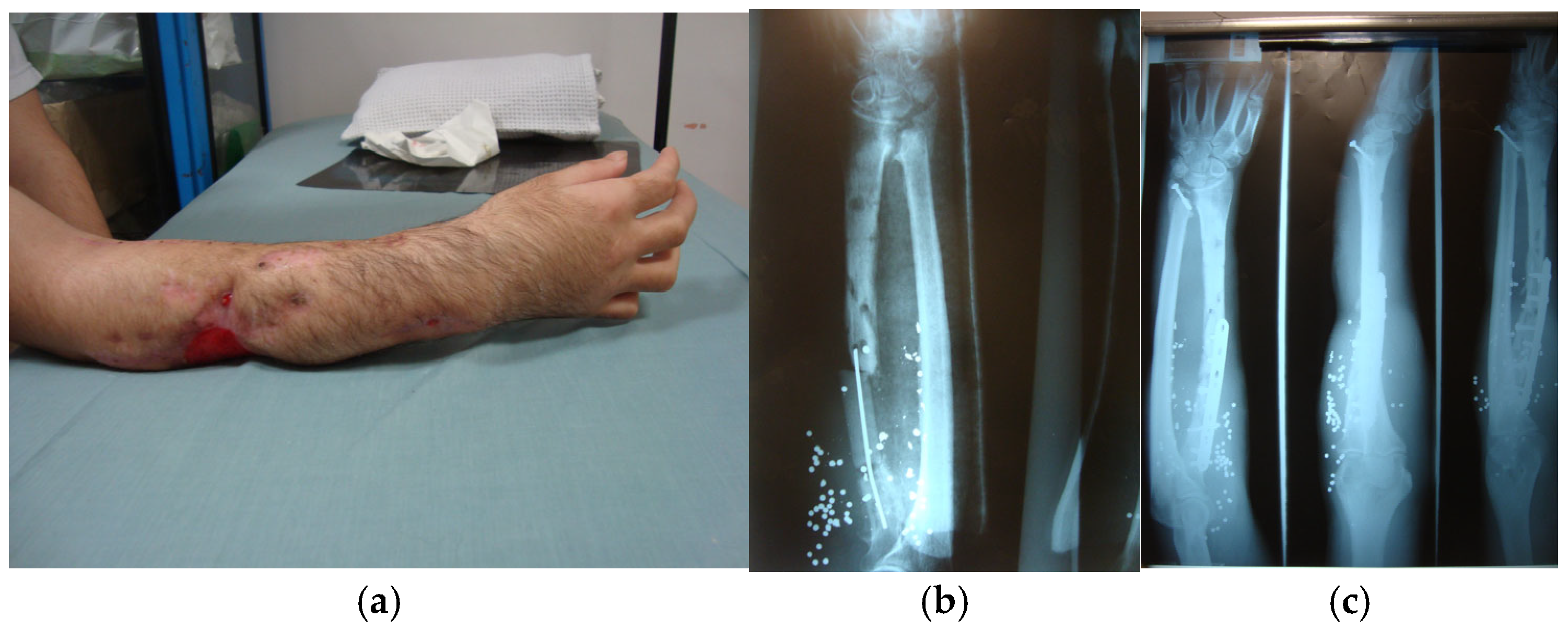
Within close distances (<3 m), the pellets are still bunched together acting as a single projectile, with their initial velocity virtually unchanged [19]. At such range, the shotgun is a formidable weapon [9] and the resultant wound involves massive tissue destruction [12,74,84,85] (Figure 8). Up to a certain point, the size of the wound increases with range [16]. The wound appearance is determined by the gauge (caliber) of the shotgun and the choke, a constriction of the barrel at the muzzle which controls the diameter of the shot pattern. The gauge influences the density of the pattern, depending on which the wound either consists of a single large cavity or contains multiple wound channels from individual pellets. The size of the wound, however, is affected by the diameter of the pattern, which is governed by the degree of choke [86]. With increasing distances within a range of 1–3 m, the wound margins appear increasingly more scalloped and ragged, reflecting the gradual divergence of pellets at the periphery of the shot, until smaller wounds from individual pellets congregate surrounding the main wound of entry [9]. The wad can also reach the target over relatively short distances, and within approximately 2 m, for as long as the whole shot enters the body through a single wound, the wad should be expected to be found within the wound as radiolucent foreign body [86]; however, it does not participate in wounding beyond distances of 5–7 m [83]. Because of the unfavorable ballistic shape of pellets, their velocity drops rapidly [9], and beyond 6 m, due to the resultant loss in their kinetic energy, they tend to cause limited tissue damage [12].
Shotgun slugs have muzzle velocities ranging approximately between 400 and 560 m/s. They are heavy projectiles made of lead or copper alloys, which within their effective range of 100 m produce extensive internal injury, demonstrating extreme shocking effects, due to rapid delivery of their kinetic energy to the target. Energy transfer to the wound is achieved because of the blunt shape of the slug associated with marked cavitation and the tendency of lead slugs to flatten or breakup [87,88]. Cavitation has no significant role in injuries from shotgun pellets [9].

2.4. Bone Injuries

Bone is significantly denser and harder than the surrounding soft tissue, also demonstrating very limited elasticity. These characteristics result in greater amounts of energy transfer following bone hit by a projectile, which sustains early destabilization with temporary cavity formation and possibly breakup [16,58]. Low-velocity projectiles with sufficient energy to completely perforate the bone cortex on both sides, producing drill-hole fractures and butterfly fractures [89,90]. Bone perforation characteristically follows a pattern known as cone cracking, resulting in a conoidal defect with the exit hole larger than the entry (beveling) due to tensile stresses around the contact area [91]. At higher velocities radiating fractures appear propagating outwards from the point of impact [92,93]. This rather typical type of fracture has been shown with 9 mm FMJ bullet perforating the diaphysis of the human femur at 360 m/s; a round entry hole was created surrounded by radiating and concentric fractures lines that resulted in comminution and frequently a square exit bone defect [94]. At high velocities (>551 m/s), the bone demonstrates behavior of a brittle material [92]. Increased amounts of energy transferred result in larger and wider radiating fractures [95].
Severe bone comminution is mainly produced by the hydrodynamic phenomena associated with cavitation within the bone marrow [23]. Rifle bullets as well as those from larger and more powerful handguns, which are capable of higher energy transfer, tend to produce fractures of an explosive character as a result of the temporary cavity they induce [69]. Because of the associated soft tissue damage and disruption of the blood supply, these fractures are often complicated with infection, delayed union, or nonunion [19].
Indirect fractures, i.e., without physical contact of the fracture site with the projectile, can be seen in high energy injuries due to the pressure wave from cavitation. These fractures, which can occur probably within a distance of 3 cm from the projectile trajectory, are characteristically linear or have a wedged shape. This is caused as the expansion of the temporary cavity bends the bone beyond its tensile strength, which results in tension failure of the cortex on the opposite side [96].

2.5. Infectious Potential of Gunshot Wounds

Bacterial colonization of gunshot wounds mainly occurs from the skin flora and clothing. The bullet transports microorganisms and textile fibers into the tissue and in high energy wounds the strong suction effect of the temporary cavity causes additional contamination [23,97,98]. Debris is also present on the bullet surface, which is not self-sterilized from the temperature developing during its passage through the barrel [16,23]. Abdominal wounds may become heavily contaminated by the spillage of fecal content; additionally, colonic injuries are associated with increased incidence of infectious complications [99] which are more common in high energy gunshot wounds [100]. As a result, gunshot wounds are considered prone to infection, with wound infection rates around 15%, whereas high energy wounds appear to carry a greater risk because of the large amount of devitalized tissue present [7].

3. Initial Assessment and Resuscitation

The ED management of gunshot wounds is a continuation of the prehospital (field) triage and resuscitation of the patient [101,102]. In both settings, evaluation and initial management of injured patients generally follows the Advanced Trauma Life Support (ATLS) guidelines [10,103,104]. Prolonged time on scene has been associated with worse outcomes, especially in patients with penetrating trauma who were also hypotensive [105,106]. However, after incidents in remote geographical areas or in regions with a lack of transport facilities, even advanced primary management of the patient may have to be performed on the scene [107].
Information that should be sought from emergency medicine service (EMS) providers includes the victim’s approximate position at the time of wounding, which can help in assessment of the bullet trajectory and the distance of shooting, particularly in the case of a shotgun wound. Any available indication about the weapon type (handgun, rifle, or shotgun) and caliber can also be helpful for the initial appraisal of the character of the wound, its extent, and length [13]. The duration of prehospital cardiopulmonary resuscitation (CPR) also needs to be reported.
Hospital resources and different policies between various trauma systems may affect management decisions; in hospitals relatively unfamiliar with gunshot injuries, direct transportation of the patient to the operating room for resuscitation is the best option. In areas with armed conflicts and in cases of mass shootings or terrorist attacks, especially in low-income countries, the surgeons and operating rooms available may soon be overwhelmed by multiple casualties arriving within a brief period. With excessively high patient volumes outstripping the hospital abilities, priority for operative management is given to patients with the best chance of recovery [103,108].

3.1. General Principles

The patient arriving at the ED with one or more gunshot wounds, in parallel with the resuscitative process, is triaged into one of two broad categories: those who need hospitalization for immediate life-saving operative management and those who can safely undergo imaging studies in the ED other than plain radiographs, which are usually the initial and often the sole imaging test required. The vital signs of the patient on admission are among the factors that determine the general direction of subsequent management (Figure 9). Hemodynamically stable patients are assessed for the possibility of serious injury with diagnostic studies or procedures (while under observation for signs of active hemorrhage or respiratory distress). Unstable patients, on the other hand, after initial resuscitation and determination of the source of hemorrhage, undergo damage control resuscitation (DCR) in the ED and subsequently damage control surgery (DCS) for definitive hemorrhage control in the operating room, unless they respond and stabilize [103].
Patients in profound shock following exsanguinating injuries or in extremis due to chest trauma or severe head injury represent triage 1 category necessitating immediate treatment before proper diagnosis is made (“treat-then-diagnose” approach). These patients should be rapidly intubated (if not already) and attempts at resuscitation are immediately initiated according to the ATLS protocol, with compression of external bleeding and bilateral chest tube placement in case of chest trauma [103].
Patients who remain unresponsive, experiencing cardiac arrest (imminent or of less than 15 min duration), with evidence of thoracic, especially transmediastinal, or thoracoabdominal trauma associated with cardiac tamponade and/or massive hemothorax, are primary candidates for the daunting resuscitative ED thoracotomy [109,110,111,112]. Resuscitation of patients in extremis from exsanguinating neck or extremity injuries may also be attempted with ED thoracotomy and aortic cross-clamping, within less than 5 min of prehospital CPR [109]. Catastrophic hemorrhage from abdominal trauma is a relative indication for ED thoracotomy [111], but DCS is the primary treatment [113]; this is also applicable to environments with limited resources [107]. Even when the injury appears to affect the chest, laparotomy is more frequently needed than thoracotomy, because injuries requiring surgical operation are much more common in the abdomen [114]. ED thoracotomy is a procedure associated with extremely low survival rates, especially following blunt trauma and when there is no cardiac activity [110,112,115]. It should be avoided during major incidents because it consumes valuable resources, also rendering ED non-operable for some time afterwards, which is critical in case of mass casualties [107].

3.2. Hemorrhage Control

Translation of previous combat experience during the wars in Iraq and Afghanistan to civilian practice has shown that, similar to war injuries, most preventable deaths from civilian trauma are due to exsanguinating hemorrhage [116,117,118]. Recent evidence suggests that prioritizing hemorrhage arrest, rather than following the traditional ABC sequence (Airway, Breathing, Circulation) used in ATLS courses for initial trauma assessment has improved outcomes [118,119]. Early control of major hemorrhage in exsanguinating trauma has been implemented for victims of terrorist attacks with the use of military-type weapons [116,120]. This paradigm shift is expressed as <C> ABC (<C>: Catastrophic hemorrhage) or XABC (X: eXsanguinating hemorrhage) [74,101,107,108,119,121,122,123]. It has further evolved to a CAB approach (Circulation, Airway, Breathing) focusing on mechanisms that preserve circulation and perfusion in hypotensive injured patients [124]. Adoption of this novel approach is particularly recommended for low- and middle-income countries, where scarcity of human resources does not allow simultaneous implementation of the ABC components, as in major trauma centers [123]; this is all the more important since in these areas penetrating injuries resulting in profound hemorrhage are more common [124].
Emphasis on hemorrhage control, along with attempts to restore impaired or failing vital functions [38], applies both to the prehospital setting and the ED, despite the potentially different techniques employed in each case [120,125]. In many cases, prioritization of hemorrhage control and circulation has been designed for patients with compressible sources of bleeding [124], such as the prehospital application of arterial tourniquet advocated for life-threatening injuries of the limbs [74,101,120,125,126,127,128,129]. Junctional hemorrhage, on the other hand, is notoriously difficult to control and while it remains theoretically compressible, it usually requires prompt intervention surgically or with endovascular balloon occlusion [103]. Junctional tourniquet devices have also been recommended for hemorrhage from the groin or the axilla [106,127], although they are more common in the military [130].
Non-compressible torso hemorrhage is approached through ED thoracotomy followed by cross-clamping of the thoracic aorta [126,130]. Resuscitative endovascular balloon occlusion of the aorta (REBOA) is a new, less invasive, alternative method for hemorrhage control in vascular injuries below the diaphragm [112,123,130,131].

3.3. Trajectory Analysis and Imaging

Radiographic assessment is essential for determining the bullet path and the organs and anatomic formations affected, and also for locating any retained bullet or fragments [12,132]. This is a critical step for evaluation of the full extent of injury [132,133], which will subsequently help the surgeon choose the appropriate incision and exposure (past history of trauma with firearms and possible retained bullets is also required to avoid confusion with the present condition) [103]. The wound trajectory is primarily influenced by the posture of the victim, and an unusual bodily position at the moment of shooting can result in a strange or “inexplicable” erratic projectile path [27,35,114,134].
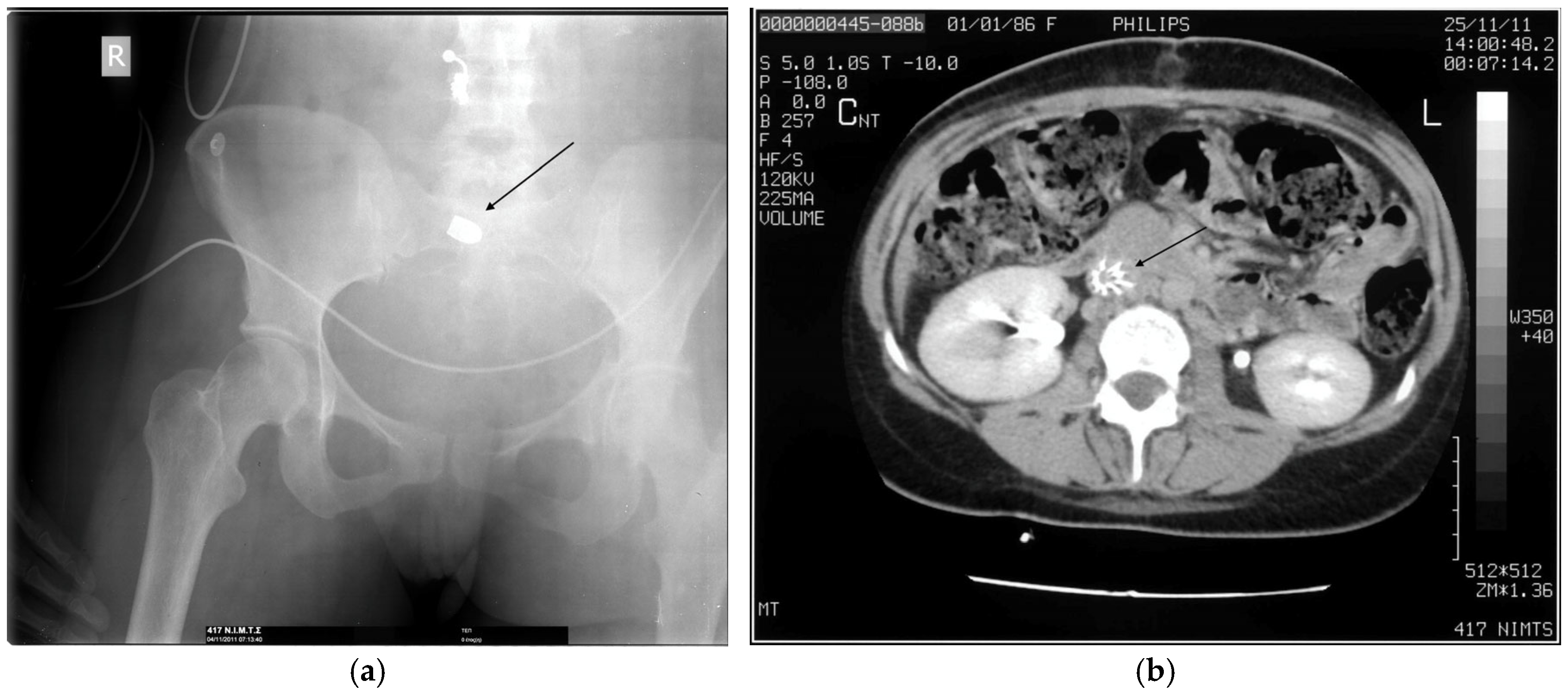
Foci of gas indicate the entry and exit wound (if it exists) [132], for which radiopaque skin markers are commonly used before obtaining a plain radiograph or a computed tomography (CT) scan [5]. While correct matching of entry and exit wounds is obviously important, speculation about which is which, based on inspection, should be avoided [74,76], unless there is unequivocal evidence from gunpowder deposits or muzzle imprint indicating the entrance side [13,135] (Figure 6a). The bullet may not follow a straight line connecting the entry and exit wound (or the retained bullet). Projectiles traveling at lower velocities may particularly be deflected off bony surfaces or strong facial layers [5,19,132]. Bullet fragments can sometimes be used to track the course of the bullet. CT analysis can clarify the trajectory with the aid of multiplanar oblique reconstructions [132].
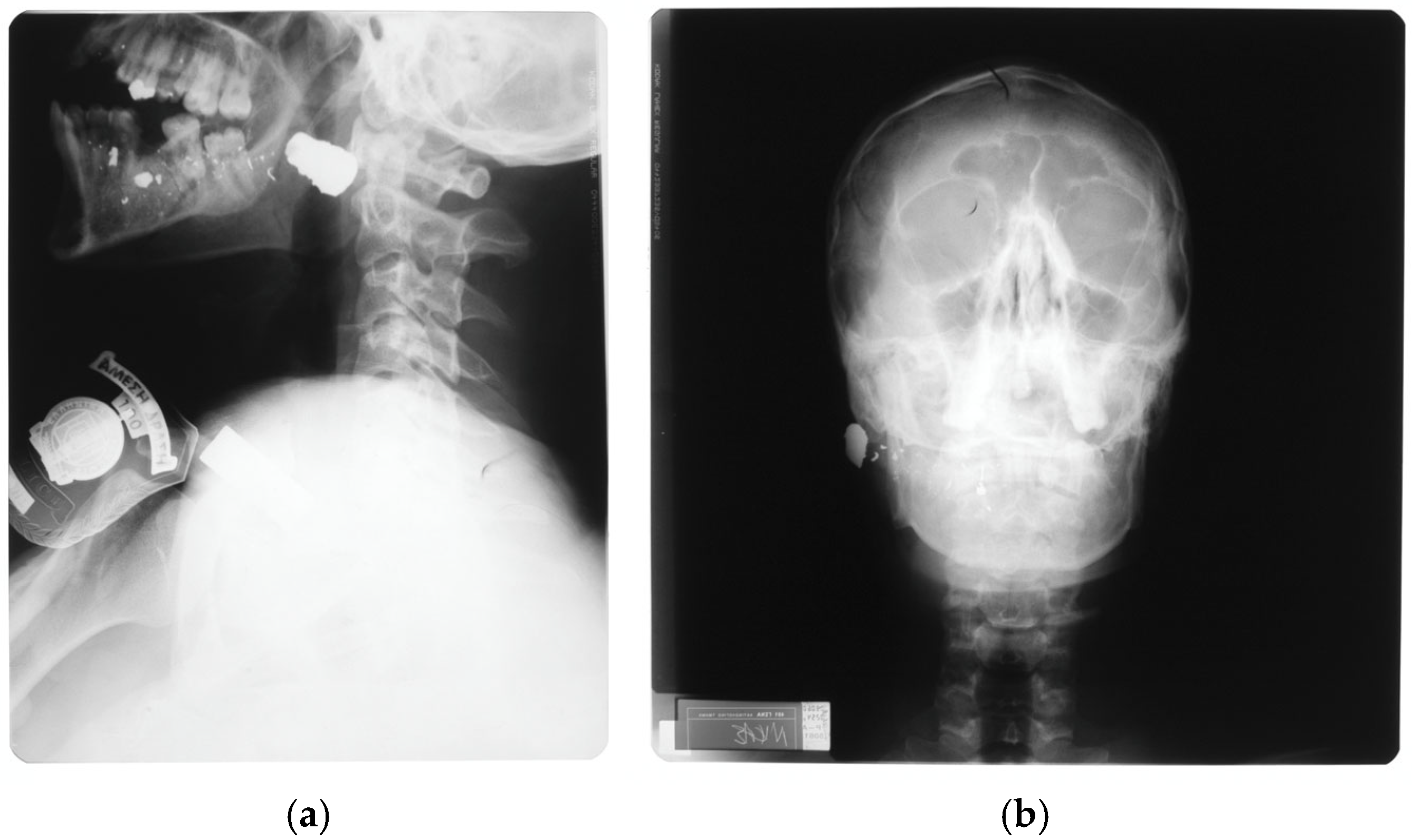
When only plain radiographs are available, the location of a retained bullet should be defined by obtaining two orthogonal views [5,19] (Figure 10). CT is superior in this respect (especially the scout CT images which provide a better overview of the wounded part) [5].
Migration of a bullet should be considered when there is no exit wound and yet it remains invisible in the diagnostic imagery available, necessitating further imaging investigation [19]. Ultrasound can be used to detect the current location of such a bullet [7]. Bullet migration occurs either by gravity or by embolization through the venous or arterial bloodstream. Bullets within serous cavities or in the subarachnoid space can move to a dependent location or to one influenced by positioning [7,19,136]. Spontaneous intracranial migration of bullets retained after gunshot injuries of the head represents a potentially life-threatening delayed complication of these injuries [137].
There are three clinical scenarios in which accurate delineation of the bullet trajectory is of critical importance. In head injuries, while the level of consciousness, estimated by the Glascow Coma Scale (GCS) score on admission, clinically remains a reliable predictor of long-term outcome; the wound trajectory, demonstrated in head CT scan, is the strongest prognostic factor of survival [138,139]. Deep bihemispheric and transventricular trajectories as well as those demonstrating multilobar or posterior fossa involvement are associated with high mortality rates [140,141,142].
Assessment of the trajectory is also important in neck wounds, because of the associated risk of vascular, neurologic, and upper aerodigestive tract injury [35,133,143], with additional damage potentially caused by cavitation [144]. Traditionally, anatomical division of the neck into three zones (I–III, from the chest up) has been used to determine the decision for surgical exploration in hemodynamically stable patients based on the level of the entry wound and the estimated possibility of involvement of deeper structures [145]. However, bullet trajectories are difficult to assess clinically, even within the limited anatomical boundaries of the neck, whereas nearly half of gunshot wounds to the neck extend to multiple zones [146,147]. Currently, the availability of computed tomographic angiography (CTA) has allowed a more selective approach to these patients, in the absence of “hard” signs of vascular trauma, such as expanding hematoma, absent carotid pulse, and carotid bruit, which mandate exploration regardless of the zone of injury [112,148].
The third clinical situation necessitating determination of the wound trajectory is a possible transmediastinal chest wound. A trajectory through the mediastinum should be suspected when there is an exit wound in the thoracic cavity opposite to the entry wound, when the bullet is found located in the thorax on the opposite side from its point of entry or in close proximity to the mediastinal space, or when the patient has sustained multiple gunshot wounds to the chest [149]. The possibility of cardiac injury should also be considered in cases with precordial or epigastric entry wounds [35,112]. CTA of the chest is the imaging study of choice for the diagnosis of a transmediastinal bullet wound [149]. Hemodynamically stable patients require evaluation of the vital structures contained in the mediastinum (heart, aorta, trachea, and esophagus) as they may harbor occult cardiovascular or aerodigestive tract injuries [76,112].
Trajectory considerations in abdominal gunshot wounds include transpelvic trajectories, associated with the risk of urologic trauma or rectal injury, and thoracoabdominal wounds. These represent absolute indications for CT imaging [112].
Shotgun wounds do not contain a single wound track that can be visualized on CT scans and surgically explored, and they represent challenging situations during the initial management [45]. Because of the presence of multiple pellets, these wounds are often complicated with numerous potential sites of vascular injury, which requires CTA for proper diagnosis. Embolization of small pellets through venous channels to the heart or causing occlusion of an artery is a serious complication that may occur as a delayed effect of these injuries [150].

3.4. Management Considerations

Gunshot wounds to the head and neck carry a high mortality rate [39,148]. Treatment of head wounds requires consultation with a neurosurgeon following stabilization of the patient [38,76]. These are complex injuries often involving extensive brain injury. Bone fragments in-driven from the entry site are commonly found within the brain and have been reported in more than one-third of civilian cases from handguns, sometimes creating secondary wound tracks [151]. The strong cavitation effect from rifle bullets [138,141] may also affect the cerebral vasculature [152].
Patients with gunshot wounds of the head who present with GCS >5–8, even with extruded brain, have an increased chance of survival [153]. Aggressive resuscitation similar to close head injuries (aiming at reversing intracranial pressure elevation) should be undertaken, with control of hemorrhage from the wound (although not a common cause of shock) and judicious fluid administration to restore blood pressure and cerebral perfusion in case of shock. Antibiotic coverage is commonly implemented [154]. CT of the head is required for assessment of the wound and the feasibility of surgical intervention. However, for want of a CT scanner, simple skull radiographs can still provide useful information [154]. Intubation is required for unconscious patients and when airway obstruction is anticipated [154]. Indications for urgent surgery with decompressive craniectomy as DCS are controversial [154,155]. Comparison of military and civilian injuries suggests that early decompressive craniectomy may contribute to better survival rates [155].
Gunshot wounds to the neck cause significant injury to underlying vital structures in approximately 50% of cases, which is higher in high energy wounds [156]. These are unique entities involving the dual danger of exsanguinating hemorrhage and airway compromise. Vascular trauma predominates in up to 40% of penetrating neck injuries [143,146], while approximately 9% of penetrating neck wounds present with some degree of airway impairment [146]. Control of external bleeding in the ED can be accomplished with direct pressure, use of hemostatic dressings or insertion, and inflation of a Foley catheter [147,156]. Intubation should be instituted in face of a developing edema or hematoma. However, if there is severe laryngotracheal injury, a surgical airway is usually performed, preferably with tracheostomy under local anesthesia to avoid further injury to the larynx [156,157,158].
Hemodynamically unstable patients with neck injuries are treated surgically or with interventional angiography for repair of vascular injuries and the same approach is used for stable patients presenting with hard signs of such injury (refractory shock, uncontrolled hemorrhage, expanding hematoma, pulse deficit, audible bruit/palpable thrill). Patients with only soft signs (minor bleeding, nonexpanding hematoma, proximity wound) undergo further investigation for vascular injury with CTA. Other indications for surgery (with immediate airway control) include signs of obvious or imminent airway compromise (e.g., stridor), massive subcutaneous emphysema, as well as positive diagnostic studies for laryngotracheal or esophageal injury [126,148].
In chest injuries, consultation with a trauma surgeon is required and tube thoracostomy should be instituted with a low diagnostic threshold [38,76]. Triage of life-threatening chest injuries should prioritize patients with airway compromise, pericardial tamponade, tension pneumothorax, and massive hemothorax, all of which can be rapidly fatal, thus necessitating immediate action without delay for confirmatory investigations [51,159,160]. Tension pneumothorax appears to be a common cause of potentially preventable death among fatalities in mass shooting incidents [161]. Therefore, both emergency physicians and EMS personnel should quickly assess gunshot victims for possible pneumothorax, which is amenable to pleural decompression through needle or tube thoracostomy [74,101].
Unlike the various possible causes of shock following a thoracic injury, hemodynamic instability in patients with intraabdominal injury can only result from major hemorrhage [162]. Early recognition of vascular injury is crucial and it is greatly facilitated by the increasing use of CTA [163]. These conditions require aggressive resuscitation with blood and blood products followed by definitive surgical treatment regardless of any associated injury [162]. Patients with abdominal gunshot wounds usually undergo exploratory laparotomy, even when they present with stable vital signs, because of the possibility of extensive intra-abdominal damage secondary to cavitation [74,76,164,165].
Nonoperative management of selected patients with abdominal gunshot wounds (hemodynamically stable, without signs of peritonitis or evisceration, with an intact level of consciousness) appears to be a safe approach in the absence of CT findings of hemorrhage, in nearly 40% of patients [112,164,166,167]. The major concern with this approach in gunshot wounds is to miss a hollow viscus injury, so the bullet trajectory in the CT of the eligible patients must not involve these structures [112]. Patients treated nonoperatively must remain under close observation for at least 24 h [168].
Thoracic wounds, particularly those below the fourth rib, or abdominal wounds, especially by high-velocity projectiles, should raise suspicion of thoracoabdominal injury traversing the diaphragm [169,170]. Thoracoabdominal gunshot injuries require laparotomy in two-thirds of the patients [112]. These injuries are often complicated by occult traumatic diaphragmatic injury, which may be clinically silent and remain undetected in the initial radiographic examination requiring a high index of clinical suspicion for its diagnosis [169].
Gunshot wounds to the extremities, in addition to fracture of long bones, may cause neurovascular injury [74,76,104]. Simple gunshot fractures can be treated conservatively, whereas those involving high energy transfer need operational management with external fixation for fracture stabilization, especially in lower extremity fractures [171,172]. Antibiotics are generally recommended despite the lack of evidence supporting their routine administration for low-energy gunshot fractures [7,104,172,173,174,175]. Articular gunshot injuries involving a joint after bullet penetration through the large intestine can lead to serious septic arthritis unless treated promptly, with washout, antibiotic therapy, and diverting colostomy to prevent fistula formation [176].
Investigation of suspected vascular trauma should be an integral part of the management of gunshot wounds in the extremities since vascular injuries often pose life- or limb-threatening conditions and require consultation with a vascular surgeon [104,177]. Active hemorrhage is easily diagnosed but ischemia is less obvious and may be missed when other injuries take precedence [178]. Hard signs providing strong evidence of vascular injury, which necessitates immediate surgical exploration, include active hemorrhage, expanding or pulsating hematoma, loss of distal pulses, and palpable thrill or audible bruit [104,126,179,180,181,182]. However, in patients with multilevel trauma to an extremity, e.g., from multiple gunshot wounds or shotgun injury, making uncertain the exact location of the vascular damage, diagnostic imaging may be required prior to surgery [180,181].
In the absence of hard signs, a history of significant hemorrhage, proximity of the injury site to major vessels, bruising or small stable hematoma, diminished distal pulses, and the presence of neurologic deficit (from injury to an anatomical related nerve) represent soft signs of possible vascular injury [104,180]. These signs prompt vascular workup, usually by means of arterial pressure indices (API)—with the exception of hemodynamically unstable patients who should also be transported to the operating room [180,182]. CTA, which is currently the gold standard for imaging of extremity vascular injury [150,181,183], can usually be reserved for patients with a pathologic API measurement (<0.9) [180,182,184,185]. When no other signs of vascular injury are detected, a history of high energy trauma represents an independent indication for investigation [180].
An updated approach to patients with suspected vascular injury of extremities [186] has suggested clinical evaluation on the basis of hemorrhagic and ischemic presenting signs. Hemorrhagic signs (active/history of significant hemorrhage, systemic hypotension, pulsatile mass/palpable thrill in proximity to the injury site, expanding hematoma) usually predominate in penetrating trauma and their presence may indicate a potentially life-threatening hemorrhage; ischemic signs (diminished pulse, abnormal API, cool limb, pallor, or impaired motor or sensory nerve function distal to the injury site) more likely appear from blunt trauma suggesting more extensive vascular pathology [178,187]. Vascular injury presenting with ischemic signs may be the result of cavitation. Patients with ischemic signs can also benefit from CTA, as it is critical for preoperative planning [178].

3.5. Soft Tissue Wounds

Soft tissue gunshot wounds produced by conventional low-velocity bullets but also wounds from high-velocity bullets creating a wound track within the limits of the narrow channel (Table 1) may be considered “low energy” injuries and treated conservatively as they are amenable to simple wound excision and irrigation [115,188,189]. These wounds are characterized by entry and exit holes smaller than 1 cm, connected by a short, narrow channel-type wound track, without any appreciable internal cavity or evidence of bullet fragmentation [188]. In the absence of fracture, the risk of wound infection appears to be low in these injuries [115], although administration of prophylactic antibiotics is generally recommended [189,190]. Tetanus prophylaxis is given as indicated [115,190,191].
High energy injuries require debridement and antibiotic coverage because of the extensive contamination along the wound track and the subsequent necrosis. In view of this necrotic tendency, surgical exploration and debridement may be planned as a serial procedure repeated every 24–48 h until the wound bed appears healthy [189,190,191,192] or performed in one stage with the aim to remove all necrotic and ischemic non-viable muscle tissue at once [193]. Traditionally, intraoperative estimation of muscle tissue viability is empirically based on the “4 C’s” classic criteria of color, consistency, contractility, and capillary bleeding [108,172,188]. These indicators used by inexperienced clinicians may lead to unnecessary excision of potentially survivable tissue as they have been reported to correlate poorly with the microscopic appearance of excised muscle [194]. It should be noted, however, that necrotic muscle fibers my appear “normal” by light microscopy several days post-injury due to the delayed phagocytic response as a result of devascularization [195].
Soft tissue gunshot wounds should be left open to heal by secondary intention or treated with delayed closure [108]. Facial wounds are an exception to this rule and, unless infected, they should generally be closed primarily because of the esthetic requirements and the rich blood supply of the area providing increased resistance to infection. Early debridement of these wounds, preferably in one stage, allows for primary definitive reconstruction of soft and hard tissues of the face, avoiding scar contraction [196,197].

3.6. Removal of Retained Projectiles

Removal of asymptomatic retained bullets should not be attempted routinely, except in cases of direct accessibility, because wound exploration may cause further injury and result in significant blood loss. It is indicated when the bullet is located within or near a joint, within the cerebrospinal fluid (CSF), or when there is concern about migration and damage to a nearby neurovascular structure [5,7,115,172,198].
Although the risk of lead poisoning from retained bullets is generally low, rarely may this condition result in chronic symptoms from various systems, such as anemia and neuropathy, which may appear long after the incident [5,198]. Bullets within a fracture site should preferably be removed, as lead projectiles have been shown to inhibit ossification and their presence may result in nonunion [104].

3.7. Management of Gunshot Wounds in Regions with Limited Resources

The same principles as above apply to environments with insufficient resources and to health systems of middle- or low-income countries. Lessons learnt from the treatment of war injuries in field hospitals are useful when logistics are restricted regarding supply of antibiotics and availability of imaging modalities [107,191]. Lack of resources or intense needs are not an excuse for suboptimal management of patients following mass casualties or in the presence of limited supplies [191].
In rural areas and developing countries, significant delays in transportation to the hospital may be the rule, whereas wounds from high-velocity projectiles possibly contaminated from the environment are often encountered. These wounds are prone to deterioration and require antibiotic prophylaxis for invasive infection [199]. The International Committee of the Red Cross (ICRC) antibiotic protocol [200] is recommended, which favors the use of a simple broad-spectrum antibiotic, such as cefazolin, for soft tissue wounds <72 h from injury. Old (>72 h from injury) soft tissue wounds and also abdominal wounds require additional coverage for Gram-negative (gentamycin) and anaerobic bacteria (metronidazole) [200]. Prolonged administration of prophylactic antibiotics is to be condemned.
It should be remembered that antibiotics are only supplemental to surgical excision of the wound, and they should not be regarded as a substitute for good surgical practice [108,199]. Trauma and orthopedic surgeons who have worked in humanitarian missions have emphasized the overwhelming importance of devitalized tissue removal and comprehensive washout of the wound, until healthy granulation tissue covers the wound bed. In most cases this may take at least 5 to 7 days. Moreover, necrotic soft tissue and bone should be removed as early as possible for antibiotics to effectively act through unhindered circulation. Wound debridement should preferably be carried out in one session, including tissue demonstrating questionable viability (unless part of an important structure), in order to spare operating time and resources and also to reduce unnecessary blood loss [108,193,199]. Inadequate debridement requiring subsequent revisions does not constitute proper surgical treatment [109].
Most clinical scenarios involving gunshot wounds do not require more advanced imaging than plain radiographs [134], and clinicians and radiologists working with limited resources should be aware that in certain cases CT imaging is superfluous as the necessary information can be obtained using good clinical judgement, for example, assessing the bullet trajectory without the need for extensive CT imaging, since most of these patients require operative intervention. Critical situations involving isolated gunshot wounds that do not necessitate CT imaging (or even preclude it because of the patient’s condition), include neck wound with hard signs of vascular or aerodigestive tract injury, chest wound with massive hemothorax, abdominal wound with signs of peritonitis or evisceration, and extremity wound with hard signs of vascular trauma or compartment syndrome [134].
Nonoperative management of abdominal gunshot wounds without indications for emergency operation may also be an attractive perspective in view of limited surgical resources. This decision, however, which should be made on an individual basis, needs to be balanced against the potential risks. Available resources for clinical observation should be taken into careful consideration, as these patients must be observed by experienced personnel in a close and continuous manner. Moreover, direct access to CT is important to support this policy [107].
Fractures, including those involving bone loss, may need to be initially treated with improvised orthopedic hardware in these environments [108] (Figure 8b). Antibiotics are routinely administered to these wounds considered prone to infection. Gunshot fractures may become infected, generally as a result of late presentation and establishment of early infection, or persistence of dead bone in the wound leading to osteomyelitis (late infection). In low-income countries, patients who develop osteomyelitis with chronic discharging sinus are often malnourished, which also needs to be corrected [199]. Local infections that are amenable to surgery do not require additional antibiotics other than the prophylactic regimen aimed at combating bacteremia due to the surgical trauma. However, patients that do not respond well to this treatment should receive full antibiotic therapy, which is often empirical, as laboratory results from culture and sensitivity testing have been found to have little relationship with clinical results [199].

4. Conclusions

The literature of gunshot wounds extends to a vast number of studies, which makes any non-systematic review attempting to summarize them prone to overestimating some of the results while underestimating others. In this review, an attempt was made to focus on studies containing specific reference to the ballistic aspects of wounding, which, however, further predisposes to risk of bias for two reasons. First, these studies tend to look at wounds from high-velocity projectiles, as in these cases the wound ballistics becomes both more interesting and important. In this way, other studies concerning important aspects of treatment, such as the nonoperative management of abdominal gunshot injuries, which applies mostly to wounds from low-velocity civilian weapons, may be largely overlooked. Second, studies that contain ballistic analysis of gunshot wounds may convey the impression that this is more important in the clinical setting than it actually is. Keeping these issues in mind, some useful conclusions can be drawn.
A knowledge of wound ballistics provides a platform for understanding the unique wounding mechanisms of penetrating projectiles, particularly the high-velocity ones. These are important considerations during interpretation of imaging data with clinical findings, since the presentation and the initial appearance of the wound usually do not reveal the full extent of the damage in deep structures. Identification of high energy gunshot injuries should also be supported by a background in ballistic characteristics of these wounds and their destructive potential, with clinical implications in cases involving vital organs or the vasculature. Recognition of the energy transfer effects in soft tissue wounds allows stratification of patients regarding the need for wound exploration and possibly more extensive or repeated debridement, and also the inherent risk for infectious complications. Wound ballistics, however, does not substitute for sound clinical judgment about cases that may be treated non-surgically or as outpatients on a selective basis and more serious injuries requiring life-preserving operations and extensive orthopedic procedures.

Author Contributions

Conceptualization, P.K.S., G.A.B., C.B., A.S.N. and G.M.; literature search: P.K.S., C.B., A.S.N., C.P. and S.A.; ballistic data curation: P.K.S. and N.E.T.; writing—original draft preparation: P.K.S., C.B. and E.S.; writing—review and editing: P.K.S., A.S.N., C.P., N.E.T., E.S., S.A. and D.A.G.; figure preparation: P.K.S., G.A.B., N.E.T. and S.A. All authors have read and agreed to the published version of the manuscript.

Funding

This research received no external funding.

Institutional Review Board Statement

Not applicable.

Informed Consent Statement

Not applicable.

Data Availability Statement

No new data were created or analyzed in this study.

Acknowledgments

The first author wishes to thank Thanos Chalkias, for his assistance with and critical review of the algorithm for the initial management of gunshot wounds. He is also grateful to Małgorzata (Gosia) Warmiñska-Marczak for kindly providing an inspection copy of the Textbook of Adult Emergency Medicine, Fifth edition, Elsevier, 2020, and a digital copy of the chapter on Hunting and Fishing Injuries of Auerbach’s Wilderness Medicine, Seventh edition, Elsevier, 2017.

Conflicts of Interest

The authors declare that they have no competing interests.

References

  1. Johns Hopkins Bloomberg School of Public Health. Firearm Violence in the United States. Available online: https://publichealth.jhu.edu/center-for-gun-violence-solutions/research-reports/gun-violence-in-the-united-states#:~:text=Evidence%20consistently%20shows%20that%20access%20to%20firearms%20increases%20the%20risk%20of%20suicide.&text=Access%20to%20a%20firearm%20in,suicide%20more%20than%20three%2Dfold.&text=Firearms%20are%20dangerous%20when%20someone,most%20lethal%20suicide%20attempt%20method (accessed on 1 July 2025).
  2. Centers for Disease Control and Prevention. Fast Facts: Firearm Injury and Death. 5 July 2024. Available online: https://www.cdc.gov/firearm-violence/data-research/facts-stats/index.html (accessed on 1 July 2025).
  3. Rozenfeld, M.; Givon, A.; Rivkind, A.; Bala, M.; Peleg, K.; Alfici, R.; Bahouth, H.; Becker, A.; Jeroukhimov, I.; Karawani, M.; et al. New trends in terrorism-related injury mechanisms: Is there a difference in injury severity? Ann. Emerg. Med. 2019, 74, 697–705. [Google Scholar] [CrossRef]
  4. Binkley, J.M.; Kemp, K.M. Mobilization of resources and emergency response on the national scale. Surg. Clin. N. Am. 2022, 102, 169–180. [Google Scholar] [CrossRef]
  5. Ditkofsky, N.; Nair, J.R.; Frank, Y.; Mathur, S.; Nanda, B.; Moreland, R.; Rotman, J.A. Understanding ballistic injuries. Radiol. Clin. N. Am. 2023, 61, 119–128. [Google Scholar] [CrossRef]
  6. Ferreira, C.; Correia, M. Surgical frontiers in war zones: Perspectives and challenges of a humanitarian surgeon in conflict environments. Trauma Surg. Acute Care Open 2024, 9, e001234. [Google Scholar] [CrossRef]
  7. Baum, G.R.; Baum, J.T.; Hayward, D.; MacKay, B.J. Gunshot wounds: Ballistics, pathology, and treatment recommendations, with a focus on retained bullets. Orthop. Res. Rev. 2022, 14, 293–317. [Google Scholar] [CrossRef] [PubMed]
  8. Rhee, P.M.; Moore, E.E.; Joseph, B.; Tang, A.; Pandit, V.; Vercruysse, G. Gunshot wounds: A review of ballistics, bullets, weapons, and myths. J. Trauma Acute Care Surg. 2016, 80, 853–867. [Google Scholar] [CrossRef] [PubMed]
  9. DiMaio, V.J.M. Gunshot Wounds: Practical Aspects of Firearms, Ballistics, and Forensic Techniques, 3rd ed.; CRC Press: Boca Raton, FL, USA, 2016; ISBN 978-1-4987-2569-9. [Google Scholar]
  10. Stewart, R.M. ATLS® Advanced Trauma Life Support® Student Course Manual, 10th ed.; American College of Surgeons: Chicago, IL, USA, 2018; ISBN 78-0-9968262-3-5. [Google Scholar]
  11. Strong, B.L.; Lauerman, M.H.; Scalea, T.M. Sequelae and care after firearm injury. JAMA 2025, 334, 726–727. [Google Scholar] [CrossRef]
  12. Cestero, R.; Plurad, D.; Demetriades, D. Ballistics. In Color Atlas of Emergency Trauma, 3rd ed.; Demetriades, D., Chudnofsky, C.R., Benjamin, E.R., Eds.; Cambridge University Press: Cambridge, UK, 2021; pp. 247–262. [Google Scholar] [CrossRef]
  13. Bledsoe, B.E. Mechanism of injury. In Bledsoe’s Paramedic Care: Principles & Practice, 6th ed.; Bledsoe, B.E., Porter, R.S., Cherry, R.A., Eds.; Pearson: Upper Saddle River, NJ, USA, 2023; Volume 2, pp. 1757–1793. ISBN 978-013-691459-4. [Google Scholar]
  14. Walker, J. Halliday & Resnick Fundamentals of Physics, 12th ed.; Wiley: Hoboken, NJ, USA, 2022; Volume 1, Chapter 7: Kinetic energy and work; pp. 156–185. ISBN 978-1-119-80115-3. [Google Scholar]
  15. McAninch, J.W.; Santucci, R.A. Renal and ureteral trauma. In Campbell-Walsh Urology, 9th ed.; Kavoussi, L.R., Novick, A.C., Partin, A.W., Peters, C.A., Eds.; Saunders: Philadelphia, PA, USA, 2007; Volume 2, pp. 1274–1292. ISBN 978-0-7216-0798-5. [Google Scholar]
  16. Karger, B. Forensic ballistics: Injuries from gunshots, explosives and arrows. In Handbook of Forensic Medicine, 2nd ed.; Madea, B., Ed.; Wiley: Chichester, UK, 2022; Volume 2, pp. 459–503. [Google Scholar] [CrossRef]
  17. Burkhalter, W.E. Orthopedic Surgery in Vietnam; Office of the Surgeon General: Washington, DC, USA, 1994. [Google Scholar]
  18. Fackler, M.L. Wound ballistics. A review of common misconceptions. JAMA 1988, 259, 2730–2736. [Google Scholar] [CrossRef] [PubMed]
  19. Hollerman, J.J.; Fackler, M.L. Wound ballistics. In Tintinalli’s Emergency Medicine: A Comprehensive Study Guide, 7th ed.; Tintinalli, J.E., Ed.; McGraw-Hill: New York, NY, USA, 2011; pp. e38–e43. ISBN 978-0-07-148480-0. [Google Scholar]
  20. DeMuth, W.E., Jr. Bullet velocity and design as determinants of wounding capability: An experimental study. J. Trauma 1966, 6, 222–232. [Google Scholar] [CrossRef]
  21. Bruner, D.; Gustafson, C.G.; Visintainer, C. Ballistic injuries in the emergency department. Emerg. Med. Pract. 2011, 13, 1–32. [Google Scholar]
  22. Ommundsen, H.; Robinson, E.H. Rifles and Ammunition; Cassell: London, UK, 1915; Chapter 10: Sporting Rifle Ammunition; pp. 179–185. [Google Scholar]
  23. Kneubuehl, B.P. General wound ballistics. In Wound Ballistics: Basics and Applications, 2nd ed.; Kneubuehl, B.P., Ed.; Springer: Berlin, Germany, 2022; pp. 87–163. [Google Scholar] [CrossRef]
  24. Kneubuehl, B.P. Wound ballistics and international agreements. In Wound Ballistics: Basics and Applications, 2nd ed.; Kneubuehl, B.P., Ed.; Springer: Berlin, Germany, 2022; pp. 337–358. [Google Scholar] [CrossRef]
  25. Anatoliyovych, I.L.; Yuriyovych, O.U.; Valentynovych, O.H. Surgical treatment features of liver gunshot wound with a dumdum bullet (expanding bullet). Int. J. Emerg. Med. 2022, 15, 57. [Google Scholar] [CrossRef]
  26. Gumeniuk, K.; Lurin, I.A.; Tsema, I.; Malynovska, L.; Gorobeiko, M.; Dinets, A. Gunshot injury to the colon by expanding bullets in combat patients wounded in hybrid period of the Russian-Ukrainian war during 2014–2020. BMC Surg. 2023, 23, 23. [Google Scholar] [CrossRef] [PubMed]
  27. Mendelson, J.A. The relationship between mechanisms of wounding and principles of treatment of missile wounds. J. Trauma 1991, 31, 1181–1202. [Google Scholar] [CrossRef] [PubMed]
  28. Owen-Smith, M.S. High-velocity and military gunshot wounds. In Management of Gunshot Wounds; Ordog, G.J., Ed.; Elsevier: New York, NY, USA, 1988; pp. 61–94. ISBN 0-444-01246-X. [Google Scholar]
  29. Kneubuehl, B.P. Ballistics: Theory and Practice; Rawcliffe, S., Translator; Springer: Berlin, Germany, 2024; Chapter 2: Physical basics; p. 34. [Google Scholar] [CrossRef]
  30. Haag, L.C. Base deformation of full metal-jacketed rifle bullets as a measure of impact velocity and range of fire. Am. J. Forensic Med. Pathol. 2015, 36, 16–22. [Google Scholar] [CrossRef]
  31. Janzon, B. Projectile-material interactions: Simulants. In Scientific Foundations of Trauma; Cooper, G.J., Dudley, H.A.F., Gann, D.S., Little, R.A., Maynard, R.L., Eds.; Butterworth Heinemann: Oxford, UK, 1997; pp. 26–36. ISBN 0 7506 1585 0. [Google Scholar]
  32. Fackler, M.L.; Surinchak, J.S.; Malinowski, J.A.; Bowen, R.E. Bullet fragmentation: A major cause of tissue disruption. J. Trauma 1984, 24, 35–39. [Google Scholar] [CrossRef] [PubMed]
  33. Berlin, R.H.; Janzon, B.; Lidén, E.; Nordström, G.; Schantz, B.; Seeman, T.; Westling, F. Terminal behaviour of deforming bullets. J. Trauma 1988, 28, S58–S62. [Google Scholar] [CrossRef]
  34. MacPherson, D. Bullet Penetration: Modelling the Dynamics and the Incapacitation Resulting from Wound Trauma; Ballistic Publications: El Segundo, CA, USA, 2005; Chapter 4: Incapacitation of bullet wounds; p. 52. ISBN 0-9643577-1-2. [Google Scholar]
  35. Reginelli, A.; Russo, A.; Maresca, D.; Martiniello, C.; Cappabianca, S.; Brunese, L. Imaging assessment of gunshot wounds. Semin. Ultrasound CT MR 2015, 36, 57–67. [Google Scholar] [CrossRef]
  36. Altheimer, I.; Schaible, L.M.; Klofas, J.; Comeau, M. Victim characteristics, situational factors, and the lethality of urban gun violence. J. Interpers. Violence 2019, 34, 1633–1656. [Google Scholar] [CrossRef]
  37. Moritz, A.R. The Pathology of Trauma, 2nd ed.; Lea & Febiger: Philadelphia, PA, USA, 1954; p. 64. [Google Scholar]
  38. Riddez, L. Wounds of war in the civilian sector: Principles of treatment and pitfalls to avoid. Eur. J. Trauma Emerg. Surg. 2014, 40, 461–468. [Google Scholar] [CrossRef]
  39. Braga, A.A.; Cook, P.J. The association of firearm caliber with likelihood of death from gunshot injury in criminal assaults. JAMA Netw. Open 2018, 1, e180833. [Google Scholar] [CrossRef]
  40. Tracqui, A.; Deguette, C.; Delabarde, T.; Delannoy, Y.; Plu, I.; Sec, I.; Hamza, L.; Taccoen, M.; Ludes, B. An overview of forensic operations performed following the terrorist attacks on November 13, 2015, in Paris. Forensic Sci. Res. 2020, 5, 202–207. [Google Scholar] [CrossRef] [PubMed]
  41. Spitz, W.U.; Diaz, F.J. Spitz and Fisher’s Medicolegal Investigation of Death: Guidelines for the Application of Pathology to Crime Investigation, 5th ed.; Charles C Thomas: Springfield, IL, USA, 2020; Chapter 8: Injury by gunfire; pp. 313–403. ISBN 9780398093129. [Google Scholar]
  42. DeMuth, W.E., Jr.; Smith, J.M. High-velocity bullet wounds of muscle and bone: The basis of rational early treatment. J. Trauma 1966, 6, 744–755. [Google Scholar] [CrossRef]
  43. Amato, J.L.; Billy, L.J.; Lawson, N.S.; Rich, N.M. High velocity missile injury. An experimental study of the retentive forces of tissue. Am. J. Surg. 1974, 127, 454–459. [Google Scholar] [CrossRef]
  44. Wen, Y.; Xu, C.; Jin, Y.; Batra, R.C. Rifle bullet penetration into ballistic gelatin. J. Mech. Behav. Biomed. Mater. 2017, 67, 40–50. [Google Scholar] [CrossRef] [PubMed]
  45. Stefanopoulos, P.K.; Mikros, G.; Pinialidis, D.E.; Oikonomakis, I.N.; Tsiatis, N.E.; Janzon, B. Wound ballistics of military rifle bullets: An update on controversial issues and associated misconceptions. J. Trauma Acute Care Surg. 2019, 87, 690–698. [Google Scholar] [CrossRef]
  46. Fackler, M.L.; Surinchak, J.S.; Malinowski, J.A.; Bowen, R.E. Wounding potential of the Russian AK-74 assault rifle. J. Trauma 1984, 24, 263–266. [Google Scholar] [CrossRef]
  47. Karademir, K.; Gunhan, M.; Can, C. Effects of blast injury on kidneys in abdominal gunshot wounds. Urology 2006, 68, 1160–1163. [Google Scholar] [CrossRef]
  48. Owers, C.; Garner, J. Intra-abdominal injury from extra-peritoneal ballistic trauma. Injury 2014, 45, 655–658. [Google Scholar] [CrossRef]
  49. Chai, L.F.; Nahass, M.M.; Iacono, S.A.; Grover, K.; Shan, Y.; Ferraro, J.; Ikegami, H.; Hanna, J.S. Delayed cardiac tamponade secondary to blast injury from gunshot wound. Trauma Case Rep. 2023, 47, 100914. [Google Scholar] [CrossRef] [PubMed]
  50. Serafetinides, E.S. Renal and ureteric trauma. In Primer on Urology; Chapple, C.R., Wein, A.J., Eds.; Springer: Cham, Switzerland, 2025; pp. 1013–1024. [Google Scholar] [CrossRef]
  51. Khandar, S.J.; Johnson, S.B.; Calhoon, J.H. Overview of thoracic trauma in the United States. Thorac. Surg. Clin. 2007, 17, 1–9. [Google Scholar] [CrossRef]
  52. Fischer, R.P.; Geiger, J.P.; Guernsey, J.M. Pulmonary resections for severe pulmonary contusions secondary to high-velocity missile wounds. J. Trauma 1974, 14, 293–302. [Google Scholar] [CrossRef]
  53. Nicolaou, N.; Conlan, A.A. High-velocity missile injuries of the thorax. S. Afr. Med. J. 1982, 28, 324–328. [Google Scholar]
  54. Spencer Netto, F.; Pannell, D.; Tien, H.C. Hollow-point ammunition and handguns: The potential for large temporary cavities. Inj. Extra 2008, 39, 50–52. [Google Scholar] [CrossRef]
  55. Jorgensen, J.J.; Naess, P.A.; Gaarder, C. Injuries caused by fragmenting rifle ammunition. Injury 2016, 47, 1951–1954. [Google Scholar] [CrossRef]
  56. Zhang, J.; Yoganandan, N.; Pintar, F.A.; Gennarelli, T.A. Temporal cavity and pressure distribution in a brain simulant following ballistic penetration. J. Neurotrauma 2005, 22, 1335–1347. [Google Scholar] [CrossRef]
  57. Oehmichen, M.; Meissner, C.; König, H.G.; Gehl, H.B. Gunshot injuries to the head and brain caused by low-velocity handguns and rifles: A review. Forensic Sci. Int. 2004, 146, 111–120. [Google Scholar] [CrossRef]
  58. Janzon, B.; Hull, J.B.; Ryan, J.M. Projectile-material interactions: Soft tissue and bone. In Scientific Foundations of Trauma; Cooper, G.J., Dudley, H.A.F., Gann, D.S., Little, R.A., Maynard, R.L., Eds.; Butterworth Heinemann: Oxford, UK, 1997; pp. 37–52. ISBN 0 7506 1585 0. [Google Scholar]
  59. Myerson, M.S.; Sammarco, V.J. Penetrating and lacerating injuries of the foot. Foot Ankle Clin. 1999, 4, 647–672. [Google Scholar]
  60. Shin, E.H.; Sabino, J.M.; Nanos, G.P., III; Valerio, I.L. Ballistic trauma: Lessons learned from Iraq and Afghanistan. Semin. Plast. Surg. 2015, 29, 10–19. [Google Scholar] [CrossRef]
  61. Adams, M.H.; Gaviria, M.; Sabbag, C.M. Military ballistic injuries of the upper extremity. Hand Clin. 2025, 41, 269–280. [Google Scholar] [CrossRef] [PubMed]
  62. Dahlgren, B.; Berlin, R.; Janzon, B.; Nordström, G.; Nylöf, U.; Rybeck, B.; Schantz, B.; Seeman, T. The extent of muscle tissue damage following missile trauma one six and twelve hours after the infliction of trauma, studied by the current method of debridement. Acta Chir. Scand. Suppl. 1979, 489, 137–144. [Google Scholar] [PubMed]
  63. Wang, Z.G.; Feng, J.X.; Liu, Y.Q. Pathomorphological observations of gunshot wounds. Acta Chir. Scand. Suppl. 1982, 508, 185–195. [Google Scholar]
  64. Fackler, M.L.; Breteau, J.P.; Courbil, L.J.; Taxit, R.; Glas, J.; Fievet, J.P. Open wound drainage versus wound excision in treating the modern assault rifle wound. Surgery 1989, 105, 576–584. [Google Scholar] [PubMed]
  65. Popov, V.A.; Vorob’ev, V.V.; Pitenin, I.Y. Microcirculatory changes in tissues surrounding a gunshot wounds. Bull. Exp. Biol. Med. 1990, 109, 437–441. [Google Scholar] [CrossRef]
  66. Vuong, P.N.; Berry, C. The Pathology of Vessels; Springer: Berlin, Germany, 2002; Chapter 6: Vascular trauma; pp. 119–137. [Google Scholar] [CrossRef]
  67. Le Gros Clark, W.E.; Blomfield, L.B. The efficiency of intramuscular anastomoses, with observations on the regeneration of devascularized muscle. J. Anat. 1945, 79, 15–32. [Google Scholar]
  68. Watson, J.D.B.; Tai, N.R.M.; Martin, M.J. Military vascular injuries; considerations and techniques. In Vascular Injury: Endovascular and Open Surgical Management; DuBose, J.J., Teixeira, P.G., Rajani, R.R., Eds.; McGraw-Hill: New York, NY, USA, 2023; pp. 273–284. ISBN 978-1-26-426983-9. [Google Scholar]
  69. Ragsdale, B.D.; Josselson, A. Experimental gunshot fractures. J. Trauma 1988, 28, S109–S115. [Google Scholar] [CrossRef]
  70. Wang, Z.G.; Qian, C.W.; Zhan, D.C.; Shi, T.Z.; Tang, C.G. Pathological changes of gunshot wounds at various intervals after wounding. Acta Chir. Scand. Suppl. 1982, 508, 197–210. [Google Scholar]
  71. Fackler, M.L. Wounding patterns of military rifle bullets. Int. Defense Rev. 1989, 22, 159–165. [Google Scholar]
  72. Penn-Barwell, J.G.; Sargeant, I.D.; Bennett, P.; Fries, C.; Kendrew, J.; Midwinter, M.; Bishop, J.; Rickard, R.; Porter, K.; Rowlands, T.; et al. Gun-shot injuries in UK military casualties—Features associated with wound severity. Injury 2016, 47, 1067–1071. [Google Scholar] [CrossRef] [PubMed]
  73. Manta, A.M.; Petrasso, P.E.Y.; Tomassini, L.; Piras, G.N.; De Maio, A.; Cappelletti, S.; Straccamore, M.; Siodambro, C.; De Simone, S.; Peonim, V.; et al. The wounding potential of assault rifles: Analysis of the dimensions of entrance and exit wounds and comparison with conventional handguns. A multicentric study. Forensic Sci. Med. Pathol. 2024, 20, 896–909. [Google Scholar] [CrossRef]
  74. Sanders, M.J.; McKenna, K.D. Sander’s Paramedic Textbook, 6th ed.; Jones & Bartlett Learning: Burlington, MA, USA, 2025; ISBN 9781284264791. [Google Scholar]
  75. Ng, E.; Choong, A.M.T.L. External iliac artery injury secondary to indirect pressure wave effect from gunshot wound. Chin. J. Traumatol. 2016, 19, 134–135. [Google Scholar] [CrossRef]
  76. Sanchez, L.D.; Ketterer, A.R. Gunshot attacks: Mass casualties. In Ciottone’s Disaster Medicine, 3rd ed.; Ciottone, G.R., Ed.; Elsevier: Philadelphia, PA, USA, 2024; pp. 904–906. [Google Scholar] [CrossRef]
  77. Rasmussen, T.E.; Stannard, A. Injury to extremities. In Fischer’s Mastery of Surgery, 6th ed.; Fischer, J.E., Ed.; Lippincott Williams & Wilkins: Philadelphia, PA, USA, 2012; pp. 2495–2504. ISBN 978-1-60831-740-0. [Google Scholar]
  78. White, J.M.; Stannard, A.; Burkhardt, G.E.; Eastridge, B.J.; Blackbourne, L.H.; Rasmussen, T.E. The epidemiology of vascular injury in the wars in Iraq and Afghanistan. Ann. Surg. 2011, 25, 1184–1189. [Google Scholar] [CrossRef] [PubMed]
  79. Tarkunde, Y.R.; Clohisy, C.J.; Calfee, R.P.; Halverson, S.J.; Wall, L.B. Firearm injuries to the wrist and hand in children and adults: An epidemiologic study. Hand 2023, 18, 575–581. [Google Scholar] [CrossRef] [PubMed]
  80. Rich, N.M.; Spencer, F.C. Vascular Trauma; Saunders: Philadelphia, PA, USA, 1978; Chapter 3: Experimental arterial trauma; pp. 44–60. ISBN 0-7216-7580-8. [Google Scholar]
  81. Straszewski, A.J.; Schultz, K.; Dickherber, J.L.; Dahm, J.S.; Wolf, J.M.; Strelzow, J.A. Gunshot-related upper extremity nerve injuries at a level 1 trauma center. J. Hand Surg. Am. 2022, 47, 88.e1–88.e6. [Google Scholar] [CrossRef]
  82. McQuillan, T.J., III; Zelenski, N.A. Ballistic injuries of the brachial plexus. Hand Clin. 2025, 41, 351–359. [Google Scholar] [CrossRef]
  83. Otten, E.J. Hunting and fishing injuries. In Auerbach’s Wilderness Medicine, 7th ed.; Auerbach, P.S., Cushing, T.A., Harris, N.S., Eds.; Elsevier: Philadelphia, PA, USA, 2017; Volume 1, pp. 549–563. ISBN 978-0-323-35942-9. [Google Scholar]
  84. Schmidt, A.; Wise, K.; Kelly, B. The physics of trauma. In PHTLS Prehospital Trauma Life Support, 10th ed.; Pollak, A.N., Ed.; Jones & Bartlett Learning: Burlington, MA, USA, 2023; pp. 103–147. ISBN 9781284264746. [Google Scholar]
  85. Glezer, J.A.; Minard, G.; Croce, M.A.; Fabian, T.C.; Kudsk, K.A. Shotgun wounds to the abdomen. Am. Surg. 1993, 59, 129–132. [Google Scholar] [PubMed]
  86. Breitenecker, R. Shotgun wound patterns. Am. J. Clin. Pathol. 1969, 52, 258–269. [Google Scholar] [CrossRef]
  87. Gestring, M.L.; Geller, E.R.; Akkad, N.; Bongiovanni, P.J. Shotgun slug injuries: Case report and literature review. J. Trauma 1996, 40, 650–653. [Google Scholar] [CrossRef]
  88. Stefanopoulos, P.K.; Tsiatis, N.E.; Herbstein, J.A. Gunshot wounds. In Encyclopedia of Forensic Sciences, 3rd ed.; Houck, M.M., Ed.; Elsevier: Oxford, UK, 2023; Volume 3, pp. 75–98. [Google Scholar] [CrossRef]
  89. Smith, H.W.; Wheatley, K.K. Biomechanics of femur fractures secondary to gunshot wounds. J. Trauma 1984, 24, 970–977. [Google Scholar] [CrossRef]
  90. Rose, S.C.; Fujisaki, K.; Moore, E.E. Incomplete fractures associated with penetrating trauma: Etiology, appearance, and natural history. J. Trauma 1988, 28, 106–109. [Google Scholar] [CrossRef]
  91. Christensen, A.M.; Rickman, J.M. Cone cracking in human bone: A CT case review series. Forensic Imaging 2022, 30, 200510. [Google Scholar] [CrossRef]
  92. Caister, A.J.; Carr, D.J.; Campbell, P.D.; Brock, F.; Breeze, J. The ballistic performance of bone when impacted by fragments. Int. J. Leg. Med. 2020, 134, 1387–1393. [Google Scholar] [CrossRef]
  93. Pandey, P.K.; Joshi, Y.K.; Ganpule, S.G. Review of interaction of bullets and fragments with skin-bone-muscle parenchyma. J. Eng. Sci. Med. Diagn. Ther. 2025, 8, 040801. [Google Scholar] [CrossRef]
  94. Schwab, N.; Jordana, X.; Monreal, J.; Garrido, X.; Soler, J.; Vega, M.; Brillas, P.; Galtés, I. Ballistic long bone fracture pattern: An experimental study. Int. J. Leg. Med. 2024, 138, 1685–1700. [Google Scholar] [CrossRef] [PubMed]
  95. Rickman, J.M.; Painter, J.; Hazael, R. A scanning electron microscopy study of projectile entry fractures in cortical bone; genesis and microarchitectural features. Int. J. Leg. Med. 2022, 136, 629–648. [Google Scholar] [CrossRef] [PubMed]
  96. Kieser, D.C.; Carr, D.J.; Leclair, S.C.J.; Horsfall, I.; Theis, J.C.; Swain, M.V.; Kieser, J.A. Gunshot induced indirect femoral fracture: Mechanism of injury and fracture morphology. J. R. Army Med Corps 2013, 159, 294–299. [Google Scholar] [CrossRef] [PubMed]
  97. Tian, H.M.; Huang, M.J.; Liu, Y.Q.; Wang, Z.G. Primary bacterial contamination of wound track. Acta Chir. Scand. Suppl. 1982, 508, 265–269. [Google Scholar]
  98. Hinz, B.J.; Muci-Küchler, K.H.; Smith, P.M. Distribution of bacterial in simplified surrogate extremities shot with small caliber projectiles. In Proceedings of the ASME 2013 International Mechanical Engineering Congress and Exposition (IMECE2013). Volume 3A: Biomedical and Biotechnology Engineering, San Diego, CA, USA, 15–21 November 2013; pp. 1–11. [Google Scholar] [CrossRef]
  99. Lazovic, R.; Radojevic, N.; Curovic, I. Performance of primary repair on colon injuries sustained from low- versus high-energy projectiles. J. Forensic Leg. Med. 2016, 39, 125–129. [Google Scholar] [CrossRef]
  100. Jones, R.C. Surgical infections in trauma. In Infectious Diseases, 2nd ed.; Gorbach, S.L., Bartlett, J.G., Blacklow, N.R., Eds.; Saunders: Philadelphia, PA, USA, 1998; pp. 927–932. ISBN 0-7216-6119-X. [Google Scholar]
  101. Pilbery, R.; Lethbridge, K. Ambulance Care Practice, 2nd ed.; Class Professional Publishing: Bridgwater, UK, 2019; Chapter 17: Trauma; pp. 311–371. ISBN 9-781859-598542. [Google Scholar]
  102. Mosesso, V.; Holtz, M.; Greenhill, M. Patient assessment and management. In PHTLS Prehospital Trauma Life Support, 10th ed.; UK edition; Pollak, A.N., Smith, C., Eds.; Jones & Bartlett Learning: Burlington, MA, USA, 2026; pp. 169–200. ISBN 978-1-284-29914-4. [Google Scholar]
  103. Smith, B.P.; Schwab, C.W. Initial assessment and resuscitation. In The Trauma Manual: Trauma and Acute Care Surgery, 5th ed.; Peitzman, A.B., Yealy, D.M., Fabian, T.C., Schwab, C.W., Eds.; Wolters Kluwer: Philadelphia, PA, USA, 2020; pp. 34–47. ISBN 978-1-975113-04-9. [Google Scholar]
  104. Lin, J.S.; Rhee, P.C. Wrist and forearm fractures from ballistic injuries. Hand Clin. 2025, 41, 313–322. [Google Scholar] [CrossRef]
  105. Nasser, A.A.H.; Nederpelt, C.; El Hechi, M.; Mendoza, A.; Saillant, N.; Fagenholz, P.; Velmahos, G.; Kaafarani, H.M.A. Every minute counts: The impact of pre-hospital response time and scene time on mortality of penetrating trauma patients. Am. J. Surg. 2020, 220, 240–244. [Google Scholar] [CrossRef]
  106. Mosesso, V.; Holtz, M. Patient assessment and management. In PHTLS Prehospital Trauma Life Support, 10th ed.; Pollak, A.N., Ed.; Jones & Bartlett Learning: Burlington, MA, USA, 2023; pp. 173–205. ISBN 978-1-284-27227-7. [Google Scholar]
  107. Lennquist, S. Medical Response to Major Incidents and Disasters: A Practical Guide for All Medical Staff; Springer: Heidelberg, Germany, 2012; ISBN 978-3-642-21894-1. [Google Scholar]
  108. Giannou, C.; Baldan, M. War Surgery: Working with Limited Resources in Armed Conflict and Other Situations of Violence, 2nd ed.; International Committee of the Red Cross: Geneva, Switzerland, 2019; Volume 1. [Google Scholar]
  109. Burlew, C.C.; Moore, E.E.; Moore, F.A.; Coimbra, R.; McIntyre, R.C., Jr.; Davis, J.W.; Sperry, J.; Biffl, W.L. Western Trauma Association critical decisions in trauma: Resuscitative thoracotomy. J. Trauma Acute Care Surg. 2012, 73, 1359–1363. [Google Scholar] [CrossRef]
  110. Liu, A.; Nguyen, J.; Ehrlich, H.; Bisbee, C.; Santiesteban, L.; Santos, R.; McKenney, M.; Elkbuli, A. Emergency resuscitative thoracotomy for civilian thoracic trauma in the field and emergency department settings: A systematic review and meta-analysis. J. Surg. Res. 2022, 273, 22–55. [Google Scholar] [CrossRef] [PubMed]
  111. Dave, S.B.; Shriki, J. The big five—Lifesaving procedures in the trauma bay. Emerg. Med. Clin. N. Am. 2023, 41, 161–182. [Google Scholar] [CrossRef] [PubMed]
  112. Neuhaus, V.; Navsaria, P.H.; Nicol, A.J. Management of trauma for the general surgeon. In Core Topics in General and Emergency Surgery, 7th ed.; Paterson, H.M., Deans, C., Eds.; Elsevier: Oxford, UK, 2024; pp. 205–223. ISBN 978-0-7020-8474-4. [Google Scholar]
  113. Wessels, L.E.; Krzyzaniak, M.J.; Martin, M.J. Penetrating abdominal trauma. In Surgical Decision Making in Acute Care Surgery; Davis, K.A., Coimbra, R., Eds.; Thieme: Stuttgart, Germany, 2020; pp. 62–76. ISBN 978-1-68420-058-0. [Google Scholar]
  114. Wisner, D.H. Initial assessment. In Cervicothoracic Trauma, 2nd ed.; Blaisdell, F.W., Trunkey, D.D., Eds.; Thieme: New York, NY, USA, 1994; pp. 1–13. ISBN 0-86577-492-7. [Google Scholar]
  115. Clare, D.; Baxley, S. An evidence-based approach to managing gunshot wounds in the emergency department. Emerg. Med. Pract. 2023, 25, 1–28. [Google Scholar]
  116. Turner, C.D.A.; Lockey, D.J.; Rehn, M. Pre-hospital management of mass casualty civilian shootings: A systematic literature review. Crit. Care 2016, 20, 362, Erratum in Crit. Care. 2017, 21, 94. [Google Scholar] [CrossRef] [PubMed]
  117. Kuckelman, J.; Derickson, M.; Long, W.B.; Martin, M.J. MASCAL management from Baghdad to Boston: Top ten lessons learned from modern military and civilian MASCAL events. Curr. Trauma Rep. 2018, 4, 138–148. [Google Scholar] [CrossRef]
  118. Brito, A.M.P.; Schreiber, M. x-ABC versus ABC: Shifting paradigms in early trauma resuscitation. Trauma Surg. Acute Care Open 2025, 10 (Suppl. S1), e001773. [Google Scholar] [CrossRef]
  119. Ferrada, P.; Dissanaike, S. Circulation first for the rapidly bleeding trauma patient—It is time to reconsider the ABCs of trauma care. JAMA Surg. 2023, 158, 884–885. [Google Scholar] [CrossRef]
  120. Hirsch, M.; Carli, P.; Nizard, R.; Riou, B.; Baroudjian, B.; Baubet, T.; Chhor, V.; Chollet-Xemard, C.; Dantchev, N.; Fleury, N.; et al. The medical response to multisite terrorist attacks in Paris. Lancet 2015, 386, 2535–2538. [Google Scholar] [CrossRef]
  121. Galvagno, S.; Shirki, J. Shock: Pathophysiology of life and death. In PHTLS Prehospital Trauma Life Support, 10th ed.; Pollak, A.N., Ed.; Jones & Bartlett Learning: Burlington, MA, USA, 2023; pp. 49–102. ISBN 978-1-284-27227-7. [Google Scholar]
  122. Manifold, C.; Abraham, H.; Latham, S.; Weekes, T. Shock: Pathophysiology of life and death. In PHTLS Prehospital Trauma Life Support, 10th ed.; UK edition; Pollak, A.N., Smith, C., Eds.; Jones & Bartlett Learning: Burlington, MA, USA, 2026; pp. 49–97. ISBN 978-1-284-29914-4. [Google Scholar]
  123. Fedor, P.J.; Burns, B.; Lauria, M.; Richmond, C. Major trauma outside a trauma center: Prehospital, emergency department, and retrieval considerations. Emerg. Med. Clin. N. Am. 2018, 36, 203–218. [Google Scholar] [CrossRef]
  124. Ferrada, P.; Duschesne, J.; Piehl, M. Prioritizing circulation over airway in trauma patients with exsanguinating injuries: What you need to know. J. Trauma Acute Care Surg. 2025, 99, 679–683. [Google Scholar] [CrossRef]
  125. Gangidine, M.M.; Sorensen, D.M. Penetrating extremity trauma: Part I. Trauma. Reports 2019, 20, 1–11. [Google Scholar]
  126. Evans, C.; Chaplin, T.; Zelt, D. Management of major vascular injuries: Neck, extremities, and other things that bleed. Emerg. Med. Clin. N. Am. 2018, 36, 181–202. [Google Scholar] [CrossRef]
  127. Berry, C.; Gallagher, J.M.; Goodloe, J.M.; Dorlac, W.C.; Dodd, J.; Fischer, P.E. Prehospital hemorrhage control and treatment by clinicians: A joint position statement. Prehosp Emerg. Care 2023, 27, 544–551. [Google Scholar] [CrossRef] [PubMed]
  128. Callaway, D.W.; Phillips, J.P. Active-shooter response. In Ciottone’s Disaster Medicine, 3rd ed.; Ciottone, G.R., Ed.; Elsevier: Philadelphia, PA, USA, 2024; pp. 459–464. [Google Scholar] [CrossRef]
  129. Roberts, B.; James, D.; Haft, J.; Balakrishnan, N.; Prince, J.; Sathya, C. Wounding patterns in pediatric firearm fatalities. Injury 2023, 54, 154–159. [Google Scholar] [CrossRef] [PubMed]
  130. Slama, R.; Villaume, F. Penetrating vascular injury: Diagnosis and management updates. Emerg. Med. Clin. N. Am. 2017, 35, 789–801. [Google Scholar] [CrossRef]
  131. Qasim, Z. Resuscitative endovascular balloon occlusion of the aorta: A practical review. Emerg. Med. Clin. N. Am. 2023, 41, 71–88. [Google Scholar] [CrossRef]
  132. Naeem, M.; Hoegger, M.J.; Petraglia, F.W.; Ballard, D.H.; Zulfigar, M.; Patlas, M.N.; Raptis, C.; Mellnick, V.M. CT of penetrating abdominopelvic trauma. Radiographics 2021, 41, 1064–1081. [Google Scholar] [CrossRef]
  133. Hynes, A.M. Finding the missing bullet: A case report of an unusual trajectory from the left scapula into the left orbit. Trauma Case Rep. 2021, 35, 100530, Erratum in Trauma Case Rep. 2023, 45, 100820. [Google Scholar] [CrossRef]
  134. Martin, M.J.; Davis, J.W. To operate or image? (Pulling the trigger). In Front Line Surgery: A Practical Approach, 2nd ed.; Martin, M.J., Beekley, A.C., Eckert, M.J., Eds.; Springer: Cham, Switzerland, 2017; pp. 77–87. ISBN 978-3-319-56780-8. [Google Scholar]
  135. Failla, A.V.M.; Licciardello, G.; Cocimano, G.; Di Mauro, L.; Chisari, M.; Sessa, F.; Salerno, M.; Esposito, M. Diagnostic challenges in uncommon firearm injury cases: A multidisciplinary approach. Diagnostics 2025, 15, 31. [Google Scholar] [CrossRef]
  136. Ledgerwood, A.M. The wandering bullet. Surg. Clin. N. Am. 1977, 57, 97–109. [Google Scholar] [CrossRef] [PubMed]
  137. Rammo, R.A.; DeFazio, M.V.; Bullock, M.R. Management of migrating intracranial bullets: Lessons learnt from surviving an AK-47 bullet through the lateral brainstem. World Neurosurg. 2012, 77, 591.e19–591.e24. [Google Scholar] [CrossRef]
  138. Cooper, P.R. Gunshot wounds to the brain. In Head Injury, 3rd ed.; Cooper, P.R., Ed.; Williams & Wilkins: Baltimore, MD, USA, 1993; pp. 355–371. ISBN 0-683-02108-7. [Google Scholar]
  139. Aarabi, B.; Eisenberg, H. Surgical management and prognosis of penetrating brain injury. In Youmans & Winn Neurological Surgery, 8th ed.; Winn, H.R., Ed.; Elsevier: Philadelphia, PA, USA, 2023; pp. 3092–3104. ISBN 978-0-323-66192-8. [Google Scholar]
  140. Part2: Prognosis in penetrating brain injury (no authors listed). J. Trauma 2001, 51 (Suppl. S2), S44–S86.
  141. Rosenfeld, J.V.; Bell, R.S.; Armonda, R. Current concepts in penetrating and blast injury to the central nervous system. World J. Surg. 2015, 39, 1352–1362. [Google Scholar] [CrossRef]
  142. Zakrison, T.L.; Essig, R.; Polcari, A.; McKinley, W.; Arnold, D.; Beyene, R.; Wilson, K.; Rogers, S., Jr.; Matthews, J.B.; Millis, J.M.; et al. Review paper on penetrating brain injury: Ethical quandaries in the trauma bay and beyond. Ann. Surg. 2023, 277, 66–72. [Google Scholar] [CrossRef]
  143. Asensio, J.A.; Valenziano, C.P.; Falcone, R.E.; Grosh, J.D. Management of penetrating neck injuries: The controversy surrounding zone II injuries. Surg. Clin. N. Am. 1991, 71, 267–296. [Google Scholar] [CrossRef]
  144. Majors, J.S.; Brennan, J.; Holt, G.R. Management of high-velocity injuries of the head and neck. Facial Plast. Surg. Clin. North. Am. 2017, 25, 493–502. [Google Scholar] [CrossRef]
  145. Scott, B.G. Approach to penetrating injuries of the neck. In Head, Face, and Neck Trauma: Comprehensive Management; Stewart, M.G., Ed.; Thieme: New York, NY, USA, 2005; pp. 202–206. ISBN 1-58890-308-7. [Google Scholar]
  146. Bean, A.S. Trauma to the neck. In Tintinalli’s Emergency Medicine: A Comprehensive Study Guide, 9th ed.; Tintinalli, J.E., Ed.; McGraw-Hill: New York, NY, USA, 2020; pp. 1722–1729. ISBN 978-1-260-01993-3. [Google Scholar]
  147. Loss, L.; Henry, R.; White, A.; Matsushima, K.; Barrett, C.; Lammers, D.; Schreiber, M.; Inaba, K. Penetrating neck trauma: A comprehensive review. Trauma Surg. Acute Care Open 2025, 24, e001619. [Google Scholar] [CrossRef] [PubMed]
  148. Hamilton, J.M.; Chan, T.G.; Moore, C.E. Penetrating head and neck trauma: A narrative review of evidence-based evaluation and treatment protocols. Otolaryngol. Clin. N. Am. 2023, 53, 1013–1025. [Google Scholar] [CrossRef]
  149. Zhao, F.Z.; Biffl, W.L. Thoracic trauma. In Textbook of Critical Care, 8th ed.; Vincent, J.-L., Moore, F.A., Bellomo, R., Marini, J.J., Eds.; Elsevier: Oxford, UK, 2024; pp. 1255–1263. ISBN 978-0-323-75929-8. [Google Scholar]
  150. Raja, A.S. Peripheral vascular trauma. In Rosen’s Emergency Medicine: Concepts and Clinical Practice, 10th ed.; Walls, R.M., Ed.; Elsevier: Philadelphia, PA, USA, 2023; Volume 1, pp. 429–437. ISBN 978-0-323-75847-5. [Google Scholar]
  151. Kirkpatrick, J.B.; Di Maio, V. Civilian gunshot wounds of the brain. J. Neurosurg. 1978, 49, 185–198. [Google Scholar] [CrossRef] [PubMed]
  152. Dawoud, F.M.; Feldman, M.J.; Yengo-Kahn, A.M.; Roth, S.G.; Wolfson, D.I.; Ahluwalia, R.; Kelly, P.D.; Chitale, R.V. Traumatic cerebrovascular injuries associated with gunshot wounds to the head: A single-institution ten-year experience. World Neurosurg. 2021, 146, e1031–e1044. [Google Scholar] [CrossRef] [PubMed]
  153. Kennedy, F.; Gonzalez, P.; Dang, C.; Fleming, A.; Sterling-Scott, R. The Glasgow Coma Scale and prognosis in gunshot wounds to the brain. J. Trauma 1993, 35, 75–77. [Google Scholar] [CrossRef] [PubMed]
  154. Greenberg, M.S. Greenberg’s Handbook of Neurosurgery, 10th ed.; Thieme: New York, NY, USA, 2023; Chapter 65: Gunshot wounds and non-missile penetrating brain injuries; pp. 1093–1097. ISBN 978-1-68420-504-2. [Google Scholar]
  155. Benjamin, E.R.; Demetriades, D.; Owattanapanich, N.; Shackelford, S.A.; Roedel, E.; Polk, T.M.; Biswas, S.; Rasmussen, T. Therapeutic interventions and outcomes in civilian and military isolated gunshot wounds to the head: A Department of Defense Trauma Registry and ACS TQIP-matched study. Ann. Surg. 2023, 278, e131–e136. [Google Scholar] [CrossRef] [PubMed]
  156. Piaseczny, M.; La, J.; Chaplin, T.; Evans, C. Protect that neck! Management of blunt and penetrating neck trauma. Emerg. Med. Clin. N. Am. 2023, 41, 35–49. [Google Scholar] [CrossRef]
  157. Tisherman, S.A.; Bokhari, F.; Collier, B.; Cumming, J.; Ebert, J.; Holevar, M.; Kurek, S.; Leon, S.; Rhee, P. Clinical practice guideline: Penetrating zone II neck trauma. J. Trauma 2008, 64, 1392–1405. [Google Scholar] [CrossRef]
  158. Nganzeu, C.; Esce, A.; Abu-Ghanem, S.; Meiklejohn, D.A.; Sims, H.S. Laryngeal trauma. Otolaryngol. Clin. N. Am. 2023, 56, 1039–1053. [Google Scholar] [CrossRef]
  159. Schellenberg, M.; Inaba, K. Critical decisions in the management of thoracic trauma. Emerg. Med. Clin. N. Am. 2018, 36, 135–147. [Google Scholar] [CrossRef]
  160. Boffard, K.D.; White, J.O. Manual of Definitive Surgical Trauma Care: Incorporating Definitive Anaesthetic Trauma Care, 6th ed.; CRC Press: Boca Raton, FL, USA, 2024; ISBN 9781032157818. [Google Scholar]
  161. Smith, E.R.; Sarani, B.; Shapiro, G.; Gondek, S.; Rivas, L.; Ju, T.; Robinson, B.R.; Estroff, J.M.; Fudenberg, J.; Amdur, R.; et al. Incidence and cause of potentially preventable death after civilian public mass shooting in the US. J. Am. Coll. Surg. 2019, 229, 244–251. [Google Scholar] [CrossRef]
  162. Brenner, M.; Hicks, C. Major abdominal trauma: Critical decisions and new frontiers in management. Emerg. Med. Clin. N. Am. 2018, 36, 149–160. [Google Scholar] [CrossRef]
  163. Aylwin, C.; Jenkins, M. Abdominal aortic trauma, iliac and visceral vessel injuries. In Rich’s Vascular Trauma, 4th ed.; Rasmussen, T.E., Tai, N.R.M., Eds.; Elsevier: Philadelphia, PA, USA, 2022; pp. 212–225. [Google Scholar] [CrossRef]
  164. Pryor, J.P.; Reilly, P.M.; Dabrowski, G.P.; Grossman, M.D.; Schwab, C.W. Nonoperative management of abdominal gunshot wounds. Ann. Emerg. Med. 2004, 43, 344–353. [Google Scholar] [CrossRef] [PubMed]
  165. Wilkes, G. Abdominal trauma. In Textbook of Adult Emergency Medicine, 5th ed.; Cameron, P., Little, M., Mitra, B., Deasy, C., Eds.; Elsevier: Oxford, UK, 2020; pp. 92–95. ISBN 978-0-7020-7624-4. [Google Scholar]
  166. Velmahos, G.C.; Demetriades, D.; Toutouzas, K.G.; Sarkisyan, G.; Chan, L.S.; Ishak, R.; Alo, K.; Vassiliu, P.; Murray, J.A.; Salim, A.; et al. Selective nonoperative management in 1856 patients with abdominal gunshot wounds: Should routine laparotomy still be the standard of care? Ann. Surg. 2001, 234, 395–403. [Google Scholar] [CrossRef]
  167. Sander, A.; Spence, R.; Ellsmere, J.; Hoogerboord, M.; Edu, S.; Nicol, A.; Navsaria, P. Penetrating abdominal trauma in the era of selective conservatism: A prospective cohort study in a level 1 trauma center. Eur. J. Trauma Emerg. Surg. 2022, 48, 881–889. [Google Scholar] [CrossRef]
  168. Hughes, R.K. Thoracic trauma. Ann. Thorac. Surg. 1965, 1, 778–804. [Google Scholar] [CrossRef]
  169. Martin, M.J.; Brown, C.V.R.; Shatz, D.V.; Alam, H.; Brasel, K.; Hauser, C.J.; de Moya, M.; Moore, E.E.; Vercruysse, G.; Inaba, K. Evaluation and management of abdominal gunshot wounds: A Western Trauma Association critical decisions algorithm. J. Trauma Acute Care Surg. 2019, 87, 1220–1227. [Google Scholar] [CrossRef]
  170. Khomenko, I.; Tsema, I.; Humeniuk, K.; Makarov, H.; Rahushyn, D.; Yarynych, Y.; Sotnikov, A.; Slobodianyk, V.; Shypilov, S.; Dubenko, D.; et al. Application of damage control tactics and transpapillary biliary decompression for organ-preserving surgical management of liver injury in combat patient. Mil. Med. 2022, 187, e781–e786. [Google Scholar] [CrossRef]
  171. Persad, I.J.; Reddy, R.S.; Saunders, M.A.; Patel, J. Gunshot injuries to the extremities: Experience of a U.K. trauma centre. Injury 2005, 36, 407–411. [Google Scholar] [CrossRef] [PubMed]
  172. Laubscher, M.; Ferreira, N.; Birkholtz, F.F.; Graham, S.M.; Maqungo, S.; Held, M. Civilian gunshot injuries in orthopedics: A narrative review of ballistics, current concepts, and the South African experience. Eur. J. Orthop. Surg. Traumatol. 2021, 31, 923–930. [Google Scholar] [CrossRef] [PubMed]
  173. Papasoulis, E.; Patzakis, M.J.; Zalavras, C.G. Antibiotics in the treatment of low-velocity gunshot -induced fractures: A systematic literature review. Clin. Orthop. Relat. Res. 2013, 471, 3937–3944. [Google Scholar] [CrossRef] [PubMed]
  174. Sathiyakumar, V.; Thakore, R.V.; Stinner, D.J.; Obremskey, W.T.; Ficke, J.R.; Sethi, M.K. Gunshot-induced fractures of the extremities: A review of antibiotic and debridement practices. Curr. Rev. Musculoskelet. Med. 2015, 8, 276–289. [Google Scholar] [CrossRef]
  175. Omid, R.; Stone, M.A.; Zalavras, C.G.; Marecek, G.S. Gunshot wounds to the upper extremity. J. Am. Acad. Orthop. Surg. 2019, 27, e301–e310. [Google Scholar] [CrossRef]
  176. Maqungo, S.; Kauta, N.; Held, M.; Mazibuko, T.; Keel, M.J.B.; Laubscher, M.; Ahmad, S.S. Gunshot injuries to the lower extremities: Issues, controversies and algorithm of management. Injury 2020, 51, 1426–1431. [Google Scholar] [CrossRef]
  177. Kauvar, D.S.; Sarfati, M.R.; Kraiss, L.W. National trauma databank analysis of mortality and limb loss in isolated lower extremity vascular trauma. J. Vasc. Surg. 2011, 53, 1598–1603. [Google Scholar] [CrossRef]
  178. Hynes, A.M. Traumatic proximal brachial artery injury selectively managed non-operatively: A case report and review of the literature. Trauma Case Rep. 2022, 38, 100612. [Google Scholar] [CrossRef] [PubMed]
  179. de Sauza, I.S.; Benabbas, R.; McKee, S.; Zangbar, B.; Jain, A.; Paladino, L.; Boudourakis, L.; Sinert, R. Accuracy of physical examination, ankle-brachial index, and ultrasonography in the diagnosis of arterial injury in patients with penetrating extremity trauma: A systematic review and meta-analysis. Acad. Emerg. Med. 2017, 24, 994–1017. [Google Scholar] [CrossRef]
  180. Kobayashi, L.; Coimbra, R.; Goes, A.M.O., Jr.; Reva, V.; Santorelli, J.; Moore, E.; Galante, J.; Abu-Zidan, F.; Peitzman, A.B.; Ordonez, C.; et al. American Association for the Surgery of Trauma—World Society of Emergency Surgery guidelines on diagnosis and management of peripheral vascular injuries. J. Trauma Acute Care Surg. 2020, 89, 1183–1196. [Google Scholar] [CrossRef]
  181. Carpenter, C.R.; Raja, A.S. Penetrating trauma to the extremities and vascular injuries. In Evidence-Based Emergency Care: Diagnostic Testing and Clinical Decision Rules, 3rd ed.; Pines, J.M., Bellolio, F., Carpenter, C.R., Raja, A.S., Eds.; Wiley Blackwell: Chichester, UK, 2023; pp. 214–224. [Google Scholar] [CrossRef]
  182. Wahlgren, C.M.; Riddez, L. Penetrating arterial injuries below elbow/knee. In Penetrating Trauma: A Practical Guide on Operative Technique and Peri-Operative Management, 3rd ed.; Degiannis, E., Doll, D., Velmahos, G.C., Eds.; Springer: Cham, Switzerland, 2023; pp. 635–640. [Google Scholar] [CrossRef]
  183. Colip, C.G.; Gorantla, V.; LeBedis, C.A.; Soto, J.A.; Anderson, S.W. Extremity CTA for penetrating trauma: 10-year experience using a 64-detector row CT scanner. Emerg. Radiol. 2017, 24, 223–232. [Google Scholar] [CrossRef]
  184. Feliciano, D.V. Pittalls in the management of peripheral vascular injuries. Trauma Surg. Acute Care Open 2017, 2, e000110. [Google Scholar] [CrossRef]
  185. Warwick, H.; Cherches, M.; Shaw, C.; Toogood, P. Comparison of computed tomography angiography and physical exam in the evaluation of arterial injury in extremity trauma. Injury 2021, 52, 1727–1731. [Google Scholar] [CrossRef]
  186. Romagnoli, A.N.; DuBose, J.; Dua, A.; Betzold, R.; Bee, T.; Fabian, T.; Morrison, J.; Skarupa, D.; Podbielski, J.; Inaba, K.; et al. Hard signs gone soft: A croitical evaluation of presenting signs of extremity vascular injury. J. Trauma Acute Care Surg. 2021, 90, 1–10. [Google Scholar] [CrossRef]
  187. Kauvar, D.S.; Propper, B.W. Lower extremity vascular trauma. In Rich’s Vascular Trauma, 4th ed.; Rasmussen, T.E., Tai, N.R.M., Eds.; Elsevier: Philadelphia, PA, USA, 2022; pp. 273–287. [Google Scholar] [CrossRef]
  188. Guthrie, H.C.; Clasper, J.C.; Kay, A.R.; Parker, P.J. Initial extremity war wound debridement: A multidisciplinary consensus. BMJ Mil. Health 2011, 157, 170–175. [Google Scholar] [CrossRef] [PubMed]
  189. Kennedy, T.M.; Russo, C.J. Management of specific soft tissue injuries. In Reichman’s Emergency Medicine Procedures, 3rd ed.; Reichman, E.F., Ed.; McGraw-Hill: New York, NY, USA, 2019; pp. 1034–1049. ISBN 978-1-259-86192-5. [Google Scholar]
  190. Muetterties, C.; Miller, A.J.; Strohl, A. Soft tissue reconstruction of the upper extremity after ballistic injury. Hand Clin. 2025, 41, 303–312. [Google Scholar] [CrossRef] [PubMed]
  191. Kay, A. Wound management in a field hospital environment. In Field Hospitals: A Comprehensive Guide to Preparation and Operation; Bar-On, E., Peleg, K., Kreiss, Y., Eds.; Cambridge University Press: Cambridge, UK, 2020; pp. 160–179. [Google Scholar] [CrossRef]
  192. Bowyer, M.W.; Rhee, P.; DuBose, J.J. Soft tissue wounds and fasciotomies. In Front Line Surgery: A Practical Approach, 2nd ed.; Martin, M.J., Beekley, A.C., Eckert, M.J., Eds.; Springer: Cham, Switzerland, 2017; pp. 329–352. ISBN 978-3-319-56780-8. [Google Scholar]
  193. Stefanopoulos, P.K.; Aloizos, S.; Mikros, G.; Nikita, A.S.; Tsiatis, N.E.; Bissias, C.; Breglia, G.A.; Janzon, B. Assault rifle injuries in civilians: Ballistics of wound patterns, assessment and initial management. Eur. J. Trauma Emerg. Surg. 2024, 50, 2741–2751. [Google Scholar] [CrossRef]
  194. Sassoon, A.; Riehl, J.; Rich, A.; Langford, J.; Haidukewych, G.; Pearl, G.; Koval, K.J. Muscle viability revisited: Are we removing normal muscle? A critical evaluation of dogmatic debridement. J. Orthop. Trauma 2016, 30, 17–21. [Google Scholar] [CrossRef] [PubMed]
  195. Schmalbruch, H. Skeletal Muscle; Springer: Berlin, Germany, 1985; p. 282. [Google Scholar] [CrossRef]
  196. Hadlock, T.A.; Cheney, M.L. Soft tissue injuries of the face. In Facial Surgery: Plastic and Reconstructive; Cheney, M.L., Hadlock, T.A., Eds.; CRC Press: Boca Raton, FL, USA, 2015; Volume 2, pp. 671–682. ISBN 978-1-62623-665-3. [Google Scholar]
  197. Grillo, R.; Quinta Reis, B.A.; Brozoski, M.A.; Zindel Deboni, M.C.; Melhem-Elias, F. A historical perspective on ballistic evolution and its implications for maxillofacial surgery. J. Craniomaxilllofac Surg. 2025, 53, 1052–1060. [Google Scholar] [CrossRef] [PubMed]
  198. Dienstknecht, T.; Horst, K.; Sellei, R.M.; Berner, A.; Nerlich, M.; Hardcastle, T.C. Indications for bullet removal: Overview of the literature, and clinical practice guidelines for European trauma surgeons. Eur. J. Trauma Emerg. Surg. 2012, 38, 89–93. [Google Scholar] [CrossRef] [PubMed]
  199. Giannou, C.; Baldan, M.; Molde, Å. War Surgery: Working with Limited Resources in Armed Conflict and Other Situations of Violence, 2nd ed.; International Committee of the Red Cross: Geneva, Switzerland, 2021; Volume 2. [Google Scholar]
  200. ICRC Antibiotic Protocol for Weapon Wounded. 2022. Available online: https://www.icrc.org/en/publication/4593-icrc-antibiotic-protocol-weapon-wounded (accessed on 8 October 2025).
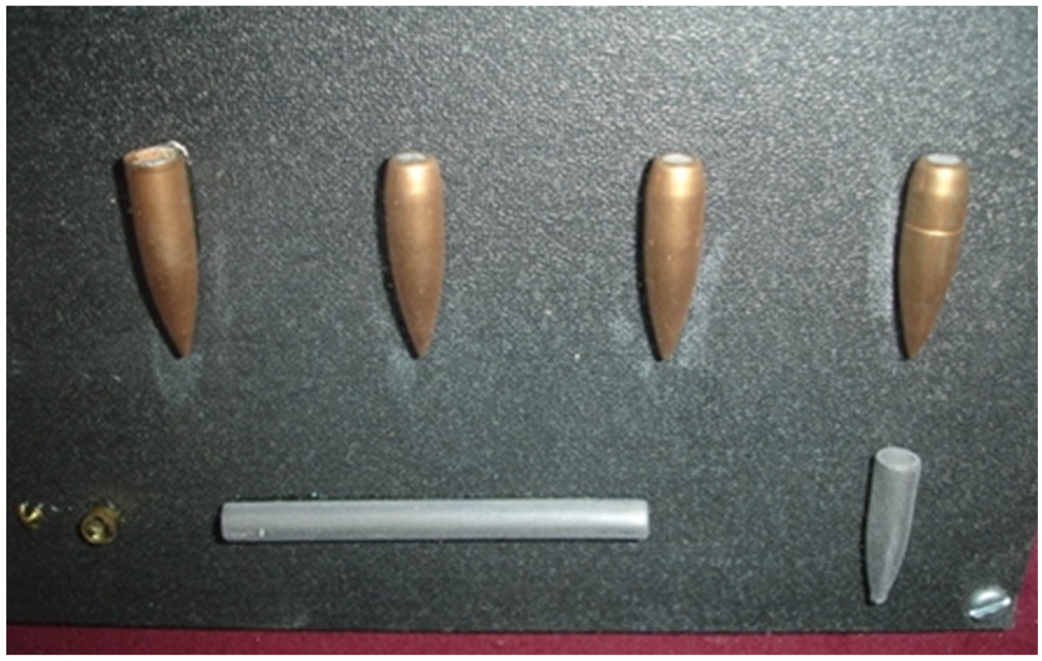
Figure 1. Various stages of construction of a full metal-jacketed military rifle bullet. The lead core (lower row) is enclosed by jacket which covers its tip but leaves its base semi-open. The canellure, around which the mouth of the cartridge case holds firmly the bullet, representing a weak point of the jacket acting as a stress concentrator, can also be seen (upper row, far right). War Museum of Athens.
Ecm 02 00052 g001
Figure 2. Destabilizing effect of the retarding force acting on a military (full metal-jacketed) rifle bullet. In the air, yaw tendency (due to inherent instability) is counteracted by the spin. During soft tissue penetration, the retarding force increases in magnitude as the bullet undergoes irreversible yaw.
Ecm 02 00052 g002
Figure 3. Abdominal gunshot wound from 0.45 cal. handgun bullet (shown with arrows), which resulted in laceration of the right internal iliac vein. (a) Emergency x-ray of the patient. (b) CT scan, 21 days later, which also reveals the construction of the retained projectile. Courtesy of D. Pinialidis, MD.
Ecm 02 00052 g003
Figure 4. Schematic depiction of the temporary cavity formation (pink) and expansion (blue arrows) in tissue by a tumbling military rifle bullet. Black arrow indicates direction of movement.
Ecm 02 00052 g004
Figure 5. Close-range hand injury from a 0.38 cal. handgun bullet, with extensive soft tissue damage from bullet deformation and cavitation as well as from bone shattering. Intraoperative photographs of the entry (a) and exit (b) wounds. (c) Emergency X-ray of the bone injury.
Ecm 02 00052 g005
Figure 6. (a) High energy gunshot wound from a 7.62 mm rifle bullet shot by Rio de Janeiro police. The bullet hit the pistol carried by the wounded (a drug dealer) (b), before entering his thigh, apparently becoming rapidly destabilized, and released its enormous kinetic energy dispersing metallic fragments from the bullets and the magazine into the soft-tissue masses of the gluteal area. The synergistic effect with the subsequent cavitation resulted in the massive exit wound. Courtesy of LTC O. Carvahlo.
Ecm 02 00052 g006
Figure 7. The onset of maximum cavitation in a block of ballistic gelatin penetrated by a 5.56 mm NATO assault rifle bullet, as the latter moves sideways just before its breakup. The high-speed image also demonstrates the relatively long distance of penetration normally required for this bullet in order to turn sideways at 90 degrees of yaw within the target (dimensions of the block: 25 × 15 × 38 cm). Arrow indicates the direction of penetration.
Ecm 02 00052 g007
Figure 8. (a) Late healing of a close-range shotgun injury of the forearm encountered in one of the humanitarian missions of the second author (G.B.) (the photos from the initial presentation are not available). The severity of the injury can be seen from the extent of the soft tissue loss during healing by secondary intention. (b) Postoperative radiograph demonstrating a handmade PMMA spacer with a nail used to reestablish bone continuity of the radius in the acute setting, following the extensive bone loss due to the nature of the wounding agent. (c) After 8 weeks, definitive reconstruction was performed with the PMMA spacer replaced with osteosynthesis using an iliac bone graft.
Ecm 02 00052 g008
Figure 9. Algorithm for initial assessment of patients with gunshot wounds in the emergency department, with diagnostic and therapeutic priorities. Boxes with arrows indicate diagnostic maneuvers; boxes in red indicate possible diagnoses necessitating immediate action. The main indication of FAST (focused assessment sonography in trauma) in gunshot wounds is the diagnosis of pericardial tamponade but focused abdominal sonography for trauma may be used as a decision-making diagnostic tool for assessing the need for laparotomy in hypotensive patients with abdominal wound. (*) Abdominal gunshot injuries may be treated nonoperatively in the absence of signs of hemorrhage, free gas on X-ray, peritonism, or evisceration. Abbreviations: ABCDE: Airway, Breathing, Circulation, Disability, Exposure; ABI: arterial brachial/pressure index; CT: computed tomography; CTA: computed tomographic angiography; CXR: chest X-ray; DPL: diagnostic peritoneal lavage/aspiration; ED: emergency department; FAST: focused assessment sonography in trauma/focused abdominal sonography for trauma; OR: operating room; REBOA: resuscitative endovascular balloon occlusion of the aorta.
Ecm 02 00052 g009
Figure 10. (a) Lateral radiograph of the neck demonstrating at the level of zone III a retained solid lead bullet shot at close range, with fragments indicating its intraoral course. (b) Posteroanterior view reveals the position of the bullet with respect to the mandible.
Ecm 02 00052 g010
Table 1. Characteristics of wounds produced by high-velocity bullets.
Bullet TypeProfile of Injury *Energy TransferDescription
FMJEcm 02 00052 i001Low energy—small exit woundThe bullet fails to yaw due to short wound channel
FMJEcm 02 00052 i002High energy—large exit woundThe bullet tumbles close to the point of exit with cavitation
FMJEcm 02 00052 i003High energy—relatively small exit woundA long wound channel allows bullet tumbling deep within the tissues with the bullet moving base-forward near the exit
JSPEcm 02 00052 i004High energy +/− exit wound(s)Expanding bullet demonstrating fragmentation with early large temporary cavity (the fragments may create multiple exit wounds)
(*) The solid black lines outline the wound borders and the maximum volume of the temporary cavity created. The synergistic effect of fragmentation with cavitation resulting in a large temporary cavity, shown here with expanding soft-point bullet, can also be seen with a high-velocity FMJ bullet (e.g., 5.56 mm NATO) sustaining breakup during mid-tumbling (see text for details). Bullet motion from left to right as indicated by arrow. Abbreviations: FMJ: full metal-jacketed; JSP: jacketed soft-point.
Disclaimer/Publisher’s Note: The statements, opinions and data contained in all publications are solely those of the individual author(s) and contributor(s) and not of MDPI and/or the editor(s). MDPI and/or the editor(s) disclaim responsibility for any injury to people or property resulting from any ideas, methods, instructions or products referred to in the content.