Article

Efficacy of Portable Fugitive Aerosol Mitigation Systems for Nebulizer Therapy During High-Flow Nasal Cannula and Non-Invasive Ventilation

by
Adithya Shyamala Pandian
1,2,3,†,
Bhavesh Patel
4,5,6,*,†,
Karam Abi Karam
1,2,
Amelia Lowell
5,7,
Kelly McKay
4,
Sabrina Jimena Mora
1,2,
Piyush Hota
1,8,
Gabriel Pyznar
1,2,
Sandra Batchelor
4,7,
Charles Peworski
7,
David Rivas
9,
Devang Sanghavi
10,
Ngan Anh Nguyen
1,8,
Aliaa Eltantawy
1,2,
Xueqi Li
1,2,
Xiaojun Xian
1,2,
Michael Serhan
1,2,8 and
Erica Forzani
1,2,8,*
1
Center for Bioelectronics and Biosensors, Biodesign Institute, Arizona State University, Tempe, AZ 85281, USA
2
Health Futures Center, Arizona State University, Phoenix, AZ 85054, USA
3
School of Electrical, Energy, and Computer Engineering, Arizona State University, Tempe, AZ 85287, USA
4
Center for Military Medicine, Mayo Clinic, Phoenix, AZ 85054, USA
5
Department of Critical Care Medicine, Mayo Clinic, Phoenix, AZ 85054, USA
6
Department of Respiratory Care, Mayo Clinic, Phoenix, AZ 85054, USA
7
Department of Nursing, Mayo Clinic, Phoenix, AZ 85054, USA
8
School of Engineering for Matter, Transport, and Energy, Arizona State University, Tempe, AZ 85287, USA
9
Facilities Engineering, Mayo Clinic, Phoenix, AZ 85054, USA
10
Department of Critical Care Medicine, Mayo Clinic, Jacksonville, FL 32224, USA
*
Authors to whom correspondence should be addressed.
These authors contributed equally to this work.
Emerg. Care Med. 2025, 2(3), 36; https://doi.org/10.3390/ecm2030036
Submission received: 4 March 2025 / Revised: 9 June 2025 / Accepted: 14 July 2025 / Published: 29 July 2025

Abstract

Objectives: This study evaluates the efficacy of existing and new aerosol mitigation methods during nebulization (Neb) in combination with high-flow nasal cannula (HFNC) oxygen supplementation and non-invasive ventilation (NIV). Methods: We recorded fugitive aerosol particle concentrations over time and assessed the peak (P) and area (A) efficacy of active and passive mitigation methods, comparing them to a no-mitigation condition. Peak efficacy was measured by the reduction in maximum aerosol concentration, while area efficacy was quantified by the reduction of the area under the aerosol concentration–time curve. Results: For HFNC with Neb, we found that active mitigation using a mask with a biofilter and a fan (referred to as the aerosol barrier mask) significantly outperformed passive mitigation with a face mask. The peak and area efficacy for aerosol reduction were 99.0% and 96.4% for active mitigation and 35.9% and 7.6% for passive mitigation, respectively. For NIV with Neb, the active mitigation method, using a box with a biofilter and fan, also outperformed passive mitigation using only the box. The peak and area efficacy for aerosol reduction were 92.1% and 85.5% for active mitigation and 53.7.0% and 25.4% for passive mitigation, respectively. Conclusion: We concluded that active mitigation set up systems advantageous for effective reduction of airborne aerosols during aerosol generated procedures.

Graphical Abstract

1. Introduction

Human respiratory infections have been the cause of lethal and economically destructive global losses. Many of these infections spread from the infected host’s respiratory system to other susceptible hosts in contact with droplets and aerosols generated by sneezing, coughing, speaking, and breathing [1,2,3,4,5,6,7,8,9]. An aerosol, in this context, is a suspension of fine particles (<10 μm, including potential infectious pathogens) in an airborne liquid mist that is not strongly affected by gravity and can be transported through ventilation systems (e.g., air conditioning) [2,7,10,11,12,13]. Ninety-nine percent of aerosols produced by humans, regardless of age, sex, weight, and height, are less than 10 μm [1,7]. For instance, researchers have found viruses [6] with virulent activity [7] in aerosols of this size [2,14,15]. Since viruses effective size can be 100 nm [16,17], they can be encapsulated within aerosols from the respiratory system of an infected person and dispersed by aerosol-generating procedures (AGPs) such as medication administered through nebulization [18], high-flow nasal cannula (HFNC) oxygen therapy, and/or non-invasive ventilation (NIV) [19] potentially increasing the latent safety risks associated with inter/intrahospital transport, timely space decontamination [20], and ultimately potentially increasing infection risk and medication exposure in close contacts and healthcare workers (HCW) [21,22,23,24]. These AGPs cause a high risk of dispersing pathogens due to the generation of aerosols with sizes less than 10 μm [7,12,25,26,27,28]. Therefore, many different systems have been developed to protect HCWs [29]. The use of personal protective equipment (PPE) may be preventative during exposure to AGPs, yet some studies estimated the risk of infection 1.8 to 4.8 times greater than controls [30,31]. Hence, using the Wells–Riley Model, a widely accepted quantitative infectious risk assessment framework [32,33], we directed our efforts at reducing the “quanta generation rate” of potentially infected aerosol to reduce the probability of a HCW infection and used fugitive aerosol detected as a surrogate marker. Although HCW susceptibility and pathogen specific data remain limited, using the measles virus as an example, with an estimated quanta generation rate of 1000 quanta per hour from an infected individual (range 480–5589 quanta per hour) [34,35], breathing at rest (respiratory rate 15, tidal volume 550 mL) in an average 50 cubic meter hospital room (1770 cubic feet) at three room air exchanges per hour with one susceptible healthcare worker at a distance of one meter for 15 min (average nebulizer treatment), the probability of infection is estimated at 57% which would be reduced to <1% if quanta generation was decreased to <10 (99% reduction). Although hospital design, architecture, and HVAC systems may be effective at reducing ambient aerosol [36], they are also capital and energy intensive; thus, we focused our study at mitigating aerosol at the source during AGPs.
In this work, we evaluate the efficiency of a new active aerosol barrier mask designed to reduce fugitive aerosols during vibrating mesh nebulization (Neb) when used with a high-flow nasal cannula (HFNC). The performance of the active aerosol barrier mask is compared with a commonly used passive solution, a surgical face mask [37,38]. Additionally, we assess the effect of the new active aerosol barrier mask on delivering aerosolized drugs during nebulization. Furthermore, we examine the efficiency of an aerosol mitigation box equipped with a biofilter and a fan to reduce fugitive aerosols during Neb treatment with non-invasive ventilation (NIV) and compare it to a mitigation box without the biofilter and the fan.

2. Materials and Methods

2.1. Overview of Aerosol-Generating Procedures, Systems, and Mitigation Methods

During our study, we used different relevant materials and methods indicated in this section. Table 1 summarizes the aerosol-generating conditions we tested. The conditions include the use of (1) high-flow nasal cannula (HFNC) and nebulization with a vibrating mesh nebulizer (Neb) in conditions of aerosol mitigation and no mitigation, and (2) non-invasive ventilation (NIV) and Neb with aerosol mitigation and no mitigation. Figure 1 illustrates the testing conditions and shows pictures of system 1 with the HFNC connected to a Neb in the cases of no mitigation (1A), passive mitigation with a surgical face mask (1B), and active mitigation with aerosol barrier mask, including a fitted oronasal silicone mask, a biofilter, and fan (fan filtration). The figure also illustrates system 2, with the NIV connected to a Neb in the cases of no mitigation (2A), passive mitigation with an aerosol mitigation box (2B), and active mitigation with the aerosol mitigation box and the biofilter and fan.

2.2. Aerosol Generating (AG) Procedures

The simulated medication nebulization (Neb) was generated via a 3.0 mL saline solution at room temperature in a vibrating mesh nebulizer (Aerogen Solo, Aerogen, Galway, Ireland) [39,40]. The nebulization lasted 8 min and was placed with post heated humidifier (850 System, Fisher Paykel Healthcare Ltd., Auckland, New Zealand). The vibrating mesh nebulizer was used for nebulization and connected to the lines of HFNC and NIV systems, which are detailed below:
High-flow nasal cannula (HFNC): The HFNC (Airvo 2, Fisher & Paykel Healthcare, Auckland, New Zealand) was used with simulated supplemental oxygen (21%) at 30 L/min with heated humidified air set to 31 degrees Celsius.
Non-invasive ventilation (NIV): The NIV system (Respironics V60, Philips, Amsterdam, The Netherlands, was used with simulated supplemental oxygen (21%) heated to 31 degrees Celsius and delivered via an oronasal mask (Amara Full-Face Mask Philips, Amsterdam, the Netherlands) with inspiratory positive airway pressure (IPAP) of 12 cm H2O and expiratory airway pressure (EPAP) of 5 cm H2O. Leak and mask fit were monitored closely during the simulator application. Air was heated and humidified with sterile water using a single limb heated wire 3-foot circuit and a Fisher Paykel 850 system (Fisher Paykel Healthcare Limited, Auckland, New Zealand). This system was used in conjunction with a manikin (see Real and Simulated Human Subjects, Section 2.4).

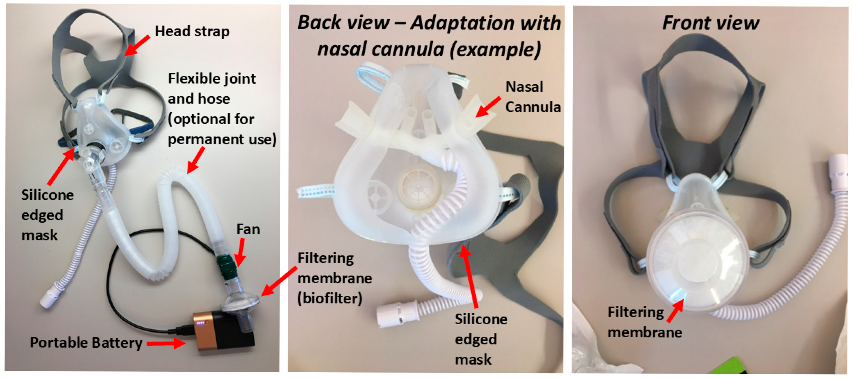
2.3. Mitigation Methods

Passive mitigation methods: One of the passive mitigation methods included the surgical face mask (Table 1 and Figure 1: case 1B) which was a Level 1 face mask (Yellow 47117, Owens and Minor Halyard, Inc, Alpharetta, GA, USA) that was used to mitigate HFNC [38] and Neb system. The other passive mitigation method included the mitigation box (Table 1 and Figure 1: case 2B), whose design was inspired by the isolation hood that has gained attention to mitigate SARS-CoV-2 pathogens. A 66-quart clear plastic box (Sterilite Corporation, MA, USA) was surrounded by a transparent plastic sheet that extended to cover the whole bed. The box was placed on a rolled blanket to level the box around the head of the manikin.
Active mitigation methods: One of the active mitigation methods included the aerosol barrier mask with an oronasal silicone mask, which is illustrated in Figure 2. This mask included a portable battery-powered axially directed fan with exhaust air filtration through standard bacterial/viral biofilter (Hudson RCI Bacterial Viral Filter # RHF605U, Teleflex, NC, USA) adapted to a fitted oronasal silicone mask (TF Health Corp. d.b.a Breezing Co, Phoenix, AZ, USA) which was customized to accommodate the HFNC without leak (Table 1 and Figure 1: case 1C). The biofilter is commonly available for mechanical ventilation with a 99.9992% bacterial efficiency, 99.990% viral efficiency, and humidity exchange capability. The reusable filtration fan had an airflow of 15–18 L/min and was powered by a rechargeable 5V USB battery pack (Duracell, Bethel, CT, USA) [30].
The other passive mitigation method included the mitigation box and consisted of a filtration fan and a biofilter (Hudson RCI Bacterial Viral Filter # RHF605U, Teleflex, Morrisville, NC, USA) that was inserted through the wall of the clear plastic mitigation box (Table 1 and Figure 1: case 2C). The fan was also powered by a rechargeable 5V USB battery pack (Duracell)[37].

2.4. Real and Simulated Human Subjects

A total of 6 test human subjects participated in this study. The human subjects were consented via ASU IRB: STUDY00006544 and were 22 to 55 years old, 5’5’’ to 6’ tall, and had body mass indexes <25. They participated in all the tests in the study for system 1 (HFNC and Neb).
A manikin (Trauma HAL S3040.10, Gaumard Scientific Company, Inc, Miami, FL, USA) was used in the study for system 2 (NIV and Neb). To seal the manikin airway system during ventilation, the airway was occluded by inserting an endotracheal tube (ETT) retrograde through a tracheostomy slot and inflating balloon in the manikin larynx. This ETT was connected to an artificial lung simulator which permitted intermittent triggering of the ventilator at breath frequency of 16 breaths per minute, a tidal volume of 400 to 1000 mL, and a resulting minute ventilation of 6 to 8 L per minute (L/min).

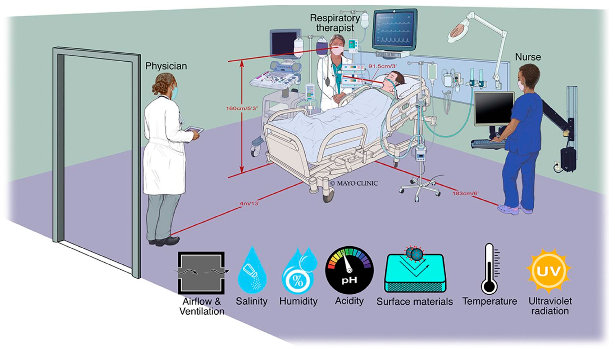
2.5. Simulated Applied Clinical Environment

In aerosol studies, it is important to ensure aerosol particle stability comparable to real settings. For this reason, we simulated an applied clinical environment where we reproduced conditions from previous studies [41]. More specifically, our simulated clinical environment had similar characteristics to a hospital’s airborne infectious isolation room (AIIR). The room has a size of 19’ 10” × 18’ 3” × 10’, an air turnover of 20 air exchange per hour rate as well as a typical humidity, temperature, surface/flooring material, lighting, and distance of bed to headwall of the AIIR. In addition, we applied procedures with nebulization conditions of salinity and pH (acidity) similar to hospital-applied procedures. Further, we selected strategic distances of 3, 6, and 13 feet for sensor placement, as these represent relevant distances among various healthcare practitioners, as illustrated in Figure 3 (see next section).

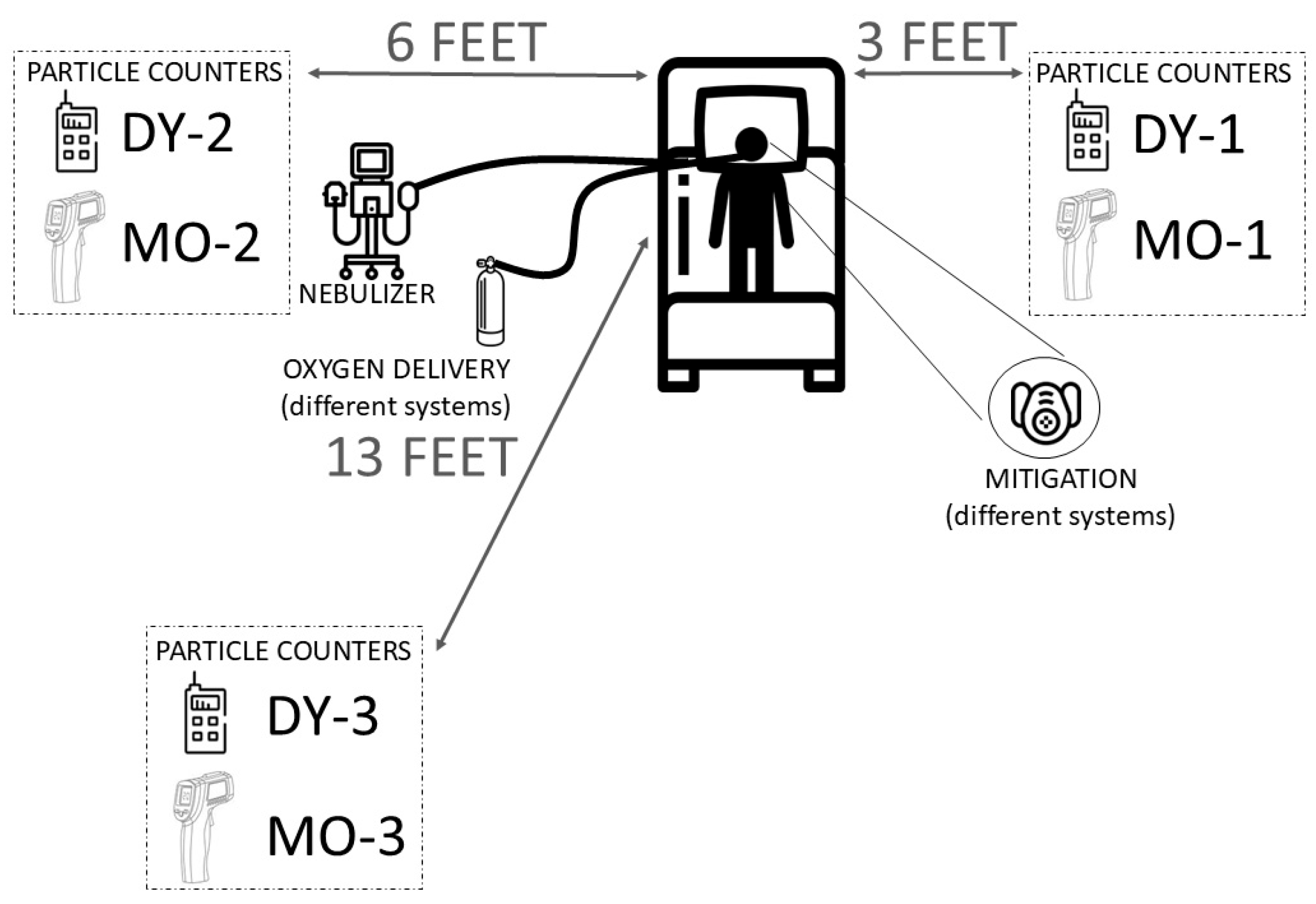
2.6. Aerosol Measurement Equipment

We used two aerosol particle counters Dylos DC1100 Pro sensors (DY) (Dylos Corporation, Riverside, CA, USA) and MET ONE HHPC2+ (MO) (Beckman Coulter, Indianapolis, IN, USA) were placed 160 cm above the ground at the 3 clinically relevant locations mentioned above: 3, 6, and 13 feet from the source. Figure 4 illustrates the aerosol particle counters: DY and MO with the respective locations. We took aerosol particle concentration measurements at 1 min intervals. For the DY sensors, we took instant readings registered every 1 min, and for the MO sensors, we took the concentration averaged over 1 min. The sensors provided measurements of a nominal size of 0.5 µm. However, we verified that the 0.5 µm measurements could also detect aerosol particles of 0.3 µm. The verification tests of this feature are provided in Supplementary Materials. In all measurements, we applied the approach of so-called “redundant sensing.” We used at least 3 sensors to confirm the presence of aerosols in the testing environment and the effect of the different mitigation methods. This approach enabled us to verify our results. In this publication, we provide reading and analysis based on the 6-foot distance since it represents the overall results assessed at 3 and 13 feet. More information on 3 and 13 feet is provided in the Supplementary Materials.

2.7. Measurement Protocol and Data Analysis

The efficacy of mitigating the nebulization aerosol dispersion was assessed via the analysis of curves of aerosol particle counts/feet3 recorded from MO and DY sensors in the presence and absence of mitigation. Figure 5 shows an example of the data of aerosol particle concentration as a function of time during a nebulization therapy with and without the mitigation system. For each experiment, the 0.5 µm particle baseline concentration was assessed for at least 5 min under steady-state conditions with the HFNC + Neb or NIV + Neb operational system inside the room before starting the nebulization. No movement (e.g., no entrance or exit of subjects into or from the room) occurred in the room during the measurement to minimize the risk of resuspension of settled particles. The room returned to baseline aerosol conditions between measurements.
We evaluated the efficacy of the aerosol mitigation, using the peak and the area under the aerosol particle counts/feet3 transient curves of the no mitigation and mitigation cases. The following equations were used in connection with transients similar to the ones shown in Figure 5 (note: Figure 5 is only for sake of representative illustration):
P e a k   E f f i c a c y % = [ ( P e a k   w i t h   n o   m i t i g a t i o n   s o l u t i o n ) ( P e a k   w i t h   m i t i g a t i o n   s o l u t i o n ) P e a k   w i t h   n o   m i t i g a t i o n   s o l u t i o n ] × 100
A r e a   E f f i c a c y % = [ ( A r e a   w i t h   n o   m i t i g a t i o n   s o l u t i o n ) ( A r e a   w i t h   m i t i g a t i o n   s o l u t i o n ) A r e a   w i t h   n o   m i t i g a t i o n   s o l u t i o n   ] × 100

2.8. High-Flow Nasal Cannula Testing Procedure

We used a high-flow nasal cannula therapy on the subjects with a fraction of inspired oxygen of 21% and a humidified airflow rate of 30 L per min, while a 3.0 mL saline solution was nebulized over 8 min using Neb. We performed the measurements with DY and MO sensors as indicated in the previous section. More specifically, we tested conditions: without mitigation (Table 1, Figure 1, case 1A), one with passive mitigation using the surgical mask (Table 1, Figure 1, case 1B), and the other with active mitigation using the aerosol barrier mask with the oronasal silicone mask customized to integrate the high-flow nasal cannula and the fan filtration (Table 1, Figure 1, case 1C).

2.9. Non-Invasive Ventilation Testing Procedure

We used a non-invasive ventilator (NIV) on a manikin with an inspiratory airway pressure of 12 cm H2O and an expiratory airway pressure of 5 cm H2O, a fraction of inspiration oxygen of 21%, breath frequency of 16 breaths per minute, and 6 to 8 L/min of ventilation. We adjusted the mask fit to keep the leak to the minimum. Neb was used to administer 3.0 mL saline solution over 8 min. We performed the measurements with DY and MO sensors as indicated in the previous section. More specifically, we tested the following conditions: without mitigation (Table 1, Figure 1, case 2A), one with passive mitigation using the mitigation box (Table 1, Figure 1, case 2B), and one with active mitigation using the mitigation box with a fan filtration (Table 1, Figure 1, case 2C).

2.10. Measurements of Drug Delivery Efficacy in Oronasal Silicone Mask with Active Mitigation System

In addition to the efficacy of fugitive aerosol mitigation, we measured the potential impact of active mitigation on drug delivery. Acknowledging the complex interaction between nebulizer type, patient interface, drug formulation, breathing pattern, lung physiology, and patient compliance [42], we focused on the fitted oronasal silicone mask (Figure 2) to deliver 4 commonly nebulized medication doses and formulations [43]. To achieve this goal, we used an “aerosol analysis filter” to collect the delivered aerosol drugs during simulated inhalation cycles and developed customized analytical methods to quantify the amount of delivered drugs in the aerosol analysis filter (see Supplementary Materials). To accurately measure the amount of aerosolized medication delivered, finding a good fit of the oronasal silicone mask to the aerosol analysis filter was necessary. Due to the complexity of obtaining a tight fitting of the HFNC to the “aerosol analysis filter,” we delivered drug aerosols using a breath-actuated nebulizer (BAN) (AeroEclipse II®, Monaghan Medical, Plattsburgh, NY, USA) as shown in Figure 6A and an experimental setup described in the next section.
Experimental setup for comparative drug efficacy: The drug delivery efficacy of the active mitigation system including the aerosol barrier mask (oronasal silicone mask and fan filtration system) (Figure 6A) was compared to the corresponding drug delivery efficacy of the BAN without the active filtration oronasal mask (Figure 6B). Figure 6A–C shows the setup, which connects the BAN to the customized, 3D-printed aerosol analysis filter (see details below) and the metabolic breath simulator (see Supplementary Materials). Using this setup, we measured the efficacy of four drugs delivered with a flow rate of 6 L/min oxygen through the BAN as follows:
  • Saline solution: 5.0 mL of 0.9% sodium chloride solution (AddiPak, Hudson RCI, Temecula, CA, USA).
  • Albuterol solution: 3.0 mL of 0.83 mg/mL Albuterol (Nephron Pharmaceutical Corporation, West Columbia, SC, USA).
  • Vancomycin solution: 5.0 mL of 50 mg/mL Vancomycin prepared from solid Vancomycin (Klonal Laboratories, Klonal, Argentina).
  • Amikacin solution: 5 mL of 100 mg/mL of Amikacin sulfate prepared from 500 mg/2 mL Amikacin sulfate (Klonal Laboratories, Klonal, Argentina).
We determined the amount of nebulized drug present in the aerosol analysis filter and calculated the percentage of the nebulized drug in this filter with respect to the total drug amount placed on the nebulizer before starting the nebulization:
D r u g   d e l i v e r y   e f f i c a c y   % = [ a m o u n t   o f   n e b u l i z e d   d r u g   p r e s e n t   i n   t h e   a e r o s o l   a n a l y s i s   f i l t e r a m o u n t   o f   d r u g   p l a c e   i n   t h e   n e b u l i z e r ] × 100

3. Results

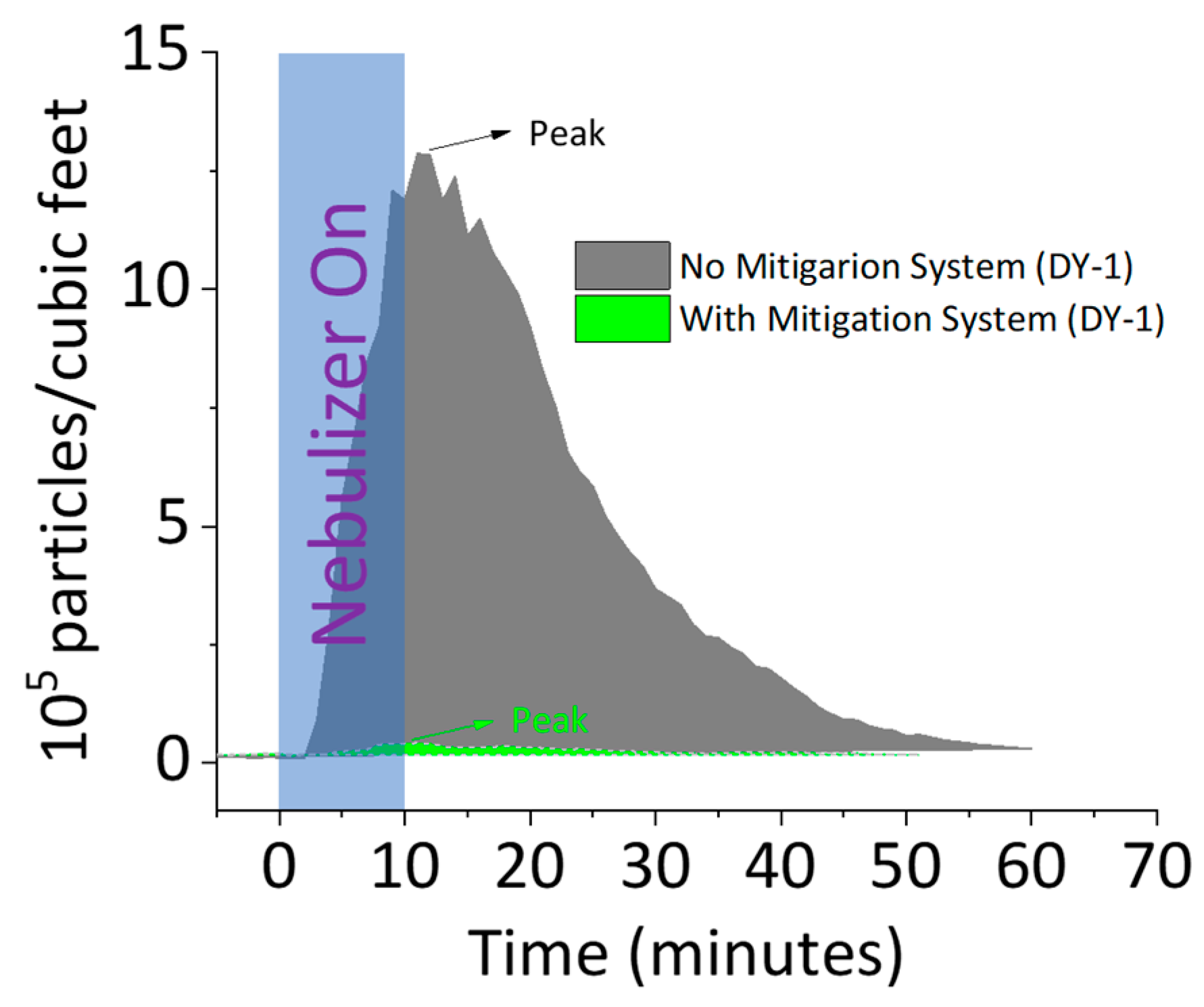
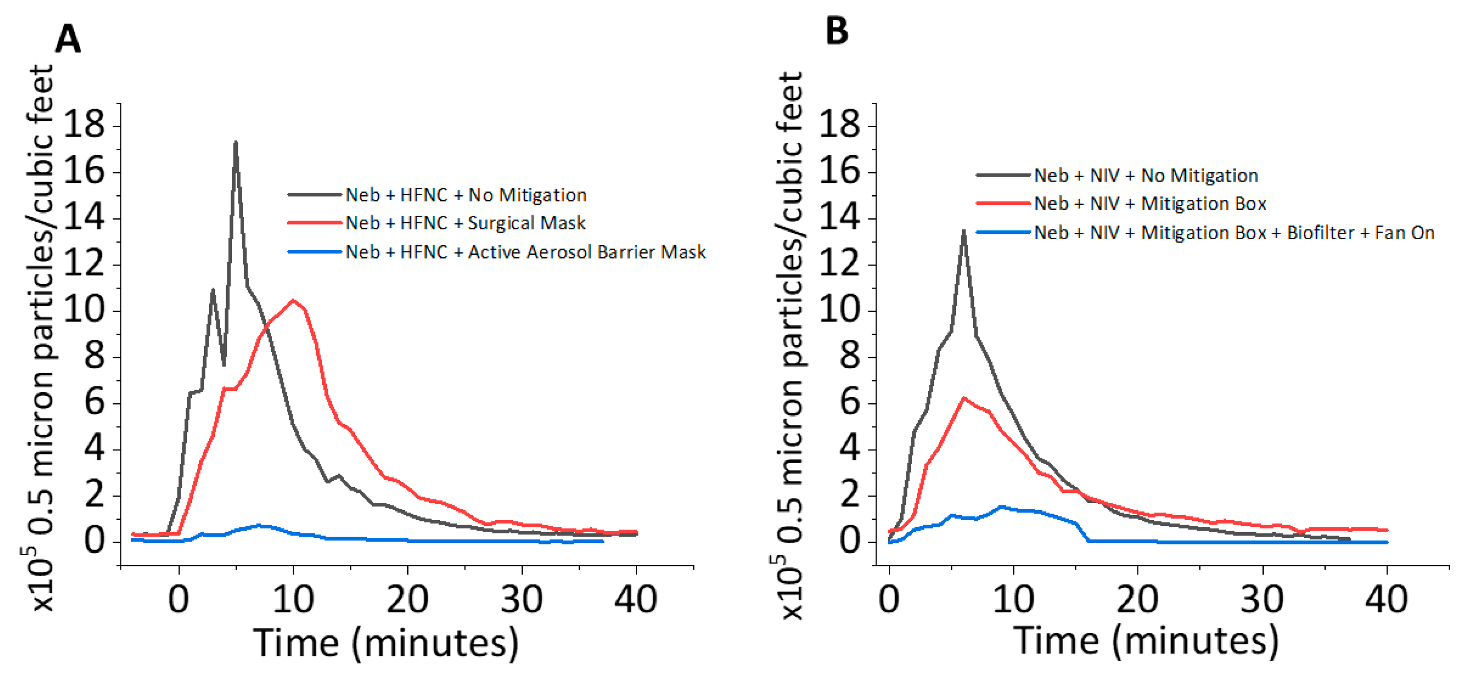
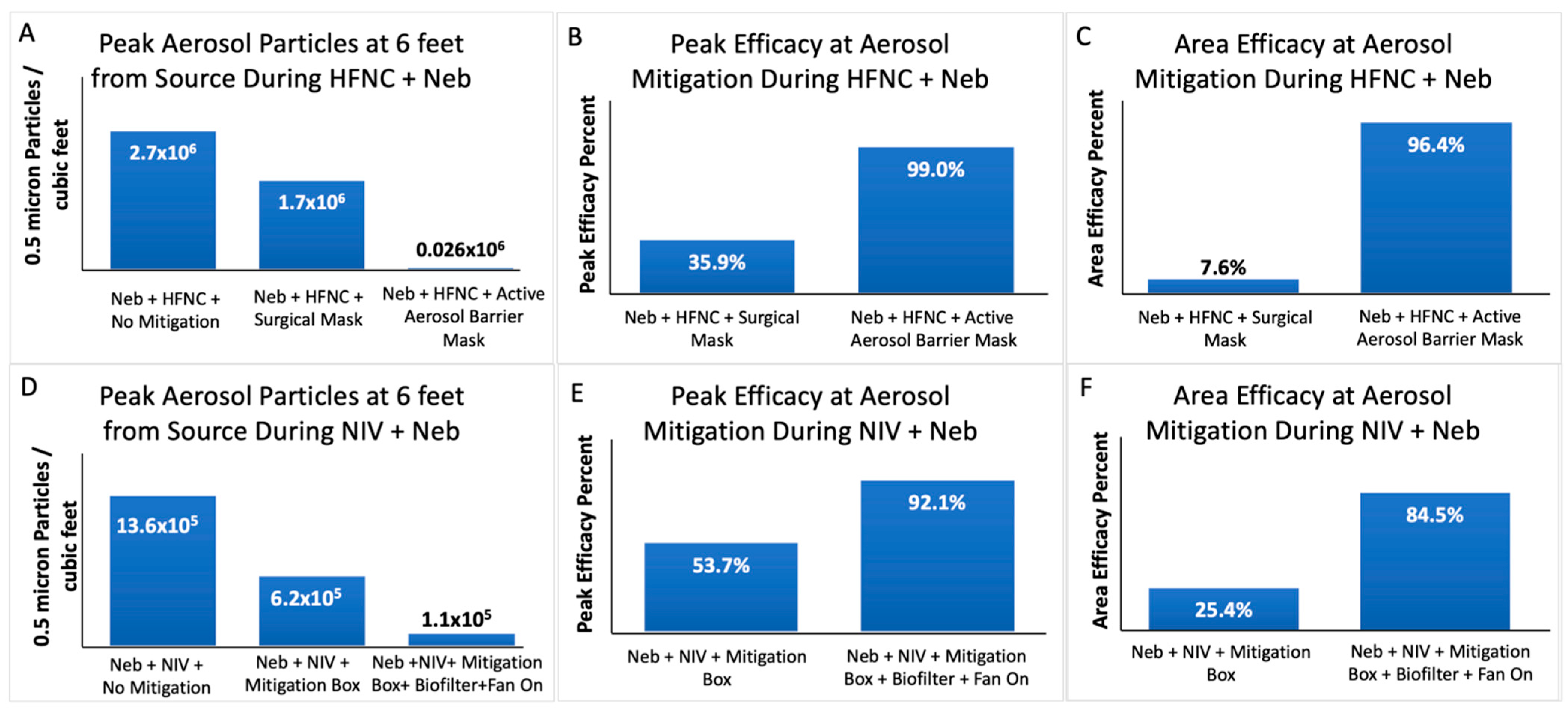
3.1. Aerosol Mitigation Efficacy

Figure 7 illustrates the particle count concentration profiles over time, measured at a distance of 6 feet from the nebulization source during simulated nebulization therapies. Figure 7A presents aerosol concentration profiles for the HFNC + Neb system under three conditions: no mitigation (Figure 1, top: 1A), passive mitigation using a surgical mask (Figure 1, top: 1B), and active mitigation with the aerosol barrier mask (Figure 1, top: 1C). Similarly, Figure 7B depicts aerosol concentration profiles for the NIV + Neb system, including no mitigation (Figure 1, bottom: 2A), passive mitigation with a containment box (fan off) (Figure 1, bottom: 2B), and active mitigation incorporating the containment box, biofilter, and fan (Figure 1, bottom: 2C).
The analysis of mitigation efficacy for each of the systems was performed on particle counts/feet3-time profiles. Figure 8 shows a summary of the aerosol particles for nebulization conditions using the HFNC (Figure 8A–C) and NIV (Figure 8D–F) systems and the averaged efficacy percentages for the different systems. The analysis results from transient aerosol profiles like those shown in Figure 7 followed the procedures presented in the experimental section. Figure 8A presents a summary of the peak aerosol particle concentrations per cubic feet for HFNC nebulization under three conditions: no mitigation, passive mitigation with a surgical mask, and active mitigation using the aerosol barrier mask. Figure 8B,C illustrate the efficacy of aerosol reduction for passive and active mitigation, quantified using the peaks and areas of aerosol transient profiles, respectively. Similarly, for NIV nebulization, Figure 8D summarizes peak aerosol particle concentrations per cubic feet for no mitigation, passive mitigation with a containment box (without a fan), and active mitigation incorporating the box, biofilter, and fan. Additionally, Figure 8E,F depict the percentage reduction in aerosol concentration for passive and active mitigation, assessed using transient profile peaks and areas, respectively.

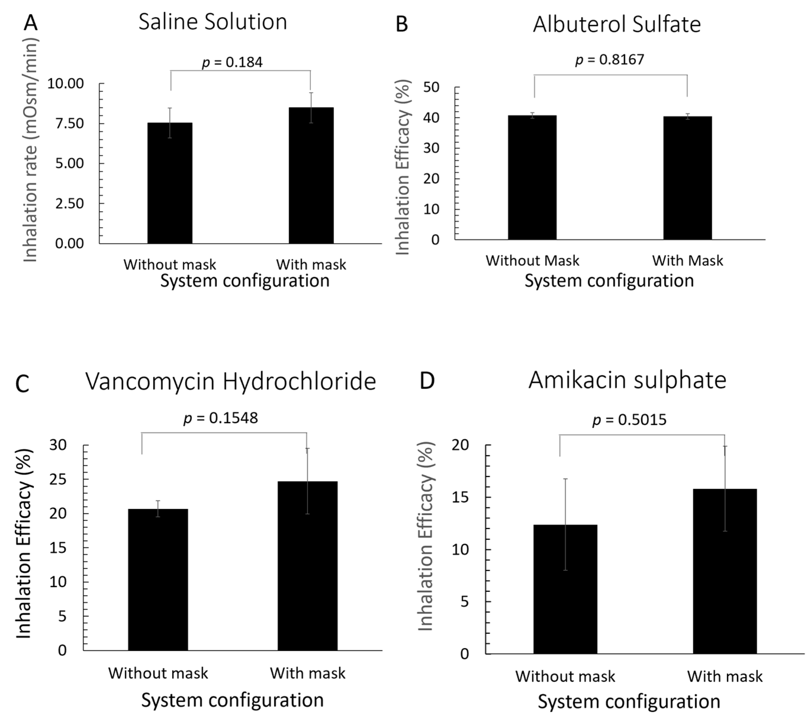
3.2. Drug Delivery Efficacy of Active Mitigation System

Figure 9 presents comparative analyses of the inhaled drug amount (Figure 9A) and the inhaled drug percentage (Figure 9B–D) during nebulization, performed with and without an active aerosol mitigation system, specifically the aerosol barrier mask, which includes an oronasal silicone mask and a filtering system. As detailed in the experimental section, nebulization was conducted using a nebulizer designed for accurate quantification of delivered drugs. Four drugs were administered: saline solution (Figure 9A), albuterol (Figure 9B), vancomycin (Figure 9C), and amikacin (Figure 9D), with drug quantification performed using customized analytical methods outlined in the Supplementary Materials. For Figure 9B–D, nebulization efficacy was assessed by calculating the percentage of the drug captured in the analysis filter during inhalation cycles relative to the total drug delivered throughout the nebulization process.

4. Discussion

4.1. Aerosol Mitigation Effect

Our study confirmed that passive mitigation systems, such as the use of a surgical mask during nebulization with a high-flow nasal cannula (HFNC), demonstrated limited efficacy in reducing fugitive aerosols. Specifically, these systems achieved only a 35.9% peak reduction and a 7.6% overall reduction in aerosol exposure (Figure 7A and Figure 8B,C), leaving over ~92% of the nebulized aerosols unmitigated [41,44]. In addition, passive mitigation using a mitigation box during nebulization with NIV demonstrated a peak reduction efficacy of 53.7% and an overall area reduction efficacy of 25.4% (Figure 7B and Figure 8E,F), leaving only 75% of fugitive aerosols unmitigated.
On the contrary, active mitigation demonstrated a 99.0% efficacy in reducing peak aerosol load and 96.4% in decreasing total aerosol exposure during nebulization with HFNC (Figure 7A and Figure 8B,C). Similarly, active mitigation during nebulization with NIV showed a 92.1% reduction in peak aerosol exposure and an 84.5% reduction in total aerosol exposure (Figure 7B and Figure 8E,F). Using the Wells–Riley framework and the measles virus hypothetical example provided in the introduction, an unmasked healthcare worker delivering a nebulizer with this degree of quanta reduction could decrease the probability of infection to <1% and 12% for HFNC and NIV respectively. Given that HFNC and NIV are preferred oxygen therapies for delaying the need for invasive mechanical ventilation in patients with airborne infections [29], active mitigation could offer a practical solution for controlling aerosols during the administration of nebulized medications, especially for patients with infectious respiratory diseases.
Aerosol dispersal patterns, particularly for particles around 0.5 µm, are influenced by various factors, including the mechanism of aerosol generation, air exchange rates, room ventilation streams, and environmental conditions such as temperature and humidity [45]. These highly variable and unpredictable aerosol patterns, combined with the limited availability of airborne isolation rooms in hospital settings, highlight the importance of point-of-dispersion mitigation techniques as a critical layer in a multi-faceted aerosol containment strategy.
Achieving active mitigation of fugitive aerosols from 99% to 100% efficacy necessitates improvements in mask sealing and optimization of differential airflow. Systems lacking an effective mask-to-face seal, or those with dispersion airflow rates exceeding the filtration system’s airflow capacity, have shown limited efficacy in aerosol containment [44]. Enhancing these factors in our tested mitigation systems significantly improved overall mitigation, ensuring minimal aerosol escape during treatments involving nebulization.
To achieve 100% efficacy in aerosol mitigation, it was observed that complete sealing of the oronasal mask is crucial, as any leaks lead to aerosol release. The modified silicone disposable mask developed for this study, as shown in Figure 2, demonstrated significant progress toward this goal. This aerosol barrier mask includes: (1) an inlet port for oxygen delivery, (2) an outlet port for expelling residual aerosols and air, equipped with a biofilter and fan, and (3) a safety/emergency inlet port with a one-way valve for inhalation of ambient air in case of oxygen therapy interruption. The combination of convective transport from the fan and filtration from the biofilter—featuring a bacterial and viral filtering membrane with a 0.2 μm pore size and multiple layers of microfiber material—proved to be optimal for aerosol capture.
Additionally, using silicone material for the mask edges allowed the mask to adapt to the user’s face, ensuring an excellent seal that prevents contaminated air from leaking. The fan facilitated the movement of aerosols through the biofilter, ensuring that all aerosols 0.3 μm and larger were captured before the main air stream exited the system. The mask’s portability, combined with predictably low energy consumption (powered by a 5V rechargeable battery with a 6 h runtime), enhances its suitability for clinical settings. Moreover, this mask could potentially be applied in home care settings [46,47] or for inter- and intrahospital transport [48].
Furthermore, applying active mitigation to transports within hospitals may decrease contaminated patient-generated aerosols in everyday use areas such as halls and elevators and promote timely space decontamination [20] of CT/MRI scanners.

4.2. Impact of Active Aerosol Mitigation on Drug Delivery Efficacy

The presence of the active mitigation system for fugitive aerosols such as the aerosol barrier mask, which includes the silicone mask and filtration system (Figure 2), did not significantly affect the nebulization efficacy compared to nebulization performed without the mitigation system (Figure 9). Statistical analysis using t-tests yielded p-values > 0.1, indicating no statistically significant difference which could be relevant for drugs such as Amikacin with a narrow therapeutic window although many factors determine clinically relevant therapeutic effect of inhaled medications [42], our data suggest active mitigation is within acceptable limits (±20%) of in vitro testing for nebulizers as outlined by the United States Pharmacopeia (USP) requirements [49]. These results confirm that the in vitro delivery of nebulized medication remains acceptably affected when using the active aerosol mitigation system. This finding suggests the system’s potential to be a component of an occupational aerosol exposure reduction bundle in close-contact situations, especially for healthcare workers in high-risk environments.

4.3. Strengths

We conducted multiple measurements at clinically relevant distances and heights from the patient/manikin to emulate healthcare provider inhalation exposure locations. In addition, we used two sensor technologies at various distances, demonstrating that results were internally consistent and reliable (see details in Supplementary Materials). Repeat measurements were also performed with different NIV air flows and air leak rates, demonstrating mitigation efficacy consistent with the presented data. In addition, we used the aerosol active mitigation system with the silicon mask and the filtration system with a mechanical nebulizer (home nebulizer) and probed the efficacy of peak aerosol and total aerosol reduction during nebulization was 98% and 98%, respectively (see Supplementary Materials, Figure S5), which reassured the applicability of the system to different nebulizers. For measurements of the drug delivery efficacy of the aerosol active mitigation system, we performed triplicate tests for each case. We analyzed each independent test by duplicating the amount of drug collected in each aerosol analysis filter.
During the pandemic, hospital personnel used the active mitigation system with the mask and filtering system under crisis conditions in over 1100 cases. The personnel provided positive feedback, indicating the system was user-friendly and improved healthcare workers’ sense of safety that was not provided by other mitigation systems, such as a surgical mask.

4.4. Limitations

Some limitations of this operational testing are notable. We tested mitigation systems using manikins and healthy subjects with stable breathing patterns, which may not effectively simulate the variable anatomy and dynamic breathing patterns of a wider variety of body mass indices, ethnicity, gender, and acute/chronic illnesses. Specifically, mask fit, and nasal cannula fit may affect the aerosol generation and mitigation effectiveness. While user tolerability and ergonomics (e.g., tissue pressure injury, fan noise, claustrophobia) were informally monitored, a formal comfort/usability assessment was not conducted. Although anecdotal feedback indicated no significant discomfort, future studies will include validated human factors assessments. In addition, we controlled aerosol-generating forces and plume dispersion forces in a simulated clinical AIIR environment, which may not represent other applied clinical settings, such as non-AIIR hospital rooms, CT scanners, patient homes, and elevators during transport or a broader range of airflows (HFNC), pressures (NIV), and duration of nebulization. For example, the HFNC testing was conducted at 30L/min, which reflects moderate HFNC use, and studies have demonstrated expiratory plumes spreading 2–3 m compared to 4.1 m at flow rates of 60 L/min [50]. While higher flow rates are common in critical care, logistical constraints prevented inclusion in this study and future in vivo reliability testing will include variable flows, pressures and other clinical environments. Finally, it should be emphasized that mitigating aerosol generation is one component of a complex quantitative microbial risk reduction strategy including dimensions such as microbial pathogenicity, PPE, environmental controls, patient density, staffing ratios, etc. to reduce infection rates in HCWs.

5. Conclusions

In summary, we demonstrate that passive methods such as surgical masks and box mitigation are less efficient than active methods for capturing aerosols generated from nebulizers during HFNC or NIV in simulated settings. The use of a biofilter and fan with the mitigation box during NIV therapy and the aerosol barrier mask used in conjunction with HFNC therapy and normal breathing significantly reduces fugitive aerosols produced during nebulization therapies which could reduce the quanta of infected aerosol exposure. While the exact infection risk involves factors such as pathogen dose-response curves, host susceptibility, HCW compliance with safety equipment and protocols, etc., aerosol suppression at the source may play a crucial part of the chain of strategies to decrease the probability of HCW infection in short duration, close contact clinical encounters. In addition, active fugitive aerosol mitigation with breath simulator, had minimal effect on the estimated nebulized drug delivery of albuterol, vancomycin, and amikacin and may decrease secondary medication exposure. Future research should evaluate the in vivo use of portable active aerosol mitigation during aerosol generating procedures within pathogen specific risk framework and determine clinical thresholds for deployment as an important component of a complex multidimensional process to reduce transmission of airborne respiratory infectious disease pathogens to healthcare workers.

Supplementary Materials

The following supporting materials can be downloaded at: https://www.mdpi.com/article/10.3390/ecm2030036/s1. Figure S1. Verification tests of performance of different commercial particle counters, Figure S2. Validation tests of Dylos DC1100 Pro (DL) and Met One HHPC2+ aerosol/particle counter to detect small particles of 0.19 µm in a simulation room, Figure S3. Picture of the manikin used in the study. The manikin’s airway system is occluded by an ETT retrograde through a tracheostomy slot and inflating a balloon in the manikin larynx, Figure S4. Particle count concentration during use of HFNC treatment with 21% of oxygen, 30 L/min of air flow rate, and nebulization with 3 mL saline physiological solution at (a) 3 feet, (b) 6 feet, and (c) 13 feet from subject, Figure S5. Particle count concentration during regular mechanical nebulizer treatment with nebulization of 3 mL saline physiological solution at (a) 3 feet, (b) 6 feet, and (c) 13 feet from subject.

Author Contributions

All authors have read and agreed to the published version of the manuscript. Conceptualization, E.F.; methodology, B.P., A.L., K.M., S.B., C.P., D.S., E.F.; software, G.P.; validation, B.P., A.L., K.M., S.B., C.P., D.S., E.F.; formal analysis, A.S.P., K.A.K., G.P., N.A.N., A.E., X.L.; investigation, B.P., A.L., K.M., S.B., C.P., D.S., E.F.; resources, E.F.; data curation, A.S.P., K.A.K., G.P., N.A.N., A.E., X.L.; writing—original draft preparation, B.P., E.F., A.L.; writing—review and editing, A.S.P., K.A.K., S.J.M., P.H., D.R., X.X., M.S.; visualization, A.S.P., K.A.K., G.P., N.A.N., A.E., X.L.; supervision, E.F.; project administration, E.F.; funding acquisition, E.F.

Funding

The research was supported by the NIH-NIBIB 5R03EB027336 award, ASU’s PLUS Alliance (https://www.plusalliance.org), ASU’s Ira Fulton Schools of Engineering, NSF- Partnership for Innovation (Project Number: 2122901), A.J. and Sigismunda Palumbo Charitable Trust, and Mayo Clinic—Arizona State University Health Alliance (Science Team Award).

Institutional Review Board Statement

The study was conducted following the Declaration of Helsinki and approved by the Institutional Review of Arizona State University (STUDY00006547). The IRB approved protocol from 4 August 2017 to 31 January 312024. The protocol was annually renewed.

Informed Consent Statement

Informed consent was obtained from all subjects involved in the study.

Data Availability Statement

The original contributions presented in this study are included in the article and Supplementary Material. Further inquiries can be directed to the corresponding authors.

Conflicts of Interest

All authors declare no conflicts of interest.

Abbreviations

The following abbreviations are used in this manuscript:
AGPsaerosol-generating procedures
Nebnebulization
µmmicrometer
nmnanometer
cm H2Ocentimeter of Water
CTcomputed tomography
MRImagnetic resonance imaging
AIIRairborne infectious isolation room
VMNvibrating mesh nebulizer
HFNChigh-flow nasal cannula
IPAPinspiratory positive airway pressure
EPAPexpiratory positive airway pressure
NIVnon-invasive ventilation
PPEPersonal Protective Equipment

References

  1. Zayas, G.; Chiang, M.C.; Wong, E.; MacDonald, F.; Lange, C.F.; Senthilselvan, A.; King, M. Cough aerosol in healthy participants: Fundamental knowledge to optimize droplet-spread infectious respiratory disease management. BMC Pulm. Med. 2012, 12, 11. [Google Scholar] [CrossRef]
  2. Correia, G.; Rodrigues, L.; Gameiro da Silva, M.; Gonçalves, T. Airborne route and bad use of ventilation systems as non-negligible factors in SARS-CoV-2 transmission. Med. Hypotheses 2020, 141, 109781. [Google Scholar] [CrossRef]
  3. Dhand, R.; Li, J. Coughs and Sneezes: Their Role in Transmission of Respiratory Viral Infections, Including SARS-CoV-2. Am. J. Respir. Crit. Care Med. 2020, 202, 651–659. [Google Scholar] [CrossRef] [PubMed]
  4. Galbadage, T.; Peterson, B.M.; Gunasekera, R.S. Does COVID-19 Spread Through Droplets Alone? Front. Public Health 2020, 8, 163. [Google Scholar] [CrossRef]
  5. Kohanski, M.A.; Lo, L.J.; Waring, M.S. Review of indoor aerosol generation, transport, and control in the context of COVID-19. Int. Forum Allergy Rhinol. 2020, 10, 1173–1179. [Google Scholar] [CrossRef]
  6. Liu, Y.; Ning, Z.; Chen, Y.; Guo, M.; Liu, Y.; Gali, N.K.; Sun, L.; Duan, Y.; Cai, J.; Westerdahl, D.; et al. Aerodynamic analysis of SARS-CoV-2 in two Wuhan hospitals. Nature 2020, 582, 557–560. [Google Scholar] [CrossRef] [PubMed]
  7. Santarpia, J.L.; Rivera, D.N.; Herrera, V.L.; Morwitzer, M.J.; Creager, H.M.; Santarpia, G.W.; Crown, K.K.; Brett-Major, D.M.; Schnaubelt, E.R.; Broadhurst, M.J.; et al. Aerosol and surface contamination of SARS-CoV-2 observed in quarantine and isolation care. Sci. Rep. 2020, 10, 12732. [Google Scholar] [CrossRef]
  8. Vuorinen, V.; Aarnio, M.; Alava, M.; Alopaeus, V.; Atanasova, N.; Auvinen, M.; Balasubramanian, N.; Bordbar, H.; Erästö, P.; Grande, R.; et al. Modelling aerosol transport and virus exposure with numerical simulations in relation to SARS-CoV-2 transmission by inhalation indoors. Saf. Sci. 2020, 130, 104866. [Google Scholar] [CrossRef] [PubMed]
  9. Workman, A.D.; Jafari, A.; Welling, D.B.; Varvares, M.A.; Gray, S.T.; Holbrook, E.H.; Scangas, G.A.; Xiao, R.; Carter, B.S.; Curry, W.T.; et al. Airborne Aerosol Generation During Endonasal Procedures in the Era of COVID-19: Risks and Recommendations. Otolaryngol. Head Neck Surg. 2020, 163, 465–470. [Google Scholar] [CrossRef]
  10. Escombe, A.R.; Ticona, E.; Chávez-Pérez, V.; Espinoza, M.; Moore, D.A.J. Improving natural ventilation in hospital waiting and consulting rooms to reduce nosocomial tuberculosis transmission risk in a low resource setting. BMC Infect. Dis. 2019, 19, 88. [Google Scholar] [CrossRef]
  11. Li, Y.; Leung, G.M.; Tang, J.W.; Yang, X.; Chao, C.Y.; Lin, J.Z.; Lu, J.W.; Nielsen, P.V.; Niu, J.; Qian, H.; et al. Role of ventilation in airborne transmission of infectious agents in the built environment - a multidisciplinary systematic review. Indoor Air 2007, 17, 2–18. [Google Scholar] [CrossRef]
  12. Schünemann, H.J.; Khabsa, J.; Solo, K.; Khamis, A.M.; Brignardello-Petersen, R.; El-Harakeh, A.; Darzi, A.; Hajizadeh, A.; Bognanni, A.; Bak, A.; et al. Ventilation Techniques and Risk for Transmission of Coronavirus Disease, Including COVID-19: A Living Systematic Review of Multiple Streams of Evidence. Ann. Intern. Med. 2020, 173, 204–216. [Google Scholar] [CrossRef]
  13. Tran, K.; Cimon, K.; Severn, M.; Pessoa-Silva, C.L.; Conly, J. Aerosol generating procedures and risk of transmission of acute respiratory infections to healthcare workers: A systematic review. PLoS ONE 2012, 7, e35797. [Google Scholar] [CrossRef]
  14. Mousavi, E.S.; Kananizadeh, N.; Martinello, R.A.; Sherman, J.D. COVID-19 Outbreak and Hospital Air Quality: A Systematic Review of Evidence on Air Filtration and Recirculation. Environ. Sci. Technol. 2021, 55, 4134–4147. [Google Scholar] [CrossRef]
  15. Centers for Disease Control and Prevention Scientific Brief: SARS-CoV-2 Transmission. Available online: https://archive.cdc.gov/www_cdc_gov/coronavirus/2019-ncov/science/science-briefs/sars-cov-2-transmission.html (accessed on 13 July 2025).
  16. Astuti, I.; Ysrafil. Severe Acute Respiratory Syndrome Coronavirus 2 (SARS-CoV-2): An overview of viral structure and host response. Diabetes Metab. Syndr. 2020, 14, 407–412. [Google Scholar] [CrossRef]
  17. Kang, S.; Yang, M.; Hong, Z.; Zhang, L.; Huang, Z.; Chen, X.; He, S.; Zhou, Z.; Zhou, Z.; Chen, Q.; et al. Crystal structure of SARS-CoV-2 nucleocapsid protein RNA binding domain reveals potential unique drug targeting sites. Acta Pharm. Sin. B 2020, 10, 1228–1238. [Google Scholar] [CrossRef]
  18. Reychler, G.; Vecellio, L.; Dubus, J.C. Nebulization: A potential source of SARS-CoV-2 transmission. Respir. Med. Res. 2020, 78, 100778. [Google Scholar] [CrossRef] [PubMed]
  19. Shaw, K.M.; Lang, A.L.; Lozano, R.; Szabo, M.; Smith, S.; Wang, J. Intensive care unit isolation hood decreases risk of aerosolization during noninvasive ventilation with COVID-19. Can. J. Anaesth. 2020, 67, 1481–1483. [Google Scholar] [CrossRef] [PubMed]
  20. Kwok, C.S.; Dashti, M.; Tafuro, J.; Nasiri, M.; Muntean, E.A.; Wong, N.; Kemp, T.; Hills, G.; Mallen, C.D. Methods to disinfect and decontaminate SARS-CoV-2: A systematic review of in vitro studies. Ther. Adv. Infect. Dis. 2021, 8, 2049936121998548. [Google Scholar] [CrossRef] [PubMed]
  21. Rosser, J.I.; Tayyar, R.; Giardina, R.; Kolonoski, P.; Kenski, D.; Shen, P.; Steinmetz, L.M.; Hung, L.Y.; Xiao, W.; Bains, K.; et al. Case-control study evaluating risk factors for SARS-CoV-2 outbreak amongst healthcare personnel at a tertiary care center. Am. J. Infect. Control 2021, 49, 1457–1463. [Google Scholar] [CrossRef]
  22. Chan, V.W.; Ng, H.H.; Rahman, L.; Tang, A.; Tang, K.P.; Mok, A.; Liu, J.H.P.; Ho, K.S.C.; Chan, S.M.; Wong, S.; et al. Transmission of Severe Acute Respiratory Syndrome Coronavirus 1 and Severe Acute Respiratory Syndrome Coronavirus 2 During Aerosol-Generating Procedures in Critical Care: A Systematic Review and Meta-Analysis of Observational Studies. Crit. Care Med. 2021, 49, 1159–1168. [Google Scholar] [CrossRef]
  23. Andrés, M.; García, M.C.; Fajardo, A.; Grau, L.; Pagespetit, L.; Plasencia, V.; Martínez, I.; Abadía, C.; Sanahuja, A.; Bella, F. Nosocomial outbreak of COVID-19 in an internal medicine ward: Probable airborne transmission. Rev. Clin. Esp. 2022, 222, 578–583. [Google Scholar] [CrossRef] [PubMed]
  24. Biney, I.N.; Ari, A.; Barjaktarevic, I.Z.; Carlin, B.; Christiani, D.C.; Cochran, L.; Drummond, M.B.; Johnson, K.; Kealing, D.; Kuehl, P.J.; et al. Guidance on Mitigating the Risk of Transmitting Respiratory Infections During Nebulization by the COPD Foundation Nebulizer Consortium. Chest 2024, 165, 653–668. [Google Scholar] [CrossRef]
  25. Pirzada, A.R.; Aleissi, S.A.; Almeneessier, A.S.; BaHammam, A.S. Management of Aerosol during Noninvasive Ventilation for Patients with Sleep-Disordered Breathing: Important Messages during the COVID-19 Pandemic. Sleep Vigil. 2020, 4, 89–94. [Google Scholar] [CrossRef]
  26. Harb, H.S.; Saeed, H.; Madney, Y.M.; Abdelrahman, M.A.; Osama, H.; Esquinas, A.M.; Abdelrahim, M.E.A. Update efficacy of aerosol therapy with noninvasive ventilator approach (non-invasive ventilation and nasal high flow). J. Drug Deliv. Sci. Technol. 2020, 59, 101922. [Google Scholar] [CrossRef]
  27. Jackson, T.; Deibert, D.; Wyatt, G.; Durand-Moreau, Q.; Adisesh, A.; Khunti, K.; Khunti, S.; Smith, S.; Chan, X.H.S.; Ross, L.; et al. Classification of aerosol-generating procedures: A rapid systematic review. BMJ Open Respir. Res. 2020, 7. [Google Scholar] [CrossRef] [PubMed]
  28. Sethi, S.; Barjaktarevic, I.Z.; Tashkin, D.P. The use of nebulized pharmacotherapies during the COVID-19 pandemic. Ther. Adv. Respir. Dis. 2020, 14, 1753466620954366. [Google Scholar] [CrossRef]
  29. Leasa, D.; Cameron, P.; Honarmand, K.; Mele, T.; Bosma, K.J.; Arntfield, R.; Basmaji, J.; Bosma, K.J.; Cameron, P.; Dashnay, I.; et al. Knowledge translation tools to guide care of non-intubated patients with acute respiratory illness during the COVID-19 Pandemic. Crit. Care 2021, 25, 22. [Google Scholar] [CrossRef]
  30. Bin Nafisah, S.A.; Mzahim, B.Y.; Aleid, B.S.; Sheerah, S.A.; Almatrafi, D.Q.; Ciottone, G.R.; AlAnazi, K.H.; Khan, A.A. The risk of coronavirus to healthcare providers during aerosol-generating procedures: A systematic review and meta-analysis. Ann. Thorac. Med. 2021, 16, 165–171. [Google Scholar] [CrossRef]
  31. Nguyen, L.H.; Drew, D.A.; Graham, M.S.; Joshi, A.D.; Guo, C.G.; Ma, W.; Mehta, R.S.; Warner, E.T.; Sikavi, D.R.; Lo, C.H.; et al. Risk of COVID-19 among front-line health-care workers and the general community: A prospective cohort study. Lancet Public Health 2020, 5, e475–e483. [Google Scholar] [CrossRef]
  32. Sze To, G.N.; Chao, C.Y. Review and comparison between the Wells-Riley and dose-response approaches to risk assessment of infectious respiratory diseases. Indoor Air 2010, 20, 2–16. [Google Scholar] [CrossRef]
  33. Edwards, A.J.; King, M.F.; Noakes, C.J.; Peckham, D.; López-García, M. The Wells-Riley model revisited: Randomness, heterogeneity, and transient behaviours. Risk Anal. 2024, 44, 2125–2147. [Google Scholar] [CrossRef] [PubMed]
  34. Riley, E.C.; Murphy, G.; Riley, R.L. Airborne spread of measles in a suburban elementary school. Am. J. Epidemiol. 1978, 107, 421–432. [Google Scholar] [CrossRef]
  35. Iwamura, N.; Tsutsumi, K. Predicting the Airborne Transmission of Measles: Impact of Indoor Carbon Dioxide (CO2) Levels and Mitigation Strategies. Cureus 2024, 16, e64882. [Google Scholar] [CrossRef]
  36. Martinot, M.; Mohseni-Zadeh, M.; Gravier, S.; Ion, C.; Eyriey, M.; Beigue, S.; Coutan, C.; Ongagna, J.C.; Henric, A.; Schieber, A.; et al. Nosocomial Coronavirus Disease 2019 during 2020–2021: Role of Architecture and Ventilation. Healthcare 2023, 12, 46. [Google Scholar] [CrossRef]
  37. Abi Karam, K.; Hota, P.; Mora, S.J.; Lowell, A.; McKay, K.; Xian, X.; Patel, B.; Forzani, E. Development of a new aerosol barrier mask for mitigation of spread of SARS-CoV-2 and other infectious pathogens. Respir. Med. 2021, 181, 106381. [Google Scholar] [CrossRef]
  38. Li, J.; Fink, J.B.; Elshafei, A.A.; Stewart, L.M.; Barbian, H.J.; Mirza, S.H.; Al-Harthi, L.; Vines, D.; Ehrmann, S. Placing a mask on COVID-19 patients during high-flow nasal cannula therapy reduces aerosol particle dispersion. ERJ Open Res. 2021, 7. [Google Scholar] [CrossRef]
  39. Sidler-Moix, A.L.; Di Paolo, E.R.; Dolci, U.; Berger-Gryllaki, M.; Cotting, J.; Pannatier, A. Physicochemical aspects and efficiency of albuterol nebulization: Comparison of three aerosol types in an in vitro pediatric model. Respir. Care 2015, 60, 38–46. [Google Scholar] [CrossRef]
  40. Simones, M.P.; Loyalka, S.K.; Duffy, C.; MacLoughlin, R.; Tatham, A.; Power, P. Measurement of the size and charge distribution of sodium chloride particles generated by an Aeroneb Pro® pharmaceutical nebulizer. Eur. J. Nanomed. 2014, 6, 29–36. [Google Scholar] [CrossRef]
  41. Li, J.; A, A.A.; L, J.H.; Fink, J.B.; Dhand, R. Mitigating Fugitive Aerosols During Aerosol Delivery via High-Flow Nasal Cannula Devices. Respir. Care 2022, 67, 404–414. [Google Scholar] [CrossRef] [PubMed]
  42. Yan, R.; Zou, C.; Yang, X.; Zhuang, W.; Huang, Y.; Zheng, X.; Hu, J.; Liao, L.; Yao, Y.; Sun, X.; et al. Nebulized inhalation drug delivery: Clinical applications and advancements in research. J. Mater. Chem. B 2025, 13, 821–843. [Google Scholar] [CrossRef]
  43. Myrianthefs, P.; Zakynthinos, G.E.; Tsolaki, V.; Makris, D. Aerosolized Antibiotics to Manage Ventilator-Associated Infections: A Comprehensive Review. Antibiotics 2023, 12, 801. [Google Scholar] [CrossRef]
  44. Harnois, L.J.; Alolaiwat, A.A.; Jing, G.; Fink, J.B.; Dhand, R.; Li, J. Efficacy of Various Mitigation Devices in Reducing Fugitive Emissions from Nebulizers. Respir. Care 2022, 67, 394–403. [Google Scholar] [CrossRef]
  45. Jayaweera, M.; Perera, H.; Gunawardana, B.; Manatunge, J. Transmission of COVID-19 virus by droplets and aerosols: A critical review on the unresolved dichotomy. Environ. Res. 2020, 188, 109819. [Google Scholar] [CrossRef] [PubMed]
  46. Gershon, R.R.M.; Pogorzelska, M.; Qureshi, K.A.; Stone, P.W.; Canton, A.N.; Samar, S.M.; Westra, L.J.; Damsky, M.R.; Sherman, M. Advances in Patient Safety Home Health Care Patients and Safety Hazards in the Home: Preliminary Findings. In Advances in Patient Safety: New Directions and Alternative Approaches (Vol. 1: Assessment); Henriksen, K., Battles, J.B., Keyes, M.A., Grady, M.L., Eds.; Agency for Healthcare Research and Quality: Rockville, MD, USA, 2008. [Google Scholar]
  47. Ollier, K.; Leppänen, M.; Wu, B.; Yermakov, M.; Newman, N.C.; Reponen, T.; Grinshpun, S.A. Inhalation Exposure and Respiratory Protection of Home Healthcare Workers Administering Aerosolized Medications (Simulation Study). Aerosol Air Qual. Res. 2019, 19, 937–944. [Google Scholar] [CrossRef]
  48. Munjal, M.; Ahmed, S.M.; Garg, R.; Das, S.; Chatterjee, N.; Mittal, K.; Javeri, Y.; Saxena, S.; Khunteta, S. The Transport Medicine Society Consensus Guidelines for the Transport of Suspected or Confirmed COVID-19 Patients. Indian. J. Crit. Care Med. 2020, 24, 763–770. [Google Scholar] [CrossRef] [PubMed]
  49. United States Pharmacopeia and National Formulary (USP 47–NF 42). General Chapter <601> Aerosols, Nasal Sprays, Metered-Dose Inhalers, and Dry Powder Inhalers; United States Pharmacopeial Convention: Rockville, MD, USA, 2024. [Google Scholar]
  50. Dellweg, D.; Kerl, J.; Gena, A.W.; Alsaad, H.; Voelker, C. Exhalation Spreading During Nasal High-Flow Therapy at Different Flow Rates. Crit. Care Med. 2021, 49, e693–e700. [Google Scholar] [CrossRef] [PubMed]
Figure 1. Aerosol-generation systems and methods for operational testing, including no mitigation (left: 1A,2A), passive mitigation (middle: 1B,2B), and active mitigation with a fan filtration system applied to vibrating mesh nebulization (Neb) (right: 1C,2C) through HFNC (top: 1A–C), and NIV (bottom: 2A–C) (see text for details).
Ecm 02 00036 g001
Figure 2. Aerosol barrier mask: Oronasal silicone mask customized to accommodate HFNC and active filtration fan with a biofilter. Mask combines convective transport from a fan and filtration from a biofilter (a filtering membrane of 0.2 µm pore size). It has silicone edges that enable connection to a HFNC while providing ergonomic adaptation to a user’s face to avoid leakage of aerosols. A fan drives the aerosols through the biofilter, assuring aerosols of 0.2 um and larger are captured before the main air stream of the therapy flow exists the system to the atmospheric environment.
Ecm 02 00036 g002
Figure 3. Schematic representation of simulated clinical environment. The testing environment ensured the aerosol particles had stability properties representative of a real clinical setting, and therefore the following conditions were controlled: airflow and ventilation, humidity, temperature, surface materials, lighting, salinity and acidity of nebulization conditions matching physiological conditions.
Ecm 02 00036 g003
Figure 4. Location of DY and MO aerosol particle sensors used in this study. DY and MO sensors were located at 3, 6, and 13 feet. The 3 and 13 foot measurements resembled the measurements of 6-foot sensors; therefore, we focused our analysis on the 6 foot measurements.
Ecm 02 00036 g004
Figure 5. Example of recorded aerosol concentration–time profile with and without mitigation system during a regular nebulization at 6 feet. Parameters corresponding to the aerosol concentration peak and area under the curve were considered to calculate the efficacy of mitigation according to Equations (1) and (2).
Ecm 02 00036 g005
Figure 6. Experimental setups used to measure drug delivery efficacy in nebulizer with (A) and without (B) an aerosol barrier mask, including oronasal silicone mask, biofilter, and fan. (C) A metabolic simulator and aerosol filter analysis were used in experiments. Aerosol analysis filter was custom-made with a 3D-printed housing to appropriately fit a filtering pad that was analyzed with custom analytical methods after nebulization.
Ecm 02 00036 g006
Figure 7. Profiles of particle count concentration vs. time obtained during simulated nebulization therapies assessed at 6 feet. (A) Aerosol concentration profiles corresponding to system with HFNC + Neb, including no mitigation (Figure 1, top: 1A), passive mitigation with a surgical mask (Figure 1, top: 1B), and active mitigation with an aerosol barrier mask (Figure 1, top: 1C). (B) Aerosol concentrations corresponding to system with NIV + Neb, including no mitigation (Figure 1, bottom: 2A), passive mitigation with a mitigation box (fan off) (Figure 1, bottom: 2B), and active mitigation with a mitigation box and a biofilter and fan on (Figure 1, bottom: 2C).
Ecm 02 00036 g007
Figure 8. (A) Maximum number of particles per cubic feet registered during Neb and HFNC, including no mitigation, passive mitigation with a surgical mask, and active mitigation with an active aerosol barrier mask (oronasal silicone mask with fan filtration system). (B) Peak and (C) area efficacy percentage of aerosol mitigation systems during Neb and HFNC for passive mitigation with a surgical mask and active mitigation with the aerosol barrier mask (oronasal silicone mask with fan filtration system). (D) Maximum number of particles per cubic feet registered during Neb and NIV including no mitigation, passive mitigation with a mitigation box, and active mitigation with a mitigation box including an active aerosol barrier system with a biofilter and a fan on. (E) Peak and (F) area efficacy percentage of aerosol mitigation systems during Neb and NIV for passive mitigation with a mitigation box, and active mitigation with a mitigation box including the active aerosol barrier system with a biofilter and a fan on. All data were collected at 6 feet of distance and with the aerosol particle sensor setting for 0.5 µm (nominal) particle size. The drug in the aerosol analysis filter of each independent test was analyzed in duplicate.
Ecm 02 00036 g008
Figure 9. Drug delivery efficacy of aerosol active mitigation system, e.g., aerosol barrier mask, comprising oronasal silicone mask and filtering system for (A) saline solution, (B) albuterol, (C) vancomycin, and (D) amikacin. Error bars represent standard deviation for three independent tests. The drug in the aerosol analysis filter of each independent test was analyzed in duplicate.
Ecm 02 00036 g009
Table 1. Tested aerosol-generating conditions of this study.
Aerosol Generating SystemsTesting Conditions
1.
High-Flow Nasal Cannula (HFNC) and Vibrating Mesh Nebulization (Neb)
1A No Mitigation1B Passive Mitigation: Surgical Mask1C Active Mitigation: Aerosol Barrier Mask = Fitted Oronasal Silicone Mask, biofilter, and fan (fan filtration)
2.
Non-Invasive Ventilation (NIV) and Vibrating Mesh Nebulization (Neb)
2A No Mitigation2B Passive Mitigation: Aerosol Mitigation Box2C Active Mitigation: Aerosol Mitigation Box with biofilter and fan (fan filtration)
Disclaimer/Publisher’s Note: The statements, opinions and data contained in all publications are solely those of the individual author(s) and contributor(s) and not of MDPI and/or the editor(s). MDPI and/or the editor(s) disclaim responsibility for any injury to people or property resulting from any ideas, methods, instructions or products referred to in the content.

