Article

No Learner Left Behind: How Medical Students’ Background Characteristics and Psychomotor/Visual–Spatial Abilities Correspond to Aptitude in Learning How to Perform Clinical Ultrasounds †

by
Samuel Ayala
1,*,
Eric R. Abrams
2,
Lawrence A. Melniker
1,
Laura D. Melville
1 and
Gerardo C. Chiricolo
3
1
New York Presbyterian-Brooklyn Methodist Hospital, Brooklyn, NY 11215, USA
2
Kaiser Permanente Medical Center, San Diego, CA 92123, USA
3
Community Medical Center Robert Wood Johnson Barnabas Health, Toms River, NJ 08755, USA
*
Author to whom correspondence should be addressed.
This paper is an extended version of our study presented the World Congress of Ultrasound in Medical Education (WCUME) meeting, Portland, OR, USA, October 2014, and the Society of Academic Emergency Medicine (SAEM) meeting, San Diego, CA, USA, May 2015.
Emerg. Care Med. 2025, 2(3), 31; https://doi.org/10.3390/ecm2030031
Submission received: 12 April 2025 / Revised: 11 June 2025 / Accepted: 18 June 2025 / Published: 25 June 2025

Abstract

Background/Objectives: The goal of educators is to leave no learner behind. Ultrasounds require dexterity and 3D image interpretation. They are technologically complex, and current medical residency programs lack a reliable means of assessing this ability among their trainees. This prompts consideration as to whether background characteristics or certain pre-existing skills can serve as indicators of learning aptitude for ultrasounds. The objective of this study was to determine whether these characteristics and skills are indicative of learning aptitude for ultrasounds. Methods: This prospective study was conducted with third-year medical students rotating in emergency medicine at the New York Presbyterian Brooklyn Methodist Hospital, Brooklyn, NY, USA. First, students were given a pre-test survey to assess their background characteristics. Subsequently, a psychomotor task (Purdue Pegboard) and visual–spatial task (Revised Purdue Spatial Visualization Tests) were administered to the students. Lastly, an ultrasound task was given to identify the subxiphoid cardiac view. A rubric assessed ability, and proficiency was determined as a 75% or higher score in the ultrasound task. Results: In total, 97 students were tested. An analysis of variance (ANOVA) was used to ascertain if any background characteristics from the pre-test survey was associated with the ultrasound task score. The student’s use of cadavers to learn anatomy had the most correlation (p-value of 0.02). Assessing the psychomotor and visual–spatial tasks, linear regressions were used against the ultrasound task scores. Correspondingly, the p-values were 0.007 and 0.008. Conclusions: Ultrasound ability is based on hand–eye coordination and spatial relationships. Increased aptitude in these abilities may forecast future success in this skill. Those who may need more assistance can have their training tailored to them and further support offered.

1. Introduction

The fundamental goal of educators is to leave no learner behind—independent of aptitude or skill set. The more complex the learned task, the more involved the pre-training assessment required to fulfill this goal. Clinical ultrasound has become integral to the practice of emergency medicine, as well as other specialties. High-quality clinical ultrasounds require excellent manual dexterity, hand–eye coordination, and training in sonographic interpretation. However, despite the technological complexity of the task, few emergency ultrasound training programs assess the inherent technical ability of trainees (i.e., physician assistants, residents, fellows, attendings). Aptitude testing is especially important in circumstances where unique training in a profession occurs over many years [1]. There are a multitude of non-medical and medical careers where an individual’s propensity to successfully perform a future task is of interest to the trainer and employer. Various non-medical professions (aviation, music, cartography, military, engineering, manufacturing) currently use intelligence testing (psychomotor and visuospatial) to assess employees [2].
In medicine, the procedural specialties of surgery, gastroenterology, anesthesia, and even pathology have begun to investigate the baseline aptitude of its learners [1,3,4,5]. To achieve competence, it takes a great deal of familiarity with numerous basic science areas (i.e., anatomy and physiology). In addition, learning how to use an instrument such as a laparoscope or endoscope takes a great deal of practice. Studies exploring trainees’ natural ability in this area have yielded some interesting results. One study by the authors Enochsson, Isaksson, Tour, Kjellin, Hedman, Wredmark, and Tsai-Felländer, they sought to determine whether background factors (handedness, computer skills, playing video games) and visuospatial ability correlated with performance using a virtual endoscopy simulator [3]. Another study evaluated the preliminary visuospatial and psychomotor abilities of beginning residents using a procedure-based performance exam. Researchers actively compared visuospatial versus psychomotor ability as predictors of performance using an ultrasound-guided phantom nerve block model for injecting regional anesthesia. Interestingly, the results indicated that visual–spatial aptitudes were better than psychomotor abilities for ultrasound aptitude [2]. These assessments are significant, as they show that individuals with higher abilities generally require less training time to reach goals [5].
Innate ability assessment may be of interest to the field of emergency medicine, where clinical ultrasound use has become critical to competency. The ultrasound skills needed to carry out a clinical examination are very unique [6]. We believe the ability to assess which trainees will be easily adept at performing ultrasounds will be very useful. Those very skilled in the practice can be trained to become specialized clinical sonographers. Also, if specific predictors can be identified, trainees who need more assistance with learning can be identified early, and support can be offered that is tailored to their needs [7]. Our goal is the inclusion of all trainees, No Learner Left Behind.
There are numerous validated studies reviewing complex medical skills that show aptitudes as predictors of competence achievement in various fields [8]. Our study proposes that background characteristics and individual motor and spatial abilities can predict a learner’s aptitude with ultrasound, with medical students as our learners. As novice individuals introduced to a new subject, the authors considered the cognitive resources that students would need to gain access into their own inherent abilities. Based on Cognitive Load Theory (CLT), cognitive resources (i.e., intelligences) are finite. As new learning occurs, there is a requirement to tap into these resources [9]. However, only limited amounts of information can be processed at a given time [8,9,10,11,12]. In this study, the abilities a student innately possesses to learn this complex skill will be assessed with pre-validated, standardized instruments. Next, their psychomotor and visual–spatial intelligences will be assessed with an ultrasound task. Their cognitive loads (i.e., working memory) will be put to the test in the context of learning sonography for the first time.
Note the following definitions for psychomotor and visual–spatial ability:
  • Psychomotor Ability/Intelligence: The ability to perform body motor movements with precision, coordination, or strength [6,8].
  • Visual–Spatial Ability/Intelligence: The cognitive ability to create, save, recall, and manipulate visual images in the mind. This requires the ability to mentally rotate and convert 2D images in order to create a series of views that represent a 3D object [6,7,8].

2. Materials and Methods

This was a prospective cohort study conducted on third-year medical students taking part in an Emergency Medicine student rotation. The aim was to determine if there was a relationship between background characteristics and psychomotor and visual–spatial abilities in learning how to perform clinical ultrasounds.

2.1. Setting

This study was conducted in the Department of Emergency Medicine in a large urban community teaching hospital.

2.2. Selection of Participants

Inclusion: Third-year medical students from Ross University and Saint George’s University Medical Schools who rotated through the Department of Emergency Medicine at New York Presbyterian Brooklyn Methodist Hospital, during their Emergency Medicine rotation.
Exclusion: Any students that had previous didactic or hands-on learning in the administered aptitude tests or any ultrasound instruction.

2.3. Data Collection and Processing

(1) Written informed consent was obtained for participation in this study, which stated participation would in no way affect the students’ grades or evaluations.
(2) Students affirmed in writing that they had no previous knowledge of the administered intelligence tests or formal ultrasound instruction.
(3) A pre-testing questionnaire (based on previous published studies) was given to each student assessing medical student background characteristics. Data were obtained on demographics (medical school, age, hand dominance), student self-rating on anatomical knowledge, the use of anatomy study resources (books, cadavers, computer images, 3D-models, pre-dissected organ models), skill at reading geographic maps, computer usage and video game playing (length of time), and personal interest and enthusiasm in ultrasound learning (see Appendix A for the scales used). (Results Section, Table 1, Table 2 and Table 3)
(4) The “Purdue Pegboard Test,” a psychomotor task, was administered. The test consists of four timed steps (Steps 1–4). Two scores were recorded for each subject. Score One was the summation of Steps 1–3, and Score Two was the end result of Step 4 (see Figure A1, Appendix B for details) [13]. (Results Section, Table 4)
(5) The “Revised Purdue Spatial Visualization Tests: Visualization of Rotations,” a visuospatial task, was administered. The test consisted of thirty questions. Each question involved an initial complex shape (Image 1) represented on paper, which was then rotated in a particular direction. The student then had to conceptually apply this same 3D rotational effect to a second complex shape (Image 2). Subsequently, five potential examples were represented, with the correct answer holding the same rotational direction as the initial image (Image 1). The student had unlimited time for the test, and final scores were recorded for each subject (see Figure A2, Appendix C for details) [14]. (Results Section, Table 4)
(6) A brief training tutorial (pre-made video totaling five minutes in length) on ultrasound instrumentation was then given, utilizing the Zonare One Pro, 2014 Model. The focus was on basic knob purpose (gain, depth, image recording), probe placement, and on-screen orientation.
(7) Lastly, each medical student was tested separately on “The Ultrasound Task”, in which the student had to obtain the best possible ultrasound image using a human model. For this study, the subxiphoid cardiac view was chosen as the ultrasound anatomical identification task. Prior to beginning the task, the optimal subxiphoid view was shown to them in a still image. Then, they had to identify the most optimal subxiphoid cardiac view of the heart on their own. A grading rubric was used to assess each student’s ability in this task, noting the following nine areas (max score of 3 in each area, perfect score of 27 (100%)): probe in the correct orientation, probe on the correct spot on the model, probe at the correct angle, identifying the heart chambers, image at best gain quality, image at best depth, image storage, total time to obtain image, and overall image quality. An ultrasound task score was generated based on the student’s performance. The same human model and rater for scoring was used for anatomy identification to allow for reproducibility (see Table A1, Appendix D for details). Note, the rater was an emergency medicine physician and ultrasound specialist in the field.
(8) The ultrasound task was validated by using ultrasound fellowship-trained emergency physicians. These were emergency medicine physicians with a minimum of 4-years’ experience in the field who had also completed a one-year specialty training in emergency ultrasound. These specialists all consistently scored >95% on the ultrasound task. This was compared with the scores of the tested medical students. As noted in Step 7 above, a perfect Ultrasound Task score is 27 (100%). A score of ≥20 (75%) or greater on the ultrasound task was determined to show proficiency in obtaining an optimal subxiphoid image on ultrasound.

2.4. Data Analysis

The student characteristics (pre-test survey) and aptitude tests (psychomotor and visual–spatial task) scores were compared with their performance on the ultrasound task. Analysis of variance (ANOVA) was used to ascertain if any background characteristics on the pre-test survey had an association with the ultrasound task score (Table 2 and Table 3). For the psychomotor and visual spatial tasks, linear regression against the ultrasound task was obtained (Table 4).

3. Results

In this department, a new cohort of third-year medical students rotated every 6 weeks. The cohort size varied (approximately 4–10 students). A total of 97 medical students were enrolled in the study over a 2-year period. One student’s psychomotor score was dropped due to previous knowledge of the testing instrument; however, their pre-test survey and visual–spatial results were still included. The pre-test survey and psychomotor and visual–spatial tasks were performed in one session. The ultrasound task was completed in another session, within a two-week period. The mean and median age of the students were 26.9/26 years of age. See below for full results.
Table 1. Student demographics with percentages out of the total number of students tested.
N% (of Total 97)
Total Students97
Medical School
Ross University2728%
Saint George’s University7072%
Hand Dominance
Left1111%
Right8689%
Table 2. Students’ self-reported pre-testing survey questions (see Appendix A). Presented (in whole numbers) are the number of students who responded “yes” to that question. Of those number of students, also presented is the chance in percentages of a student receiving a score ≥20. A score of ≥20 (75%) shows proficiency in the ultrasound task (Appendix D). p-values obtained via Chi-squared testing.
Number Students
(Out N = 97)
Percentage Chancep-Value
(Chi-Squared)
Medical School
Ross University2755.0%1.0
Saint George’s University7056.6%1.0
Hand Dominance
Left1179.8%0.14
Right8655.6%0.14
Frequency of Video Game Playing
Never746.3%0.86
Occasionally (monthly)3855.8%0.86
Often (weekly)3758.9%0.86
Daily1559.2%0.86
Length of Time/Day on Computer
Less than 2 h/day1059.8%0.95
2 to 5 h/day3656.5%0.95
5 h and more/day3156.3%0.95
Table 3. Students’ self-reported pre-testing survey questions (see Appendix A), using a Likert Scale rating from 1–5; 1 is low value, and 5 is high value. Presented are the medians and means of the students’ self-reported ratings on each question, with standard deviations. Subsequently, each student’s rating is compared to the student’s own outcome on the ultrasound task. A score ≥20 (75%) shows proficiency on the ultrasound task (see Appendix D) and is statistically represented in p-values (t-test) and 95% CI [±95%] (confidence intervals).
MedianMeanStandard
Deviation
p-Value
(t-Test)
95%CI
[±95%CI]
Rating on Anatomical Knowledge3.03.00.7790.1940.15
[3.05–3.35]
Study Resources Usefulness to Learn Anatomy
Anatomy books4.03.690.9790.7550.19
[3.50–3.88]
Cadavers5.04.350.8440.02370.17
[4.18–4.52]
Computer 2D images3.03.051.040.6920.21
[2.84–3.26]
3D models4.04.070.9790.9950.19
[3.88–4.26]
Pre-dissected organ models4.03.751.020.5180.20
[3.55–2.95]
Skill at Map Interpretation and Finding Directions4.04.050.9110.6740.18
[3.87–4.26]
Interest Level in Learning and Training on
Ultrasound
54.490.6350.1110.13
[4.36–4.62]
Table 4. Presented are the medians and means of the students’ psychomotor (see Appendix B) and visuo-spatial (see Appendix C) task scores, with standard deviations. Each student’s psychomotor and visual–spatial scores are presented in comparison to a student’s own outcome on the ultrasound task. A score ≥20 (75%) shows proficiency on the ultrasound task (see Appendix D) and is statistically represented in p-values (t-test) and 95% CI [±95%] (confidence intervals).
MedianMeanStandard
Deviation
p-Value
(t-Test)
95%CI
[±95%CI]
Psychomotor Task Score
Score 14443.64.130.190.82
[42.78–44.42]
Score 24039.55.920.006361.17
[38.33–40.67]
Visual–spatial Task Score (max 30)25245.170.007771.02
[22.98–25.02]

4. Discussion

Individuals that are more adept (i.e., knowledge, skills, coordination, etc.) at baseline for a profession that requires proficiency in a technical task frequently encounter fewer challenges when training. Those who have difficulties with certain tasks always need more support from their training program so that they can become competent in their fields. A residency program’s task is to identify those individuals and guide them—No Learner Left Behind! The challenge for a trainer or employer is finding which proficiencies and abilities are needed for that particular area.
In this study, pre-survey questions were formulated and asked of the students based on previous studies in aptitude. These analyses included investigations of complex technical tasks such as in the fields of aviation, cartography, engineering, manufacturing, and medicine [2]. Note that these questions were the students’ own self-ratings on a multitude of questions—the frequency of video game playing, length of time per day on the computer, skill at map interpretation and following directions, rating on anatomical knowledge, usefulness of different study resources to learn anatomy, and lastly their interest level on learning and training in ultrasound skills. Of note, the medical school of attendance and handedness data were recorded only for demographic reference. In no way did we infer that these factors were predictors of aptitude in learning how to perform ultrasounds.
This study showed significance for the student’s rating of using “cadavers” as a means for learning anatomy in comparison to their performance on the ultrasound task, with a p-value of 0.024, 95%CI [4.18–4.52]. This result, compared to all the other resources to learn anatomy, is notably the most tangible when understanding the human physical body. When using an ultrasound, the dissecting skills acquired during medical school human anatomy are extended, without actually cutting. Dissection itself is a skill of fine hand motions, hand–eye coordination, and the interpreting of 3D relationships. In a 2023 systematic review looking at components of ultrasound competence development, the authors looked at the relationship between relevant knowledge (image interpretation, technical aspects, and general cognitive ability) and the ability to learn or perform ultrasounds [8]. Despite heterogeneity in the studies, 8 out of 15 reported a “significant relationship” between one of the measured parameters in relevant knowledge and ultrasound skill [8]. Good understanding of anatomy was often considered under one of these variables. Consistent with this systematic review, a strong grasp of human anatomy is central to ultrasound skill acquisition. Furthermore, it is suggested that a student’s own interest in learning ultrasound aided in attaining a higher ultrasound task score; however, a p-value of only 0.11 was reached and not statistically significant. These findings indicate that the overall enthusiasm of a novice learner may possibly help them grasp this new and exciting skill.
Moreover, performing ultrasounds is a skill based in bimanual dexterity and spatial relationships. Increased aptitude in these abilities may forecast future success in ultrasound skill. Concerning the psychomotor (Score 2 only) and visual–spatial tasks, linear regressions against the ultrasound task score were used and the p-values/CIs were 0.007/95%CI [38.33–40.67] and 0.008/95%CI [22.98–25.02], respectively. Specifically, the psychomotor Score 2 was noteworthy because this score was a result of a complex unit assembled (i.e., a complex task from the Pegboard test) using both hands and also as the last learned task of the Purdue Pegboard test.
The Purdue Pegboard test is a simple assessment; however, it does include essential hand–eye coordination (i.e., complex movements) that overlaps with a sonographer’s psychomotor baseline proficiency. As the author Nicholls stated in the article “Psychomotor Skills in Medical Ultrasound Imaging,” hand motions with psychomotor testing emulate those of a sonographer. These include visualization of 3D anatomy, moving a transducer, scanning through an organ in multiple planes, and fully optimizing an image on the machine in real time [6]. The variety of movements with the pegboard are considered a “closed skill” (i.e., the approach is accomplished in rote fashion each time). Interestingly, the Purdue Pegboard instrument does show some relation to the more relevant “open skill” set of sonography (i.e., movements and motion that contain variation each time) [6]. This is evidenced through its correlation with the subxiphoid view ultrasound task that the students had to perform in this study. In this study, the same human model was used to provide typical heart anatomy, especially in regard to organ position. Early in the pathway to a learner’s skill acquisition, reproducibility is important to success. A transition from a closed to open psychomotor skillset, more typical of clinical sonography, is something that comes with time in the real-world setting.
Concerning visual–spatial skills, the Revised Purdue Spatial Visualization Test given to the students was a good representation of a fundamental skill central to the field of clinical ultrasound. The literature in medical procedural fields has shown a largely positive connection between visual spatial ability and technical performance [7,15]. Awareness of the numerous deviations of organ position and picturing a 2D image to 3D structure in the mind helps the sonographer appreciate the complexity of human anatomy. Evidenced by the authors Clem, Donaldson, Curs, Anderson and Hdeib in their study, administration of a spatial ability test can be an appropriate predictor of a student’s aptitude in learning ultrasound scans. Their study showed a “moderately strong relationship found between spatial ability and student performance in learning sonographic scanning” [7]. Similarly, this current study also found a positive correlation with visual spatial ability and ultrasound capacity.
These chosen tests (Purdue Pegboard and Revised Purdue Spatial Visualization) for this study helped demonstrate that performing ultrasounds is a skill that is associated with these fundamental aptitudes. More generally, despite the new information being presented and the complexity of the task, the neurocognitive ability (intelligences) of the students were satisfactorily examined in this investigation [15,16].

Limitations

All of the students enrolled in this study were from international medical schools. Presumably, these students learned the same material needed to prepare them for clerkships and future careers in medicine. However, training may have differed in various aspects compared to non-international medical schools. An example would be the student responses in the pre-test survey (i.e., rating regarding resources to learn anatomy). There are also aspects of a student’s background, such as undergraduate education and vocational/hands-on instruction, that could inherently provide higher visual–spatial and psychomotor skillsets. In addition, primary language (i.e., English) and ability in language acquisition have been shown to have a correlation to aptitudes [10]. Unfortunately, neither undergraduate and vocational schooling nor primary language was information that was obtained during the pre-test survey.
The ultrasound task was limited to only one anatomical ultrasound view, the subxiphoid cardiac view. Per Ackerman’s educational model of skill acquisition, proper assessment should only be done on one organ, not multiple [6,12]. Furthermore, the authors elected this view because it was considered to be both anatomically accessible in the human model and not too overly complex to visualize and understand for a beginner. A simple view such as a soft tissue ultrasound may miss complex spatial relationships. A challenging ultrasound, such as locating the gallbladder, requires advanced psychomotor maneuvers to anatomically locate. This can lead to failure and frustration. Utilizing the subxiphoid view of the heart allows for comprehension of chamber locations adjacent to other surrounding organs, satisfying the need for a unique spatial relationship. Concerning the psychomotor movements, this reflected an obtainable motion task for beginner sonographers. Understandably, this anatomical view may have been an easier task for some students versus others. Another view (i.e., kidney or vessel) may have been simpler to identify. The subxiphoid view limited generalizability, and other multiple views can be considered for future studies.
During the 6-week rotation period, students were given the pre-survey, visual spatial test (Purdue Spatial Relations Test), and psychomotor task (Purdue Pegboard Test) to complete in a single day. Considering the fact that students were accessible only during the rotation period, we were only able to organize one ultrasound task session on a separate day (within 2 weeks). The authors considered the possibility of having multiple raters score students on their ultrasound task skill of finding the subxiphoid view. However, these sessions on average took approximately 12–15 min, including initial training and rating by the single rater. Scheduling was also coordinated with the rater and the same volunteer. This allowed for reproducibility but was liable to rater bias. Undoubtedly, this approach required much coordination with student volunteers and study assistants. With ample coordination and time, this study could have benefitted from multiple raters.
Lastly, the aforementioned Purdue Spatial Relations Test and Purdue Pegboard Test were the standardized exams used for this study. From the known available resources, there are numerous applications that could have been chosen. The authors believed that they most closely represented an evaluation of the neurocognition (i.e., intelligence) needed for a beginner student to effectively conduct a sonography task. Nonetheless, with the multitude of applications available (i.e., Perspective Taking Spatial Orientation Test, Redrawn Vandenburg/Kuse MRT, Paper Folding Test, etc.), previous authors found a wide variation of methods implemented to study visual–spatial and psychomotor neuro-aptitudes [15]. Future comparison studies are needed to determine which is best for the field of ultrasound.

5. Conclusions

The indications and applications for clinical sonography are increasing exponentially. Assessing competency and training is paramount for residency programs (i.e., Council of Emergency Medicine Residency Directors—CORD) [17]. Reviewing the current available US research, there are numerous studies that focus mostly on review courses and program rotations. Pre- and post-training assessments are the only manner in which competency is based [8]. With the use of aptitude testing, programs can recognize particular skill sets needed for training and allow for personalized teaching methods necessary for some learners [6,7,17,18,19,20,21].
Currently there are very few formal aptitude evaluation opportunities in medical schools to assess student ability prior to applying to residency. Ultrasound training for medical students is a technical skill that has become increasingly important for schools to introduce early into the curriculum. Numerous medical schools formally incorporate ultrasound training, and a significant portion have a longitudinal curriculum [22]. In contrast, many still lack any formal education in their curriculum and rely on sparse introduction during the clinical years of medical school. Until more robust ultrasound education is incorporated into medical schools, the aptitude testing approach will not be fully appreciated. In relation to cognitive load theory, for some students the task of learning sonography can overwhelm their own working memory [17]. These students have a limited baseline ability to learn this new skill. Using ultrasound aptitude testing, medical schools may be able to better guide and assist these students. There is limited scientific study that defines the fundamental intelligence used in ultrasound medicine [6]. This study adds to the current literature that is available.
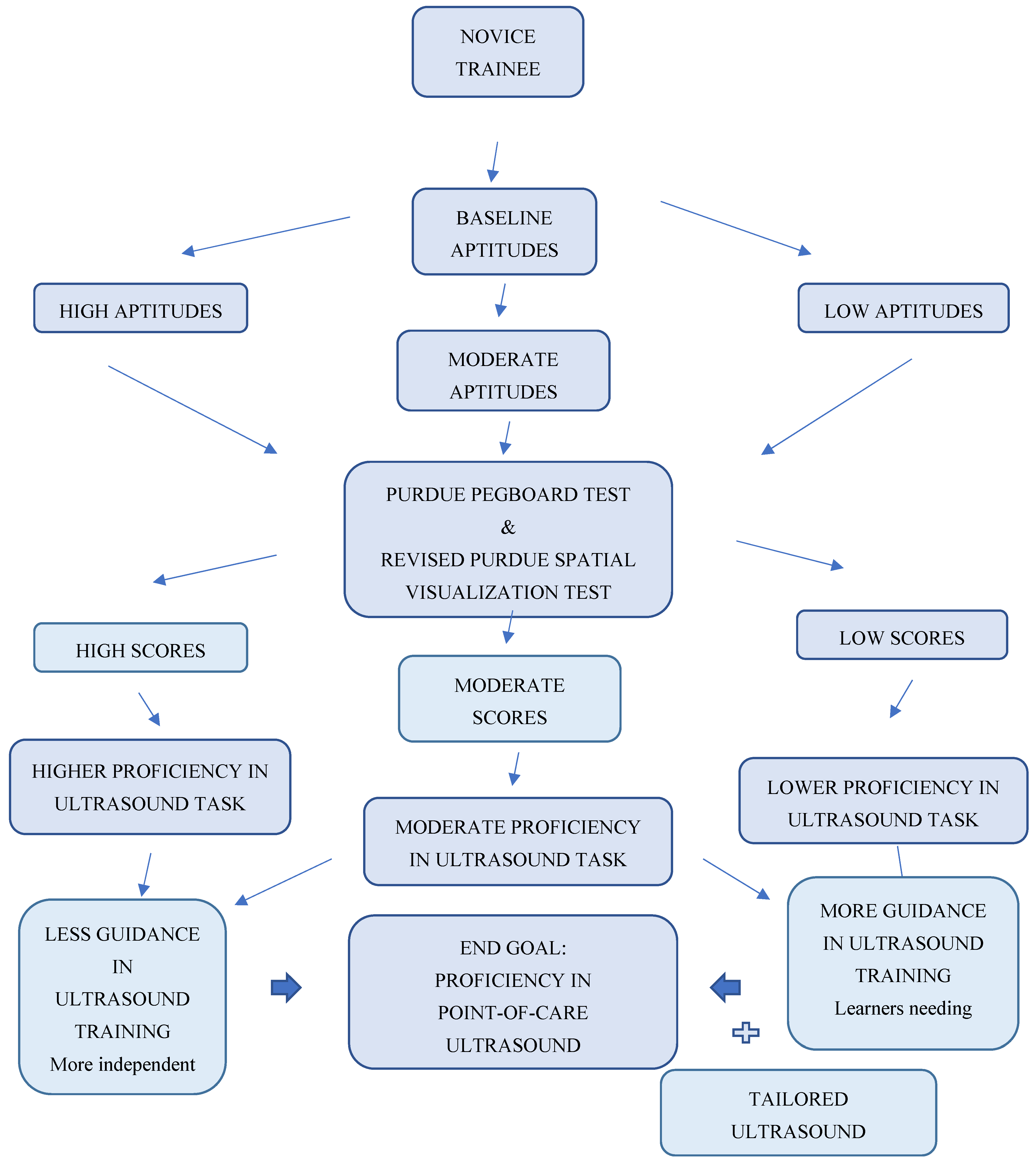
After medical school, it is further advantageous for Emergency Medicine program directors to ascertain baseline aptitudes of point-of-care ultrasounds for future residents (see Figure A3, Appendix E). Additionally, residents in other fields (i.e., internal medicine, general surgery, and numerous sub-specialties) benefit from possessing these same aptitudes when applying ultrasounds clinically in the care of a patient. This study has helped associate certain abilities in performing ultrasounds that can be identified via a simple survey and task testing. In a way, students can obtain assistance with weaker points and not be “triaged” for a specialty of their choice just based on intrinsic skill [12,18]. Consequently, with individualized training, a student’s cognitive load can be lessened, leading to more success [8]. Again, the mantra “No learner left behind” in ultrasound education is paramount early in a medical career.

Author Contributions

Conceptualization, S.A., L.A.M. and G.C.C.; methodology, S.A., E.R.A., L.A.M. and G.C.C.; formal analysis, S.A., L.A.M. and L.D.M.; investigation, S.A. and E.R.A.; resources, S.A.; data curation, S.A.; writing—original draft preparation, S.A. and L.D.M.; writing—review and editing, S.A., E.R.A., L.A.M. and L.D.M., supervision, L.A.M. and G.C.C.; project administration, S.A. All authors have read and agreed to the published version of the manuscript.

Funding

This research received no external funding.

Institutional Review Board Statement

This study was conducted in accordance with the Declaration of Helsinki and approved by the Institutional Review Board (or Ethics Committee) of the New York Presbyterian-Brooklyn Methodist Hospital (IRB #586619-3 and 3 May 2015).

Informed Consent Statement

Informed consent was obtained from all subjects involved in this study.

Data Availability Statement

The original contributions presented in this study are included in the article. Further inquiries can be directed to the corresponding author(s).

Acknowledgments

Special thanks to William M. Briggs, who assisted with statistical analysis.

Conflicts of Interest

The authors declare no conflicts of interest.

Appendix A

Pre-Testing Survey Using Likert Scale Ratings—(Rate from 1–5: 1 Is Low, 5 Is High):
  • Which medical school do you attend?
  • Age?
  • Dominant hand, left or right?
  • How would you rate your current knowledge and memory of human anatomy?
  • Rate on a scale from 1–5 the usefulness of the following study resources for learning human anatomy.
    • Anatomy books?
    • Cadavers?
    • Computer 2D images?
    • 3D models?
    • Pre-dissected organ models?
  • How skilled are you at interpreting a geographical map and finding directions?
  • How often do you play video games (of any type)?
    • Never
    • Occasionally (monthly basis)
    • Often (weekly basis)
    • Daily
  • How much time per day do you work on a computer?
    • Less than 2 h/day
    • 2 to 5 h/day
    • 5 h and more/day
  • Rate your interest level in learning and training on an ultrasound machine.

Appendix B

Figure A1. Purdue Pegboard Test: Reproduced with permission from Lafayette Instrument Company, producers of the Purdue Pegboard Test [13].
Ecm 02 00031 g0a1
Purpose: this test is used for fine and gross motor dexterity examination, first originating in assembly-line workers.
Evaluation: measures the (a) gross movement of arms and hands and (b) fingertip dexterity.
Psychometric Characteristics: high reliability (due to rigorous experimentation in a variety of industrial settings) and is considered a standard test against which other standard assessments can be compared [13].
Administration
  • Using the right hand only: First, an individual uses only their right hand to place small pins into a series of holes in a column. They have 30 s for this task. The examiner counts the number of pins placed in the holes and records this score. The board is then reset.
  • Using the left hand only: Next, the individual uses only their left hand to place small pins into the column holes. Again, they have 30 s for this task. The examiner counts the number of pins placed in the holes and records this score. The board is then reset.
  • Using both hands: Next, the individual has 30 s to place pins in two adjacent columns of holes using both hands at the same time. The examiner counts the number of pins placed in the holes on each side and records this score. The board is then reset.
  • Assembly apparatus: Lastly, using both hands, individuals assemble a four-part unit consisting of a pin, washer, collar, and washer (in that order). They have 60 s for this task. The score equals the number of each part successfully placed.
  • Two final scores are obtained. Score number 1 is the summation of steps 1, 2, and 3. Score number 2 is the final score of Step 4 only.
Time required: instruction, administration, and results should be completed within 10 min [13].

Appendix C

Revised Purdue Spatial Visualization Tests—Visualization of Rotations
The original version of the test was designed by Guay (1976). It was utilized to gauge an individual’s ability to mentally rotate a 3D image and evaluate spatial visualization intelligence. In the present day, the instrument is used extensively in the fields of science, technology, engineering, and mathematics to assess spatial ability. It has since been revised by Yoon et al. (2011) to eliminate previous figural errors and other biases [14].
Figure A2. Revised Purdue Spatial Visualization Test- Visualization of Rotations: Reproduced with permission from author Yoon S., Psychometric properties of the Revised Purdue Spatial Visualization Tests: Visualization of rotations (The Revised PSVT:R). Ph.D. Thesis, Purdue University, West Lafayette, IN, USA, 2011 [14].
Ecm 02 00031 g0a2

Appendix D

Table A1. Ultrasound Task Grading Rubric.
SCORE123
Holds probe in correct orientation2 corrections1 correctionNo correction
SCORE123
Places probe on correct spot on the model2 corrections1 correctionNo correction
SCORE123
Holds probe at correct angle2 corrections1 correctionNo correction
SCORE123
Properly identifies the chambers of the heart viewed3 guesses2 guesses1 guess
SCORE123
Captures image at best gain qualitySub-optimalAcceptableOptimal
SCORE123
Places image at best depthSub-optimalAcceptableOptimal
SCORE123
Stores image2 reminders1 reminderNo reminder
SCORE123
Total time to obtain image>5 Minutes1-5 Minutes1 Minute
SCORE123
Overall image qualityPoorGoodExcellent

Appendix E

Figure A3. No Learner Left Behind Pathway.
Ecm 02 00031 g0a3

References

  1. Cross, S.S. Aptitude testing and assessment of training progress in histopathology. Curr. Diagn. Pathol. 2005, 11, 299–307. [Google Scholar] [CrossRef]
  2. Smith, H.M.; Kopp, S.L.; Johnson, R.L.; Long, T.R.; Cerhan, J.H.; Hebl, J.R. Looking into learning: Visuospatial and psychomotor predictors of ultrasound-guided procedural performance. Reg. Anesth. Pain Med. 2012, 37, 441–447. [Google Scholar] [CrossRef] [PubMed]
  3. Enochsson, L.; Isaksson, B.; Tour, R.; Kjellin, A.; Hedman, L.; Wredmark, T.; Tsai-Felländer, L. Visuospatial skills and computer game experience influence the performance of virtual endoscopy. J. Gastrointest. Surg. 2004, 8, 874–880. [Google Scholar] [CrossRef] [PubMed]
  4. Early, D.S.; Hall, M.; Aslanian, H.R.; Casey, B.; Burbridge, R.; Chak, A.; Wani, S. A prospective, multicenter study researching the aptitude of trainees in endoscopic ultrasonography (rate US study) using cumulative sum analysis. (Abstract) Gastrointestinal Endoscopy. In Proceedings of the Conference: Digestive Disease Week, DDW 2014 ASGE, Chicago, IL, USA, 3–6 May 2014. [Google Scholar]
  5. Ritter, E.M.; McClusky, D.A.; Gallagher, A.G.; Enochsson, L.; Smith, C.D. Perceptual, visuospatial, and psychomotor abilities correlate with duration of training required on a virtual-reality flexible endoscopy simulator. Am. J. Surg. 2006, 192, 379–384. [Google Scholar] [CrossRef] [PubMed]
  6. Nicholls, D.; Sweet, L.; Hyett, J. Psychomotor Skills in Medical Ultrasound Imaging. J. Ultrasound Med. 2014, 33, 1349–1352. [Google Scholar] [CrossRef] [PubMed]
  7. Clem, D.W.; Donaldson, J.; Curs, B.; Anderson, S.; Hdeib, M. Role of Spatial Ability as a Probable Ability Determinant in Skill Acquisition for Sonographic Scanning. J. Ultrasound Med. 2013, 32, 519–528. [Google Scholar] [CrossRef] [PubMed]
  8. Mulder, T.A.; van de Velde, T.; Dokter, E.; Boekestijn, B.; Olgers, T.J.; Bauer, M.P.; Hierck, B.P. Unravelling the skillset of point-of-care ultrasound: A systematic review. Ultrasound J. 2023, 15, 19. [Google Scholar] [CrossRef] [PubMed]
  9. Jian, Y.; Bakar, J. Comparing cognitive load in learning spatial ability: Immersive learning environment vs. digital learning media. Discov. Sustain. 2024, 5, 111. [Google Scholar] [CrossRef]
  10. Mackie, R. Multiple Intelligences and Graphic Design Ability in Five North Carolina Community Colleges; Adult and Community College Education; North Carolina State University: Raleigh, NC, USA, 2005. [Google Scholar]
  11. Castro-Alonso, J.C.; Ayres, P.; Sweller, J. Instructional visualizations, cognitive load theory, and visuospatial processing. Visuospatial Process. Educ. Health Nat. Sci. 2019, 5, 111–143. [Google Scholar]
  12. Changiz, T.; Amouzeshi, Z.; Najimi, A.; Adibi, P. A narrative review of psychomotor abilities in medical sciences: Definition, categorization, tests, and training. J. Res. Med. Sci. 2021, 26, 69. [Google Scholar] [CrossRef] [PubMed]
  13. Purdue Pegboard Test Manual PDF. Model 32020A User’s Manual. Available online: www.lafayetteinstrument.com (accessed on 3 May 2015).
  14. Yoon, S.Y. Psychometric Properties of the Revised Purdue Spatial Visualization Tests: Visualization of Rotations (The Revised PSVT:R). Ph.D. Thesis, Purdue University, West Lafayette, IN, USA, 2011. Order Number: 3480934. [Google Scholar]
  15. Maurice-Ventouris, M.; Moran, H.R.M.; Alharbi, M.; Ahn, B.T.; Harley, J.M.; Lachapelle, K.J. The study of visuospatial abilities in trainees: A scoping review and proposed model. Surg. Open Sci. 2021, 5, 25–33. [Google Scholar] [CrossRef] [PubMed]
  16. Leppink, J.; Heuvel, A.V.D. The evolution of cognitive load theory and its application to medical education. Perspect. Med. Educ. 2015, 4, 119–127. [Google Scholar] [CrossRef] [PubMed]
  17. Akhigbe, T. Cognitivism and Ultrasound Skills in Emergency Medicine Training: From Theory to Practice. Int. J. Med. Rev. 2018, 5, 47–49. [Google Scholar] [CrossRef]
  18. Wani, S.; Hall, M.; Keswani, R.N.; Aslanian, H.R.; Casey, B.; Burbridge, R.; Early, D. Variation of aptitude of trainees in endoscopic ultrasonography, based on cumulative sum analysis. Clin. Gastroenterol. Hepatol. 2015, 13, 1318–1325. [Google Scholar] [CrossRef] [PubMed]
  19. Ayala, S.; Abrams, E.; Chiricolo, G. Assessing medical student background characteristics, psychomotor and visual-spatial abilities and how they correlate to aptitude in learning medical ultrasound. Academic Emergency Medicine. In Proceedings of the Conference: 2014 World Congress of Ultrasound in Medical Education (WCUME), Portland, OR, USA, 10–12 October 2014. [Google Scholar]
  20. Ayala, S.; Abrams, E.; Chiricolo, G. Assessing medical student background characteristics, psychomotor and visual-spatial abilities and how they correlate to aptitude in learning medical ultrasound. Academic Emergency Medicine. In Proceedings of the Conference: 2015 Annual Meeting of the Society for Academic Emergency Medicine, SAEM 2015, San Diego, CA, USA, 12–15 May 2015. [Google Scholar]
  21. Chapman, E. Is there a relationship between ultrasound scanning ability and visuospatial perception or psychomotor ability? Should this type of assessment be employed as a pre-selection tool for ultrasound training? Br. Med. Ultrasound Soc. 2016, 24, 214–221. [Google Scholar]
  22. Kondrashova, T.; Kondrashov, P. Integration of Ultrasonography into the Undergraduate Medical Curriculum: Seven Years of Experience. Mo. Med. 2018, 115, 38–43. [Google Scholar] [PubMed]
Disclaimer/Publisher’s Note: The statements, opinions and data contained in all publications are solely those of the individual author(s) and contributor(s) and not of MDPI and/or the editor(s). MDPI and/or the editor(s) disclaim responsibility for any injury to people or property resulting from any ideas, methods, instructions or products referred to in the content.