Article

Reversing the Trend: The Evolution of Cranial Akinesis in the Terror Birds (Cariamiformes, Phorusrhacidae)

by
Federico J. Degrange
1,*,
Claudia P. Tambussi
1 and
Lawrence M. Witmer
2
1
Centro de Investigaciones en Ciencias de la Tierra (CICTERRA), UNC, CONICET, Avenida Vélez Sársfield 1611, Córdoba X5016GCA, Argentina
2
Department of Biomedical Sciences, Heritage College of Osteopathic Medicine, Ohio University, Athens, OH 45701, USA
*
Author to whom correspondence should be addressed.
Foss. Stud. 2025, 3(3), 12; https://doi.org/10.3390/fossils3030012
Submission received: 27 June 2025 / Revised: 13 August 2025 / Accepted: 15 August 2025 / Published: 17 August 2025

Abstract

Cranial kinesis in Neornithes is enabled by a complex system of bones, muscles, ligaments, and joints, allowing variable degrees of movement between the upper jaw and the neurocranium. Particularly, cranial prokinesis—mobility of the upper jaw relative to the neurocranium—is the most widespread form. It has been stated that Phorusrhacidae (Aves, Cariamiformes) were incapable of performing cranial kinesis. To assess potential functional differences within the clade, all known cranial flexion zones were analyzed and compared with the kinetic systems of extant Neornithes. In Psilopterinae and Mesembriornithinae, fusion of the ventral palatal flexion zones indicates a rigid, immobile palate. In larger taxa such as Patagornithinae and Phorusrhacinae, the pronounced thickening and fusion of the craniofacial flexion zone confirms the loss of prokinetic capabilities. The functional implications of such akinesis include simplification of food processing and a significant increase in bite force, which correlates with the predatory skills of the group. In a phylogenetic framework, phorusrhacids evolved from a kinetic ancestral condition, representing the only truly akinetic group among Neornithes.

1. Introduction

Cranial kinesis is a hallmark of avian cranial evolution, enabling a remarkable range of skull mobility that underlies key functional, ecological, and evolutionary innovations in Neornithes. Following [1,2] cranial kinesis can be defined as the presence of movements between portions of the skull at intracranial joints. In Neornithes, cranial kinesis can involve several distinct types of intracranial movement, including prokinesis (movement between the neurocranium and the facial skeleton at the craniofacial hinge), neurokinesis (movement between the neurocranium and the palate at the basipterygoid articulation), streptostyly (mobility of the quadrate bone), rhynchokinesis (flexibility within the upper beak), and streptognathism (lateromedial movement of the lower jaw). However, in most discussions of avian cranial kinesis, the term typically refers to prokinesis.
The morphofunctional complex that acts in prokinesis consists of several bones joined by mobile unions or zonae flexoriae [3,4,5,6]. These mobile areas include the otic synovial joint, the quadratum–pterygoid and quadratum–arcus jugalis articulations, pterygopalatine articulation, basal synovial joint (when present), the zona flexoria arcus jugalis, zona flexoria palatina, and the zona flexoria craniofacialis [7]. These mobile points rotate mainly by the action of the musculus protractor pterygoidei et quadrati, which originates on the interorbital septum and inserts on the mesial surface of the os quadratum and on the dorsal surface of the os pterygoideum [8,9]. When this muscle contracts, it pushes the os quadratum forward (which rotates at the level of the otic synovial joint), pushing forward the pterygopalatine bones and the jugal arch. Thanks to the ventral zonae flexoriae (palatina et arcus jugalis), the upper jaw can rotate relative to the jugal arch and the palatine bones [4,10,11]. This rotation is dorsal due to the rostral thrust of the palatines and jugal arch and the presence of the dorsal flexion zone, the zona flexoria craniofacialis. Altogether, the coordinated action of contraction of the protractors and the consequent rotation at the flexion zones results in upper jaw elevation, allowing a greater oral opening or gape. It has been stated that m. depressor mandibulae also play a role, in that lowering the mandible also pushes the os quadratum rostrally, further engaging the push-rod system to elevate the upper jaw [4,5].
A widely accepted framework identifies four anatomical criteria required to assess the presence of cranial kinesis [1]: (1) an otic synovial articulation (quadratosquamosal articulation); (2) a basal synovial articulation (basipterygoid articulation); (3) the presence of protractor muscles, (4) permissive kinematic linkages. According to the fulfillment of one or several of these criteria, the authors recognize three states. The “Partially kinetically competent” state includes fulfillment of criteria 1, 2 or 3, but not 4, and lacks in vivo validation. The “Fully kinetically competent” state includes all four criteria, also without in vivo validation. The “Kinetic” state requires that all criteria are fulfilled and that there is in vivo validation. The authors also acknowledge the possible existence of a fourth state, “Akinetic”, but they note that the term is ambiguous, as it may encompass partially or fully kinetically competent states. In birds, criteria 1 and 3 are consistently fulfilled, but criteria 2 and 4 may not be.
The case of terror birds (Cariamiformes, Phorusrhacidae) is of wide interest, as they represent one of the most iconic groups of birds of the South American avifauna, occupying the ecological role of apex terrestrial predators during much of the Cenozoic. Phorusrhacids were mid- to large-body-size birds that filled the apex predatory niche [12,13] during most of the Cenozoic (middle Eocene to upper Pleistocene [14,15]). Phylogenetically related to extant seriemas (Cariamiformes, Cariamidae), they had poor-to-absent flying capabilities, but were highly cursorial [16,17]. While some authors propose a sister relationship between Cariamidae and Phorusrhacidae [18,19], recent phylogenetic analyses proposed a sister relationship between Bathornis+Cariamidae and Phorusrhacidae [20] or between Ameghinornis+Phorusrhacidae, with a successive sister relationship with Bathornis and Cariamoidea (a taxon that includes Cariamidae, Dynamopterus, and Elaphrocnemus, see [21]).
The characteristically high, narrow, and elongated beaks of phorusrhacids, often ending in a pronounced distal hook, have long been considered as a distinctive trait. However, unlike other Neornithes, terror birds lost the ability to rotate their upper beaks dorsoventrally, that is, they lack the cranial kinesis typical of most modern birds. This cranial immobility, unknown in other predatory avians, represents a derived condition within this clade. It is thought to have enabled terror birds to subdue their prey through powerful vertical strikes delivered with their beaks [12,13].
Here, we present an extended anatomical description of the cranial regions associated with the loss of kinesis in phorusrhacids. Our aim is to investigate the evolution of this trait within a phylogenetic framework in order to assess potential relationships between cranial morphology, trophic adaptations, and diversification of the group. By integrating anatomical and evolutionary perspectives, this study contributes to a better understanding of the functional and ecological implications of cranial immobility in this iconic clade of extinct predatory birds.

2. Materials and Methods

Osteological nomenclature follows [7] except when noted. The myological description follows [9] for the adductor muscles and [8] for the rest of the muscles.

3. Results

3.1. Otic Synovial Joint

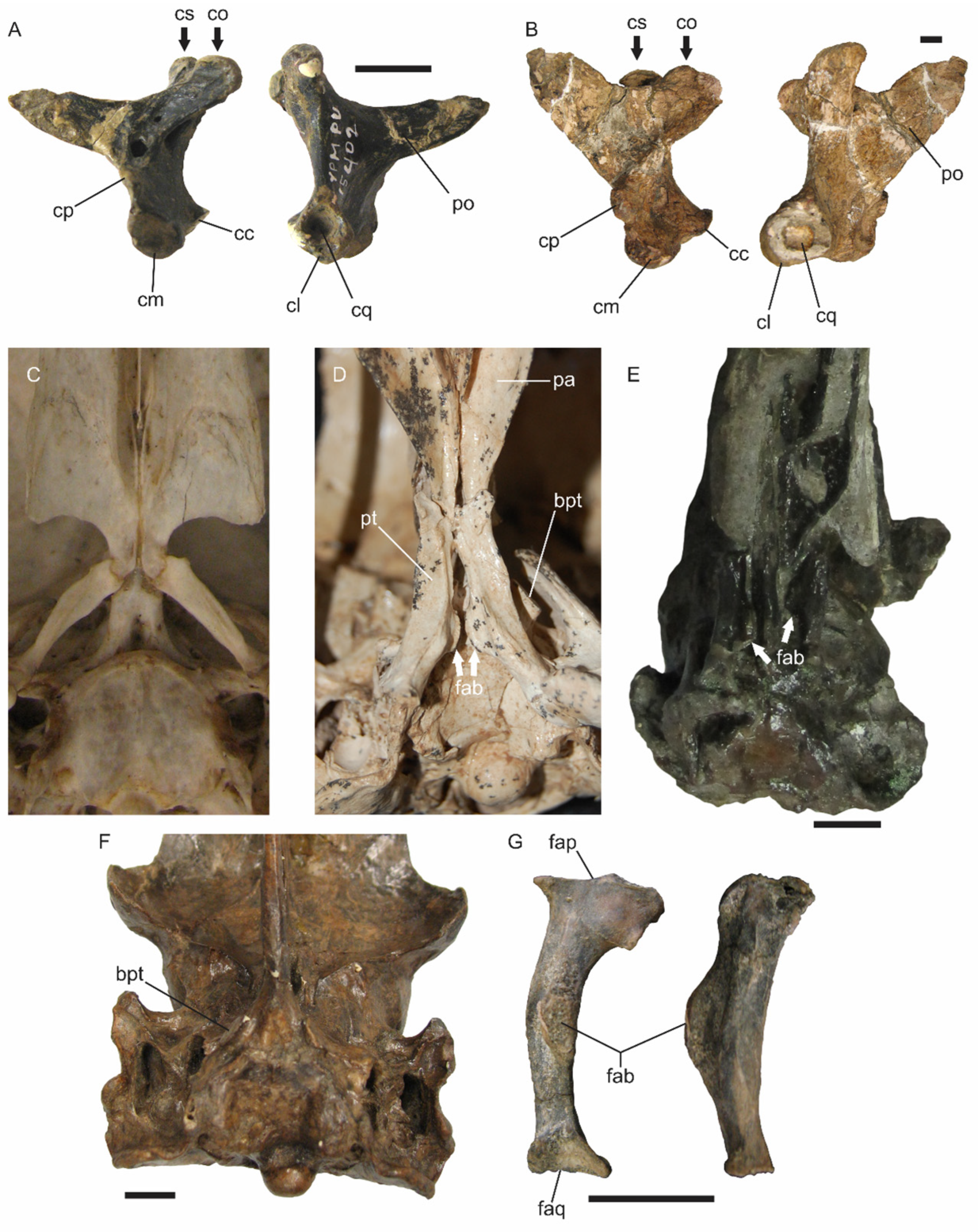
In Neognathae, the quadrate (os quadratum) bears two capitula, each enclosed in its respective synovial capsules, that articulate with the cotylae of the otic region. Terror birds share this dual-headed morphology with their living relatives, the Cariamidae [12]. Both capitula lie in the same horizontal plane—mirroring the cotyla—yet the capitulum squamosum sits rostral to the capitulum oticum, a configuration especially pronounced in Patagornithinae (Figure 1B). The smooth, soft, and rounded surface of both capitula point to the presence of a broad coating of hyaline capsules, with only a narrow capsular fibrous rim (especially notorious in Patagornis marshi MLP 20-122), matching the constricted pattern noted by [1]. Collectively, these traits indicate that the quadrate–otic articulation in phorusrhacids remained mobile, consistent with streptostyly. In the extinct Bathornis CM9377, most of the otic region is not preserved. Only the oval and deep cotyla quadratica squamosi remains, while from the os quadratum, only the capitulum squamosum is partially preserved (see [19]).

3.2. Basal Synovial Joint

Although this articulation is characteristic of Palaeognathae, and basal Neognathae such as Galliformes and Anseriformes (e.g., [22]), it is also present in several Neoaves groups such as Cathartiformes, Procellariiformes, Strigiformes, and Trogoniformes, among others [23], in which the processus basipterygoidei are developed. In phorusrhacids, this articulation is highly developed, in stark contrast with seriemas (Figure 1C), where it is absent. In Bathornis, the basis cranii externa was reconstructed by [24], but the processus is actually not preserved [19]. In phorusrhacids, the processus basipterygoidei are oval, flat, and pedunculated, rostroventrolaterally directed [12], and they articulate with a similar process located on the body of the os pterygoideum [12,25], a feature considered unique to phorusrhacids (Figure 1D–F). This processus is particularly well developed in Patagornis marshi NMHUK A516, in which the surrounding of the articulation facet is very well expanded. However, the connection between both structures does not appear to be particularly robust. Unlike in Palaeognathae, where a convex processus basipterygoideus fits into a concave facet on the os pterygoideus, the articulation in phorusrhacids consists of two flat surfaces in contact, likely joined by synovial capsules.

3.3. Protractor Muscles

Protractor musculature plays a key role in cranial kinesis [26]. In Neornithes, the musculus protractor pterygoidei et quadrati is the primary muscle responsible for initiating kinetic movement. Its singular origin is located on the septum interorbitale, ventral to the foramen opticum, but its insertion is double, being on the os pterygoideum and more broadly on the medial surface of the processus orbitalis of the os quadratum [27]. This muscle is present in Cariamidae (pers. obs.), and although its presence would be expected in Bathornis, this cannot be corroborated, as the bony regions associated with its attachment are not preserved in the available fossil material.
In observable phorusrhacids, this muscle was present and well developed. However, its origin can be fully appreciated only in specimens of Psilopterus lemoinei (AMNH 9257), Patagornis marshi (MLP 20-122), and Llallawavis scagliai (MMP 5050) (Figure 2). It is located in an oval area on the interorbital septum, clearly delimited by ridges, rostroventral to the foramen maxillomandibularis, ventral to the foramen nervus optici, and dorsal to the processus basipterygoideus. Additionally, it is situated within a small caudal notch, rostrodorsomedial to the ridge or lip formed by the ala parasphenoidalis (see [12,25]).
The insertion is more complex to reconstruct. The ossa pterygoidea of Andalgalornis (FM-P14357) are highly fragmentary, and in Psilopterus lemoinei (AMNH 9257), there is no pit or mark that clearly defines the site of insertion on this bone. However, in Llallawavis (MMP 5050), a small pit is observed posterior to the processus of the facies articularis basipterygoidea. This muscle inserts dorsal to a medial crest that marks the origin of the m. pterygoideus ventralis, where a second pit associated with the insertion is also found. The insertion of this muscle on the os quadratum is clearer, as it attaches to a fossa located on the medial aspect of the processus orbitalis, rostral to the medial pneumatic foramina located on the corpus ossa quadrati (see [12]). In Andalgalornis (FM-P14357) and Psilopterus lemoinei (YPM-PU15402), this pit is poorly marked (Figure 2). In Llallawavis (MMP 5050), a subhorizontal medial crest ventrally delimits the insertion of this muscle.

3.4. Permissive Kinematic Linkages

The zonae flexoriae are flattened, thin areas of bone that allow bending and thus function as points of rotation. The zona flexoria craniofacialis is located dorsally and is represented by a thin area on the processus nasalis of the os premaxillare and the adjacent processus premaxillaris of the os nasale, in continuity with the os frontale [11].
The palatal or ventral flexion zones include the zona flexoria palatina and the zona flexoria arcus jugalis, which correspond to thin regions at the palatine–maxilla and jugal–maxilla junctions, respectively [28]. Seriemas possess the three zonae flexoriae (Figure 3B), whereas in Bathornis at least the palatal zonae flexoria are present (the zona flexoria craniofacialis is not preserved; Figure 3A). Nevertheless, in Cariamidae, a large ligament runs rostrocaudally between the os palatinum and the os maxillare [29], limiting movement at the zona flexoria palatina. This ligament appears to be absent in Bathornis, which exhibits a narrower processus rostralis of the os palatinum.
In phorusrhacids, two or even all three of these zones may be absent (Figure 3C). The absence of zonae flexoria palatina et arcus jugalis results from the fusion of the processus rostralis of the os palatinum with both the os maxillare and the arcus jugalis. In the former case, the caudal portion of the os premaxillare is also incorporated into the fusion. These absences produce immobility of the palate in the morphotype I phorusrhacid skull, which includes Psilopterus and Llallawavis (psilopterine type, see [12]). In contrast, in the morphotype II phorusrhacid skull (terror bird type), which includes Patagornis, Andalgalornis, Kelenken, and Phorusrhacos, among others, the thickening of the bony laminae in the zona flexoria craniofacialis prevents any bending, functionally eliminating this zone (Figure 3C). This condition indicates that these birds lacked prokinetic mobility (see [12,13,30]).

3.5. Ligamentum Postorbitale

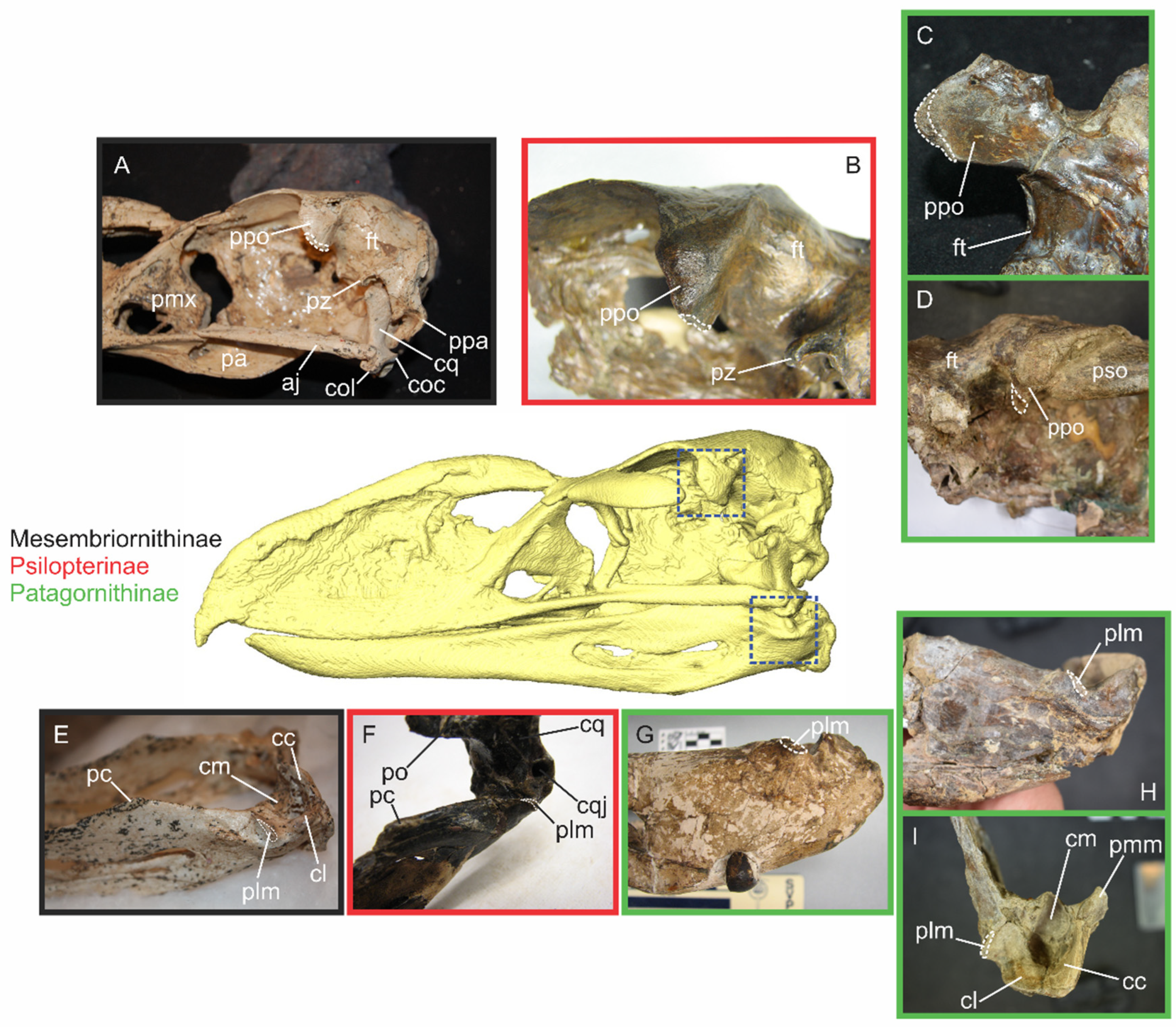
Although not originally proposed as a requisite attribute for cranial kinesis by [1], the presence or absence of the ligamentum postorbitale should be considered as an additional criterion in the kinetic analysis of Neornithes. It was extensively studied by [4], who showed that its main function is to couple mandibular and maxillary movements during cranial kinesis. While this ligament is absent in Palaeognathae [31], it is present in many Neognathae taxa [4,32,33]—Neognathae that lack this ligament present decoupled cranial kinesis [6], meaning that they are able to depress the jaw without simultaneously elevating the maxilla. The ligamentum postorbitale is present in extant Cariamidae, and its presence in Bathornis can be inferred from a small scar on the caudal edge of the processus postorbitalis, indicating is proximal attachment. Unfortunately, the processus lateralis mandibulae, where the distal attachment would be located, is not preserved. In phorusrhacids, the presence of this ligament can also be inferred (Figure 2E and Figure 4). The proximal attachment is rostrolaterally broad, attaching on a roughened surface of the processus postorbitalis. The distal attachment is located within a small circular notch on the rostral side of the processus lateralis mandibulae.

4. Discussion

Some anatomical features of the Phorusrhacidae are congruent with kinetic capabilities—such as streptostyly and the presence of protractor muscles—reflecting a retained kinetic heritage. However, other derived traits, such as the absence of zonae flexoriae, clearly certify that phorusrhacids were incapable of mobilizing the maxilla (i.e., of performing prokinesis). In other words, phorusrhacids were akinetic.
The function of the processus basipterygoideus and its contact with the os pterygoideum, forming a presumably synovial basal joint, remains difficult to establish [4]. However, it has been hypothesized that these processes serve as guides [10] or as stops during kinetic movements [4,10,34]. Gussekloo and Bout [35] proposed that the processus basipterygoidei in Palaeognathae restrict caudal movements of ossa pterygoidei. Consequently, these processi prevent depression of the maxilla below its resting position, which occurs during contraction of the pterygoideus muscles and musculus pseudotemporalis profundus [4,10,27,36]. The peculiar and intimate association that exists between the components of the basal joint—the os pterygoideum and the processus basipterygoideus—in phorusrhacids is difficult to explain. This feature has not been recorded in other Cariamiformes (absent in Cariamidae, not preserved in Bathornis). Although it is plausible that it was absent in basal Cariamiformes, its absence in extant Cariamidae could alternatively represent a cariamid apomorphy. It is noteworthy that seemingly “open” and synovial basal joints occur in various akinetic archosaur clades, leading [1] to suggest that the basal joint may plesiomorphically represent a growth zone rather than a kinetic joint. Basal joints and basipterygoid processes seem to have arisen multiple times in Neornithines. However, the presence of a patent basal joint in phorusrhacids appears to be related to the immobilization of the palate, a phenomenon likely associated with their evolution of akinesis.
The function of the protractor musculature in the phorusrhacids is also complex to elucidate. Holliday and Witmer [1] demonstrated that, similar to the basal joint, protractor muscles were widely present in archosaur clades that are almost universally considered akinetic; this pattern also holds true for theropods along the stem leading to birds. They proposed that the presence of these muscles, that do not produce movement, may have evolved instead to moderate and redirect forces generated by the jaw adductor musculature. Following the loss of certain bones in the avian lineages (e.g., postorbital, ectopterygoid) as part of a suite of morphological changes near the origin of birds—including expansion of the brain, reorientation of jaw muscles (see [2])—kinematic linkages became permissive of cranial kinesis, allowing the protractor musculature to assume an active role in powering prokinesis.
The basic avian prokinetic apparatus was already well established by the time Cariamiformes, including phorusrhacids, evolved. Therefore, the presence of protractor muscles in phorusrhacids represents a relic of their kinetic ancestry. The loss of cranial kinesis in phorusrhacids, despite the retention of the protractor musculature, suggests that these muscles may have reverted to their ancestral functions observed in more distant akinetic non-avian ancestors. Such functions could include shock absorption, impact mitigation, or compensating for forces exerted by the external adductor muscles, consistent with the estimated large bite force in phorusrhacids. Moreno et al. [37] suggested that the musculus protractor pterygoideus et quadrati controls part of the strain experienced by the os pterygoideum during contraction of the pterygoid musculature. Given that the pterygoid muscles of phorusrhacids are highly developed [25], it plausible to hypothesize that the musculus protractor pterygoideus et quadrati functioned as a shock dissipator, compensating any strain or stress generated by the contraction of the pterygoid muscles or even the total bite force.
The nature of the articulations between skull bones is critically important for understanding the mechanical behavior of the skull, as the connections either permit or restrict intracranial movements [1] and influence the distribution of stress and deformation [38,39]. In phorusrhacids, the absence of zonae flexoriae indicates that no form of cranial kinesis is possible. In morphotype II skulls (see [12]), this condition is further reinforced by the presence of additional rigid elements: the processus orbitalis of the os lacrimale is fused with the os lacrimale communicans [13], constituting a structural column that links the os lacrimale with the arcus jugalis. In Andalgalornis, a medial lip at the contact area with the arcus jugalis ensures a rigid articulation. In this taxon, the os lacrimale also fuses cranially with both the os nasale and os frontale, while in Kelenken the processus supraorbitalis of the os lacrimale is completely fused with the os frontale [30]. A similar condition is observed in Bathornis grallator, where the os lacrimale is fused with the os frontale and the os ectethmoidale, forming a solid structural unit [19].
According to [4], the coupling produced by the ligamentum postorbitale, when present, prevents depression of the lower jaw beyond its resting (closed-mouth) position unless the upper jaw is elevated. In other words, its absence allows independent movements of the upper and lower jaws. However, the validity of this coupling mechanism has been debated and even refuted by [6]. Several species possessing this ligament are able of independent movement of the jaw and maxilla (see [40,41]), and experimental data show that the ligament offers minimal resistance to mandibular depression when the maxilla is held stationary [42]. Zusi [5] argued that, even when the upper jaw is fixed, the minor caudal displacement of the mandible resulting from contraction of the m. depressor mandibulae is sufficient to reduce system coordination, thereby allowing increased mandibular depression. The author also proposed that this ligament may serve functions beyond jaw coupling: specifically, it could act to stabilize and protect the os quadratum–mandibular joint by maintaining both surfaces in close apposition [5,6]. The predatory behavior of phorusrhacids supports this stabilizing function, as the powerful bite forces exerted on active prey could otherwise compromise the integrity of the beak [13].

4.1. The Phorusrhacids as Akinetic Neornithes

As [1] pointed out, the term “akinetic” can be ambiguous, as it may encompass more than one of the kinetic states they recognized. It is therefore necessary to distinguish between “morphologically or anatomically akinetic” and “functionally akinetic”. This conceptual distinction was briefly outlined, though not fully developed, by [4], and more recently mentioned by [43] to characterize cranial function in Tyrannosaurus. Phorusrhacids represent an excellent example for this differentiation: although some intracranial articulations that might allow movements are retained (e.g., os squamosum–os quadratum and os pterygoideum–os palatinum articulations), most of the mobile cranial regions present in Neornithes are absent in phorusrhacids. This includes the zonae flexoriae arcus jugalis and palatina et craniofacialis. Due to extensive bone fusion and thickening, these regions are anatomically akinetic, incapable of any cranial movement. On the other hand, the streptostylic os quadratum is theoretically capable of rotating and therefore is anatomically kinetic.
According to [1]’s classification of kinetic states, phorusrhacids may be considered “partially kinetically competent”, as they fulfill criteria 1, 2, and 3. However, they do not meet criterion 4, which requires the presence of permissive kinematic joints. This last criterion is ultimately exclusive, as it directly relates to the dorsoventral movement of the upper jaw characteristic of prokinesis. In smaller phorusrhacids, the palate itself is the limiting structure, incapable of flexing rostrally to allow movement of the upper jaw. In larger phorusrhacids, both the palate and the upper jaw are rigid: the latter cannot flex dorsally due to the absence of the zona flexoria craniofacialis. This condition is best described as “functionally akinetic”, because—although certain skull elements may be anatomically capable of performing kinesis—the system as a whole is unable to execute such motions, and the kinetically competent joints remain immobile. This functional immobility characterizes all known phorusrhacid skulls, which in this sense, are akinetic. The term “partially kinetically competent” may lead to confusion, as it does not explicitly indicate which elements are theoretically mobile, despite ultimately referring to an akinetic system.
Here, the term “akinetic” is used in reference to both functional and anatomical immobility, depending on the scale of analysis (i.e., individual elements vs. the entire skull). A skull that is anatomically akinetic, with no mobile components, is by necessity functionally akinetic. But a functionally akinetic skull may still contain individual joints that could move if they are not mechanically constrained by other elements, thereby rendering the system immobile as a whole. In any case, within Neornithes, the term “akinetic” is appropriately applied to skulls that lack the ability to perform prokinetic movements, and this is the sense that the term is used in here.

4.2. The Functional Significance of Akinesis in Phorusrhacids

The loss of cranial kinesis in phorusrhacids is a remarkable derived feature, but the interpretation of this loss is complicated by the uncertainties about the functional role of cranial kinesis. It is possible that there is no single explanation for its evolution and function in all Neognathae [31]. Bock [4] suggested six potential roles for avian cranial kinesis: (1) passive maintenance of the jaw in closed position, (2) an increase in opening force in “gapers”, (3) maintenance of the primary visual orientation axis, (4) faster closure of the beak, (5) a shock absorption mechanism, (6) an increase in the attachment area for jaw musculature. Posteriorly, Zusi [5,11] proposed that the fundamental advantage of cranial kinesis is the potential diversity and versatility that it gives to the beak (maxilla and mandible) as a manipulative tool. Hoese and Westneat [41] proposed that kinesis could be linked to singing ability. Zweers et al. [28] related kinesis to the development of a greater strength of mandibular closure due to a better use of the horizontal component of the pterygoid musculature, which is highly developed in Neornithes. However, they also postulated that the kinetic mechanism could be the result of selective pressures during the development of flight: larger ocular and cerebral development and a lightening of the skull due to development of dermatocranial fenestration, as well as large nostrils, would be exaptations for cranial kinesis. Following this hypothesis, Bout and Zweers [6] stated that the adaptive meaning of avian cranial kinesis remains uncertain and that it could be the result of the cranial design, especially associated with the enlargement of the eye. Finally, Wilken et al. [2] recently found that cranial kinesis in birds is actually the result of the expansion of the neurocranium (as a result of the brain increment) which led to a change in jaw arrangement which influenced intracranial hinges by reorienting the protractor muscles in order to maximize the transmission of forces through the os pterygoideum.
One of the trade-offs of cranial kinesis is its relationship with bite force: although kinesis increases versatility for manipulating food items, it reduces the bite force compared with an akinetic system [6,35,39]. Cranial kinesis constrains the efficiency of the mandibular muscles and therefore limits the potential for generating high bite forces. Any morphological adaptation that increases the rigidity of the mandibular apparatus, such as the ossification or transformation of flexion zones into immobile regions, will consequently enhance the theoretical maximum bite force [44,45]. Akinetic skulls are capable of producing bite forces up to 1.3 times greater than those of kinetic avian skulls [6]. These considerations are consistent with both the muscular reconstructions and the high bite forces estimated for phorusrhacids [25], suggesting that the jaw closure mechanism in these birds was optimized for bite force generation at the expense of closure speed.
Phorusrhacids, particularly those with a morphotype II skull—the so-called “terror bird” type sensu [12]—exhibit a highly rigid cranial architecture associated with functional akinesis. This condition has profound biomechanical implications, including the capacity to deliver stronger bite forces and a simplification of food-handling mechanics. Importantly, these advantages are not strictly dependent on body size: the rigidity of the morphotype II skull likely enabled greater bite forces than those achievable by phorusrhacids with a morphotype I (“psilopterine”-type) skull [12], which is comparatively less robust.

4.3. Evolution of Cranial Akinesis in Phorusrhacids

While [46] proposed that the transformation of cranial flexion zones into syndesmoses or diarthroses is typically associated with increased body size—as exemplified by Gastornis giganteus (Gastornithiformes)—Phorusrhacidae exhibit the opposite pattern. In this lineage, akinesis is not a consequence of size alone, but rather the result of a secondary transformation from a kinetically capable skull. Moreover, within Phorusrhacidae, the degree of cranial rigidity appears to correlate positively with size, such that the largest taxa display the most structurally reinforced and akinetic skulls (Figure 5). This reversal highlights the unique evolutionary pathway of phorusrhacids, in which increased cranial stiffness may have been favored to maximize bite force and stabilize the skull during predatory strikes.
The primitive trait of Cariamiformes is the presence of cranial kinesis (i.e., prokinesis), meaning that the ancestor of all these taxa was capable of dorsal flexion of the upper beak. Akinesis is, therefore, a derived trait of the Phorusrhacidae. Both Bathornis and members of Cariamidae exhibit skull morphologies consistent with prokinetic capabilities. Although Cariamidae lack processus basipterygoidei—and their presence cannot be confirmed in Bathornis [19]—the emergence of constraints on palatal mobility likely began in the common ancestor of Bathornis, Cariamidae, and Phorusrhacidae. It appears probable that presence of the processus basipterygoidei, one of the proposed synapomorphies of Phorusrhacidae [18], evolved prior to the complete fusion of the palatal flexion zones. The processes would have played a key role in restricting palatal retraction during contraction of the pterygoid musculature—which is especially developed in phorusrhacids—even at a time when limited motion of the zonae flexoriae was still possible. This suggests that the evolution of these processes was an early step toward the progressive akinesis observed in the lineage.
In Phorusrhacidae, the bones that constitute the zona flexoria palatina and the zona flexoria arcus jugalis are completely fused, creating rigid unions that preclude any degree palatal movement. This complete ossification and immobilization of flexion zones constitutes a synapomorphy of the Phorusrhacidae clade [18]. When compared with the condition observed in Cariamidae (see [29]), it is likely that the zona flexoria palatina was the first to be suppressed. Subsequently, as the arcus jugalis became more robust and extended, it likely established broader contact with the os palatinum and os maxillare, resulting in the loss of the zona flexoria arcus jugalis.
Psilopterinae and Mesembriornithinae retain the zona flexoria craniofacialis, and the contact between the os lacrimale and the arcus jugalis is mediated by a novel element, the os lacrimale communicans (also present in Cariamidae). The condition becomes even more derived in the larger Phorusrhacidae such as Patagornithinae and Phorusrhacinae, which not only lack both palatal flexion zones, but also lose the zona flexoria craniofacialis, fully abolishing any prokinetic mobility, an apomorphic condition within the clade. These taxa are also characterized by another derived trait: the processus orbitalis of the os lacrimale forms a firm contact with the arcus jugalis through its fusion with the os lacrimale communicans [12,13]. In Andalgalornis and Kelenken, this ossification pattern is taken even further, with the os lacrimale fusing with the ossa nasale and frontale, making a completely immobile and rigid structure. Thus, phorusrhacids show a unique evolutionary reversal: from ancestrally kinetic skulls to secondarily akinetic architectures, where increasing rigidity correlates with both phylogenetic position and body size, culminating in some of the most robust cranial constructions among Neornithes.

5. Concluding Remarks

Cariamiformes represent a unique lineage of Neornithes that exhibits parallel trends toward terrestriality, body mass enlargement, and progressive cranial immobilization. While the association between body enlargement and terrestriality has already been noted [17], the link between body mass enlargement and the evolution of akinesis is particularly striking. Skull immobilization seems to have arisen through different anatomical solutions across the group: while seriemas present extensive contact between the palatine bones and the jugal bars, Bathornis shows fusion of the lacrimals–ecthetmoid–frontals, and the processus maxillopalatinus with the processus maxillaris of the os palatinum.
Nevertheless, this trend reaches its most extreme expression in phorusrhacids, where the suppression of cranial flexion zones is most pronounced in the largest taxa. This condition seems to be closely related to their unique predatory hunting strategy: active prey was dispatched with powerful, hatchet-like strokes delivered with the beak [13,47]. Cranial akinesis likely evolved in tandem with this behavior, enabling and reinforcing a feeding mechanism that departs radically from other Neornithes.
It is plausible to propose that akinesis not only permitted the evolution of this peculiar predatory behavior but also played a key role in the ecological success and adaptive radiation of phorusrhacids. Since no other group of Neornithes possesses such extreme cranial modifications, Phorusrhacidae stand as the only truly akinetic clade among crown birds. In summary, the terror birds exemplify how form and function can co-evolve in unexpected ways—transforming a kinetic ancestral skull into a rigid predatory apparatus, and turning a constraint into a singular evolutionary opportunity.
Author Contributions

Conceptualization: F.J.D.; investigation: F.J.D., C.P.T., and L.M.W.; project administration: F.J.D.; resources: F.J.D., C.P.T., and L.M.W.; visualization: F.J.D.; writing—original draft: F.J.D.; writing—review and editing; F.J.D., C.P.T., and L.M.W. All authors have read and agreed to the published version of the manuscript.

Funding

Funding to L.M. Witmer was provided by the US National Science Foundation (IOB-0517257, IOS-1050154, IOS-1456503), the Ohio University Heritage College of Osteopathic Medicine, and the Swedish Research Council: SRC 2021-02973. This is a contribution to the projects PIP-CONICET 2021, PICT 2019-0771, and PUE 2016—CONICET—CICTERRA.

Data Availability Statement

All the data are provided in the text.

Acknowledgments

The basis of the contents of this work is part of the doctoral thesis of the first author, with modifications and suggestions from the co-authors. Access to fossil specimens was kindly provided by C. Mehling, D. Brinkman, M. Reguero, A. Kramarz, A. Tauber, and A. Dondas. For support with CT scanning, we thank G. Alcuaz and J. I. Cuesta (Hospital San Juan de Dios, La Plata, Argentina), C. Capiel (Instituto Radiológico, Mar del Plata, Argentina), and H. Rockhold (OhioHealth O’Bleness Hospital, Athens, OH, USA). M. Taglioretti and F. Scaglia prepared the holotype of Llallawavis for scanning. We thank the three anonymous reviewers for their helpful comments and suggestions.

Conflicts of Interest

The authors declare no conflicts of interest.

Institutional Abbreviations

AMNH, American Museum of Natural History, New York City, NY, USA; CM, Carnegie Museum of Natural History, Pittsburgh, PA, USA; FM, Field Museum of Natural History, Chicago, IL, USA; MLP, Museo de La Plata, La Plata, Argentina; MMP, Museo Municipal Lorenzo Scaglia, Mar del Plata, Argentina; NHMUK, Natural History Museum of United Kingdom, London, UK; YPM-VPPU, Yale Peabody Museum, New Haven, CT, USA.

References

  1. Holliday, C.M.; Witmer, L.M. Cranial kinesis in dinosaurs: Intracranial joints, protractor muscles, and their significance for cranial evolution and function in diapsids. J. Vertebr. Paleontol. 2008, 28, 1073–1088. [Google Scholar] [CrossRef]
  2. Wilken, A.T.; Sellers, K.C.; Cost, I.N.; Davis, J.; Middleton, K.M.; Witmer, L.M.; Holliday, C.M. Avian cranial kinesis is the result of increased encephalization during the origin of birds. Proc. Natl. Acad. Sci. USA 2025, 122, e2411138122. [Google Scholar] [CrossRef] [PubMed]
  3. Bock, W.J. Secondary Articulation of the avian mandible. Auk 1960, 77, 19–55. [Google Scholar] [CrossRef]
  4. Bock, W.J. Kinetics of the avian skull. J. Morphol. 1964, 114, 1–41. [Google Scholar] [CrossRef]
  5. Zusi, R.L. The role of the depressor mandibulae muscle in kinesis of the avian skull. Proc. United States Natl. Mus. 1967, 123, 1–28. [Google Scholar] [CrossRef]
  6. Bout, R.G.; Zweers, G.A. The role of cranial kinesis in birds. Comp. Biochem. Physiol. A 2001, 131, 197–205. [Google Scholar] [CrossRef] [PubMed]
  7. Baumel, J.J.; Witmer, L.M. Osteologia. In Handbook of Avian Anatomy: Nomina Anatomica Avium; Baumel, J.J., King, A.S., Breazile, J.E., Evans, H.E., Vanden Berge, J.C., Eds.; Nuttall Ornithological Club: Cambridge, MA, USA, 1993; pp. 45–132. [Google Scholar]
  8. Vanden Berge, J.C.; Zweers, G.A. Myologia. In Handbook of Avian Anatomy: Nomina Anatomica Avium; Baumel, J.J., King, A.S., Breazile, J.E., Evans, H.E., Vanden Berge, J.C., Eds.; Nuttall Ornithological Club: Cambridge, MA, USA, 1993; pp. 189–247. [Google Scholar]
  9. Holliday, C.M.; Witmer, L.M. Archosaur adductor chamber evolution: Integration of musculoskeletal and topological criteria in jaw muscle homology. J. Morphol. 2007, 268, 457–484. [Google Scholar] [CrossRef] [PubMed]
  10. Beecher, W.J. The biomechanics of the bird skull. Bull. Chic. Acad. Sci. 1962, 11, 10–33. [Google Scholar]
  11. Zusi, R.L. Patterns of diversity in the avian skull. In The Skull. Volume 2: Patterns of Structural and Systematic Diversity; Hanken, J., Hall, B.K., Eds.; The University of Chicago Press: Chicago, IL, USA, 1993; pp. 391–437. [Google Scholar]
  12. Degrange, F.J. A revision of Phorusrhacidae (Aves, Cariamiformes) skull morphology. J. Vertebr. Paleontol. 2021, 40, e1848855. [Google Scholar] [CrossRef]
  13. Degrange, F.J.; Tambussi, C.P.; Moreno, K.; Witmer, L.M.; Wroe, S.; Turvey, S.T. Mechanical Analysis of Feeding Behavior in the Extinct “Terror Bird” Andalgalornis steulleti (Gruiformes: Phorusrhacidae). PLoS ONE 2010, 5, e11856. [Google Scholar] [CrossRef] [PubMed]
  14. Jones, W.; Rinderknecht, A.; Alvarenga, H.; Montenegro, F.; Ubilla, M. The last terror birds (Aves, Phorusrhacidae): New evidence from the late Pleistocene of Uruguay. PalZ 2017, 92, 365–372. [Google Scholar] [CrossRef]
  15. Hospitaleche, C.A.; Jones, W. Insights on the oldest terror bird (Aves, Phorusrhacidae) from the Eocene of Argentina. Hist. Biol. 2024, 37, 391–399. [Google Scholar] [CrossRef]
  16. Degrange, F.J.; Noriega, J.I.; Vizcaíno, S.F. Morphology of the forelimb of Psilopterus bachmanni (Aves, Cariamiformes) (Early Miocene of Patagonia). PalZ 2015, 89, 1087–1096. [Google Scholar] [CrossRef]
  17. Degrange, F.J. The hind limb morphometry of terror birds (Aves, Cariamiformes, Phorusrhacidae): Their functional implications for substrate preferences and locomotory lifestyle. Earth Environ. Sci. Trans. R. Soc. Edinb. 2017, 106, 257–276. [Google Scholar] [CrossRef]
  18. Degrange, F.J.; Tambussi, C.P.; Taglioretti, M.L.; Dondas, A.; Scaglia, F. A new Mesembriornithinae (Aves, Phorusrhacidae) provides new insights into the phylogeny and sensory capabilities of terror birds. J. Vertebr. Paléontol. 2015, 35, e912656. [Google Scholar] [CrossRef]
  19. Mayr, G. Osteology and phylogenetic affinities of the middle Eocene North American Bathornis grallator—One of the best represented, albeit least known Paleogene cariamiform birds (seriemas and allies). J. Paleontol. 2016, 90, 357–374. [Google Scholar] [CrossRef]
  20. LaBarge, T.W.; Gardner, J.D.; Organ, C.L. The evolution and ecology of gigantism in terror birds (Aves, Phorusrhacidae). Proc. R. Soc. B 2024, 291, 20240235. [Google Scholar] [CrossRef] [PubMed]
  21. Agnolin, F.L.; Chafrat, P.; Álvarez-Herrera, G.P. New specimens of Patagorhacos terrificus Agnolín and Chafrat, 2015 (Aves) shed light on the phylogeny and evolution of the Phorusrhacidae. Hist. Biol. 2025, 37, 1744–1756. [Google Scholar] [CrossRef]
  22. Bailleul, A.M.; Witmer, L.M.; Holliday, C.M. Cranial joint histology in the mallard duck (Anas platyrhynchos): New insights on avian cranial kinesis. Am. J. Anat. 2017, 230, 444–460. [Google Scholar] [CrossRef]
  23. Elzanowski, A. On the role of basipterygoid processes in some birds. Verhandlungen Der Anat. Ges. 1977, 71, 1303–1307. [Google Scholar]
  24. Wetmore, A. A new terrestrial vulture from the Upper Eocene deposits of Wyoming. Ann. Carnegie Mus. 1944, 30, 57–69. [Google Scholar] [CrossRef]
  25. Degrange, F.J. Morfología del Cráneo y Complejo Apendicular en Aves Fororracoideas: Implicancias en la Dieta y Modo de Vida. Ph.D. Dissertation, La Plata University, La Plata, Argentina, 2012; 388p. [Google Scholar]
  26. Holliday, C.M. New insights into dinosaur jaw muscle anatomy. Anat. Rec. 2009, 292, 1246–1265. [Google Scholar] [CrossRef] [PubMed]
  27. Bhattacharyya, B.N. Avian jaw function: Adaptation of the seven–muscle system and a review. Proc. Zool. Soc. 2013, 66, 75–85. [Google Scholar] [CrossRef]
  28. Zweers, A.G.; Vanden Berge, J.C.; Berkhoudt, H. Evolutionary patterns of avian trophic diversification. Zoology 1997, 100, 25–57. [Google Scholar]
  29. Degrange, F.J.; Tambussi, C.P. Re-examination of Psilopterus lemoinei (Moreno and Mercerat 1891), a late early Miocene little terror bird from Patagonia (Argentina). J. Vertebr. Paleontol. 2011, 31, 1080–1092. [Google Scholar] [CrossRef]
  30. Degrange, F.J.; Eddy, D.; Puerta, P.; Clarke, J. New skull remains of Phorusrhacos longissimus (Aves, Cariamiformes) from the Miocene of Argentina: Implications for the morphology of Phorusrhacidae. J. Paleontol. 2019, 93, 1221–1233. [Google Scholar] [CrossRef]
  31. Gussekloo, S.W.S.; Bout, R.G. The kinematics of feeding and drinking in palaeognathous birds in relation to cranial morphology. J. Exp. Biol. 2005, 208, 3395–3407. [Google Scholar] [CrossRef] [PubMed]
  32. Meekangvan, P.; Barhosrst, A.A.; Chatterjee, S.; Schovanec, L. Nonlinear dynamical model of avian cranial kinesis. J. Theor. Biol. 2006, 240, 32–47. [Google Scholar] [CrossRef]
  33. Ferreira, M.M.D.; Tambussi, C.P.; Degrange, F.J.; Pestoni, S.; Tirao, G.A. The cranio-mandibular complex of the nightjar Systellura longirostris (Aves, Caprimulgiformes): Functional relationship between osteology, myology and feeding. Zoology 2019, 132, 6–16. [Google Scholar] [CrossRef] [PubMed]
  34. Fisher, H.I. Some aspects of the kinetics in the jaws of birds. Wilson Bull. 1955, 67, 175–188. [Google Scholar]
  35. Gussekloo, W.S.; Bout, R.G. Cranial kinesis in palaeognathus birds. J. Experminetal Biol. 2005, 208, 3409–3419. [Google Scholar] [CrossRef] [PubMed]
  36. Bühler, P. Functional anatomy of the avian jaw apparatus. In Form and Function in Birds; King, A.S., McClelland, J., Eds.; Academic Press: New York, NY, USA, 1981; Volume 2, pp. 439–468. [Google Scholar]
  37. Moreno, K.; Wroe, S.; Clausen, P.; McHenry, C.; D’amore, D.C.; Rayfield, E.J.; Cunningham, E. Cranial performance in the Komodo dragon (Varanus komodoensis) as revealed by high-resolution 3-D finite element analysis. Am. J. Anat. 2008, 212, 736–746. [Google Scholar] [CrossRef] [PubMed]
  38. Rayfield, E.J. Using finite-element analysis to investigate suture morphology: A case study using large carnivorous dinosaurs. Anat. Rec. Part A Discov. Mol. Cell. Evol. Biol. 2005, 283A, 349–365. [Google Scholar] [CrossRef]
  39. Moazen, M.; Curtis, N.; O’HIggins, P.; Jones, M.E.; E Evans, S.; Fagan, M.J. Assessment of the role of sutures in a lizard skull: A computer modelling study. Proc. R. Soc. B Biol. Sci. 2009, 276, 39–46. [Google Scholar] [CrossRef] [PubMed]
  40. Van Gennip, E.M.S.J.; Berkhoudt, H. Skull mechanics in the pigeon, Columba livia, a threedimensional kinetic model. J. Morphol. 1992, 213, 197–224. [Google Scholar] [CrossRef] [PubMed]
  41. Hoese, W.J.; Westneat, M.W. Biomechanics of cranial kinesis in birds: Testing linkage models in the White-throated Sparrow Zonotrichia albicollis. J. Morphol. 1996, 227, 305–320. [Google Scholar] [CrossRef]
  42. Nuijens, F.W.; Bout, R.G. The role of two jaw ligaments in the evolution of passerines. Zoology 1998, 101, 24–33. [Google Scholar]
  43. Cost, I.N.; Middleton, K.M.; Sellers, K.C.; Scott Echols, M.; Witmer, L.M.; Davis, J.L.; Holliday, C.M. Palatal Biomechanics and its Significance for Cranial Kinesis in Tyrannosaurus rex. Anat. Rec. 2020, 303, 999–1017. [Google Scholar] [CrossRef] [PubMed]
  44. Wroe, S.; Moreno, K.; Clausen, P.D.; McHenry, C.R. High-resolution computer simulation of hominid cranial mechanics. Anat. Rec. 2007, 290, 1248–1255. [Google Scholar] [CrossRef] [PubMed]
  45. Wroe, S.; Huber, D.R.; Lowry, M.; McHenry, C.; Moreno, K.; Clausen, P.; Ferrara, T.L.; Cunningham, E.; Dean, M.N.; Summers, A.P. Three dimensional computer analysis of white shark jaw mechanics: How hard can a great white bite? J. Zool. 2008, 276, 336–342. [Google Scholar] [CrossRef]
  46. Witmer, L.M.; Rose, K.D. Biomechanics of the jaw apparatus of the gigantic Eocene bird Diatryma: Implications for diet and mode of life. Paleobiology 1991, 17, 95–120. [Google Scholar] [CrossRef]
  47. Tambussi, C.P.; de Mendoza, R.; Degrange, F.J.; Picasso, M.B.; Evans, A.R. Flexibility along the Neck of the Neogene Terror Bird Andalgalornis steulleti (Aves Phorusrhacidae). PLoS ONE 2012, 7, e37701. [Google Scholar] [CrossRef] [PubMed]
Figure 1. Synovial joints in Cariamiformes. (A,B) Phorusrhacid quadrates in medial and lateral views. (A) Right quadrate of Psilopterus lemoinei YPM-VPPU15402; (B) left quadrate (mirrored) of Andalgalornis steulleti FM-P14357, note the increment in the distance between the cotylae demarcated by the black arrows. (CG) Basal synovial joint. (C) Cariama cristata FM105635, note the absence of processus basipterygoideus; (D) palate and basicranium of Llallawavis scagliai MMP 5050; (E) basicranium of Psilopterus bachmanni YPM-VPPU15904; (F,G) Psilopterus lemoinei AMNH 9257. (F) Neurocranium in ventral view; (G) right pterygoid in dorsal and right lateral view. Abbreviations: bpt, processus basipterygoideus; cc, condylus caudalis; cl, condylus lateralis; cm, condylus medialis; co, capitulum oticum; cp, condylus pterygoideus; cq, cotyla quadratojugalis; cs, capitulum squamosum; fab, facies articularis basipterygoidea; fap, facies articularis palatina; faq, facies articularis quadratica; pa, os palatinum; po, processus orbitalis of the os quadratum; pt, os pterygoideum. Scale = 1 cm.
Fossstud 03 00012 g001
Figure 2. Protractor muscle reconstruction in phorusrhacids. (AC) Sites of origin and insertion of the musculus protractor pterygoidei et quadrati in Psilopterus lemoinei. (A) Origin (in continuous line) in the neurocranium of AMNH 9257; (B) insertion (interrupted line) on the right quadrate YPM-VPPU15402 in medial view; (C) insertion on the dorsal aspect of the right pterygoid of AMNH5297. (D) Insertion (interrupted line) on the left quadrate of Andalgalornis steulleti FM-P14357 in medial view. (E) Protractor muscle reconstruction in Llallawavis scagliai MMP 5050. Abbreviations: lpo, ligamentum postorbitale. Scale = 1 cm.
Fossstud 03 00012 g002
Figure 3. Permissive kinematic linkages in Cariamiformes. (A) Bathornis grallator CM9377 (Bathornithidae); (B) Cariama cristata FM105635 (Cariamidae); (C) Andalgalornis steulleti FM-P14357—mirrored—(Phorusrhacidae). Abbreviations: aj, arcus jugalis; osc, os lacrimale communicans; pa, os palatinum; pmp, processus maxillopalatinus; pol, processus orbitalis of the os lacrimale; zfaj, zona flexoria arcus jugalis; zfcf, zona flexoria craniofacialis; zfp, zona flexoria palatina; 1–3, zones of bone thickening resulting in the absence of zona flexoria craniofacialis (1), palatina (2), and arcus jugalis (3). Not at scale for comparisons.
Fossstud 03 00012 g003
Figure 4. Sites of attachment of the ligamentum postorbitale in phorusrhacids indicated in Llallawavis scagliai (blue dashed boxes refer to the sites of origin and insertion of the ligamentum). (AD) Origin; (EI), insertion. (A) Neurocranium of Llallawavis scagliai MMP 5050 in left lateral view; (B) detail of the neurocranium of Psilopterus lemoinei AMNH 9257 in left lateral view; (C) orbital region of Patagornis marshi MLP 20-122 in ventral view; (D) orbital region of Patagornis marshi NMHUK A516 in right lateral view; (E) detail of the left jaw of Llallawavis scagliai MMP 5050 in left dorsolateral view; (F) detail of the left jaw of Psilopterus lemoinei YPM-VPPU15402 in left dorsolateral view; (G) detail of the left jaw of Andalgalornis steulleti FM-P14357 in left lateral view; (H,I) detail of the left jaw of Patagornis marshi NMHUK A516 in left lateral and dorsal views. Abbreviations: aj, arcus jugalis; cc, cotyla caudalis; cl, cotyla lateralis; cm, cotyla medialis; coc, condylus caudalis; col, condylus lateralis; cq, corpus ossis quadrati; cqj, cotyla quadratojugalis; ft, fossa temporalis; pa, os palatinum; pc, processus coronoideus; plm, processus laterals mandibulae; pmm, processus medialis mandibulae; pmx, processus maxillopalatinus; po, processus orbitalis; ppa, processus paroccipitalis; ppo, processus postorbitalis; pso, processus supraorbitalis; pz, processus zygomaticus. Not at scale for comparisons.
Fossstud 03 00012 g004
Figure 5. Evolution of akinesis in phorusrhacids. Main changes plotted on a simplified phylogenetic proposal modified from [18,19]. Abbreviations: aj, arcus jugalis; bpt, processus basipterygoideus; ec, os ectethmoidale; fab, facies articularis basipterygoidea; osc, os lacrimale communicans; pa, os palatinum; pax, processus maxillaris of the os palatinum; pmx, processus maxillopalatinus; pol, processus orbitalis of the os lacrimale; pso, processus supraorbitalis of the os lacrimale; pt, os pterygoideum.
Fossstud 03 00012 g005
Disclaimer/Publisher’s Note: The statements, opinions and data contained in all publications are solely those of the individual author(s) and contributor(s) and not of MDPI and/or the editor(s). MDPI and/or the editor(s) disclaim responsibility for any injury to people or property resulting from any ideas, methods, instructions or products referred to in the content.