Article

Measuring the Anesthetic Response to Chloroform and Isoflurane in General Anesthesia Mutants in Drosophila melanogaster

1
Clore Laboratory, University of Buckingham, Buckinghamshire MK18 1EG, UK
2
Institute for Fundamental Biomedical Research, Biomedical Sciences Research Center Alexander Fleming, 16672 Vari, Greece
*
Author to whom correspondence should be addressed.
Anesth. Res. 2025, 2(2), 12; https://doi.org/10.3390/anesthres2020012
Submission received: 20 March 2025 / Revised: 1 May 2025 / Accepted: 8 May 2025 / Published: 19 May 2025

Abstract

:
Objectives: Comparative analyses of anesthetic agents on mutants with altered anesthetic sensitivity remain limited in the current literature. This study examines the sensitivity of various Drosophila melanogaster wild-type strains and mutants to the volatile anesthetics chloroform and isoflurane. We utilized recently identified mutants in ion channel-encoding genes and others historically selected for anesthetic resistance, such as AGAR (autosomal general anesthesia resistant) and har (halothane-resistant). Method: Based on the principles of the conventional inebriometer assay used to isolate these mutants, we developed a new, simpler method to measure the anesthetic response in these flies. Results: Interestingly, we discovered that wild-type flies exhibit varying levels of anesthetic resistance. Contrary to previous reports, AGAR and har mutants showed little resistance to anesthesia using our method. Several ion channel mutants displayed increased resistance or sensitivity. Across all strains, isoflurane was more potent than chloroform. To ensure objectivity, all experiments were conducted double-blind. These findings highlight the variability in anesthetic sensitivity among both wild-type and mutant flies and underscore the importance of assay design in assessing resistance.

1. Introduction

General anesthesia is a reversible response observed in a variety of life forms, triggered by a distinct set of molecules with no known structural correlation in common. In the case of human exposure to volatile anesthetics, a temporary state of akinesia, amnesia, analgesia, and unconsciousness emerges [1]. This pattern extends to other animals, although due to the lack of consensus on the nature of consciousness and the limited experimental methods available for assessing analgesia and amnesia in non-human subjects, much of the research in animal models emphasizes the easily observable display of akinesia as a means to identify the general anesthetic state. It is interesting that even motile plants exhibit akinesia, and bacteria reduce their motility in response to volatile anesthetics [2,3].
Since its initial public demonstration in 1846, general anesthesia has captivated medical and scientific communities as a fascinating mystery [4]. However, it is important to emphasize that significant progress has been made in our understanding of the mechanisms of action of anesthetic drugs, leading to improvements in their clinical application. It is now well-known that the potency of an anesthetic correlates with its lipid solubility [5]. It has been discovered that while anesthetics such as nitrous oxide and xenon inhibit NMDA receptors, propofol, halothane, and isoflurane potentiate GABAA receptors [6]. Aware of this information, in clinical practice, combinations of anesthetics are administered in small doses to minimize the risk of toxicity, which could result from using a high dose of a single agent. While many negative effects are well-documented and most of the time are avoided, serious risks persist, particularly for individuals who are aged or not in optimal health [7,8,9]. As of 2025, the underlying mechanism of general anesthesia remains elusive, even though it is administered daily to approximately 60,000 individuals globally [10].
In the late twentieth century, the phenomenon of general anesthesia began to be examined using model organisms, mice, worms, and flies [11,12,13]. These studies primarily sought to identify genetic elements correlating with altered anesthetic responses. Krishnan and Nash made substantial contributions to this field in the 1990s. They identified mutant lines from ethyl methane sulfonate (EMS) mutagenesis and investigated the anesthetic resistance of these flies using the inebriometer assay. In this initial work, they identified a group of mutants known as har because of their resistance to halothane [12]. Krishnan repeated the method in 2000 to introduce a new set of halothane-resistant mutants. They named these lines AGAR mutants given their unique trait of inheriting resistance in an autosomal dominant manner [14].
Table 1 presents a detailed summary of the literature on Drosophila mutants, emphasizing their varying sensitivities to different volatile anesthetics. It is noteworthy to point out that half of the Drosophila mutants associated with altered anesthetic response are ion channel mutants, with sensitivity as their response. In addition, har and AGAR are the only fly strains selected for their resistance to halothane, but since the initial study, no further reports or repeated studies have verified their resistance. Several challenges exist within the current body of work on this subject. Initially, there is an absence of replication for identical mutant lines being exposed to different anesthetic agents. Moreover, claims about anesthetic resistance rely on discordant endpoints [15]. This results in the incorrect labeling of a fly strain as resistant or sensitive. Furthermore, this complicates the evaluation of the potency of anesthetics for flies. Considering the variety in methodologies, assessing the literature in a comparative manner is challenging.
In this study, we evaluate the anesthetic response to isoflurane and chloroform of a diverse array of Drosophila melanogaster mutants using a simple methodology. Initially, we establish the response of the three different wild-type flies: Canton-S, Oregon-R, and w1118. Then, we focus on the two main anesthesia-resistant groups, har and AGAR. Additionally, we examine several ion channel mutants, some of which have been previously reported in the literature, to broaden our understanding.

2. Materials and Methods

2.1. Drosophila Stocks and Culture

D. melanogaster stocks were grown and maintained on a medium containing the following ingredients per 8.5 L of distilled water: 140 g semolina, 180 g whole wheat flour, 180 g brown sugar, 72 g fructose, 15 g soy flour, 6 g CaCl2, 210 g dry yeast, 45 g agar, 15 g nipagen, and 25 g propionic acid. All the flies were raised at 25 °C in 200 mL bottles containing 38 mL of medium. All experiments were performed with flies that were at least 1 day old. Table 2 shows the genotypes used in the project. The flies were kept at 25 °C and 70% relative humidity with a 12 h light–dark cycle.

2.2. Instrument

All assays were carried out with equipment that mimics the basic principle of an inebriometer. Figure 1 depicts the components of this simple apparatus, which was constructed in our laboratory. It is mainly a wooden rod equipped with four clamps that enable the attachment of four vials. To ensure uniformity in anesthetic exposure across all samples and minimize potential disparities caused by fly movement, the vials were maintained in a horizontal orientation. Conversely, due to negative geotaxis, the vertical positioning of the vials encouraged the flies to move away from the anesthetics.

2.3. Protocol

For each experiment, male flies were collected from the bottles by immobilizing them using carbon dioxide. Five flies of each genotype were placed in fly vials containing food. Flies were allowed to rest for ~24 h prior to experimentation. Approximately one hour before the experiment, the flies were transferred to transparent vials (Figure 1). To deliver the anesthetic agent, precisely 40 μL of anesthetic was applied to standard perfume smelling strips (Figure 1). All strips were marked at 3 cm from their tip in order to specify the region to apply the anesthetics. This was designed to ensure that the anesthetics evaporate from the same location on the strips throughout each trial, maintaining consistent distribution of the molecules inside the vial. Subsequently, the smelling strip, now imbued with the anesthetic, was placed directly inside the vials with flies. Then, the vials were fastened horizontally to the wooden rod using the clamps. Flies were visually examined, such that when a fly became motionless or supine, a gentle tap was applied to the vial to confirm the loss of the postural response. The process was recorded to confidently determine the time it takes for an individual fly to become motionless or supine, and was reported as “falling time”.
After the experiment concluded, the recovery of all subjects was observed to verify appropriate anesthetic dosage to avoid excess that would kill or damage the flies (Supplementary Materials Figure S1 provides a dose–response curve). Each fly’s falling time was determined using video analysis. Falling time was defined as the number of seconds it took for each fly to lose postural control and become motionless or supine. The time of inserting the strip into a vial was set to zero, and the time at which akinesia was detected for each fly was recorded as the respective falling time. Three groups of flies were assessed in each experiment: one control and two mutant strains. These groups were subjected to the identical experimental conditions and tested at for least three days.
We established three sets of wild-type controls based on the genetic background of their respective mutant lines as experimental groups. All experiments were performed blindly, with both the performer and the video analyst being unaware of the genotype of the flies in each vial.

2.4. Statistics

In hypothesis testing, the average falling time for each vial was calculated by dividing the sum of each fly’s falling times by the total number of flies in the vial. Then, the data for each vial were compared with the performance of their genetic controls to determine the statistical difference between groups. Because of the lack of a normal distribution and the existence of long tails and outliers, we used non-parametric tests to compare the mutants to their wild-type counterparts. We used the Wilcoxon rank test and performed the analysis with Bonferroni correction. Statistical significance is indicated by asterisks in the figures: ** p < 0.01, *** p < 0.001. We used the data analysis program Igor to display the data.

3. Results

Initially, we established a baseline response with the wild-type flies, followed by assessing the mutants. We used Canton-S, Oregon-R, and w1118 for the wild-type strains.

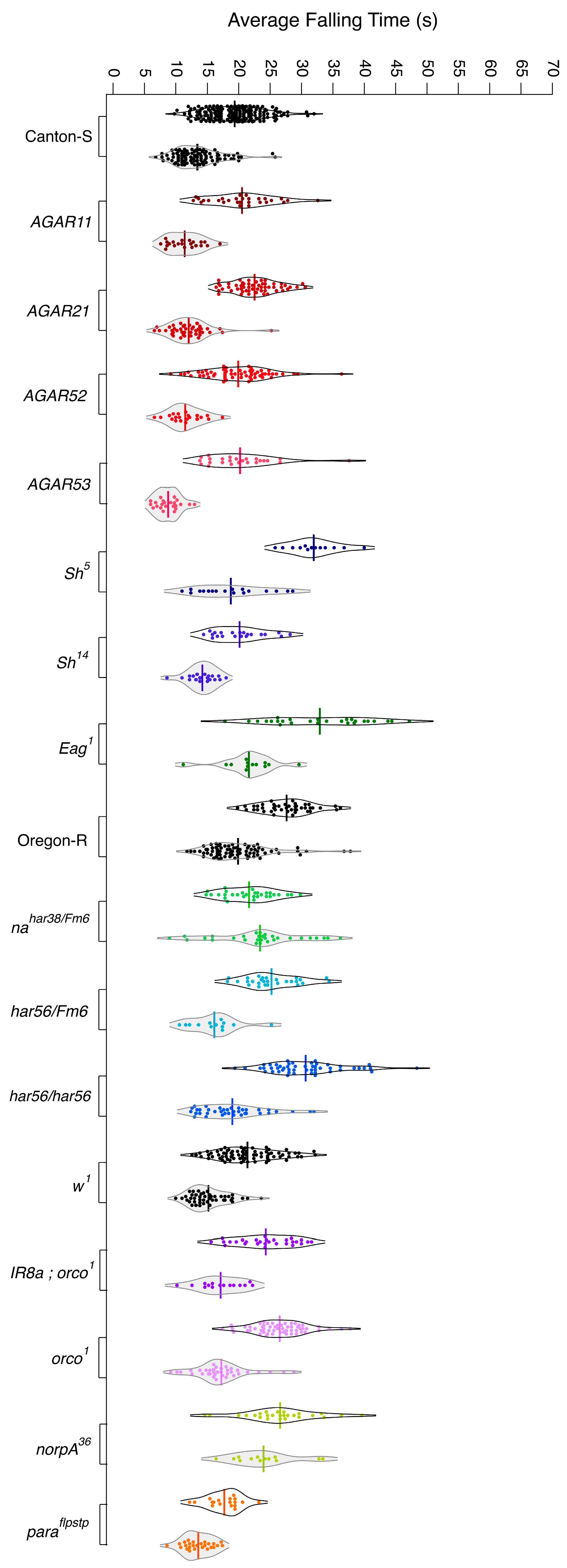
3.1. Wild-Type Flies Present with Differing Levels of Resistance to Anesthetics

Using our behavioral assay, we assessed the anesthetic resistance of Oregon-R, Canton-S, and w1118 strains. A total of 40 μL of chloroform knocked down wild-type flies on average within 10 to 50 s. The Oregon-R strain was more resistant than w1118, and w1118 was more resistant than Canton-S (Figure 2). It took ~30 s for Oregon-R flies to become anesthetized with chloroform, ~25 s for w1118, and 20 s for Canton-S. Interestingly, this order of resistance was consistent across both anesthetics, while the difference between the average falling time narrowed. All fly strains responded ~10 s faster to isoflurane than to chloroform. The response to chloroform had a broader distribution compared to isoflurane. Conveniently, the distribution profile of the strains was also conserved in chloroform and isoflurane, with Canton-S presenting with the least variation.

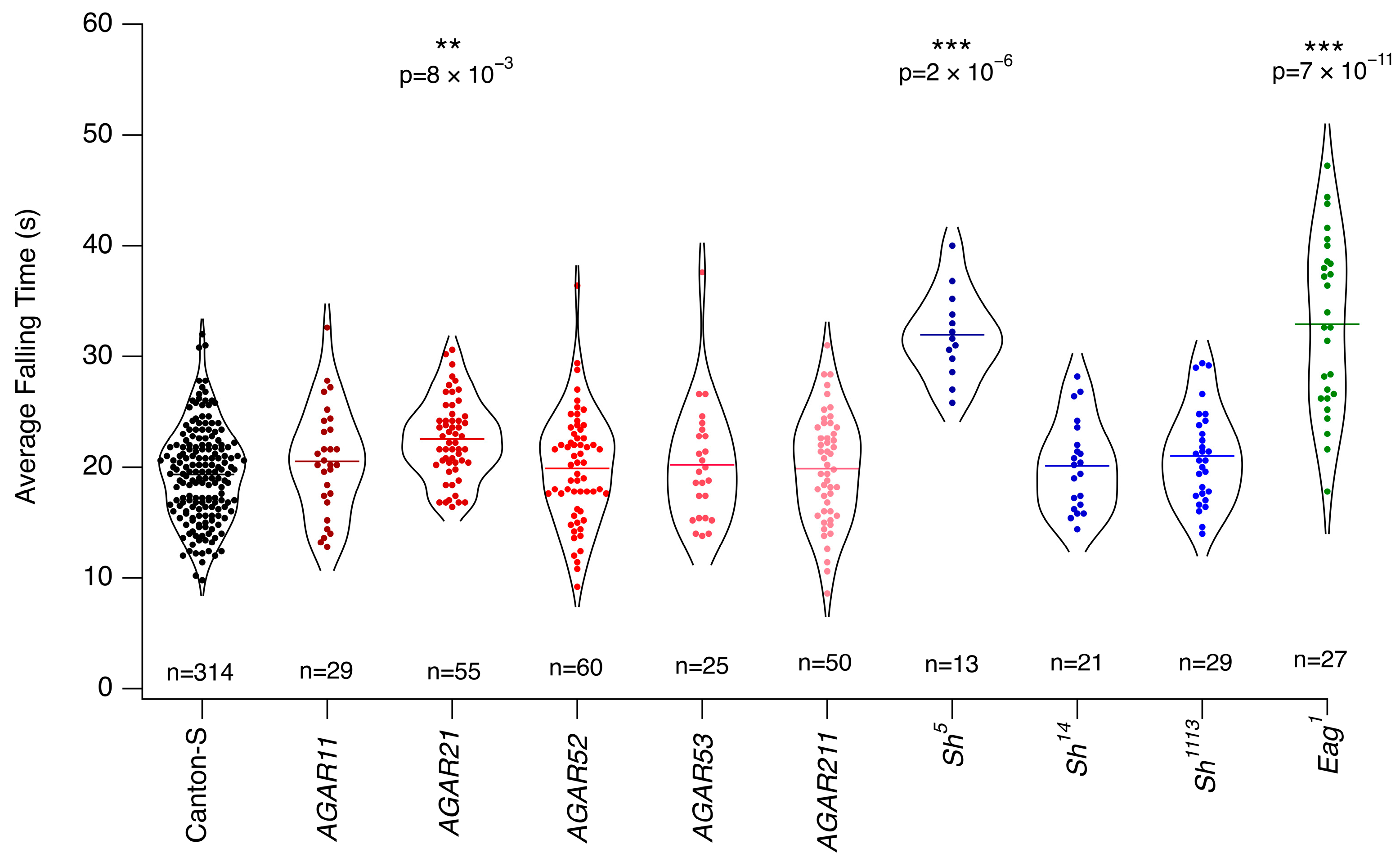
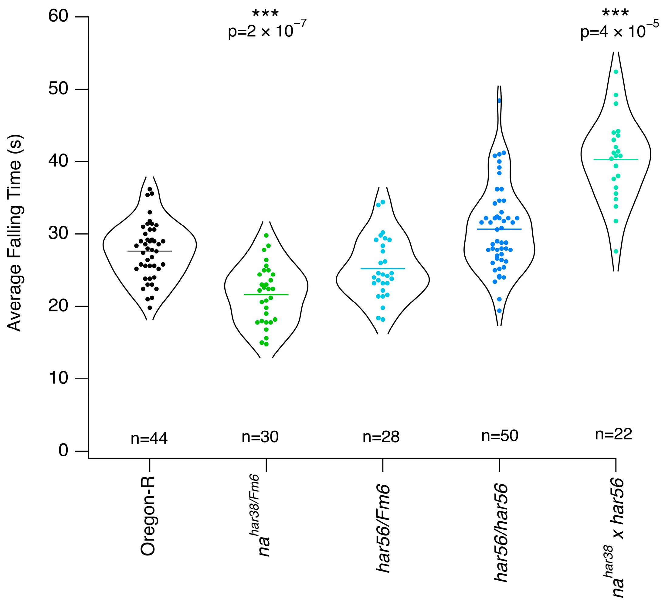
3.2. Mutants with Altered Response to Chloroform

Our first experimental set included mutants with a Canton-S background, such as AGAR, and voltage-gated potassium channel mutants, Shaker and ether-a-go. One of the AGAR mutants, AGAR21, slightly outperformed Canton-S in terms of resistance (Figure 3). Only Sh5 showed substantial differences from the other flies with Shaker mutations. Flies with this mutation were anesthetized ~10 s later than the control on average. Eag1 also exhibited higher resistance, quantitatively similar to Sh5.
Our second experimental set included mutants in the Oregon-R background and consisted primarily of the har series of mutants. When compared to their proper control, Oregon-R, har mutants did not present with high resistance (Figure 4). In fact, one of the heterozygous mutants, nahar38/fm6, displayed sensitivity. Flies with this genetic background were anesthetized ~8 s earlier than the control. Another surprising result for this set came from the crosses among har mutants, which showed drastically higher resistance. We obtained this cross by mating nahar38/Fm6 females with har56/har56 males and examined their female offspring. This group exhibited a delayed anesthetic response of ~40 s on average, surpassing the control by 10 s. On the other hand, the sensitive group was anesthetized ~5 s earlier than the control.
In the third experimental set, we wanted to compare the mutant para to its genetic background, w1118 (Figure 5). We incorporated additional strains as neutral controls. Surprisingly, among them, OR8a; orco1 had a slightly longer average falling time. The difference was subtle, but it prompted us to investigate the orco1 mutants alone in order to identify the likely cause of the increased resistance in this mutant. Surprisingly, orco1 mutants demonstrated greater resistance than OR8a; orco1, suggesting that the OR8a mutation may in fact act in the opposite manner, increasing sensitivity.

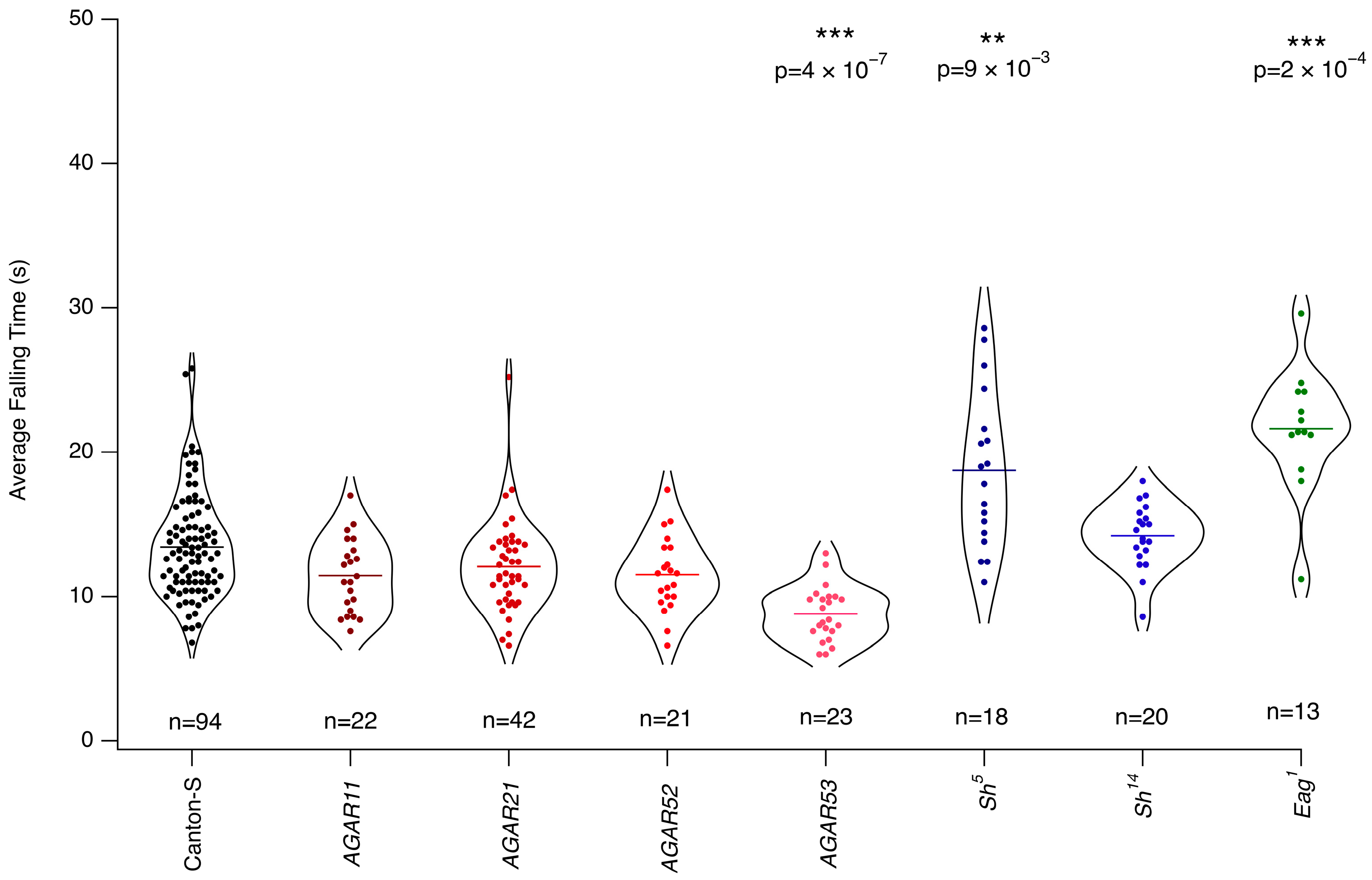
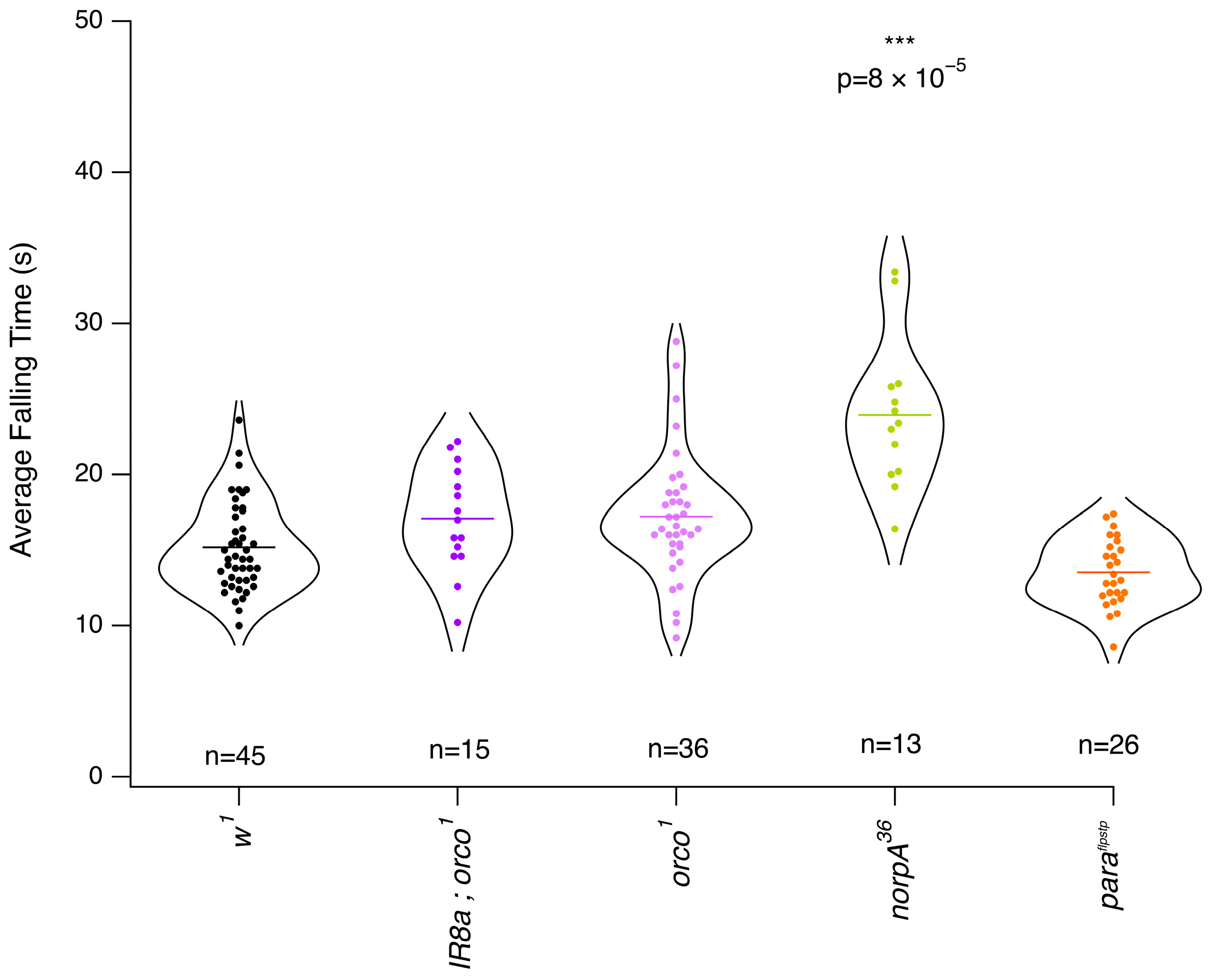
3.3. Mutants with Altered Response to Isoflurane

In this section, we evaluated the response of several mutant groups to isoflurane, primarily those previously tested with chloroform. AGAR53 flies exhibited a more rapid response to isoflurane than Canton-S, with a difference of approximately 5 s. In contrast, Sh5 and Eag1 mutants required longer to be knocked down by isoflurane, with delays of ~7 and ~10 s, respectively (Figure 6). Unexpectedly, har mutants showed no significant difference from wild-type flies (Figure 7). Likewise, w1118 mutants displayed minimal variation in their response to isoflurane relative to their controls (Figure 8).

3.4. Overall Isoflurane Is More Potent than Chloroform

Following our analysis of anesthetic resistance across 3 wild-type and 14 mutant Drosophila strains, we compared their response times to isoflurane and chloroform. Nearly all strains exhibited a faster response to isoflurane than to chloroform (Figure 9). However, two exceptions were observed, norpA36 and nahar38/Fm6, which showed no significant difference in their response times between the two anesthetics. Notably, the response to chloroform displayed greater variability, with a wider distribution of response times and more pronounced outliers compared to isoflurane.

4. Discussion

In this study, we aimed to systematically evaluate anesthetic resistance in Drosophila melanogaster using a newly developed behavioral assay. Our approach allowed us to compare the response of wild-type and mutant strains to two volatile anesthetics, isoflurane and chloroform. In contrast to our expectations, our findings revealed substantial variability in anesthetic sensitivity even among wild-type strains, with Oregon-R exhibiting the highest resistance and the greatest variation (Figure 2). Furthermore, we observed a consistent trend in which all strains responded to isoflurane more rapidly, and their responses were more consistent across different experiments (Figure 9).
The literature on the characterization of these wild-type strains is limited, making it difficult to determine the cause of the difference in the anesthetic sensitivity. However, previously, w1118 was reported to be more resistant to isoflurane than Canton-S [31]. Lifespan and ROS (reactive oxygen species) responses are known to differ between Oregon-R and Canton-S [32]. This might be an important link due to documented interplay between mitochondrial function and the anesthetic response [9,33].
Our study revisited well-characterized anesthetic-resistant mutants, such as har and AGAR, which were originally selected for their resistance to halothane. Although these strains have been previously described as highly resistant, our results did not align with these earlier findings. Notably, the literature lacks comparative analyses assessing multiple anesthetics in these strains, and AGAR mutants, in particular, have not been characterized further since their initial isolation. Although we previously used AGAR flies in our laboratory and published studies on their electron spin response to anesthetics, we did not confirm their resistance before conducting those experiments [34,35]. These studies were performed 7 years and 11 years ago. There are three possible explanations for the disparity between the older results and those given here. Firstly, it could be that over the years, the mutant strains drifted genetically and lost their initial phenotypes. However, given its autosomal dominant inheritance, this seems unlikely for the AGAR mutants, but nevertheless, their response to anesthetics could, in principle, be recovered by extensive backcrossing. Secondly, the methodological differences between our study and previous research could have contributed to discrepancies in reported anesthetic resistance. The conventional inebriometer assay, commonly used in earlier studies, measures loss of postural control over an extended period, with exposure times ranging from 75 s to over tens of minutes. This differs from the timing for the induction of general anesthesia, on the scale of seconds. In contrast, our method assessed the time for postural control loss to be within 5 to 50 s. Inebriometer drawbacks have been extensively documented in the literature [36]. The drawback of our method is the potential inhomogeneous distribution of gas within the exposure vial. The method of applying to a particular area of the smelling strip a fixed amount of anesthetic liquid provides reproducibility and simplicity, but lacks assurance of equal distribution of the anesthetic gas in the total volume of the vial, and therefore to each individual fly. Lastly, it is also possible that the observed resistance in these mutants is specific to halothane, which was not tested in this study, and isoflurane and chloroform may exert their effects through distinct and possibly non-overlapping molecular pathways.
It has been reported that har mutants exhibit variable responses to different anesthetics; this further complicates their classification as universally resistant [12,16,18,19]. Our findings support the notion that resistance to one anesthetic agent does not necessarily translate to resistance against others, challenging the generalization of anesthetic resistance across different compounds. Our finding with increased resistance of the har cross points to the possibility of mutually exclusive molecular paths to create an anesthesia-resistant phenotype (Figure 4). However, it is crucial to underline another aspect of our results: a uniformity in anesthetic response among most strains, as those that displayed resistance to chloroform also exhibited resistance to isoflurane. This supports the generalization of the anesthetic resistance. The two notable exceptions, norpA36 and nahar38/Fm6, did not follow this pattern, suggesting that certain mutations may differentially affect sensitivity to distinct anesthetic agents. Additionally, some fly lines exhibited resistance to chloroform, but not to isoflurane. Given that isoflurane induced a more rapid response across all strains, it is possible that the resistance of these mutants was overlooked due to the narrower response distribution associated with isoflurane exposure. This raises important considerations regarding the potential for methodological biases when assessing anesthetic resistance.
In summary, our study (i) introduced a simple and cost-effective method for evaluating anesthetic resistance in Drosophila; (ii) underscored the necessity of using appropriate genetic controls when assessing mutant sensitivity to anesthetics; (iii) highlighted the potential for discrepancies in anesthetic resistance depending on the specific agent used; and (iv) identified genetic candidates that may play a role in anesthetic response. Our findings challenge existing assumptions regarding anesthetic resistance and suggest that further research is needed to fully understand the genetic and physiological mechanisms underlying differential anesthetic sensitivity in Drosophila melanogaster.

Supplementary Materials

The following supporting information can be downloaded at: https://www.mdpi.com/article/10.3390/anesthres2020012/s1, Figure S1: The figure shows the dose–response curve for isoflurane (bottom) and chloroform (top); Table S1: Statistical comparisons between groups, including effect size estimates (Cohen’s r) are summarized for the wild-type flies. CH: chloroform, ISO: isoflurane, CS: Canton-S, OR: Oregon-R, w1 = w1118; Table S2: Statistical comparisons between groups, including effect size estimates (Cohen’s r), are summarized for the chloroform experiments. Table S3: Statistical comparisons between groups, including effect size estimates (Cohen’s r), are summarized for the chloroform experiments.

Author Contributions

Conceptualization, E.D., L.T. and E.M.C.S.; methodology, E.D., L.T. and E.M.C.S.; formal analysis, E.D.; data curation, E.D.; writing—original draft preparation, E.D.; writing—review and editing, E.D., L.T. and E.M.C.S.; supervision, L.T. and E.M.C.S. All authors have read and agreed to the published version of the manuscript.

Funding

This research received no external funding.

Institutional Review Board Statement

Not applicable.

Informed Consent Statement

Not applicable.

Data Availability Statement

Data will be made available on request.

Acknowledgments

E.D. thanks Anna Bourouliti for the input on the methodology and Maro Loizou for expert technical help with fly husbandry.

Conflicts of Interest

The authors declare no conflicts of interest.

Abbreviations

The following abbreviations are used in this manuscript:
AGARAutosomal general anesthesia-resistant
harHalothane-resistant
EMSEthyl methane sulfonate
w1w1118
CSCanton-S
OROregon-R
ABCATP-binding cassette

References

  1. Antognini, J.F.; Carstens, E. In vivo characterization of clinical anaesthesia and its components. Br. J. Anaesth. 2002, 89, 156–166. [Google Scholar] [CrossRef] [PubMed]
  2. Grémiaux, A.; Yokawa, K.; Mancuso, S.; Baluška, F. Plant anesthesia supports similarities between animals and plants: Claude Bernard’s forgotten studies. Plant Signal. Behav. 2014, 9, e27886. [Google Scholar] [CrossRef]
  3. Chamberlain, M.; Koutsogiannaki, S.; Schaefers, M.; Babazada, H.; Liu, R.; Yuki, K. The Differential Effects of Anesthetics on Bacterial Behaviors. PLoS ONE 2017, 12, e0170089. [Google Scholar] [CrossRef] [PubMed]
  4. Saidman, E.I.E.; Westhorpe, L.J. The Wondrous Story of Anesthesia; Springer: New York, NY, USA, 2014. [Google Scholar]
  5. Keating, V. Advances in Anaesthesia. Sci. News 1951, 20, 51–52. [Google Scholar]
  6. Covarrubias, M.; Barber, A.F.; Carnevale, V.; Treptow, W.; Eckenhoff, R.G. Mechanistic insights into the modulation of voltage-gated ion channels by inhalational anesthetics. Biophys. J. 2015, 109, 2003–2011. [Google Scholar] [CrossRef]
  7. Risks Associated with General Anaesthesia. The Royal College of Anaesthetists. Available online: https://www.rcoa.ac.uk/patients/patient-information-resources/anaesthesia-risk/risks-associated-general-anaesthesia (accessed on 18 January 2025).
  8. Lee, J.J.; Choi, G.J.; Kang, H.; Baek, C.W.; Jung, Y.H.; Shin, H.Y.; Park, Y.H.; Woo, Y.C. Relationship between Surgery under General Anesthesia and the Development of Dementia: A Systematic Review and Meta-Analysis. Biomed. Res. Int. 2020, 2020, 3234013. [Google Scholar] [CrossRef]
  9. Niezgoda, J.; Morgan, P.G. Anesthetic considerations in patients with mitochondrial defects. Paediatr. Anaesth. 2013, 23, 785–793. [Google Scholar] [CrossRef]
  10. Waking Up to Anesthesia, NIH News in Health. 2017. Available online: https://newsinhealth.nih.gov/2011/04/waking-up-anesthesia (accessed on 1 March 2025).
  11. Quinlan, J.J.; Homanics, G.E.; Firestone, L.L. Anesthesia sensitivity in mice that lack the beta3 subunit of the gamma-aminobutyric acid type A receptor. Anesthesiology 1998, 88, 775–780. [Google Scholar] [CrossRef] [PubMed]
  12. Krishnan, K.S.; Nash, H.A. A genetic study of the anesthetic response: Mutants of Drosophila melanogaster altered in sensitivity to halothane. Proc. Natl. Acad. Sci. USA 1990, 87, 8632–8636. [Google Scholar] [CrossRef]
  13. Morgan, P.G.; Sedensky, M.M. Mutations conferring new patterns of sensitivity to volatile anesthetics in Caenorhabditis elegans. Anesthesiology 1994, 81, 888–898. [Google Scholar] [CrossRef]
  14. Madhavan, M.C.; Kumar, R.A.; Krishnan, K.S. Genetics of anesthetic response: Autosomal mutations that render Drosophila resistant to halothane. Pharmacol. Biochem. Behav. 2000, 67, 749–757. [Google Scholar] [CrossRef] [PubMed]
  15. Karunanithi, S.; Troup, M.; van Swinderen, B. Using Drosophila to understand general anesthesia: From synapses to behavior. Methods Enzymol. 2018, 602, 153–176. [Google Scholar]
  16. Campbell, D.B.; Nash, H.A. Use of Drosophila mutants to distinguish among volatile general anesthetics. Proc. Natl. Acad. Sci. USA 1994, 91, 2135–2139. [Google Scholar] [CrossRef] [PubMed]
  17. Mir, B.; Iyer, S.; Ramaswami, M.; Krishnan, K.S. A genetic and mosaic analysis of a locus involved in the anesthesia response of Drosophila melanogaster. Genetics 1997, 147, 701–712. [Google Scholar] [CrossRef]
  18. Guan, Z.; Scott, R.L.; Nash, H.A. A new assay for the genetic study of general anesthesia in Drosophila melanogaster: Use in analysis of mutations in the X-chromosomal 12E region. J. Neurogenet. 2000, 14, 25–42. [Google Scholar] [CrossRef] [PubMed]
  19. Leibovitch, B.A.; Campbell, D.B.; Krishnan, K.S.; Nash, H.A. Mutations that affect ion channels change the sensitivity of Drosophila melanogaster to volatile anesthetics. J. Neurogenet. 1995, 10, 1–13. [Google Scholar] [CrossRef]
  20. Tanaka, Y.; Gamo, S. Sensitivity to diethylether anesthesia of fruit flies primarily depends on the genotypes of the sodium channel gene rather than the states of the membranes and the mechanisms might be different from heat-induced paralysis. Colloids Surf. B Biointerfaces 2001, 22, 39–53. [Google Scholar] [CrossRef]
  21. Campbell, J.L.; Nash, H.A. Volatile general anesthetics reveal a neurobiological role for the white and brown genes of Drosophila melanogaster. J. Neurobiol. 2001, 49, 339–349. [Google Scholar] [CrossRef]
  22. Cheng, Y.; Nash, H.A. Visual mutations reveal opposing effects of illumination on arousal in Drosophila. Genetics 2008, 178, 2413–2416. [Google Scholar] [CrossRef]
  23. Gao, S.; Sandstrom, D.J.; Smith, H.E.; High, B.; Marsh, J.W.; Nash, H.A. Drosophila ryanodine receptors mediate general anesthesia by halothane. Anesthesiology 2013, 118, 587–601. [Google Scholar] [CrossRef]
  24. Zalucki, O.H.; Menon, H.; Kottler, B.; Faville, R.; Day, R.; Bademosi, A.T.; Lavidis, N.; Karunanithi, S.; van Swinderen, B. Syntaxin1A-mediated Resistance and Hypersensitivity to Isoflurane in Drosophila melanogaster. Anesthesiology 2015, 122, 1060–1074. [Google Scholar] [CrossRef]
  25. Troup, M.; Zalucki, O.H.; Kottler, B.D.; Karunanithi, S.; Anggono, V.; van Swinderen, B. Syntaxin1A neomorphic mutations promote rapid recovery from isoflurane anesthesia in Drosophila melanogaster. Anesthesiology 2019, 131, 555–568. [Google Scholar] [CrossRef] [PubMed]
  26. Olufs, Z.P.G.; Loewen, C.A.; Ganetzky, B.; Wassarman, D.A.; Perouansky, M. Genetic variability affects absolute and relative potencies and kinetics of the anesthetics isoflurane and sevoflurane in Drosophila melanogaster. Sci. Rep. 2018, 8, 2348. [Google Scholar] [CrossRef] [PubMed]
  27. Gamo, S.; Tomida, J.; Dodo, K.; Keyakidani, D.; Matakatsu, H.; Yamamoto, D.; Tanaka, Y. Calreticulin mediates anesthetic sensitivity in Drosophila melanogaster. Anesthesiology 2003, 99, 867–875. [Google Scholar] [CrossRef]
  28. Gamo, S. Studies on target genes of general anesthetics. Curr. Drug Targets 2002, 3, 31–41. [Google Scholar] [CrossRef]
  29. Humphrey, J.A.; Hamming, K.S.; Thacker, C.M.; Scott, R.L.; Sedensky, M.M.; Snutch, T.P.; Morgan, P.G.; Nash, H.A. A putative cation channel and its novel regulator: Cross-species conservation of effects on general anesthesia. Curr. Biol. 2007, 17, 624–629. [Google Scholar] [CrossRef]
  30. Joiner, W.J.; Friedman, E.B.; Hung, H.-T.; Koh, K.; Sowcik, M.; Sehgal, A.; Kelz, M.B. Genetic and anatomical basis of the barrier separating wakefulness and anesthetic-induced unresponsiveness. PLoS Genet. 2013, 9, e1003605. [Google Scholar] [CrossRef]
  31. Zalucki, O.; Day, R.; Kottler, B.; Karunanithi, S.; van Swinderen, B. Behavioral and electrophysiological analysis of general anesthesia in 3 background strains of Drosophila melanogaster. Fly 2015, 9, 7–15. [Google Scholar] [CrossRef] [PubMed]
  32. Malacrida, S.; De Lazzari, F.; Mrakic-Sposta, S.; Vezzoli, A.; Zordan, M.A.; Bisaglia, M.; Menti, G.M.; Meda, N.; Frighetto, G.; Bosco, G.; et al. Lifespan and ROS levels in different Drosophila melanogaster strains after 24 h hypoxia exposure. Biol. Open 2022, 11, bio059386. [Google Scholar] [CrossRef]
  33. Rodriguez, E.; Peng, B.; Lane, N. Anaesthetics disrupt complex I-linked respiration and reverse the ATP synthase. Biochim. Biophys. Acta Bioenerg. 2025, 1866, 149511. [Google Scholar] [CrossRef]
  34. Turin, L.; Skoulakis, E.M.C.; Horsfield, A.P. Electron spin changes during general anesthesia in Drosophila. Proc. Natl. Acad. Sci. USA 2014, 111, E3524–E3533. [Google Scholar] [CrossRef] [PubMed]
  35. Turin, L.; Skoulakis, E.M.C. Electron Spin Resonance (EPR) in Drosophila and General Anesthesia. Methods Enzymol. 2018, 603, 115–128. [Google Scholar] [PubMed]
  36. Zalucki, O.; van Swinderen, B. What is unconsciousness in a fly or a worm? A review of general anesthesia in different animal models. Conscious. Cogn. 2016, 44, 72–88. [Google Scholar] [CrossRef] [PubMed]
Figure 1. The set-up for the behavior assay.
Anesthres 02 00012 g001
Figure 2. The falling time of three strains of wild-type flies is plotted. Pink represents w1118, green Canton-S, and blue Oregon-R. The first three sets in the plots are the data from the chloroform experiments (black violin border), and the last three sets are for isoflurane (gray violin border). The p-value between the response to chloroform and isoflurane of all strains was highly significant (<0.0005). The difference between the strains for the same anesthetic is shown in the upper part of the figure. CS stands for Canton-S, OR for Oregon-R, and w1 for w1118. Additional statistical details, including effect and sample sizes, are provided in the Supplementary Material (Table S1).
Anesthres 02 00012 g002
Figure 3. The falling time of the mutant lines with the Canton-S background after chloroform exposure is shown. Additional statistical details, including effect size, are provided in the Supplementary Material (Table S2). ** p < 0.01, *** p < 0.001.
Anesthres 02 00012 g003
Figure 4. The falling time of the indicated mutant lines with the Oregon-R background after chloroform exposure is shown. Additional statistical details, including effect size, are provided in the Supplementary Material (Table S2). *** p < 0.001.
Anesthres 02 00012 g004
Figure 5. The falling time of the indicated mutant lines with the w1118 background after chloroform exposure is shown. Additional statistical details, including effect size, are provided in the Supplementary Material (Table S2). ** p < 0.01, *** p < 0.001.
Anesthres 02 00012 g005
Figure 6. The falling time of the mutant lines with Canton-S background after isoflurane exposure is shown. Additional statistical details, including effect size, are provided in the Supplementary Material (Table S3). ** p < 0.01, *** p < 0.001.
Anesthres 02 00012 g006
Figure 7. The falling time of the mutant lines with Oregon-R background after isoflurane exposure is shown. Additional statistical details, including effect size, are provided in the Supplementary Material (Table S3).
Anesthres 02 00012 g007
Figure 8. The falling time of the mutant lines with w1118 background after isoflurane exposure is shown. Additional statistical details, including effect size, are provided in the Supplementary Material (Table S3). *** p < 0.001.
Anesthres 02 00012 g008
Figure 9. Average falling time of fly groups when exposed to isoflurane and chloroform is re-plotted. From every pair aligned in the x-axis, the one on the left side represents the fly group exposed to chloroform, and the one on the right to isoflurane.
Anesthres 02 00012 g009
Table 1. Literature summary on the anesthetic response of Drosophila mutants. Pink denotes flies with resistance, green represents sensitivity, blue is used to indicate unclear results, and white indicates no difference. Reference to each study is indicated with a corresponding number in the box.
MutantsGeneral Anesthetics
GenotypeGeneChromosomeFunctionDiethyl EtherChloroformTrichloroethyleneHalothaneEnfluraneIsofluraneSevofluraneDesfluraneMethoxyflurane
nahar85Narrow abdomenXNa+ leak channel[16][16][16][12,16,17,18][16][16] [16][16]
nahar38Narrow abdomenXNa+ leak channel[16][16][16][12,16,17,18][16][16] [16][16]
har56UnknownXUnknown[16][16][16][12,16][16][16] [16][16]
har63UnknownXUnknown[16][16][16][12,16,17,18][16][16] [16][16]
ShKS133ShakerXVoltage-gated K+ channel [19][19][19]
Sh5ShakerXVoltage-gated K+ channel [19][19][19]
ShKO120ShakerXVoltage-gated K+ channel [19][19][19]
Slo1Slowpoke3Voltage-gated K+ channel [19][19][19]
Slo4Slowpoke3Voltage-gated K+ channel [19][19](19)
Eag1Ether-a-go-goXVoltage-gated K+ channel [19][19][19]
Eaghd14Ether-a-go-goXVoltage-gated K+ channel [19][19][19]
Eaghd15Ether-a-go-goXVoltage-gated K+ channel [19][19][19]
Parats1paralyticXVoltage-gated Na+ channel[20][19][19][19]
Parahd839ParalyticXVoltage-gated Na+ channel[20]
Parats3ParalyticXVoltage-gated Na+ channel[20]
mlenap-ts1Maleless2 [19][19][19]
bw1Brown2ABC transporters [21][21]
w1WhiteXABC transporters [21][21]
trpTransient receptor potential3Non-selective cation channel [22]
RyRRyanodine receptor2Intracellular calcium channel [23][23][23][23]
Syx1AH3-CSyntaxin 1A3Neurotransmitter release [24,25]
Syx1AKARRAASyntaxin 1A3Neurotransmitter release [24]
ND2360114NADH dehydrogenase subunit3NADH dehydrogenase subunit [26][26]
Calreth-as311Calreticulin3 [27] [27] [27]
ScbVolScab2Encodes the α-S3 integrin[28]
rutabagaAdenylate cyclase 1XAdenylate cyclase 1[28]
amnAmnesiacXNeuropeptide precursor[28]
Unc79Uncoordinated 793Locomotor rhythms [29] [30]
AGAR Resistance to halothane [14]
Table 2. Table shows the genotypes of the flies that were used with their corresponding background. BDSC: Bloomington Drosophila Stock Center.
GenotypeBackgroundReference
Canton-S
w1
Oregon-R
AGAR11Canton-SBDSC 27588
AGAR21Canton-SBDSC 27329
AGAR52Canton-SBDSC 27589
AGAR53Canton-SBDSC 26703
AGAR211Canton-SBDSC 27330
Sh5Canton-SBDSC 111
Sh14Canton-SBDSC 4741
Sh1113Canton-S
Eag1Canton-SBDSC 3661
har56Oregon-RBDSC 26699
har56/har56Oregon-R
nahar38Oregon-RBDSC 26704
Orco1w1BDSC 23129
IR8a; Orco1w1
Paraflpstpw1BDSC 67680
NorpA36w1BDSC 9048
Disclaimer/Publisher’s Note: The statements, opinions and data contained in all publications are solely those of the individual author(s) and contributor(s) and not of MDPI and/or the editor(s). MDPI and/or the editor(s) disclaim responsibility for any injury to people or property resulting from any ideas, methods, instructions or products referred to in the content.

