Case Report

Severe Postoperative Complications Following Bilateral DIEP Flap Breast Reconstruction in a High-Risk Patient: A Case Report

Plastic Surgery Unit, Department of Neurosciences, University Hospital of Padua, 35128 Padua, Italy
*
Author to whom correspondence should be addressed.
Complications 2025, 2(2), 12; https://doi.org/10.3390/complications2020012
Submission received: 13 March 2025 / Revised: 22 April 2025 / Accepted: 27 April 2025 / Published: 2 May 2025

Abstract

:
Background/Objectives: Deep inferior epigastric perforator (DIEP) flap reconstruction is considered the gold standard for autologous breast reconstruction due to its favorable aesthetic results and low donor site morbidity. Nevertheless, it remains associated with potentially life-threatening complications such as deep vein thrombosis (DVT) and pulmonary embolism (PE). This report aims to describe a complex clinical case in which severe thromboembolic and ischemic complications occurred despite adherence to standard prophylactic protocols. Methods: We present the case of a 65-year-old female with multiple thromboembolic risk factors—including obesity, a history of heavy smoking, hormone therapy, and prior COVID-19 infection—who underwent immediate bilateral breast reconstruction with DIEP flaps following mastectomy. Results: Within the first 24 h postoperatively, the patient developed a massive pulmonary embolism requiring intensive care management. Despite appropriate anticoagulation and supportive measures, she subsequently experienced full-thickness necrosis of the central portion of the abdominal flap. Thrombophilia screening and diagnostic imaging did not reveal peripheral venous thrombosis, raising the hypothesis of a hypercoagulable state potentially related to prior SARS-CoV-2 infection. Conclusions: This case underscores the importance of individualized risk stratification and suggests that current prophylaxis protocols may be insufficient for patients with overlapping thrombotic risk factors. The findings advocate for further investigation into the long-term vascular effects of COVID-19 and support reconsidering extended or intensified prophylaxis in high-risk populations undergoing complex microsurgical procedures.

1. Introduction

1.1. Evolution of Breast Reconstruction and the Rise of the DIEP Flap

The journey of breast reconstruction began in the late 19th century, with the pioneering work of Italian surgeon Iginio Tansini. In 1896, Tansini introduced the latissimus dorsi myocutaneous flap, marking the first use of autologous tissue for breast reconstruction. This technique laid the foundation for future advancements, emphasizing the importance of using the patient’s own tissue to achieve more natural and durable results [1]. The latter half of the 20th century witnessed major innovations in reconstructive techniques. One of these was the introduction of the transverse rectus abdominis myocutaneous (TRAM) flap in the 1980s by Hartrampf et al., which enabled the transfer of abdominal tissue to the chest, providing improved breast contour and volume [2]. In 1979, Holstrom described a similar concept with his “free abdominoplasty flap” for breast reconstruction [3]. Despite its advantages, the TRAM flap sacrifices abdominal muscle, often resulting in donor site morbidity such as weakness or hernia. This is particularly concerning for middle-aged females, who may be at greater risk of rectus diastasis and abdominal wall complications. For this reason, the TRAM flap should be used selectively [4].
To overcome these issues, the deep inferior epigastric perforator (DIEP) flap was introduced in the early 1990s. Allen and Treece were the first to successfully perform a DIEP flap in 1992, transferring abdominal skin and fat while preserving the rectus abdominis muscle [5]. This evolution provided comparable soft tissue reconstruction with significantly less donor site morbidity, reduced postoperative pain, and shorter recovery time [6].
Since its introduction, the DIEP flap has undergone continuous refinement, transforming into the gold standard for autologous breast reconstruction. Key advancements include the widespread use of preoperative vascular imaging—especially computed tomography angiography (CTA)—which has improved perforator mapping and reduced operative times [7]. Intraoperatively, the use of venous couplers has improved anastomotic success and reduced ischemia time, while internal mammary perforators are now preferred recipient vessels due to superior flow and positioning benefits [8].
The implementation of intraoperative indocyanine green (ICG) angiography has allowed real-time assessment of flap perfusion and identification of areas at risk for fat necrosis [9]. Moreover, inclusion of multiple perforators rather than one has been shown to enhance flap perfusion and reduce the risk of pedicle twisting, albeit with more intramuscular dissection [7]. Recent strategies also incorporate lymph node transfer from the inguinal region for patients with postmastectomy lymphedema [10].
On the postoperative side, the adoption of modern protocols has significantly improved patient comfort and enabled earlier hospital discharge. Pain is better managed with multimodal regimens, and flap monitoring has been simplified using handheld Doppler devices rather than more invasive tools [7].
More recently, robot-assisted techniques have been explored to further refine DIEP flap harvest. Robotic approaches aim to reduce donor site trauma by allowing for more precise intramuscular dissection and improved visualization, potentially expanding the indications for DIEP flaps and enhancing recovery outcomes [11].

1.2. Patients’ Selection for DIEP Flap

Autologous breast reconstruction (ABR) offers superior long-term outcomes, a more natural appearance and feel, and a greater potential for sensory restoration compared with implant-based breast reconstruction [12]. Although it involves higher costs, a more complex surgical procedure, and a longer recovery time, recent studies report more satisfactory aesthetic outcomes and a longer time to recurrence, when compared with implant-based reconstruction [13,14,15]. Indications for using abdominal-based flaps (like the DIEP or the TRAM flap) for breast reconstruction often include the patient’s preference, severe soft tissue damage (especially secondary to radiation therapy), overweight patients, unilateral reconstruction, and even failed or unpleasant implant reconstruction [14,16,17]. It preserves the relative integrity of the rectus abdominis muscle and provides an abdominoplasty effect, which may represent a further cosmetic improvement for the patient [18]. Over the past two decades, DIEP flap has become the gold standard for autologous breast reconstruction due to its ability to achieve a natural breast appearance and texture, reduced donor site morbidity, and long-term durability. Advances in preoperative imaging and surgical techniques have further improved the efficacy, safety, and aesthetic outcomes of this procedure. The suitability of the DIEP flap depends on the availability of adequate abdominal tissue and vascular supply. In slender patients seeking bilateral reconstruction, limited abdominal bulk may necessitate the use of alternative flaps to avoid suboptimal aesthetic results. The choice of donor site should be guided by the patient’s body habitus, preoperative computed tomography scans, desired breast volume, and potential concerns about donor site scarring [19].
Absolute contraindications for abdominally based autologous breast reconstruction are limited but significant [17]. These include patients who are medically unsuitable for surgery or whose anatomy prevents the procedure from being performed safely. Assessing a patient’s fitness for surgery should involve a comprehensive discussion with the perioperative team, including the surgeon and anesthesiologist. Surgical histories that compromise the vascular integrity of the abdominal region, such as procedures transecting the superficial and deep inferior epigastric systems, pose a major barrier [20]. Additionally, unrealistic patient expectations, despite thorough informed consent, should be considered an absolute contraindication. In cases where reconstruction would cause significant delays in cancer treatment, opting for an alternative approach or delaying reconstruction is advised.
Relative contraindications depend on the surgeon’s experience, evidence from the literature, and the tolerance for potential complications. Most of these issues are modifiable factors, and a meticulous preoperative plan may minimize their impact on the procedure [17]. A history of abdominal surgeries does not universally preclude the use of abdominal flaps, but it may necessitate alterations in flap design or technique based on preoperative assessment [21]. For example, while midline laparotomies can increase donor site complications, they do not significantly raise flap failure rates if managed properly [22]. Likewise, patients with previous abdominoplasty or liposuction may still be candidates for abdominally based flaps. Preoperative imaging techniques such as Doppler ultrasound and CT angiography allow precise evaluation of residual vascular anatomy after procedures like abdominoplasty or liposuction. By identifying well-positioned and adequately sized perforators, these tools help overcome previous contraindications and support safe flap planning [23,24].
Smoking is another relative contraindication due to its adverse effects on wound healing and increased risk of complications, making smoking cessation essential at least four weeks before surgery. Some authors reported a significant increase in donor site morbidity, in hernia and bulging rate, and in vascular sufferance of the mastectomy flaps [25]. Obesity also elevates the risk of various complications, including wound infections, seroma formation, and abdominal bulge, although satisfaction with autologous reconstruction remains high in this population [26].
Coagulation disorders represent a critical relative contraindication for abdominally based autologous breast reconstruction. Hypercoagulability, whether due to genetic predispositions such as factor V Leiden, protein C deficiency, or antiphospholipid antibody syndrome, significantly elevates the risk of both intraoperative and postoperative thrombotic events [27]. Studies have reported a high incidence of flap thrombosis in patients with thrombophilia, with a notable proportion of cases leading to complete flap loss. Additionally, certain medications, particularly selective estrogen-receptor modulators like tamoxifen, have been identified as prothrombotic and may further complicate surgical outcomes [28]. While the evidence regarding aromatase inhibitors is less conclusive, the potential risks associated with hypercoagulability necessitate careful preoperative assessment and management to minimize the likelihood of thrombosis and ensure the viability of the flap [29].
The main features regarding selection criteria and contraindications for DIEP flap are summarized in Table 1.

1.3. Complications of DIEP Flap Surgery

One of the most significant flap-related complications is venous congestion, which typically occurs distal to the vascular tip and may result in partial or total flap necrosis [30]. To mitigate this risk, some surgeons perform a secondary venous anastomosis between the superficial inferior epigastric vein and a recipient vein, which has been shown to significantly improve venous outflow and reduce congestion [31]. In addition, meticulous preoperative planning with Doppler ultrasound, computed tomography angiography CTA, and infrared thermography aids in the identification of reliable perforators and atypical venous connections, minimizing the risk of inadequate drainage. Intraoperatively, ensuring tension-free microvascular anastomoses and avoiding excessive compression at the inset site are also essential steps in preventing venous compromise [32,33].
Fat induration or necrosis, commonly presenting as hardened areas within the reconstructed breast, results from insufficient perfusion of adipose tissue. The use of intraoperative ICG fluorescent angiography has been demonstrated to reduce the incidence of fat necrosis by allowing real-time assessment of tissue perfusion and guiding the selective excision of poorly vascularized areas [34]. Additional strategies include minimizing flap thickness in high-risk zones and ensuring the preservation of robust perforators during flap elevation.
Infectious complications, wound dehiscence, and seroma formation represent additional risks. Preventive measures encompass the use of perioperative antibiotic prophylaxis tailored to patient risk factors and institutional flora, meticulous surgical technique to minimize dead space, and routine placement of closed-suction drains at both donor and recipient sites to control fluid accumulation. The use of fibrin sealants and quilting sutures may further reduce seroma rates. Systemic complications such as DVT and PE remain among the most serious adverse events in DIEP flap breast reconstruction [35]. Compared with implant-based reconstruction, autologous procedures carry a higher VTE risk due to prolonged operative time, increased intra-abdominal pressure from donor site closure, and limited postoperative mobility [36]. In a retrospective cohort of 192 patients undergoing DIEP flap reconstruction, Modarressi et al. reported a 3.1% overall VTE rate, with PE occurring in 2.1% and DVT in 1% of patients. Notably, all events occurred in patients stratified as high risk according to the Caprini risk assessment model (score ≥ 5), with the VTE rate increasing to 12.1% among patients with a Caprini score of 7 [35].
A large population-based analysis by Masoomi et al. using the Nationwide Inpatient Sample (NIS) database showed an overall VTE incidence of 0.13% among 35,883 patients undergoing autologous breast reconstruction, with higher rates observed in pedicled TRAM flaps (0.26%) and DIEP flaps (0.19%), compared with other flap types. Importantly, the same study identified immediate reconstruction (adjusted odds ratio [AOR] 5.4), age > 65 (AOR 4.2), obesity (AOR 3.7), and prior chemotherapy (AOR 3.5) as independent predictors of VTE [36]. Patient-specific risk factors—such as a personal or family history of VTE, active malignancy, high BMI, smoking, estrogen therapy, and advanced age—further contribute to thrombotic risk [35]. To address these risks, a thorough preoperative risk stratification using validated tools such as the Caprini risk assessment model (RAM) is recommended [37]. This scoring system incorporates both procedure-related and patient-specific variables to categorize patients into low, moderate, high, or highest risk of thromboembolic events. According to the score, preventive measures can be tailored accordingly. In patients with a low Caprini score, mechanical prophylaxis alone—such as graduated compression stockings (GCS) or intermittent pneumatic compression (IPC) devices—is typically sufficient. For those with moderate to high scores, the addition of pharmacologic prophylaxis, such as low-molecular-weight heparin (LMWH), is generally advised, starting preoperatively or immediately postoperatively. In very high-risk individuals (e.g., Caprini ≥ 5), a combined approach using both mechanical and pharmacologic strategies is strongly recommended, and extended prophylaxis (up to 4 weeks post-discharge) may be considered based on the presence of persistent risk factors. Moreover, perioperative protocols should emphasize early ambulation, ideally within 12–24 h after surgery, as this has been shown to significantly reduce VTE incidence. Minimizing operative time through dual team approaches and careful planning, as well as optimizing intraoperative hydration and hemodynamic stability, are additional key components in reducing thrombotic risk. Regular staff education and adherence to institution-specific VTE prevention protocols further contribute to improved safety in these complex procedures [38].

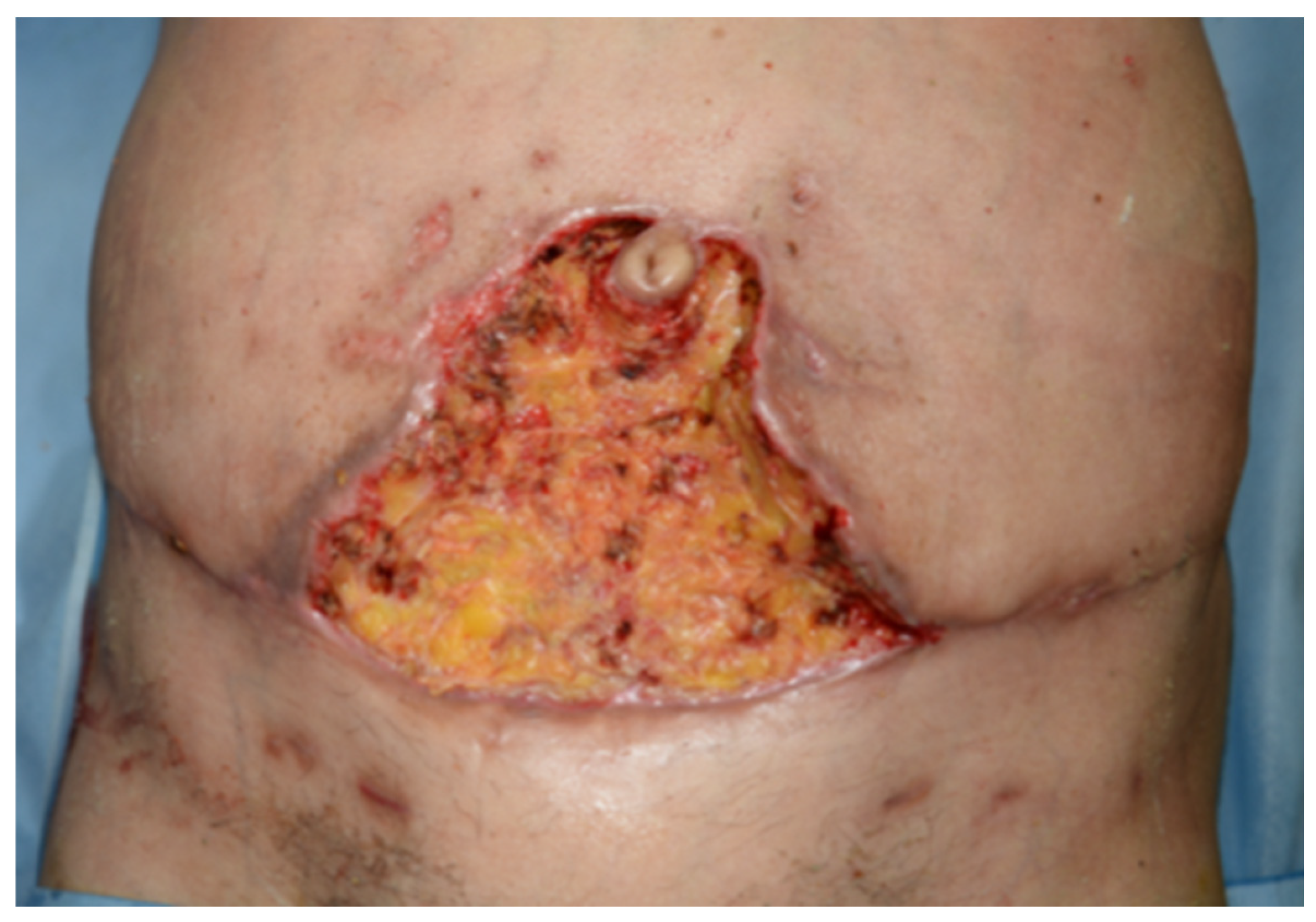
2. Case Presentation

A 65-year-old female patient, weighing 63 kg and measuring 162 cm in height, presented to the breast unit of our University Hospital with a medical history of HTN, GERD, MGUS, and a prior diagnosis of right breast cancer. The initial breast cancer, treated in 2008, involved a quadrantectomy followed by five years of tamoxifen therapy and adjuvant radiotherapy. The patient had a substantial smoking history of 30 pack-years, having quit in 2019. She also reported scoliosis and a previous COVID-19 infection contracted in October 2023. In December 2023, following a right breast cancer relapse, the patient underwent a right mastectomy with sentinel lymph node biopsy and placement of a mammary tissue expander. During subsequent follow-up, a contralateral late metachronous breast cancer was identified. At this point, the patient was referred to our plastic surgery unit. We proposed a comprehensive surgical approach involving a left mastectomy, removal of the right breast tissue expander, and immediate bilateral breast reconstruction with DIEP flaps. The preoperative clinical condition and assessment are shown in Figure 1 and Table 2.

2.1. Surgical Procedure

The procedure took place on 20 June 2024, and it was initiated following standard aseptic protocols, with preoperative cutaneous markings performed using Doppler ultrasound to identify suitable abdominal perforators as shown in Figure 2.
The breast surgery started with a left mastectomy through an omega-shaped infra-areolar incision. Superior and inferior mastectomy flaps were developed, and glandular tissue was meticulously dissected from the pectoralis major fascia. A sentinel lymph node biopsy was performed, resulting in the excision of three lymph nodes, which were confirmed negative for metastatic disease upon intraoperative pathological examination.
Successive histological diagnosis showed an invasive ductal carcinoma (G3) associated with high-grade cribriform ductal carcinoma in situ, with focal lymphovascular invasion and low stromal lymphocytic infiltration (TIL 0–10%). Hormone receptors were strongly positive (ER/PR 95%), HER2 was negative, and Ki67 was 25–30%; there were no lymph node metastases (0/3), and the pathological stage was pT1b N0 (sn) M0. On the right side, the previously placed tissue expander and the periprosthetic capsule were removed for histological analysis.
Microsurgical reconstruction followed, beginning with the preparation of recipient mammary vessels. This involved the elevation of the third costal cartilage to expose one artery and one vein on each side. Bilateral DIEP flaps were then harvested, with the right flap based on a single medial row perforator and the left flap on a single lateral row perforator. Intramuscular dissection was carried out to isolate the vascular pedicles. The flaps were sequentially transferred to the respective breast sites, where termino-terminal microvascular anastomoses were performed using 8/0 nylon sutures under surgical microscopy. Adequate perfusion of the flaps was confirmed intraoperatively with indocyanine green angiography. The right flap underwent partial disepithelialization, while the left flap was completely disepithelialized, employing the “buried flap” technique. Abdominal closure was performed by repairing the rectus muscle defects with retromuscular placement of biological prostheses (Egis © Joint S.r.l., Mestre, Italy; distributed by DECOmed S.r.l., Marcon, Italy). Dual-layer fascial plication was applied, complemented by the use of fibrin glue to enhance tissue adherence and minimize dead space. The donor site was closed utilizing the abdominoplasty technique. The drain placement included two per breast, one per abdominal muscle pocket, and two in the subcutaneous abdominal region to manage postoperative serous fluid accumulation. The immediate postoperative result is shown in Figure 3.
The total surgical time was 8 h and 20 min. As planned, the patient was transferred to the intensive care unit (ICU) for postoperative monitoring. Postoperative antibiotics and prophylactic anticoagulant therapy were scheduled.

2.2. Postoperative Course

The patient’s immediate postoperative course was uneventful, and she was transferred to the plastic surgery ward on 21 June at 12:42 P.M. During the night of 21 June to 22, while flaps condition were stable, she began to experience slow and progressive oxygen desaturation. On the morning of 22 June, her condition appeared stable, she was afebrile, and her SpO2 was at 96% while receiving 2 L/min of oxygen. By 2:37 P.M., her SpO2 had dropped to 88% despite an increase to 3 L/min of oxygen. This sudden desaturation prompted urgent diagnostic investigations, including an ECG, Doppler ultrasound of the lower limbs, and a CTPA. By 5:30 P.M., a reservoir mask delivering 10 L/min of oxygen was able to elevate her SpO2 from 89% to 98%. Later that evening, her oxygen requirement increased to 14 L/min, and troponin levels were drawn to assess for potential cardiac involvement.
The CTPA confirmed the presence of a pulmonary embolism, revealing thrombi in multiple lung segments, bilateral pleural effusions, and nearly complete atelectasis of the lower lobes, more pronounced on the right side. In response to this diagnosis, the patient was transferred to the internal medicine unit for specialized management. Therapeutic anticoagulation with enoxaparin (Inhixa) at a dose of 6000 IU subcutaneously twice daily was initiated. Oxygen therapy was administered through a high-flow nasal cannula (HFNC) with gradual weaning based on the patient’s tolerance and oxygenation status. A compression ultrasound (CUS) of the lower limbs performed on 28 June was negative for DVT. Throughout this period, the patient’s hemodynamic parameters remained stable, with mild tachycardia at 90 bpm and a blood pressure of 140/80 mmHg. By 29 June, the patient had improved sufficiently to be transferred back to the plastic surgery unit while receiving 3 L/min of oxygen. This support was gradually reduced until she was able to maintain stable SpO2 levels on ambient air. During a routine surgical evaluation on 24 June, early signs of tissue suffering in the abdominal region were documented, leading to the initiation of conservative treatments, including regular dressings and the application of incisional negative pressure wound therapy (iNPWT). Anticoagulation therapy and oxygen support were continued, allowing for a gradual improvement in the patient’s condition. The patient’s hemoglobin levels were found to be decreasing, reaching 8.4 g/dL, which prompted a blood transfusion. Following stabilization and the cessation of oxygen therapy, the patient was discharged on 3 July with instructions for outpatient follow-up. Pain management was provided as needed, and oral antibiotic therapy was prescribed with amoxicillin–potassium clavulanate (875/125 mg), three times daily for six days. Thromboprophylaxis with enoxaparin (Inhixa) 6000 IU subcutaneously twice daily was continued until further evaluation. The patient was also advised to wear a supportive bra and a compression garment for at least 60 days post-surgery. Treatment included cycles of advanced dressings and NPWT. The condition of the abdominal tissue continued to worsen progressively until 15 July, when an eschar measuring approximately 10 × 5 cm was documented (see Figure 4). Despite this, the umbilicus remained pink and vital, and the other surgical scars displayed normal physiological healing.
A surgical procedure was proposed for eschar debridement and the application of NPWT. Informed consent was obtained, and the patient was scheduled for surgery. During the waiting period for surgery, the patient underwent regular wound dressings with topic collagenase unguent and antiseptic treatments. The surgical debridement was performed on August 6th until viable tissue was exposed, as shown in Figure 5. Hemostasis was meticulously achieved, and a biopsy sample was obtained for microbiological analysis. The umbilical pedicle remained viable. A vacuum-assisted closure device was applied to the abdominal region and maintained at −125mmhg. Microbiological analysis yielded negative results.
The patient subsequently underwent multiple NPWT dressing changes on a weekly basis until a well-vascularized granulating tissue bed was achieved.
On September 13th, the patient underwent a secondary surgical intervention for abdominal wound repair using partial-thickness skin grafts harvested from the lateral side of the right thigh. Outpatient dressing care continued postoperatively, and by September 28th, the graft was fully integrated. The patient was deemed completely healed.
Clinical photographs at four months postoperatively are shown in Figure 6, showing complete healing of the abdominal site. The patient has had no recurrence of disease to date and continues hormonal therapy with regular surgical, oncological, and breast follow-up.

3. Discussion

Reconstructive breast surgery using the DIEP flap has become increasingly refined over the past decades; however, it remains associated with a number of well-documented complications. These range from venous congestion and fat necrosis to general surgical and systemic thromboembolic events. In order to contextualize the present findings and support the discussion with current evidence, Table 3 summarizes key studies from the literature that address the pathophysiology, risk factors, and preventive strategies related to these complications. The decision to perform bilateral microvascular breast reconstruction using deep inferior epigastric perforator (DIEP) flaps was based on a comprehensive assessment of the patient’s clinical history and anatomical factors, including abdominal fat and skin excess. The patient had previously undergone a quadrantectomy, followed by a mastectomy and tissue expander placement, resulting in moderate capsular contracture. Prior radiotherapy further compromised the quality of the local tissues, increasing the risk of complications related to implant-based reconstruction.
Given the patient’s preference for single-stage autologous bilateral reconstruction, the DIEP flap technique was selected, consistent with the current literature recommendations [14,17]. This choice also allowed for simultaneous panniculectomy, addressing abdominal tissue excess and contributing to an improved body contour.
Breast cancer treatment has increasingly evolved into a multidisciplinary model, replacing the former surgeon-centered approach. This shift has been shown to improve patient outcomes, reduce medical errors, and enhance the precision of care delivery [39]. In this case, a collaborative planning process involved breast oncological and plastic surgeons, anesthesiologists, and oncology specialists. The involvement of a dedicated breast unit team proved essential, as emphasized by Mutebi et al. [40], ensuring a comprehensive strategy addressing both oncological and reconstructive aspects.
Given the procedure’s expected duration—over six hours, including mastectomy, expander removal, pocket adjustments, and microsurgical flap inset—there was a high concern for thromboembolic events. Females undergoing immediate post-mastectomy breast reconstruction face multiple established risk factors for VTE, including age over 40, cancer diagnosis, tamoxifen therapy, and prolonged surgery [41,42].
The cumulative impact of cancer-related and procedural risks places patients undergoing autologous reconstruction at significantly increased risk. Studies report VTE rates up to 1.1% in patients treated for breast cancer and 1.5% among those receiving reconstructive procedures [41,43]. Free flap techniques, in particular, may elevate this risk twofold to threefold, with VTE incidence ranging from 2.2% to 3.4% [44].
A recent meta-analysis by Wormald et al. compared outcomes between bilateral and unilateral DIEP flap reconstructions. Involving 400 bilateral and 562 unilateral flaps, the study found a significantly increased complication rate in bilateral cases, with a more than threefold higher risk of total flap failure (RR 3.3; 95% CI 1.5–7.3; p = 0.003) [45]. The higher complication rate was attributed to extended surgical time, and surgeon fatigue was identified as a potential contributing factor. To address this, a two-team surgical approach was adopted. Additionally, preoperative vascular mapping with CTA and Doppler ultrasound allowed for precise perforator identification, minimizing intraoperative delays and supporting optimal flap design [46]. Previous studies have already demonstrated that microsurgical breast reconstruction can be performed safely in patients with prothrombotic conditions, provided that appropriate perioperative precautions and standardized anticoagulation protocols are implemented [47].
Recent evidence reinforces the notion that DIEP flap breast reconstruction is associated with a significant thromboembolic risk, which is further amplified by patient-specific factors and systemic inflammatory conditions. In a large retrospective study of 424 patients, Awaida et al. reported a 2.1% incidence of VTE following DIEP flap procedures, confirming that autologous breast reconstruction increases the odds of thromboembolism by over 100% compared with mastectomy alone. The same study found that a Caprini score ≥ 8 significantly predicted VTE risk (OR 11.0), aligning with recommendations from the ASPS VTE Task Force [48]. Despite appropriate risk mitigation strategies, including preoperative thoracic and abdominal CTA and Doppler ultrasound, the patient developed a pulmonary embolism on postoperative day one. Risk stratification was performed using the Caprini risk assessment score [49], which included risk factors such as long-term smoking history, active neoplasm, previous malignancy, radiotherapy, hormonal therapy, age over 65 years, BMI > 25, and major surgery.
Prophylaxis followed the current guidelines [50], with intraoperative and postoperative use of compression boots and pharmacologic anticoagulation with 4000 IU subcutaneously of low-molecular-weight heparin (LMWH), initiated preoperatively and repeated within 24 h of surgery. Additionally, mechanical garments were used throughout hospitalization. However, these preventive measures did not avert the onset of a pulmonary embolism.
On postoperative day one, the patient developed tachycardia and progressive dyspnea. Pulmonary CTA confirmed a bilateral embolism, which was promptly treated without long-term sequelae. While the patient responded well to treatment, this case highlights the limitations of standard prophylactic protocols in high-risk patients. The absence of thrombi in the heart and lower extremities on imaging raised questions about the embolism’s origin.
Multiple hypotheses were considered [51]:
  • Pelvic or abdominal thrombosis due to vessel injury or stasis during dissection.
  • Post-COVID-19 hypercoagulable state as the patient had an asymptomatic infection eight months earlier.
  • Intraoperative thrombosis due to venous clamping or impaired revascularization.
  • Paradoxical embolism via a potential patent foramen ovale.
  • In situ pulmonary thrombosis as a result of systemic coagulopathy.
A comprehensive cardiological workup and thrombophilia screen were inconclusive. Nevertheless, the possibility of a persistent prothrombotic state following COVID-19 infection remains relevant. A multicenter study by Johns Hopkins University reported significantly increased risks of wound complications and thromboembolic events in patients undergoing breast reconstruction within six months of COVID-19 infection [52]. Although the infection in this case occurred more than eight months prior, the long-term vascular implications remain uncertain [53]. Furthermore, the patient developed full-thickness necrosis of the median abdominal donor site. Although partial dehiscence and delayed healing are well documented, this was the first case of complete necrosis in our series. Intraoperative evaluation included Doppler ultrasound and ICG angiography to assess flap perfusion.
While ICG angiography effectively guided inset planning and reduced the risk of fat necrosis [34], it may not have fully predicted the extent of donor site ischemia. Subtle perfusion deficits in the central abdominal region, possibly missed due to transient vasoconstriction or early vascular compromise, could have contributed to the complication. Further studies are warranted to clarify whether delayed or repeated ICG assessments during closure could enhance predictive accuracy.
The abdominal closure followed Hamdi’s technique using an abdominoplasty flap, with careful preservation of the muscular fascia [54]. Nevertheless, the development of necrosis suggests that even technically sound closure may not overcome the impacts of patient-specific vascular vulnerabilities.
This case underscores the importance of individualized perioperative planning in high-risk patients. While the current standard of care recommends combined mechanical and pharmacologic prophylaxis, the presence of overlapping risk factors—such as prior COVID-19, smoking, hormonal therapy, and advanced age—may require more aggressive or extended preventive strategies. Further research is needed to refine perioperative VTE risk models and improve outcomes in microsurgical breast reconstruction.
Moreover, the temporal distribution of thrombotic events shows a peak within the first three postoperative weeks, although cases have been documented beyond 30 days. This suggests that a persistently elevated prothrombotic state may play a role in delayed events [55]. In this context, COVID-19 infection has gained increasing attention as a prolonged hypercoagulable trigger. Although few cases have been formally described in patients undergoing breast reconstruction, the pathophysiological basis—chronic endothelial dysfunction, platelet activation, and dysregulated coagulation—makes this a credible mechanism [56]. While studies like that of Rochlin et al. suggest that over 65% of VTE events occur post-discharge, Awaida’s cohort contradicts this trend, with 77% of events occurring during hospitalization. Notably, all events were confined to the first 28 days, highlighting the relevance of early postoperative prophylaxis [57]. However, there is no current consensus on whether extended chemoprophylaxis offers superior protection: while Pittelkow et al. suggested a trend toward lower VTE with prolonged regimens, no statistically significant difference was found in the incidence of DVT/PE or hematoma rates [58].
The recent literature has also drawn attention to the potential vascular implications of SARS-CoV-2 infection in patients undergoing reconstructive procedures. Gao W. et al. [59] demonstrated that DIEP flap breast reconstruction can be performed safely in the post-COVID-19 era; however, they reported a small but notable incidence of VTE (0.8%) despite standard prophylaxis. This suggests that previous COVID-19 infection may act as a compounding risk factor. Complementary findings by Gao Y-P et al. [60] showed that endothelial function, assessed through brachial artery flow-mediated dilation (FMD), remained significantly impaired in COVID-19 survivors nearly one year after recovery. This persistent endothelial dysfunction, linked to elevated TNF-α levels, provides a plausible pathophysiological mechanism that may explain thromboembolic complications in high-risk surgical patients. These findings highlight the need to consider prior SARS-CoV-2 infection in perioperative VTE risk assessment, particularly in oncologic and reconstructive settings.
Table 3. Summary of the key literature included in the study on DIEP flap surgery complications.
TopicReference
Venous congestion management and surgical refinementsPignatti et al. [31] proposed multiple venous drainage configurations and a decision-making algorithm to reduce venous congestion in DIEP flaps.
Preoperative vascular imaging and detection of venous anomaliesHennessy et al. [32] reviewed the utility of infrared thermography for perforator mapping; Davis et al. [33] demonstrated that CTA can identify atypical venous connections predictive of congestion.
Fat necrosis and perfusion analysisYoo et al. [34] showed that intraoperative ICG angiography significantly reduces fat necrosis by guiding resection of poorly perfused areas.
General surgical complications (infection, seroma, dehiscence) and individual risk factorsModarressi et al. [35] highlighted systemic and procedural risk factors influencing postoperative morbidity in a large DIEP flap cohort.
VTE risk in immediate reconstruction after mastectomyPannucci et al. [41] identified increased thrombotic risk in patients undergoing immediate autologous breast reconstruction; Mandalà et al. [42] and Chew et al. [43] discussed the role of cancer therapy in enhancing VTE incidence.
Higher VTE risk in autologous vs. implant-based reconstructionLemaine et al. [44] quantified VTE rates in DIEP patients on LMWH prophylaxis; Masoomi et al. [36] identified predictive risk factors for VTE in autologous reconstructions.
Elevated risk in bilateral DIEP flap proceduresWormald et al. [45] conducted a meta-analysis showing increased flap failure and complications in bilateral vs. unilateral DIEP reconstructions; Schaverien et al. [46] provided prospective data confirming this risk.
Importance of Caprini score and healthcare team trainingPannucci et al. [37] validated the Caprini risk assessment model in reconstructive surgery; Gould et al. [38] provided guideline-based VTE prevention strategies in surgical patients.
Comparison of short vs. extended chemoprophylaxis in DIEP flap patientsAwaida et al. [54], in a cohort of 424 patients, found no significant difference in VTE incidence between short-term and extended prophylaxis but confirmed higher risk with Caprini ≥ 8.
Temporal pattern of VTE and implications for prophylaxis durationRochlin et al. [57], in a cohort of 12,778 patients, reported that over 65% of VTE events occurred post-discharge, mostly within the first 24 days.
Post-COVID prothrombotic state as emerging risk factorGao W et al. [59] demonstrated that DIEP flap reconstruction post-COVID remains safe overall, although 0.8% of post-COVID patients developed VTE despite standard prophylaxis.
Persistent endothelial dysfunction post-COVID and implications for microvascular riskGao Y-P et al. [60] showed significantly reduced flow-mediated dilation (FMD) in COVID-19 survivors nearly one year after infection, linked to TNF-α elevation, suggesting long-term endothelial damage and higher thrombotic potential.

4. Conclusions

Despite adherence to established guidelines, thromboembolic complications remain a significant concern in microvascular breast reconstruction, with a non-negligible incidence for a procedure that is both widely performed and steadily increasing worldwide.
In conclusion, this case emphasizes the importance of personalized risk assessment and rigorous postoperative monitoring in patients undergoing complex microsurgical reconstruction. Particular attention should be given to identifying potential risk factors that may predispose to thromboembolic events, including a history of COVID-19 infection. While most studies focus on the prothrombotic effects of the virus in the acute phase or shortly thereafter, the persistence of a hypercoagulable state months after infection remains a topic of debate. Could prior viral infection be a determining risk factor in high-risk surgical patients?
Further research is needed to optimize prophylactic protocols, particularly for individuals with an elevated thromboembolic risk profile. A deeper understanding of the long-term impact of COVID-19 on coagulation could play a crucial role in minimizing the incidence of severe complications such as pulmonary embolism in these patients.

Author Contributions

Conceptualization, F.M., T.B. and M.G.; formal analysis and investigation, F.M. and M.G.; writing—original draft preparation, F.M. and A.D.C.; writing—review and editing, F.M., M.G. and A.D.C.; visualization, T.B., F.M. and F.B.; supervision, T.B. and F.B. All authors have read and agreed to the published version of the manuscript.

Funding

This research received no external funding.

Institutional Review Board Statement

Ethical review and approval were waived for this study due to the retrospective nature of the study and the minimal risk to participants.

Informed Consent Statement

Informed consent was obtained from the patient and healthcare proxy/decision-maker for the publication of this case report and the accompanying images.

Data Availability Statement

The data are not publicly available due to patient privacy and confidentiality. The data presented in this study are available on request from the corresponding author.

Conflicts of Interest

The authors declare no conflicts of interest.

References

  1. Champaneria, M.C.; Wong, W.W.; Hill, M.E.; Gupta, S.C. The Evolution of Breast Reconstruction: A Historical Perspective. World J. Surg. 2012, 36, 730–742. [Google Scholar] [CrossRef] [PubMed]
  2. Hartrampf, C.R.; Scheflan, M.; Black, P.W. Breast reconstruction with a transverse abdominal island flap. Plast. Reconstr. Surg. 1982, 69, 216–225. [Google Scholar] [CrossRef] [PubMed]
  3. Holstrom, H. The free abdominoplasty flap and its use in breast reconstruction. An experimental study and clinical case report. Scand. J. Plast. Reconstr. Surg. 1979, 13, 423–427. [Google Scholar]
  4. Blondeel, N.; Vanderstraeten, G.G.; Monstrey, S.J.; Van Landuyt, K.; Tonnard, P.; Lysens, R.; Boeckx, W.D.; Matton, G. The donor site morbidity of free DIEP flaps and free TRAM flaps for breast reconstruction. Br. J. Plast. Surg. 1997, 50, 322–330. [Google Scholar] [CrossRef]
  5. Allen, R.J.; Treece, P. Deep inferior epigastric perforator flap for breast reconstruction. Ann. Plast. Surg. 1994, 32, 32. [Google Scholar] [CrossRef]
  6. Futter, C.M.; Webster, M.H.C.; Hagen, S.; Mitchell, S.L. A retrospective comparison of abdominal muscle strength following breast reconstruction with a free TRAM or DIEP flap. Br. J. Plast. Surg. 2000, 53, 578–583. [Google Scholar] [CrossRef] [PubMed]
  7. Nahabedian, M.Y. The deep inferior epigastric perforator flap: Where we started and where we are now. Gland. Surg. 2023, 12, 696–703. [Google Scholar] [CrossRef] [PubMed] [PubMed Central]
  8. O’connor, E.F.; Rozen, W.M.; Chowdhry, M.; Patel, N.G.; Chow, W.T.; Griffiths, M.; Ramakrishnan, V.V. The microvascular anastomotic coupler for venous anastomoses in free flap breast reconstruction improves outcomes. Gland. Surg. 2016, 5, 88–92. [Google Scholar]
  9. Newman, M.I.; Samson, M.C. The application of laser-assisted indocyanine green fluorescent dye angiography in microsurgical breast reconstruction. J. Reconstr. Microsurg. 2009, 25, 21–226. [Google Scholar] [CrossRef]
  10. Winters, H.; Tielemans, H.; Hummelink, S.; Slater, N.; Ulrich, D. DIEP flap breast reconstruction combined with vascularized lymph node transfer for patients with breast cancer-related lymphedema. Eur. J. Surg. Oncol. 2022, 48, 1718–1722. [Google Scholar] [CrossRef]
  11. Aiello, C.; Choe, J.; Suri, K.; Smith, M.L.; Selber, J.C.; Sugiyama, G.; Tanna, N. Advancing breast reconstruction: Next-generation robotic-assisted deep inferior epigastric perforator flap techniques. Int. Microsurg. J. 2024, 8, 3. [Google Scholar] [CrossRef]
  12. Garza, R.I.; Ochoa, O.M.; Chrysopoulo, M.M. Post-mastectomy breast reconstruction with autologous tissue: Current methods and techniques. Plast. Reconstr. Surg. Glob. Open 2021, 9, e3433. [Google Scholar] [CrossRef]
  13. Stefura, T.; Rusinek, J.; Wątor, J.; Zagórski, A.; Zając, M.; Libondi, G.; Wysocki, W.M.; Koziej, M. Implant vs. autologous tissue-based breast reconstruction: A systematic review and meta-analysis of the studies comparing surgical approaches in 55,455 patients. J. Plast. Reconstr. Aesthetic Surg. 2023, 77, 346–358. [Google Scholar] [CrossRef]
  14. Song, Y.; Zeng, J.; Tian, X.; Zheng, H.; Wu, X. A review of different breast reconstruction methods. Am. J. Transl. Res. 2023, 15, 3846–3855. [Google Scholar] [PubMed] [PubMed Central]
  15. Tallroth, L.; Velander, P.; Klasson, S. A short-term comparison of expander prosthesis and DIEP flap in breast reconstructions: A prospective randomized study. J. Plast. Reconstr. Aesthetic Surg. 2021, 74, 1193–1202. [Google Scholar] [CrossRef] [PubMed]
  16. Borrero, M.; Hilaire, H.S.; Allen, R. Modern approaches to abdominal-based breast reconstruction. Clin. Plast. Surg. 2023, 50, 267–279. [Google Scholar] [CrossRef]
  17. Murphy, B.D.; Kerrebijn, I.; Farhadi, J.; Masia, J.; Hofer, S.O.P. Indications and Controversies for Abdominally-Based Complete Autologous Tissue Breast Reconstruction. Clin. Plast. Surg. 2018, 45, 83–91. [Google Scholar] [CrossRef] [PubMed]
  18. Ménez, T.; Michot, A.; Tamburino, S.; Weigert, R.; Pinsolle, V. Multicenter evaluation of quality of life and patient satisfaction after breast reconstruction, a long-term retrospective study. Ann. Chir. Plast. Esthet. 2018, 63, 126–133. [Google Scholar] [CrossRef]
  19. Opsomer, D.; Vyncke, T.; Depypere, B.; Stillaert, F.; Blondeel, P.M.; Van Landuyt, K.M. Lumbar Flap versus the Gold Standard: Comparison to the DIEP Flap. Plast. Reconstr. Surg. 2020, 145, 706e–714e. [Google Scholar] [CrossRef]
  20. Bar-Meir, E.D.; Reish, R.G.; Yueh, J.H.; McArdle, C.; Tobias, A.M.; Lee, B.T. The Maylard incision: A low transverse incision variant seen in DIEP flap breast reconstruction. J. Plast. Reconstr. Aesthetic Surg. 2009, 62, e447–e452. [Google Scholar] [CrossRef]
  21. Hsieh, F.; Kumiponjera, D.; Malata, C.M. An algorithmic approach to abdominal flap breast reconstruction in patients with pre-existing scars—Results from a single surgeon’s experience. J. Plast. Reconstr. Aesthetic Surg. 2009, 62, 1650–1660. [Google Scholar] [CrossRef] [PubMed]
  22. Ribuffo, D.; Marcellino, M.; Barnett, G.R.; Houseman, N.D.; Scuderi, N. Do prior abdominal surgeries increase complications in abdominally based breast reconstructions? Ann. Plast. Surg. 2015, 75, 526–533. [Google Scholar]
  23. Ribuffo, D.; Marcellino, M.; Barnett, G.R.; Houseman, N.D.; Scuderi, N. Breast reconstruction with abdominal flaps after abdominoplasties. Plast. Reconstr. Surg. 2000, 108, 1604–1608. [Google Scholar] [CrossRef]
  24. De Frene, B.; Van Landuyt, K.; Hamdi, M.; Blondeel, P.; Roche, N.; Voet, D.; Monstrey, S. Free DIEAP and SGAP flap breast reconstruction after abdominal/gluteal liposuction. J. Plast. Reconstr. Aesthetic Surg. 2006, 59, 1031–1036. [Google Scholar] [CrossRef]
  25. Klasson, S.; Nyman, J.; Svensson, H.; Velander, P. Smoking increases donor site complications in breast reconstruction with DIEP flap. J. Plast. Surg. Hand Surg. 2016, 50, 331–335. [Google Scholar] [CrossRef] [PubMed]
  26. Schaverien, M.V.; McCulley, S.J. Effect of obesity on outcomes of free autologous breast reconstruction: A meta-analysis. Microsurgery 2014, 34, 484–497. [Google Scholar] [CrossRef]
  27. Wang, T.Y.; Serletti, J.M.; Cuker, A.; McGrath, J.; Low, D.W.; Kovach, S.J.; Wu, L.C. Free tissue transfer in the hypercoagulable patient: A review of 58 flaps. Plast. Reconstr. Surg. 2012, 129, 443–453. [Google Scholar] [CrossRef]
  28. Ellis, A.J.; Hendrick, V.M.; Williams, R.; Komm, B.S. Selective estrogen receptor modulators in clinical practice: A safety overview. Expert. Opin. Drug Saf. 2015, 14, 921–934. [Google Scholar] [CrossRef]
  29. Mirzabeigi, M.N.; Nelson, J.A.; Fischer, J.P.; Kovach, S.J.; Serletti, J.M.; Wu, L.C.; Kanchwala, S. Tamoxifen (selective estrogen-receptor modulators) and aromatase inhibitors as potential perioperative thrombotic risk factors in free flap breast reconstruction. Plast. Reconstr. Surg. 2015, 135, 670e–679e. [Google Scholar] [CrossRef]
  30. Pignatti, M.; Pinto, V.; Giorgini, F.A.; Lozano Miralles, M.E.; Cannamela, G.; D’Arpa, S.; Cipriani, R.; De Santis, G. Meta-analysis of the effects of venous super-drainage in deep inferior epigastric artery perforator flaps for breast reconstruction. Microsurgery 2021, 41, 186–195. [Google Scholar] [CrossRef]
  31. Pignatti, M.; Pinto, V.; Giorgini, F.A.; Lozano Miralles, M.E.; D’Arpa, S.; Cipriani, R.; De Santis, G. Different hydraulic constructs to optimize the venous drainage of DIEP flaps in breast reconstruction: Decisional algorithm and review of the literature. J. Reconstr. Microsurg. 2021, 37, 216–226. [Google Scholar] [CrossRef] [PubMed]
  32. Hennessy, O.; Potter, S.M. Use of infrared thermography for the assessment of free flap perforators in autologous breast reconstruction: A systematic review. JPRAS Open 2020, 23, 60–70. [Google Scholar] [CrossRef]
  33. Davis, C.R.; Jones, L.; Tillett, R.L.; Richards, H.; Wilson, S.M. Predicting venous congestion before DIEP breast reconstruction by identifying atypical venous connections on preoperative CTA imaging. Microsurgery 2019, 39, 24–31. [Google Scholar] [CrossRef] [PubMed]
  34. Yoo, A.; Palines, P.A.; Mayo, J.L.; Bartow, M.J.; Danos, D.M.; St Hilaire, H.; Wise, M.W.; Stalder, M.W. The impact of indocyanine green angiography on fat necrosis in deep inferior epigastric perforator flap breast reconstruction. Ann. Plast. Surg. 2022, 88, 415–419. [Google Scholar] [CrossRef] [PubMed]
  35. Modarressi, A.; Schettini, A.V.; Rüegg, E.M.; Pittet-Cuénod, B. Venous thromboembolism events after breast reconstructions with DIEP free flaps in 192 consecutive case. Ann. Chir. Plast. Esthet. 2018, 63, 11–19. [Google Scholar] [CrossRef] [PubMed]
  36. Masoomi, H.; Paydar, K.Z.; Wirth, G.A.; Aly, A.; Kobayashi, M.R.; Evans, G.R. Predictive risk factors of venous thromboembolism in autologous breast reconstruction surgery. Ann. Plast. Surg. 2014, 72, 30–33. [Google Scholar] [CrossRef] [PubMed]
  37. Pannucci, C.J.; Bailey, S.H.; Dreszer, G.; Fisher Wachtman, C.; Zumsteg, J.W.; Jaber, R.M.; Hamill, J.B.; Hume, K.M.; Rubin, J.P.; Neligan, P.C.; et al. Validation of the Caprini risk assessment model in plastic and reconstructive surgery patients. J. Am. Coll. Surg. 2011, 212, 105–112. [Google Scholar] [CrossRef] [PubMed] [PubMed Central]
  38. Gould, M.K.; Garcia, D.A.; Wren, S.M.; Karanicolas, P.J.; Arcelus, J.I.; Heit, J.A.; Samama, C.M. Prevention of VTE in nonorthopedic surgical patients: Antithrombotic Therapy and Prevention of Thrombosis, 9th ed: American College of Chest Physicians Evidence-Based Clinical Practice Guidelines. Chest 2012, 141, e227S–e277S, Erratum in Chest 2012, 141, 1369. [Google Scholar] [CrossRef] [PubMed] [PubMed Central]
  39. Kesson, E.M.; Allardice, G.M.; George, W.D.; Burns, H.J.; Morrison, D.S. Effectsofmulti- disciplinary team working on breast cancer survival: Retrospective, compara- tive, interventional cohort study of 13 722 women. BMJ 2012, 344, e2718. [Google Scholar] [CrossRef]
  40. Mutebi, M.; Anderson, B.O.; Duggan, C.; Adebamowo, C.; Agarwal, G.; Ali, Z.; Bird, P.; Bourque, J.; DeBoer, R.; Gebrim, L.H.; et al. Breast cancer treatment: A phased approach to implementation. Cancer 2020, 126 (Suppl. S10), 2365–2378. [Google Scholar] [CrossRef] [PubMed]
  41. Pannucci, C.J.; Chang, E.Y.; Wilkins, E.G. Venous thromboembolic disease in autogenous breast reconstruction. Ann. Plast. Surg. 2009, 63, 34–38. [Google Scholar] [CrossRef] [PubMed]
  42. Mandalà, M.; Tondini, C. Adjuvant therapy in breast cancer and venous thromboembolism. Thromb. Res. 2012, 130 (Suppl. S1), S66–S70. [Google Scholar] [CrossRef] [PubMed]
  43. Chew, H.K.; Wun, T.; Harvey, D.J.; Zhou, H.; White, R.H. Incidence of venous thromboembolism and the impact on survival in breast cancer patients. J. Clin. Oncol. 2007, 25, 70–76. [Google Scholar] [CrossRef] [PubMed]
  44. Lemaine, V.; McCarthy, C.; Kaplan, K.; Mehrara, B.; Pusic, A.L.; Cordeiro, P.G.; Disa, J.J. Venous thromboembolism following microsurgical breast reconstruction: An objective analysis in 225 consecutive patients using low-molecular-weight heparin prophylaxis. Plast. Reconstr. Surg. 2011, 127, 1399–1406. [Google Scholar] [CrossRef] [PubMed]
  45. Wormald, J.C.; Wade, R.G.; Figus, A. The increased risk of adverse outcomes in bilateral deep inferior epigastric artery perforator flap breast reconstruction compared to unilateral reconstruction: A systematic review and meta-analysis. J. Plast. Reconstr. Aesthetic Surg. 2014, 67, 143–156. [Google Scholar] [CrossRef]
  46. Schaverien, M.V.; Butler, C.E. Complications in DIEP Flap Breast Reconstruction After Mastectomy for Breast Cancer: A Prospective Cohort Study Comparing Unilateral and Bilateral Reconstructions. Ann. Surg. Oncol. 2017, 24, 1451–1453. [Google Scholar] [CrossRef] [PubMed]
  47. Veeramani, A.; McCarty, J.C.; Vieira, B.L.; Karinja, S.; Pusic, A.L.; Carty, M.J.; Erdmann-Sager, J. Safety of DIEP Flap Reconstruction in Patients with Factor V Leiden: A Retrospective Cohort Study. Plast. Reconstr. Surg. Glob. Open. 2022, 10, e4244. [Google Scholar] [CrossRef] [PubMed] [PubMed Central]
  48. Awaida, C.; Trabelsi, N.; Bou-Merhi, J.; Bernier, C.; Gagnon, A.; Harris, P.; Tchakmakian, A.; Dragomir, A.; Odobescu, A. Short-term versus extended chemoprophylaxis against venous thromboembolism in DIEP flap breast reconstruction: A retrospective study of 424 patients. Ann. Chir. Plast. Esthet. 2024, 69, 384–390. [Google Scholar] [CrossRef]
  49. Caprini, J.A.; Arcelus, J.I.; Reyna, J.J. Effective risk stratification of surgical and nonsurgical patients for venous thromboembolic disease. Semin. Hematol. 2001, 38 (Suppl. S5), 12–19. [Google Scholar] [CrossRef] [PubMed]
  50. Ricci, J.A.; Crawford, K.; Ho, O.A.; Lee, B.T.; Patel, K.M.; Iorio, M.L. Practical Guidelines for Venous Thromboembolism Prophylaxis in Free Tissue Transfer. Plast. Reconstr. Surg. 2016, 138, 1120–1131. [Google Scholar] [CrossRef] [PubMed]
  51. Goldhaber, S.Z. Pulmonary embolism. Lancet 2004, 363, 1295–1305. [Google Scholar] [CrossRef] [PubMed]
  52. Karius, A.K.; Chen, J.; Tiongco, R.F.P.; Lagziel, T.; Cooney, C.M.; Broderick, K.P. Prior COVID-19 Infection Predisposes to Worse Outcomes After Autologous Breast Reconstruction: A Propensity Score-Matched Analysis. Ann. Plast. Surg. 2023, 90 (Suppl. S5), S639–S644. [Google Scholar] [CrossRef] [PubMed]
  53. Angelini, D.E.; Kaatz, S.; Rosovsky, R.P.; Zon, R.L.; Pillai, S.; Robertson, W.E.; Elavalakanar, P.; Patell, R.; Khorana, A. COVID-19 and venous thromboembolism: A narrative review. Res. Pract. Thromb. Haemost. 2022, 6, e12666. [Google Scholar] [CrossRef] [PubMed] [PubMed Central]
  54. Hamdi, M.; Kapila, A.K.; Waked, K. Current status of autologous breast reconstruction in Europe: How to reduce donor site morbidity. Gland. Surg. 2023, 12, 1760–1773. [Google Scholar] [CrossRef] [PubMed] [PubMed Central]
  55. Sweetland, S.; Green, J.; Liu, B.; de González, A.B.; Canonico, M.; Reeves, G.; Beral, V.; Mws on behalf of the Million Women Study collaborators. Duration and magnitude of the postoperative risk of venous thromboembolism in middle-aged women: Prospective cohort study. BMJ 2009, 339, b4583. [Google Scholar] [CrossRef]
  56. Fischer, J.P.; Wes, A.M.; Tuggle, C.T.; Wu, L.C. Venous thromboembolism risk in mastectomy and immediate breast reconstruction: Analysis of NSQIP data sets. Plast. Reconstr. Surg. 2014, 133, 263e–273e. [Google Scholar]
  57. Rochlin, D.H.; Sheckter, C.C.; Pannucci, C.; Momeni, A. Venous thromboembolism following microsurgical breast reconstruction: A longitudinal analysis of 12,778 patients. Plast. Reconstr. Surg. 2020, 146, 465–473. [Google Scholar] [CrossRef]
  58. Pittelkow, E.M.; DeBrock, W.C.; Mailey, B.; Ballinger, T.J.; Socas, J.; Lester, M.E.; Hassanein, A.H.M. Evaluation of an extended-duration chemoprophylaxis regimen for VTE after microsurgical breast reconstruction. Plast. Reconstr. Surg. Glob. Open. 2021, 9, e3741. [Google Scholar] [CrossRef] [PubMed]
  59. Gao, W.; Lu, Y.; Huang, O.; Chen, X.; He, J.; Chen, W.; Li, Y.; Xu, H.; Wu, J.; Shen, K. Safety analysis of immediate breast reconstruction with a deep inferior epigastric perforator (DIEP) flap in the post-COVID-19 era: A comparison between pre- and post-pandemic cohorts. Gland. Surg. 2023, 12, 1475–1484. [Google Scholar] [CrossRef] [PubMed] [PubMed Central]
  60. Gao, Y.-P.; Zhou, W.; Huang, P.-N.; Liu, H.-Y.; Bi, X.-J.; Zhu, Y.; Sun, J.; Tang, Q.-Y.; Li, L.; Zhang, J.; et al. Persistent Endothelial Dysfunction in Coronavirus Disease-2019 Survivors Late After Recovery. Front. Med. 2022, 9, 809033. [Google Scholar] [CrossRef] [PubMed] [PubMed Central]
Figure 1. Preoperative pictures. (a) Front view (b) and (c) side views.
Complications 02 00012 g001
Figure 2. Preoperative marking and identification of the perforator vessels on the skin surface.
Complications 02 00012 g002
Figure 3. Immediate postoperative picture after flaps inset and sutures.
Complications 02 00012 g003
Figure 4. Dry necrosis of the inferomedial abdominal wall 25 days post-surgery.
Complications 02 00012 g004
Figure 5. Wound bed after surgical debridement.
Complications 02 00012 g005
Figure 6. Pictures showing four months postoperative results: (a) front view and (b) lateral view.
Complications 02 00012 g006
Table 1. This table summarizes the main favorable selection criteria and absolute/relative contraindications for the DIEP flap based on the literature and the authors’ experience.
Indications Contraindications
Patient preferenceUnderweight patients (without abdominal fat and skin suitable for surgery) or obese and severe obese patients
Severe soft tissue damage (ex: after radiation therapy)Anatomical unsuitability (vasculopathic patients or patients with previous abdomen surgery)
Overweight patients with excess of abdominal skin and fat)Strong active smokers
Unilateral reconstructionMedical unsuitability for surgery
Failed or unpleasant implant reconstructionDelays in oncologic treatment
Table 2. This table summarizes the preoperative investigations and procedures performed for the patient.
ParameterValue
Caprini risk score8 (high risk for thromboembolic events)
ThromboprophylaxisEnoxaparin (Inhixa) 4000 IU SC pre-op, elastic compression stockings, pneumatic compression devices, postoperative enoxaparin 4000 IU daily
ASA classificationIII
Mallampati scoreI
Home medicationsLetrozole, ramipril, omeprazole
Laboratory findings (06/14/2024)Hb: 14.1 g/dL, Hct: 43.2%, PLT: 299 × 109/L, PT: 1.18, aPTT: 1.13
ECG (06/14/2024)Sinus bradycardia (heart rate: 55 bpm) and first-degree atrioventricular block (1° AV block).
Chest X-ray (06/14/2024)No significant findings
CT neck, chest, abdomen (06/12/2024)No metastatic lesions; mild pericardial effusion and small hepatic nodule (9 mm, S2)
Disclaimer/Publisher’s Note: The statements, opinions and data contained in all publications are solely those of the individual author(s) and contributor(s) and not of MDPI and/or the editor(s). MDPI and/or the editor(s) disclaim responsibility for any injury to people or property resulting from any ideas, methods, instructions or products referred to in the content.

