Case Report

Ocular Fat Embolism Syndrome Following Surgical Repair of a Long Bone Fracture

by
Lauren P. A. Hughes
1,
Ryan M. Dragoman
2,
Kirk A. J. Stephenson
1,* and
Andrew W. Kirker
1
1
Department of Ophthalmology and Visual Sciences, University of British Columbia, Vancouver, BC V6T 1Z4, Canada
2
Department of Radiology, University of British Columbia, Vancouver, BC V6T 1Z4, Canada
*
Author to whom correspondence should be addressed.
Complications 2025, 2(1), 6; https://doi.org/10.3390/complications2010006
Submission received: 26 December 2024 / Revised: 17 February 2025 / Accepted: 27 February 2025 / Published: 3 March 2025
(This article belongs to the Special Issue Complications in Ophthalmology)

Abstract

:
Fat embolism syndrome (FES) is a rare multisystem disorder caused by the dispersion of fat emboli in the systemic circulation. FES typically occurs after orthopedic trauma and classically manifests as a triad of respiratory failure, neurologic impairment, and petechial rash. Ophthalmic involvement is uncommon in the absence of cardiac or pulmonary arteriovenous shunts and presents with diffuse retinal hemorrhages and accompanying visual disturbances. We report a case of FES and Purtscher-like retinopathy following the surgical repair of a comminuted femur fracture in a previously healthy 19-year-old male without a known predisposing cause and document the course of successful recovery.

1. Introduction

Fat embolism syndrome (FES) is a rare multisystem disorder (<1% of orthopedic trauma) caused by the dissemination of fat emboli into the systemic circulation. While the exact pathophysiology remains unclear, both mechanical and biochemical theories have been proposed. The mechanical theory suggests that trauma or long bone fractures disrupt marrow fat and rupture intraosseous blood vessels, allowing fat globules to enter the circulation. Increased intramedullary pressure during orthopedic procedures can further force fat into the vasculature [1]. Once in the circulation, fat emboli can cause microvascular arterial or capillary occlusion. Initially, it was thought that fat emboli could only pass from the venous to the arterial circulation through a patent foramen ovale or atrioventricular septal defect (AVSD), but systemic fat embolization has been documented in patients without such anatomical variants. Alternative explanations include passage through arteriovenous malformations (AVMs) in the lungs or the ability of fat globules to deform and traverse capillaries.
The biochemical theory argues that insult or trauma mobilizes fat from body stores into tissues. When bone marrow fat enters the lungs, it triggers the release of lipase, which breaks down fat into a toxic excess of free fatty acids. This leads to endothelial damage, edema, hemorrhage, and the release of proinflammatory cytokines in end organs (e.g., lung, brain, retina, skin). Marrow fat is also prothrombotic, quickly activating the coagulation cascade, and elevated acute-phase reactants cause the agglutination of lipids in the bloodstream. FES likely results from a combination of the occlusion of microvasculature (i.e., mechanical pathway) and a robust inflammatory response (i.e., biochemical pathway) in affected end organs.
FES is most commonly associated with orthopedic trauma, particularly long bone (majority femur) fractures [2]. Classic clinical manifestations include a triad of respiratory failure, neurologic impairment, and petechial rash, which typically develops on non-dependent areas such as skin folds of the neck and axillae as well as conjunctiva and oral mucous membranes [3]. Symptom onset typically occurs between one to three days post-injury. Although FES is usually self-limiting and managed with supportive care, severe cases can be fatal due to complications such as massive pulmonary embolism or stroke, which have been reported following spinal surgery and femoral fractures [4,5,6,7].
Ophthalmic signs of FES, primarily diffuse intraretinal hemorrhages, are uncommon in the absence of an AVSD/AVM [8,9]. However, rare cases of ocular FES without predisposing anatomic variants have been reported, with variable structural and functional recovery [8,10].
Here, we present a case of FES with Purtscher-like retinopathy (PLR) following the surgical repair of a comminuted femur fracture in a patient without a predisposing anatomical variant and document the course of recovery.

2. Materials and Methods

Clinical records were reviewed to ascertain pertinent clinical details. A literature review was carried out on PubMed (inclusion dates January 1980 to November 2024) to identify the current literature, and a discussion was made. The patient provided written informed consent for the publication of anonymous clinical details and imaging. This study abides by the institutional ethics board guidelines for case studies.

3. Case Presentation

3.1. History of Injury and Clinical Course

A 19-year-old male with no prior medical or ophthalmic issues sustained a right open femur fracture in a skiing accident. He was hemodynamically stable, and the neurovascular function in his injured leg was intact. The following day, the patient underwent intramedullary nail fixation with irrigation and debridement of the open wound. No intraoperative surgical complications were reported. Approximately four hours later, the patient developed bilateral blurred vision and multiple scattered gray scotomata. He subsequently developed mild fever, non-productive cough, and mild dyspnea, with modest reduction in oxygen saturation (91%) that did not require supplemental oxygen. There were no associated focal neurologic deficits; however, the patient’s family reported that he was ‘dazed’ and cognitively slower than his baseline. No cutaneous petechiae were observed.

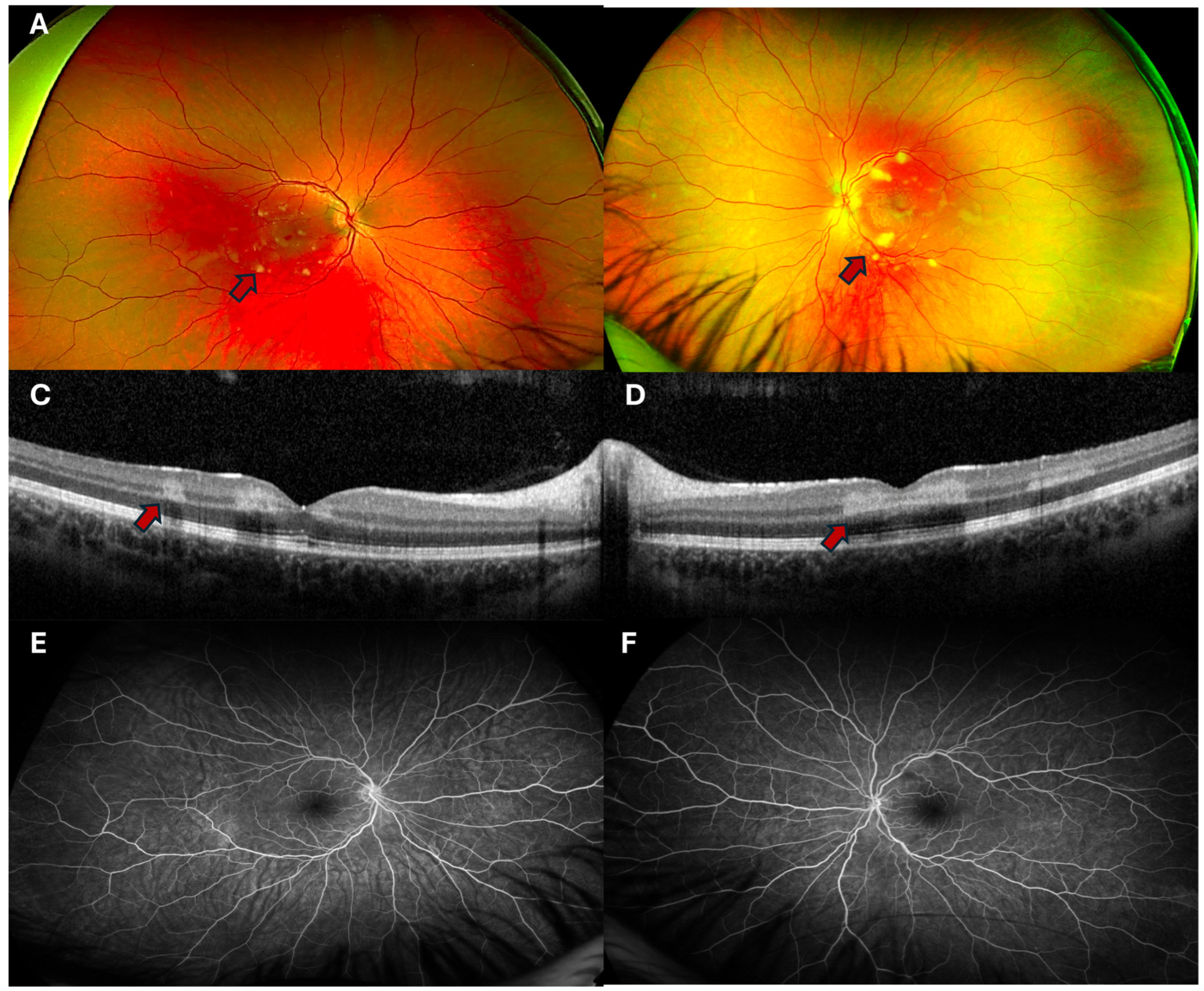
3.2. Ophthalmic Findings

Ophthalmic examination showed uncorrected visual acuity (VA) of 20/30 in the right eye (OD) and 20/25 in the left eye (OS) with intraocular pressures of 11 and 13 mmHg OD and OS, respectively. Anterior segment examination was unremarkable in both eyes, with no conjunctival hemorrhages or vascular changes. Dilated fundus examination was significant for bilateral cotton wool spots (CWSs), which were more prominent in the OS (Figure 1A,B). Macular optical coherence tomography (OCT) showed hyperreflectivity of the middle retinal layers in both eyes, consistent with paracentral acute middle maculopathy (PAMM), a sign of retinal ischemia (Figure 1C,D). Despite this, fluorescein angiography (FA) did not show any frank ischemia (Figure 1E,F), suggesting that vascular occlusion was limited to terminal arterioles and capillaries. The ophthalmic findings were consistent with PLR.

3.3. Clinical Workup

The patient received two computed tomography pulmonary angiography (CTPA) studies; the first immediately following the trauma that excluded pulmonary embolism, and the second study on day two post-fracture repair following new hypoxia. The second study revealed the interval development of peripheral ground glass and centrilobular nodules, in the absence of any infectious symptoms or aspiration events. These likely represent microscopic fat emboli, with no corresponding filling defect seen in the large pulmonary arteries (Figure 2). No pulmonary AVM were noted on the CTPA study, and there was no known family history of hereditary hemorrhagic telangiectasia. An echocardiogram excluded congenital heart disease, including patent foramen ovale and AVSD.
Non-contrast magnetic resonance imaging (MRI) of the brain revealed multiple small T2/fluid-attenuated inversion recovery (FLAIR) hyperintense foci corresponding to small white matter infarcts, without corresponding diffusion restriction on diffusion-weighted imaging sequences (Figure 3A). On susceptibility-weighted MRI angiography sequences, innumerable areas of blooming artifact were visible in the splenium of the corpus callosum and basal ganglia, corresponding to areas of microhemorrhage (Figure 3B).

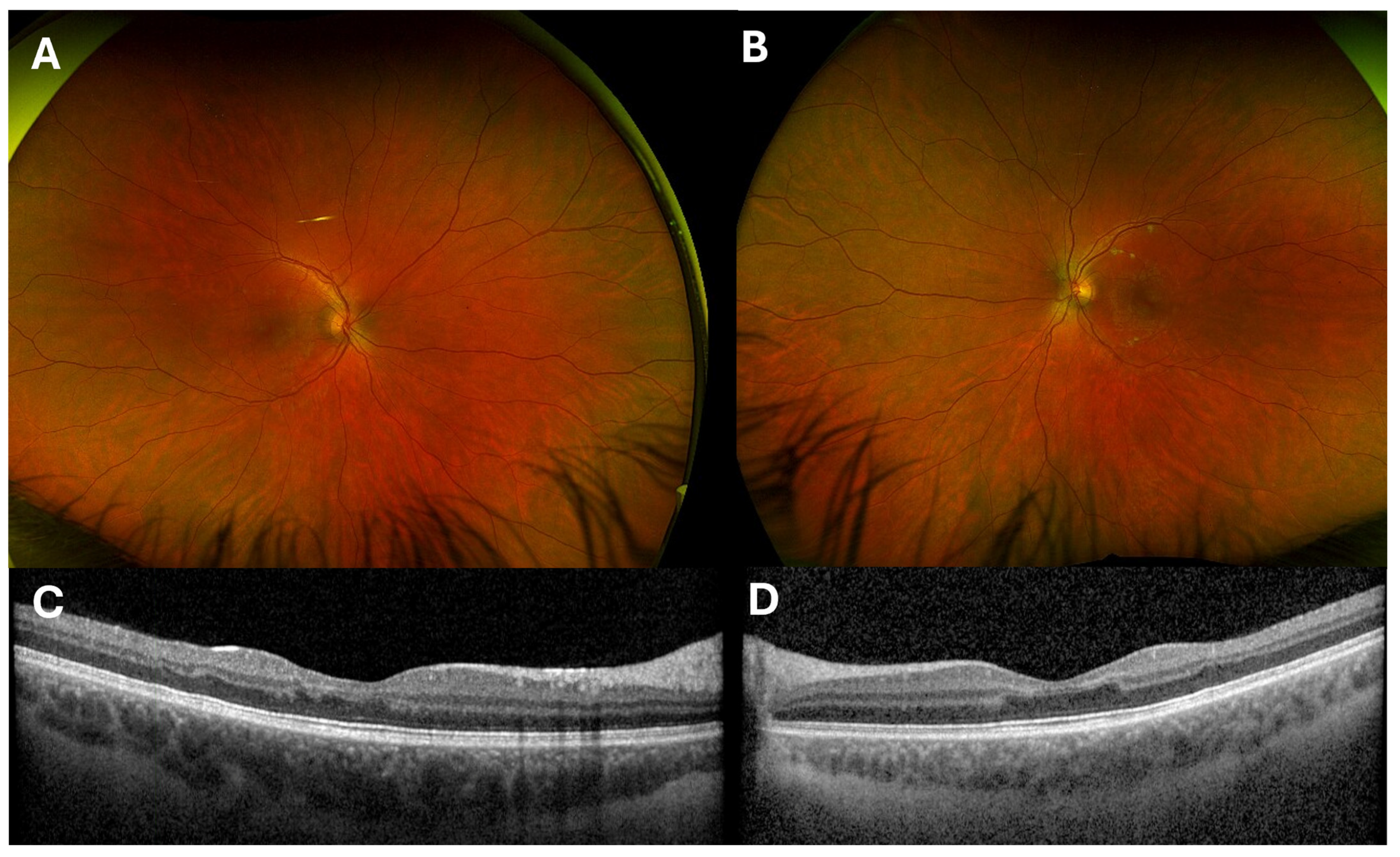
3.4. Diagnosis, Differential and Course of Recovery

His overall clinical presentation was consistent with FES secondary to the repair of long bone fracture with retinal, brain, and pulmonary involvement. Other differential diagnoses were considered less likely, including central retinal vein occlusion (due to the patient’s young age, lack of risk factors, absence of venular tortuosity, and bilaterality of disease), diabetic retinopathy (as the patient was not diabetic), and Roth spots (given the absence of endocarditis, anemia or myelodysplastic features). With conservative supportive measures, his respiratory and cognitive symptoms resolved within several days. Intervention was deferred due to the patient’s excellent baseline health and the relative risk of wound complications with systemic steroids. Vision significantly improved by six weeks without ophthalmic intervention and, at the most recent review (nine months), the progress continued; however, blur was noted when reading or occluding one eye. Uncorrected VA was 20/20 in both eyes. Acute features (e.g., CWS) had resolved (Figure 4A,B), though there was residual attenuation of the parafoveal inner retinal layers in areas previously affected by PAMM (Figure 4C,D). The Humphrey visual field 10-2 test was unable to identify focal visual field deficits.

4. Discussion

We present an unusual case of FES with significant ocular manifestations, where the patient was otherwise healthy and lacked any predisposing anatomic anomalies that would facilitate the dissemination of fat emboli into the arterial system. Further, his extraocular sequelae were mild and self-resolving. The timing of symptom onset suggests that the repair of the patient’s femoral fracture triggered the dissemination of numerous fat emboli to the systemic circulation though delayed symptom presentation, which has been reported up to three days post-fracture. Fortunately, the patient made a good functional recovery of his visual, respiratory, and neurocognitive faculties, likely due to his young age and good physiological reserve.
Purtscher retinopathy (PR), a sight-threatening retinal microangiopathy, was first described in 1910 in the setting of severe head trauma. The patient’s ophthalmic findings were in line with PLR, which is a similar condition but refers to cases associated with systemic diseases and traumatic injuries not involving the head. A variety of causes have been described including pancreatitis, systemic lupus erythematosus, long bone fracture, chest compression injuries, and embolic disease (e.g., air, amniotic fluid) [11].
The annual incidence of PR/PLR is approximately 0.24 individuals per million population, but the true incidence may be higher given many cases are asymptomatic or masked by the systemic sequelae of head trauma (e.g., loss of consciousness/coma, death) [11]. Although injury rates are higher among young males (e.g., long bone fractures in <50-year-olds are twice as common in males), incidence is similar when considering all age groups [12,13]. A recent systematic review found an equal distribution of PR between males and females [11].
The pathogenesis of PR/PLR is multifactorial and depends on the underlying etiology. Several mechanisms have been implicated, including an acute rise in intracranial pressure, the extravasation of lymphatic fluid, venous dilatation from raised intrathoracic pressure (i.e., transient increased resistance to venous outflow), vasculitis due to excess free fatty acids, vascular endothelial damage, and complement-induced leukoembolization, all of which have an effect on small caliber vasculature (e.g., retina, brain, lungs, skin) [14]. The likelihood of FES and the severity of end-organ functional sequelae (e.g., PLR) depend on factors such as the anatomical location of trauma (e.g., femur fractures have the greatest association with FES), characteristics of emboli (e.g., size, distensibility), patient age (e.g., children have less bone marrow fat than adults), and the presence of direct arteriovenous shunts [1]. In FES, the route by which fat emboli reach the retinal circulation can be either direct (i.e., via AVSD or AVM) or indirect (i.e., no detectable direct arteriovenous shunt). While AVSD is a well-understood cause, in some cases of retinal arterial occlusive disease, emboli may enter the systemic arterial circulation via pulmonary capillaries or dynamic intrapulmonary arteriovenous anastomoses [15,16]. The latter mechanism is rarely encountered in ophthalmic practice, and few case reports are available [8,10].
Clinical findings of PR/PLR are typically bilateral and include scattered CWS and intraretinal hemorrhages, with polygonal areas of retinal whitening (Purtscher flecken), corresponding to areas of PAMM on OCT [17]. PAMM is an imaging finding that represents retinal inflammation resulting from ischemia in the distribution of the middle and/or deep retinal capillary plexi. Such transient non-perfusion can be caused by vessel wall pathology, emboli, or arterial hypotension [18]. Though Purtscher flecken are considered pathognomonic for PR/PLR, they are present in only 50% of cases [14]. The visual course and recovery in PR/PLR are variable. In a study conducted by Agrawal and McKibbin reviewing 24 eyes of 15 patients with PR/PLR, 23% showed VA improvement by ≥4 Snellen lines and 50% by 2 lines at six months with observation alone [19]. Another study reported a favorable prognosis in 53% of patients, defined as any improvement in final best-corrected VA compared to presentation [14]. Long-term sequelae can include optic atrophy, thinning of the retinal nerve fiber layer, attenuation or sheathing of retinal vessels, and macular pigment mottling or atrophy, the severity of which is dependent on the degree of acute localized ischemic and inflammatory damage [2,20]. The visual sequelae of FES may be multifactorial with the embolic/ischemic disruption of both the retina and the cortical visual pathways [21]. While this has rarely been documented, it may be under-reported due to concomitant injuries (e.g., coma, death).
Mortality in FES occurs in 7–10% of cases, with acute respiratory distress syndrome and cerebral edema as the leading causes [2]. In FES secondary to long bone fractures, survival improves with prompt surgical repair [2]. In this case, FES presented immediately following the surgical repair of a long bone fracture, although both the inciting trauma and the surgical intervention may have contributed. The rate of ophthalmic involvement in severe FES may be higher than reported, likely masked in fatal or neurologically debilitating cases due to the delay in or absence of ophthalmic examination, as the ophthalmic clinical features can be transient or only identifiable by detailed imaging (e.g., OCT).

5. Conclusions

In summary, we report a case of FES with retinal involvement (i.e., PLR) and spontaneous recovery following a long bone fracture and orthopedic surgery. A synthesis of the available data suggests fat emboli may have entered the arterial circulation by deforming through pulmonary capillaries, given no predisposing anatomic variants were found on a thorough work-up. An ophthalmic examination (±FA) is recommended in all cases of PR/PLR, as abnormalities in the clinically visible retinal microvasculature could prompt screening investigations for other vaso-occlusive pathology, such as in the brain and lungs. The patient reported in this case fortunately had mild disease; however, early ophthalmic assessment is critical to determine whether visual symptoms are attributable to retinal or visual pathway embolic disease.

Author Contributions

Conceptualization, A.W.K. and K.A.J.S.; methodology, L.P.A.H. and K.A.J.S.; data curation, L.P.A.H.; writing—original draft preparation, L.P.A.H. and K.A.J.S.; writing—review and editing, L.P.A.H., R.M.D., K.A.J.S. and A.W.K.; visualization, K.A.J.S. and R.M.D.; supervision, A.W.K.; project administration, K.A.J.S. All authors have read and agreed to the published version of the manuscript.

Funding

This research received no external funding.

Institutional Review Board Statement

This study was conducted in accordance with the Declaration of Helsinki. Ethical review and approval were waived for this study in line with the institutional policy of the University of British Columbia for case reports.

Informed Consent Statement

Informed consent was obtained from the subject involved in this study.

Data Availability Statement

All source data are represented in this manuscript.

Acknowledgments

The authors wish to acknowledge the patient for their gracious participation and the ophthalmic imaging department (Section F) of Vancouver General Hospital.

Conflicts of Interest

The authors declare no conflicts of interest.

Abbreviations

The following abbreviations are used in this manuscript:
AVMArteriovenous malformation
AVSDAtrioventricular septal defect
CTPAComputed tomography pulmonary angiogram
CWSCotton wool spots
FAFluorescein angiography
FESFat embolism syndrome
FLAIRFluid-attenuated inversion recovery
MRIMagnetic resonance imaging
OCToptical coherence tomography
PAMMParacentral acute middle maculopathy
PRPurtscher retinopathy
PLRPurtscher-like retinopathy

References

  1. Rothberg, D.L.; Makarewich, C.A. Fat Embolism and Fat Embolism Syndrome. J. Am. Acad. Orthop. Surg. 2019, 27, e346–e355. [Google Scholar] [CrossRef] [PubMed]
  2. Adeyinka, A.; Pierre, L. Fat Embolism. In StatPearls; StatPearls Publishing LLC.: Treasure Island, FL, USA, 2024. [Google Scholar]
  3. Mellor, A.; Soni, N. Fat embolism. Anaesthesia 2001, 56, 145–154. [Google Scholar] [CrossRef] [PubMed]
  4. Bracco, D.; Favre, J.B.; Joris, R.; Ravussin, A. Fatal fat embolism syndrome: A case report. J. Neurosurg. Anesthesiol. 2000, 12, 221–224. [Google Scholar] [CrossRef] [PubMed]
  5. Morimoto, T.; Kobayashi, T.; Yoshihara, T.; Tsukamoto, M.; Kai, K.; Mawatari, M. Fatal fat embolism syndrome during posterior spinal fusion surgery: A case report and literature review. Medicine 2021, 100, e28381. [Google Scholar] [CrossRef] [PubMed]
  6. Mohar, J. Fatal Fulminant Fat Embolism Syndrome in Adult Spine Deformity Surgery: A Case Report. JBJS Case Connect. 2022, 12, e22. [Google Scholar] [CrossRef] [PubMed]
  7. Hirata, Y.; Inokuchi, G.; Tsuneya, S.; Hoshioka, Y.; Chiba, F.; Yoshida, M.; Makino, Y.; Iwase, H. A case of fatal fulminant fat embolism syndrome following multiple fractures resulting from a fall. J. Forensic Sci. 2022, 67, 2115–2121. [Google Scholar] [CrossRef] [PubMed]
  8. Nentwich, M.M.; Remy, M.; Schaller, U.C. Ocular fat embolism syndrome. Int. Ophthalmol. 2011, 31, 15–16. [Google Scholar] [CrossRef] [PubMed]
  9. Lyons, L.J.; Yarrabolu, T.; Kuffel, R.R., Jr.; Bishop, J.E. Juvenile Central Retinal Artery Occlusion Associated with Atrial Septal Defect. J. Pediatr. Ophthalmol. Strabismus 2019, 56, e73–e75. [Google Scholar] [CrossRef] [PubMed]
  10. Li, Y.S.; Liu, Y.H.; Chou, H.D.; Tseng, H.J.; Fu, Y.C.; Liu, W.C. Concomitant post-traumatic ocular and cerebral fat embolism syndrome and thrombotic pulmonary embolism: A case report. Medicine 2022, 101, e29331. [Google Scholar] [CrossRef] [PubMed]
  11. Roden, D.; Fitzpatrick, G.; O’Donoghue, H.; Phelan, D. Purtscher’s retinopathy and fat embolism. Br. J. Ophthalmol. 1989, 73, 677–679. [Google Scholar] [CrossRef] [PubMed]
  12. Meling, T.; Harboe, K.; Søreide, K. Incidence of traumatic long-bone fractures requiring in-hospital management: A prospective age- and gender-specific analysis of 4890 fractures. Injury 2009, 40, 1212–1219. [Google Scholar] [CrossRef] [PubMed]
  13. Almigdad, A.; Mustafa, A.; Alazaydeh, S.; Alshawish, M.; Bani Mustafa, M.; Alfukaha, H. Bone Fracture Patterns and Distributions according to Trauma Energy. Adv. Orthop. 2022, 2022, 8695916. [Google Scholar] [CrossRef] [PubMed]
  14. Serhan, H.A.; Abuawwad, M.T.; Taha, M.J.J.; Hassan, A.K.; Abu-Ismail, L.; Delsoz, M.; Alrawashdeh, H.M.; Alkorbi, H.A.; Moushmoush, O.; Elnahry, A.G. Purtscher’s and Purtscher-like retinopathy etiology, features, management, and outcomes: A summative systematic review of 168 cases. PLoS ONE 2024, 19, e0306473. [Google Scholar] [CrossRef] [PubMed]
  15. Burrowes, K.S.; Clark, A.R.; Tawhai, M.H. Blood flow redistribution and ventilation-perfusion mismatch during embolic pulmonary arterial occlusion. Pulm. Circ. 2011, 1, 365–376. [Google Scholar] [CrossRef] [PubMed]
  16. Lovering, A.T.; Elliott, J.E.; Beasley, K.M.; Laurie, S.S. Pulmonary pathways and mechanisms regulating transpulmonary shunting into the general circulation: An update. Injury 2010, 41 (Suppl. S2), S16–S23. [Google Scholar] [CrossRef] [PubMed]
  17. Tripathy, K.; Patel, B.C. Purtscher Retinopathy. In StatPearls; StatPearls Publishing LLC.: Treasure Island, FL, USA, 2024. [Google Scholar]
  18. Meng, Y.; Sawut, A.; Tian, M.; Li, Y.; Cai, L.; Xiao, D.; Yi, Z.; Chen, C. Purtscher-like retinopathy and paracentral acute middle maculopathy associated with improper antihypertensive drug use: A case report. Front. Med. 2024, 11, 1394614. [Google Scholar] [CrossRef] [PubMed]
  19. Agrawal, A.; McKibbin, M. Purtscher’s retinopathy: Epidemiology, clinical features and outcome. Br. J. Ophthalmol. 2007, 91, 1456–1459. [Google Scholar] [CrossRef] [PubMed]
  20. Beckingsale, A.B.; Rosenthal, A.R. Early fundus fluorescein angiographic findings and sequelae in traumatic retinopathy: Case report. Br. J. Ophthalmol. 1983, 67, 119–123. [Google Scholar] [CrossRef] [PubMed]
  21. Mirza, K.; Acharya, P.U.; Austine, J. Transient cortical blindness in fat embolism syndrome—A diagnostic enigma. Chin. J. Traumatol. 2021, 24, 79–82. [Google Scholar] [CrossRef] [PubMed]
Figure 1. Retinal imaging: (A,B): Optos ultra-widefield (UWF) pseudocolor fundus images of the right (A) and left (B) eyes, demonstrating cotton wool spots (red arrows) that are more noticeable in the left eye. (C,D): Optical coherence tomography (OCT) of the right (C) and left (D) maculae showing hyperreflectivity in the middle retinal layers consistent with paracentral acute middle maculopathy, red arrows. Late stage UWF fluorescein angiography images (E,F) excluding macrovascular retinal ischemia.
Complications 02 00006 g001
Figure 2. Computed tomography pulmonary angiography on lung windows demonstrating extensive bilateral ground-glass changes and diffuse centrilobular nodularity. Given the clinical context, findings represent extensive microscopic fat emboli throughout the lung parenchyma.
Complications 02 00006 g002
Figure 3. Axial magnetic resonance imaging (MRI) of the brain on T2 FLAIR sequence (A) demonstrating multiple small white matter infarcts (red arrows) throughout the bilateral centrum ovale and SWAN sequences (B) showing innumerable miniscule parenchymal microhemorrhages (red arrows) focused in the splenium of the corpus callosum.
Complications 02 00006 g003
Figure 4. Retinal imaging at nine-month follow-up visit: (A,B): UWF fundus photos show no visible remnants of stigmata of PLR. (C,D): Inner retinal attenuation and disorganization corresponding to the areas previously affected by PAMM.
Complications 02 00006 g004
Disclaimer/Publisher’s Note: The statements, opinions and data contained in all publications are solely those of the individual author(s) and contributor(s) and not of MDPI and/or the editor(s). MDPI and/or the editor(s) disclaim responsibility for any injury to people or property resulting from any ideas, methods, instructions or products referred to in the content.

