
Arteriovenous Fistula with Pseudoaneurysm and Facial Palsy Following Bilateral Sagittal Split Osteotomy: A Case Report

 , , and
Abstract
1. Introduction
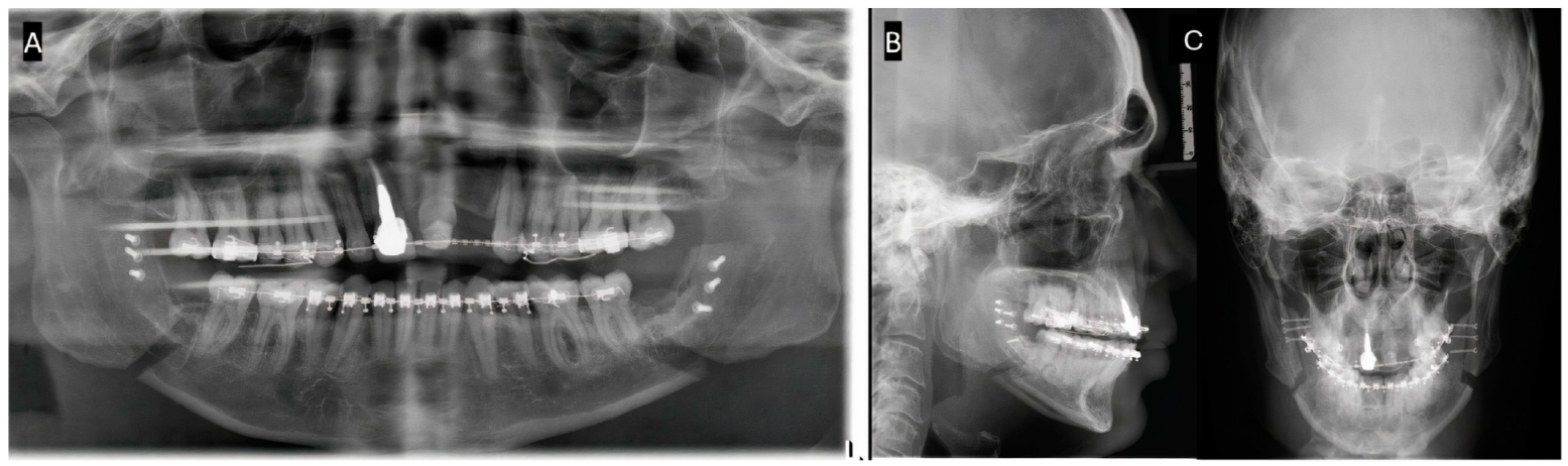
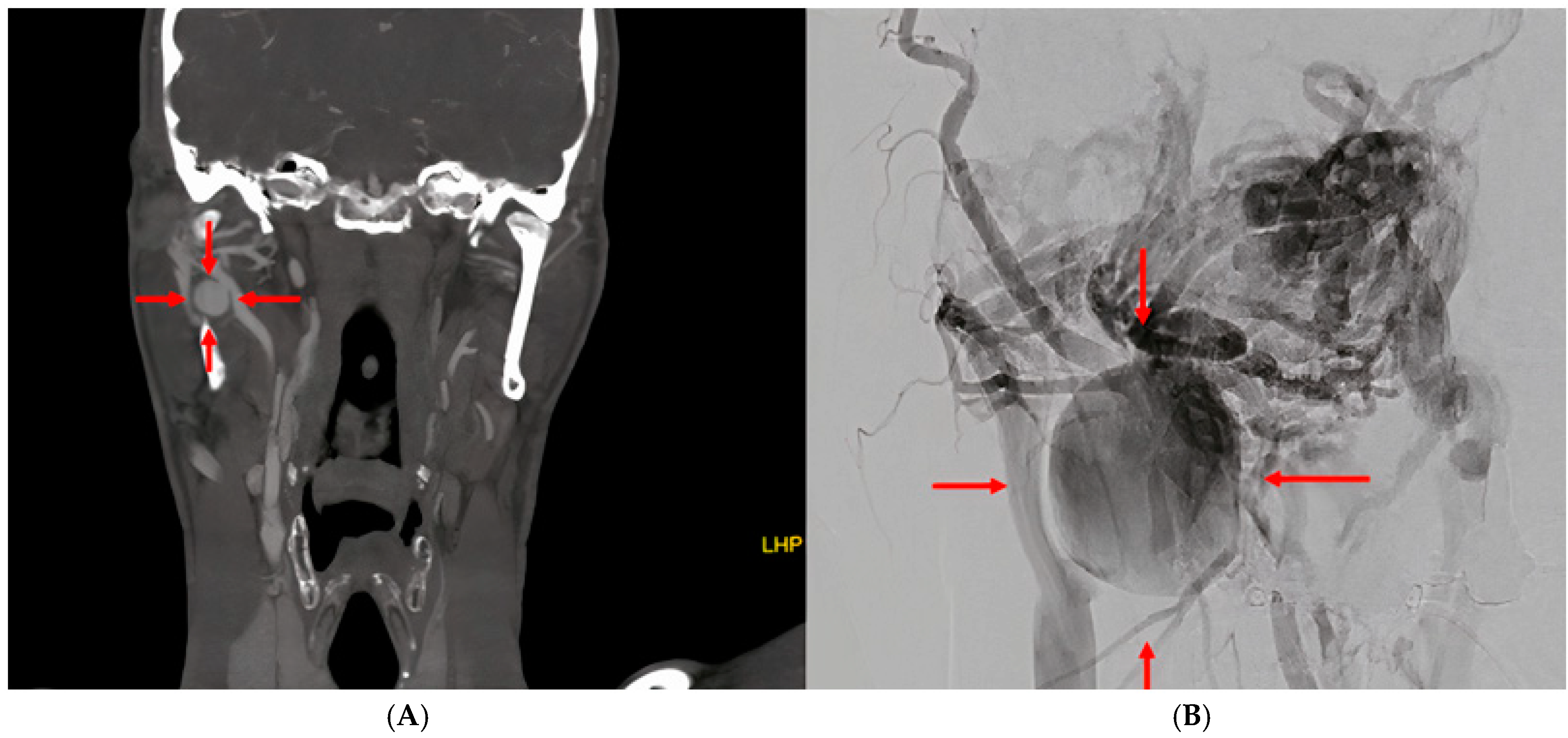
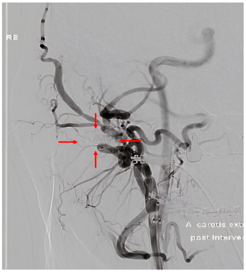
2. Case Report
3. Discussion
4. Conclusions
Supplementary Materials
Author Contributions
Funding
Institutional Review Board Statement
Informed Consent Statement
Data Availability Statement
Conflicts of Interest
References
- Seifert, L.B.; Langhans, C.; Berdan, Y.; Zorn, S.; Klos, M.; Landes, C.; Sader, R. Comparison of two surgical techniques (HOO vs. BSSO) for mandibular osteotomies in orthognathic surgery—A 10-year retrospective study. Oral Maxillofac. Surg. 2023, 27, 341–351. [Google Scholar] [CrossRef] [PubMed]
- Kumar, A.; Kaur, A.; Singh, M.; Rattan, V.; Rai, S. Signs and Symptoms Tell All-Pseudoaneurysm as a Cause of Postoperative Bleeding after Orthognathic Surgery-Report of a Case and a Systematic Review of Literature. J. Maxillofac. Oral Surg. 2021, 20, 345–355. [Google Scholar] [CrossRef]
- Silva, A.C.; O’Ryan, F.; Beckley, M.L.; Young, H.Y.; Poor, D. Pseudoaneurysm of a branch of the maxillary artery following mandibular sagittal split ramus osteotomy: Case report and review of the literature. J. Oral Maxillofac. Surg. 2007, 65, 1807–1816. [Google Scholar] [CrossRef]
- Hunsuck, E. A modified intraoral sagittal splitting technic for correction of mandibular prognathism. J. Oral Surg. 1968, 26, 249–252. [Google Scholar]
- Epker, B. Modifications in the sagittal osteotomy of the mandible. J. Oral Surg. 1977, 35, 157–159. [Google Scholar] [PubMed]
- Möhlhenrich, S.C.; Kniha, K.; Peters, F.; Ayoub, N.; Goloborodko, E.; Hölzle, F.; Fritz, U.; Modabber, A. Fracture patterns after bilateral sagittal split osteotomy of the mandibular ramus according to the Obwegeser/Dal Pont and Hunsuck/Epker modifications. J. Cranio-Maxillofac. Surg. 2017, 45, 762–767. [Google Scholar] [CrossRef] [PubMed]
- Brusati, R.; Fiamminghi, L.; Sesenna, E.; Gazzotti, A. Functional disturbances of the inferior alveolar nerve after sagittal osteotomy of the mandibular ramus: Operating technique for prevention. J. Maxillofac. Surg. 1981, 9, 123–125. [Google Scholar] [CrossRef]
- Ow, A.; Cheung, L.K. Skeletal Stability and Complications of Bilateral Sagittal Split Osteotomies and Mandibular Distraction Osteogenesis: An Evidence-Based Review. J. Oral Maxillofac. Surg. 2009, 67, 2344–2353. [Google Scholar] [CrossRef] [PubMed]
- Kobayashi, T.; Izumi, N.; Kojima, T.; Sakagami, N.; Saito, I.; Saito, C. Progressive condylar resorption after mandibular advancement. Br. J. Oral Maxillofac. Surg. 2012, 50, 176–180. [Google Scholar] [CrossRef] [PubMed]
- Piñeiro-Aguilar, A.; Somoza-Martín, M.; Gandara-Rey, J.M.; García-García, A. Blood loss in orthognathic surgery: A systematic review. J. Oral Maxillofac. Surg. 2011, 69, 885–892. [Google Scholar] [CrossRef] [PubMed]
- Schwaiger, M.; Wallner, J.; Edmondson, S.-J.; Mischak, I.; Rabensteiner, J.; Gary, T.; Zemann, W. Is there a hidden blood loss in orthognathic surgery and should it be considered? Results of a prospective cohort study. J. Cranio-Maxillofac. Surg. 2021, 49, 545–555. [Google Scholar] [CrossRef] [PubMed]
- Avelar, R.L.; Goelzer, J.G.; Becker, O.E.; de Oliveira, R.B.; Raupp, E.F.; de Magalhães, P.S.C. Embolization of pseudoaneurysm of the internal maxillary artery after orthognathic surgery. J. Craniofacial Surg. 2010, 21, 1764–1768. [Google Scholar] [CrossRef] [PubMed]
- Clark, R.; Lew, D.; Giyanani, V.L.; Gerlock, A. False aneurysm complicating orthognathic surgery. J. Oral Maxillofac. Surg. 1987, 45, 57–59. [Google Scholar] [CrossRef]
- Steel, B.J.; Cope, M.R. Unusual and rare complications of orthognathic surgery: A literature review. J. Oral Maxillofac. Surg. 2012, 70, 1678–1691. [Google Scholar] [CrossRef]
- AbuKaraky, A.; Al Mousa, M.; Samara, O.A.; Baqain, Z.H. Pseudoaneurysm in the inferior alveolar artery following a bad split in bilateral sagittal split osteotomy. Int. J. Oral Maxillofac. Surg. 2021, 50, 798–800. [Google Scholar] [CrossRef]
- Bykowski, M.R.; Hill, A.; Garland, C.; Tobler, W.; Losee, J.E.; Goldstein, J.A. Ruptured Pseudoaneurysm of the Maxillary Artery and Its Branches Following Le Fort I Osteotomy: Evidence-Based Guidelines. J. Craniofac. Surg. 2018, 29, 998–1001. [Google Scholar] [CrossRef] [PubMed]
- Rosenberg, I.; Austin, J.C.; Wright, P.G.; King, R.E. The effect of experimental ligation of the external carotid artery and its major branches on haemorrhage from the maxillary artery. Int. J. Oral Surg. 1982, 11, 251–259. [Google Scholar] [CrossRef] [PubMed]
- Keeling, A.N.; McGrath, F.P.; Lee, M.J. Interventional Radiology in the Diagnosis, Management, and Follow-Up of Pseudoaneurysms. CardioVascular Interv. Radiol. 2009, 32, 2–18. [Google Scholar] [CrossRef] [PubMed]
- Cornwall, J.W.; Png, C.Y.M.; Han, D.K.; Tadros, R.O.; Marin, M.L.; Faries, P.L. Endovascular techniques in the treatment of extracranial carotid artery aneurysms. J. Vasc. Surg. 2021, 73, 2031–2035. [Google Scholar] [CrossRef] [PubMed]
- Obayi, I.J.; Cornwall, J.W.; Rao, A.G.; Han, D.K.; Tadros, R.O.; Marin, M.L.; Faries, P.L. Advancements in Endovascular Treatment of Extracranial Carotid Artery Aneurysms. Ann. Vasc. Surg. 2024; ahead of print. [Google Scholar] [CrossRef]
Disclaimer/Publisher’s Note: The statements, opinions and data contained in all publications are solely those of the individual author(s) and contributor(s) and not of MDPI and/or the editor(s). MDPI and/or the editor(s) disclaim responsibility for any injury to people or property resulting from any ideas, methods, instructions or products referred to in the content. |
© 2025 by the authors. Licensee MDPI, Basel, Switzerland. This article is an open access article distributed under the terms and conditions of the Creative Commons Attribution (CC BY) license (https://creativecommons.org/licenses/by/4.0/).
Share and Cite
Ivanic-Sefcikova, M.; Starke, V.; Groessing, L.; Augustin, M.; Schwaiger, M.; Zemann, W. Arteriovenous Fistula with Pseudoaneurysm and Facial Palsy Following Bilateral Sagittal Split Osteotomy: A Case Report. Complications 2025, 2, 3. https://doi.org/10.3390/complications2010003
Ivanic-Sefcikova M, Starke V, Groessing L, Augustin M, Schwaiger M, Zemann W. Arteriovenous Fistula with Pseudoaneurysm and Facial Palsy Following Bilateral Sagittal Split Osteotomy: A Case Report. Complications. 2025; 2(1):3. https://doi.org/10.3390/complications2010003
Chicago/Turabian StyleIvanic-Sefcikova, Michala, Vasco Starke, Lukas Groessing, Michael Augustin, Michael Schwaiger, and Wolfgang Zemann. 2025. "Arteriovenous Fistula with Pseudoaneurysm and Facial Palsy Following Bilateral Sagittal Split Osteotomy: A Case Report" Complications 2, no. 1: 3. https://doi.org/10.3390/complications2010003
APA StyleIvanic-Sefcikova, M., Starke, V., Groessing, L., Augustin, M., Schwaiger, M., & Zemann, W. (2025). Arteriovenous Fistula with Pseudoaneurysm and Facial Palsy Following Bilateral Sagittal Split Osteotomy: A Case Report. Complications, 2(1), 3. https://doi.org/10.3390/complications2010003