Review

Protein Kinases in Mediating Phage-Bacteria Interactions

by
Yong Everett Zhang
Department of Biology, University of Copenhagen, DK-2200 Copenhagen, Denmark
Kinases Phosphatases 2025, 3(3), 14; https://doi.org/10.3390/kinasesphosphatases3030014
Submission received: 9 April 2025 / Revised: 23 June 2025 / Accepted: 23 June 2025 / Published: 25 June 2025

Abstract

Protein kinases and phosphatases are essential for post-translational regulation, enabling bacteria to adapt to environmental stresses and modulate virulence. While prior reviews have broadly covered their roles in stress response, antibiotic resistance, and virulence, this article updates specifically on the roles of histidine kinases (HKs) and serine/threonine kinases (STKs) in mediating phage-bacteria interactions. A key aspect is phage-encoded kinases, which hijack bacterial signalling by phosphorylating and disrupting host processes to promote infection. Despite their importance, significant gaps remain in understanding these regulatory networks. This microreview highlights both the unresolved mechanisms and the therapeutic potential of targeting kinase pathways—for instance, by disrupting phage evasion strategies or enhancing phage-based antimicrobial therapies.

1. Introduction

Bacterial survival in dynamic environments relies on sophisticated signalling systems, including particularly histidine kinases (HKs) and serine/threonine kinases (STKs) (for a more exhaustive classification and discussion of bacterial kinases refer to [1]), which govern adaptive responses to stressors ranging from nutrient scarcity to antimicrobial threats. HKs, central to two-component systems (TCSs) (e.g., Escherichia coli EnvZ/OmpR), transduce extracellular signals via histidine phosphorylation, driving stress adaptation, antibiotic tolerance, resistance [2] and virulence [3]. STKs, resembling eukaryotic kinases, regulate intracellular processes such as Staphylococcus aureus PknB-dependent efflux pump activation [4] and E. coli HipA-induced persister cell formation [5,6], linking kinase activity to antibiotic tolerance and resistance (refer to [7] for further clarifications of their differences).
While these kinases are well-documented in stress resilience and therapeutic evasion [1,2,8], their roles in phage-bacteria interactions remain poorly understood. However, investigating these roles is critical given the therapeutic promise of phages as potential alternatives to antibiotics. For instance, Marinomonas mediterranea TCS BarA/UvrY modulates CRISPR-Cas defenses [9], and phages like T7 deploy kinase mimics (Gp0.7) to subvert host signalling [10]. Moreover, most studies of phage-bacteria interactions fail to account for environmental stress conditions, which dynamically reshape bacterial physiology and may concurrently influence phage infectivity and antibiotic susceptibility. This knowledge gap remains unresolved, even if multidrug-resistant pathogens escalate into a global health crisis and phage therapy emerges as a promising therapeutic alternative.
By synthesizing recent advances, this article highlights the dual significance of HKs/STKs in stress-driven antibiotic resistance and more their nascent, yet critical, intersections with phage defense—a triad of interactions essential for deciphering bacterial adaptability and guiding next-generation antimicrobial strategies.

2. Brief Sum of HKs and STKs in Bacterial Stress Response and Antibiotic Tolerance/Resistance

Bacterial HKs and STKs are master regulators of stress adaptation and antibiotic tolerance/resistance. Their roles have been extensively reviewed across various organisms, including in several recent reviews [1,2,11,12]. Therefore, only a brief summary is provided here to illustrate their distinct structural features and mechanistic strategies for counteracting environmental stress and therapeutic interventions. HKs, as core components of TCSs, are typically transmembrane proteins with a sensor domain that detects extracellular stressors such as osmotic shifts, nutrient deprivation, or antimicrobial presence. Upon signal perception, HKs autophosphorylate a conserved histidine residue within their cytoplasmic kinase domain, subsequently transferring the phosphate group to an aspartate residue on a cognate response regulator (RR) [13,14]. This phosphorylation activates the specific RR, enabling it to modulate transcription of stress-response genes. For instance, E. coli HK EnvZ phosphorylates RR OmpR to adjust porin expression in response to osmolarity, indirectly influencing antibiotic influx [15,16]. In more complex signaling networks, additional phosphotransfer intermediates such as DHp and Hpt domains are involved, and signal crosstalk between different TCS pathways can occur, even though HK-RR specificity is generally maintained [13,14]. For instance, Pseudomonas aeruginosa LadS, an HK autophosphorylates and passes on the phosphate to a second HK GacS, then to RR GacA, leading to a switch from the acute to the chronic virulence program and drug tolerance [17].
In contrast, STKs operate through eukaryotic-like phosphorylation cascades, dynamically modifying substrate proteins by transferring phosphate groups from ATP to specific serine/threonine residues to orchestrate diverse cellular processes, including cell division, sporulation, biofilm formation, stress responses, and host interactions [1]. For instance, in Bacillus subtilis, PrkC directs cell wall remodeling and sporulation [18]; Pseudomonas aeruginosa Stk1 modulates virulence and antibiotic resistance through transcription factor phosphorylation [19]; and Mycobacterium tuberculosis STK PknG regulates metabolic adaptation by redirecting carbon flux via GarA phosphorylation [20].
Among STKs is another group of toxin-like kinase exemplified by the E. coli HipA STK and its homologs/paralogs [21]. HipA is a component of the HipBA toxin-antitoxin (TA) system, orchestrating the formation of antibiotic-tolerant persister cells through a sophisticated phosphorylation cascade (reviewed in [22]). Under stress conditions, HipA phosphorylates glutamyl-tRNA synthetase (GltX) at Ser239 [5], a critical enzyme responsible for charging tRNAGlu with glutamate during translation. This modification disrupts GltX’s function, leading to the accumulation of uncharged tRNAGlu. The uncharged tRNA activates the stringent response via the alarmone (p)ppGpp, a global regulator of stress adaptation. Elevated (p)ppGpp levels halt ribosomal RNA synthesis [23], repress translation machinery assembly, and arrest growth-related processes [24,25], effectively inducing a metabolically dormant state. This transient shutdown allows a subpopulation of cells (persisters) to evade bactericidal antibiotics (e.g., fluoroquinolones [26]) that target active cellular functions. While not genetically resistant, persisters survive treatment by entering this quiescent state, later resuscitating to repopulate the bacterial community post-stress. Alternatively, persisters could obtain mutations, becoming resistant to the applied antibiotics [27]. The HipA-GltX axis exemplifies how TA systems drive phenotypic heterogeneity [28], highlighting a key mechanism behind chronic infections and antibiotic recalcitrance [22,27].
While HK/STK pathways are well-established modulators of bacterial metabolism and envelope integrity—directly shaping antibiotic susceptibility by altering drug targets or access—their interplay with phage infections remains poorly characterized. Recent breakthroughs in phage-bacteria interaction studies have unveiled sophisticated defense and counter-defense systems [29], yet the involvement of kinase signalling in these dynamics is strikingly understudied. Given that phages predominantly infect bacteria living under various environmental stresses—conditions where HK/STK networks are supposed to be active—it is logical to hypothesize that kinase activity influences phage predation efficacy, either by priming host defenses or by reshaping physiological states critical for viral replication. This highlights a pivotal gap in our understanding of the tripartite interactions between bacteria, antibiotics, and phages. Addressing this knowledge gap could reveal novel strategies to potentiate phage therapy or disrupt stress-induced antibiotic tolerance. Therefore, the following section explores how HK/STKs influence the outcomes of phage infections.

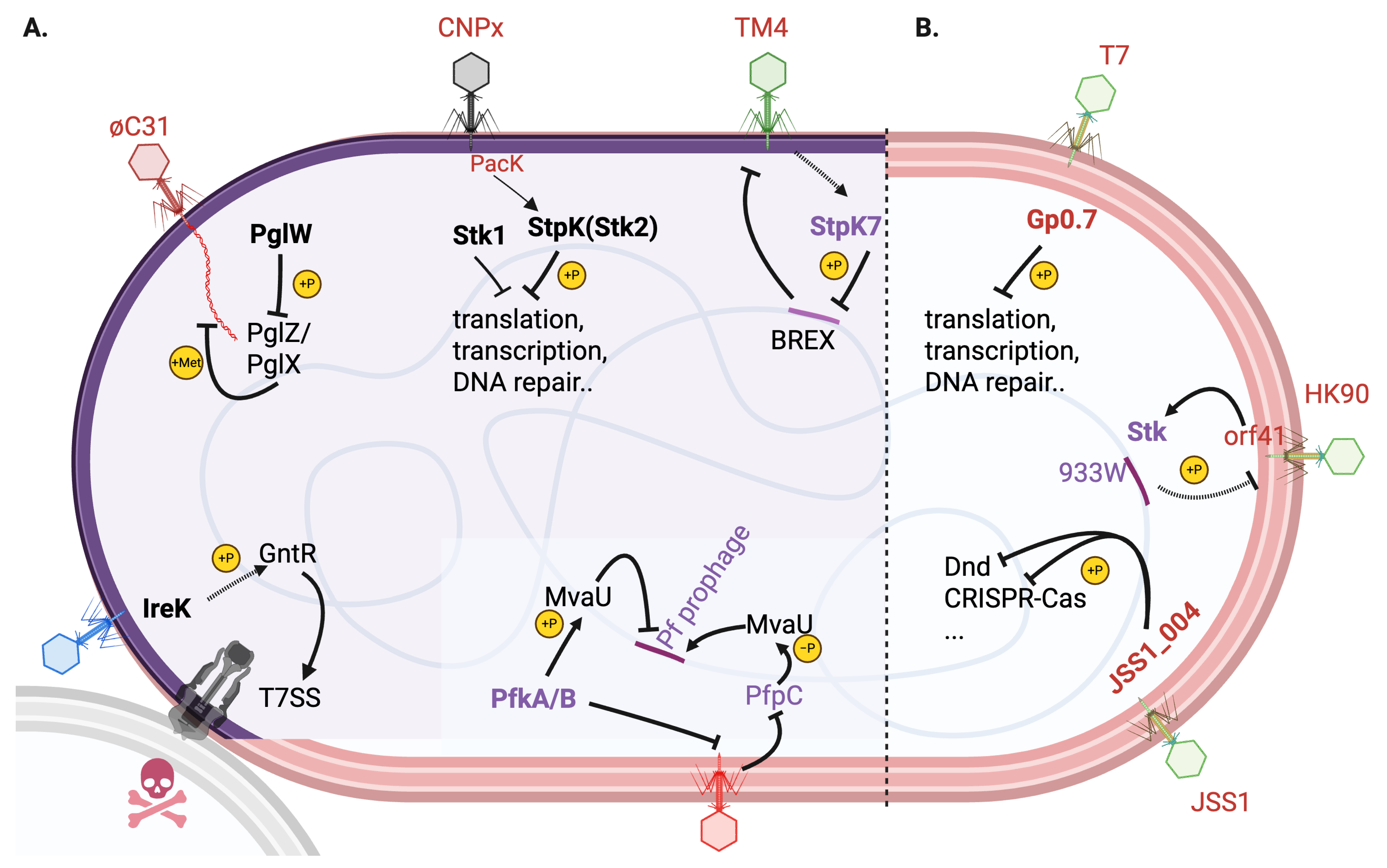
3. Bacteria-Encoded STKs in Phage-Bacteria Interactions

So far, there are very few studies (Table 1; Figure 1A) reporting the involvement of host bacteria-encoded STKs in mediating interaction with phages.
Table 1. List of kinases discussed in this article. STK, serine/threonine kinase; HK, histidine kinase.
Kinases Origin and Types FunctionsReference
StpK7 (MSMEG_1200)Mycobacterium smegmatis BREX; STKMediates TM4 phage infection[30]
StpKStaphylococcus aureus; STKActivated by phage protein PacK to mediate abortive infection[31]
PglWStreptomyces coelicolor; STKLinked to phage growth limitation (Pgl) and BREX anti-phage systems[32]
IreKEnterococcus faecalis; STKMediates stress-responsive activation of the type VIIb secretion system (T7SS) and contact-dependent antagonism of bystander bacteria[33]
PfkA/BPseudomonas aeruginosa; STKs (PfkA/B) and phosphatase (PfpC)Coordinates prophage activation and antiphage defense[34]
PfpC
Gp0.7E. coli T7 phage; STKPhosphorylates multiple host E. coli proteins to hijack cellular processes[35,36,37,38,39]
Stk933Wlambdoid phage 933W; tyrosine kinaseExclusion of secondary phage infection[40,41]
JSS1_004Salmonella phage JSS1; STKPhosphorylate and dismantle multilayered host defences e.g., CRISPR-Cas[42]
RcsSerratia sp. ATCC 39006; HKBalancing innate and adaptive immunity[43]
BarA/UvrYMarinomonas mediterranea MMB-1; HKRegulating the dual CRISPR-Cas systems (PAM- dependent or -independent) to confer phage resistance[9]
CpxKlebsiella pneumoniae, etc; HKDual role in enhancing carbapenem resistance and conjugative plasmid transfer; potential interference with phage infection[44,45]

3.1. Mycobacterium smegmatis StpK7: A Pro-Phage Kinase [30]

In M. smegmatis, the STK StpK7 (MSMEG_1200) is uniquely exploited by the TM4 phage to counteract host defenses. StpK7 resides within a BREX (bacteriophage exclusion)-like gene island (MSMEG_1191 to MSMEG_1200) and phosphorylates to inhibit a transcription factor (MSMEG_1198) in this locus, silencing this host defense system. This phosphorylation sustains TM4 phage adsorption to host cells by maintaining normal glycolipid surface receptors and suppresses bacterial cell death, thereby enabling phage replication. However, the exact activating signal of StpK7 remains unknown.

3.2. Staphylococcus aureus StpK: Altruistic Suicide via Phosphorylation [31]

In Staphylococcus, infection by a temperate phage CNPx triggers the activation of StpK by the viral protein PacK, a P-loop NTPase domain protein. Activated StpK phosphorylates many essential host proteins involved in translation, DNA repair, and central metabolism, inducing rapid cell death. This altruistic suicide limits phage CNPx propagation, protecting neighboring populations of Staphylococcus cells—a defense mechanism akin to abortive infection systems. Intriguingly, StpK (Stk2) seems to collaborate with a second conserved kinase Stk1 (or PknB) (linked to cell wall integrity, antimicrobial resistance [4,46] and virulence [47] of Staphylococcus), forming a phosphorylation cascade reminiscent of eukaryotic antiviral pathways.

3.3. Streptomyces PglW: Component of Pgl/BREX Defense [32]

Streptomyces coelicolor STK PglW is linked to the phage growth limitation (Pgl) and BREX anti-phage systems, which block phage ϕC31 DNA replication. In Pgl, PglW’s kinase activity is essential for signaling during phage infection, likely phosphorylating downstream targets like PglZ (a phosphatase-like antitoxin) and PglX (a toxin DNA methyltransferase) to execute phage restriction. This system uniquely combines toxin-antitoxin regulation with DNA modification: PglX methylates phage DNA during the first-round infection, marking progeny phages for attenuation in subsequent infections of Pgl+ host cells. Unlike classical Restriction-Modification systems, Pgl tolerates transient modification failures but restricts phage spread through phase variation [48,49,50,51], which is regulated by a hypervariable G-tract in pglX gene to balance defense costs and evade phage counteradaptation.

3.4. Enterococcus faecalis IreK: Sensing Phage Infection Signals [33]

IreK, a STK in Enterococcus faecalis, plays a central role in mediating stress-responsive activation of the type VIIb secretion system (T7SS), which drives collateral damage to non-target bacteria in multi-species communities. During lytic phage infection, membrane damage triggers IreK-dependent signaling, leading to transcriptional upregulation of T7SS genes in coordination with OG1RF_11099, a GntR-family transcription factor. This activation induces contact-dependent antagonism of bystander Gram-positive bacteria through toxin (LXG)-mediated activity, while immunity proteins protect kin cells.

3.5. Pseudomonas aeruginosa KKP: Dual-Function Module Balancing Prophage Activation and Antiphage Defense [34]

The KKP (kinase-kinase-phosphatase) module comprising two Ser/Thr kinases (PfkA, PfkB) and a phosphatase (PfpC) is a dual-function regulatory system in Pseudomonas aeruginosa Pf filamentous prophages that coordinates prophage activation and antiphage defense. The KKP system controls virion production of co-resident Pf prophages by modulating phosphorylation level of MvaU, a host nucleoid-binding protein and prophage silencer. The kinase activity increases MvaU phosphorylation to repress prophage gene expression thereby sustaining lysogeny, while the opposing phosphatase activity reduces MvaU phosphorylation to derepress prophage gene expression and trigger virion release. Additionally, KKP functions as a tripartite toxin-antitoxin system, providing defense against diverse lytic phages: when lytic phages infect, a conserved phage replication protein (e.g., T4 phage Gp59) inhibits PfpC phosphatase activity, unleashing toxic kinase activity that blocks lytic phage replication. The conservation of KKP-like modules across >1000 temperate prophages suggest widespread roles in balancing lysogeny and lytic defense, with implications for biofilm formation, virulence, and phage competition in bacterial populations.
These examples underscore divergent STK roles: while Staphylococcus, Pseudomonas and Streptomyces STKs mediate anti-phage defenses, M. smegmatis StpK7 is subverted by phages to disable host immunity.

4. Phage-Encoded STKs in Phage-Bacteria Interactions [10]

Not surprisingly, phages also encode kinases, playing critical roles in subverting host machinery to facilitate viral replication (Table 1; Figure 1B). A well-characterized example is T7 phage Gp0.7, a STK that phosphorylates multiple host E. coli proteins to hijack cellular processes. Gp0.7 modifies the β’ subunit of host RNA polymerase at Thr1068, promoting transcription termination at phage early genes and redirecting resources to viral middle/late gene expression [35,36]. It also phosphorylates RNase III, enhancing its activity to process phage transcripts, and targets translation machinery—including elongation factor G, ribosomal protein S6, and initiation factors—to prioritize phage mRNA translation [36,37]. Additionally, Gp0.7 destabilizes host RNA degradosome components like RNase E, stabilizing viral RNA [38]. These modifications optimize T7 replication, particularly under stress conditions (e.g., nutrient scarcity) [52], where Gp0.7 becomes essential for phage infection [10]. A recent preprint reported a much broad range of substrates of Gp0.7 and suggest that Gp0.7 is a hyper-promiscuous STK [39].
Another example is the lambdoid phage 933W, which encodes a unique tyrosine-phosphorylating kinase Stk933W, contrasting with typical serine/threonine-phosphorylating STKs. Stk933W is activated during infection by the heterologous phage HK97, which expresses the protein Orf41 [40]. This interaction triggers Stk933W autophosphorylation, leading to phage exclusion and protecting the bacterial population from secondary HK97 infections.
A third example is the phage-encoded kinase JSS1_004 in Salmonella phage JSS1, functioning as a versatile immune evasion factor that disrupts multiple bacterial defense systems to facilitate viral proliferation [42]. JSS1_004 employs its N-terminal Ser/Thr/Tyr kinase activity to phosphorylate key components of the host’s DNA degradation (Dnd) system—specifically targeting the DndFGH complex—thereby subverting phosphorothioate-mediated self/non-self discrimination and DNA cleavage. Beyond Dnd, JSS1_004 broadly phosphorylates (likely similar to T7 Gp0.7) diverse bacterial immune pathways, including CRISPR–Cas, QatABCD, SIR2 + HerA, and DUF4297 + HerA, effectively neutralizing their defensive capabilities. This multisite phosphorylation strategy allows JSS1_004 to dismantle layered host defenses, enhancing phage survival even against combinatorial bacterial immunity. The widespread phylogenetic distribution of JSS1_004 homologs suggests that such kinase-driven immune evasion represents a conserved countermeasure among phages, enabling them to overcome complex resistance mechanisms and thrive in hostile host environments.
These examples highlight the dual roles of phage kinases: T7 Gp0.7 reprograms transcription and translation for viral proliferation, while Stk933W acts more as a defensive sentinel. These and the above mechanisms underscore the importance of phosphorylation in phage-host conflicts, offering insights into bacterial stress adaptation and potential therapeutic targets to facilitate phage replication.
Notably, it is clear from above descriptions that defining the origin of kinases as strictly bacterial or phage-encoded presents a significant challenge due to the blurred boundaries between host and viral genomes. For instance, prophages are viral DNA integrated into bacterial chromosomes and complicate categorization, as their genes may become permanently fixed in the host genome over evolutionary time, functioning as bacterial genes. Similarly, phage defense islands often cluster with mobile genetic elements [53], suggesting that many of these genes originate from ancient prophages, transposons and integrative conjugative elements that are co-opted by bacteria and repurposed to combat related or novel phages. Regardless of their evolutionary origin, STKs exemplify their functional versatility, acting as potent tools to manipulate host physiology or disrupt phage activity during their dynamic interplay. This genetic fluidity underscores the difficulty in disentangling “bacterial” from “phage” contributions, reflecting the intertwined evolutionary histories of microbes and their viruses.

5. Bacteria HKs in Phage-Bacteria Interactions

There are surprisingly fewer reports (Table 1; Figure 2) of canonical HK/RRs systems that are involved in phage-bacteria interactions, despite the large body of research of them in bacterial stress response and antibiotic tolerance and resistance [12,54]. Only three examples were found and described below, i.e., the Rcs, BarA/UvrY and Cpx pathways.

5.1. The Rcs (Regulator of Capsule Synthesis) System: A Multifaceted Envelope Stress Response Pathway [55]

The Rcs system is a complex bacterial signalling network that responds to diverse envelope stresses, including perturbations in lipopolysaccharide (LPS) charge/fluidity [56,57,58], peptidoglycan biosynthesis defects [59], impaired lipoprotein trafficking [60], and loss of periplasmic osmoregulated glycans [61]. Central to this system is the outer membrane lipoprotein RcsF, which acts as a stress sensor. In its unique conformation, RcsF threads through outer membrane proteins (OMPs), positioning its signalling domain in the periplasm to detect LPS alterations via electrostatic interactions and potentially sense peptidoglycan disruptions [58]. Activation of Rcs involves a multi-step phosphorelay distinct from simpler two-component systems: under stress, RcsF relieves IgaA-mediated repression of the HK RcsC [62,63], enabling autophosphorylation and subsequent phosphotransfer through RcsD to the RR RcsB. Phosphorylated RcsB regulates transcription either as a homodimer or by forming heterodimers with auxiliary regulators like RcsA, which is controlled post-translationally via proteolysis and folding dynamics [55]. The Rcs regulon drives diverse physiological adaptations, including upregulation of colanic acid capsule synthesis, biofilm formation [64], and stress resistance (via RpoS activation through the sRNA RprA [65]), while repressing motility genes [66]. RcsB also interacts with non-canonical partners (e.g., BglJ, GadE) [55] to fine-tune responses. Despite its well-characterized roles, many Rcs-regulated genes remain unannotated [55], underscoring the system’s complexity in bridging envelope integrity monitoring with global transcriptional reprogramming.
The Rcs phosphorelay system in Serratia sp. ATCC 39006 was found to act as a dual regulator of phage defense, balancing innate and adaptive immunity [43] (Figure 2A). Mutations activating Rcs (e.g., ∆igaA/∆mdoG) induce cell-surface modifications via RcsB-RcsA, blocking phage adsorption through innate mechanisms, while concurrently suppressing CRISPR-Cas adaptive immunity. Suppressed CRISPR-Cas system instead facilitates plasmid-mediated antibiotic resistance gene transfer. This trade-off prioritizes rapid, energy-efficient innate protection under stress over costly CRISPR-Cas activity, enhancing survival in dynamic environments. Therefore, Serratia seems to have repurposed Rcs to strategically allocate defenses. These findings position Rcs as a central integrator of environmental signals and defense prioritization, with implications for therapeutic design and understanding conserved stress-immune crosstalk in Gram-negative bacteria.

5.2. BarA/UvrY (PpoS/PpoR) System in Phage Resistance via CRISPR-Cas in Marinomonas mediterranea [9]

The BarA/UvrY TCS system, termed PpoS/PpoR in Marinomonas mediterranea MMB-1, plays a critical role in regulating the bacterium’s dual CRISPR-Cas systems (type I-F and III-B) to confer phage resistance (Figure 2B). PpoS, a hybrid HK, initiates a phosphorelay cascade by sensing environmental signals, likely related to stress or phage presence, and phosphorylates the RR PpoR. Activated PpoR subsequently drives the transcriptional upregulation of cas operons for both CRISPR-Cas systems, even in the absence of phage infection, ensuring constitutive expression of defense machinery. This regulatory mechanism primes the bacteria for rapid CRISPR-Cas activity, enabling cooperative targeting of phages by the I-F system (PAM-dependent) and the III-B system (PAM-independent), which compensates for phage escape mutations. Mutants lacking ppoS or ppoR exhibit reduced cas gene expression and heightened phage susceptibility, underscoring the system’s necessity for maintaining CRISPR-Cas readiness. Additionally, the PpoS/PpoR cascade involves homologs of the CsrA regulatory protein and small RNAs (CsrB/CsrC), suggesting a layered regulatory network akin to BarA/UvrY-Csr systems in other bacteria [67], though the primary mechanism here is transcriptional. By integrating stress signaling with CRISPR-Cas activation, the PpoS/PpoR system optimizes M. mediterranea’s adaptive immunity, balancing metabolic costs with robust antiviral defense in dynamic marine environments.

5.3. The Cpx Envelope Stress Response System

For a nice recent review of Cpx pathway please refer to [68]. Below only the core functions, and recent progress especially its connection with plasmid horizontal transfer is summarized.

5.3.1. Molecular Mechanisms and Physiological Relevance

The Cpx system in E. coli serves as a critical sentinel of inner membrane and periplasmic stress, distinct from the sigma-E system that monitors outer membrane integrity [54]. Activated by diverse perturbations—including pH shifts [69], osmolarity changes [70], peptidoglycan biosynthesis defects [71], copper exposure [72], and misfolded inner membrane/periplasmic proteins—the Cpx response employs the sensor HK CpxA and its cognate RR CpxR. Stress signals, such as structural misfolding of CpxA’s periplasmic domain or mislocalization of lipoproteins like NlpE to the inner membrane, trigger CpxA autophosphorylation, followed by phosphotransfer to CpxR. Activated CpxR then transcriptionally modulates stress-mitigation pathways: upregulating chaperones (e.g., DegP), proteases, peptidoglycan modifiers, and efflux pumps, while repressing non-essential inner membrane complexes (e.g., nuo, cyo) [73] to alleviate metabolic burden. A self-regulating negative feedback loop involves CpxR-induced expression of cpxP [74], which inhibits CpxA [75], and the sRNA CpxQ [76], which fine-tunes CpxP and chaperone Skp levels [77]. By dynamically balancing protein folding, redox/metal homeostasis, and membrane integrity, the Cpx system ensures bacterial survival under envelope stress, exemplifying its role as a versatile orchestrator of inner membrane adaptation.
The Cpx system critically modulates bacterial virulence by spatial-temporally regulating toxin and adhesion factor expression [78,79] (e.g., in Salmonella and E. coli), coordinating pathogenicity island activity, and balancing biofilm formation with host invasion [68]. The Cpx system ensures proper folding of virulence factors like P-pili in uropathogenic E. coli (UPEC) [80,81,82], contributing to successful host colonization. It also drives antimicrobial resistance via upregulation of efflux pumps (e.g., AcrAB-TolC) [83,84,85], β-lactamase expression [86], and cell wall modifications [87]. Additionally, the Cpx system maintains redox homeostasis by countering oxidative stress [88,89] and facilitates acid adaptation by modulating membrane fatty acids [90]. Remarkably, it engages in inter-kingdom signalling by sensing host-derived molecules like serotonin and indole [91,92], fine-tuning virulence gene expression in intestinal pathogens. By integrating stress adaptation, virulence, resistance, and host-microbe crosstalk, the Cpx system exemplifies a central hub in bacterial survival, offering therapeutic targets for combating infections and antibiotic resistance.

5.3.2. Recent Update of Cpx in Antibiotic Hetero-Resistance and Resistance

The first study [86] demonstrates that hyperactivation of the Cpx pathway, through a gain-of-function mutation in the sensor kinase CpxA (Y144N), confers resistance to β-lactams (e.g., imipenem) and aminoglycosides (e.g., amikacin). This resistance arises from two key mechanisms: downregulation of outer membrane porins like OmpF, which reduces β-lactam influx, and upregulation of efflux pumps such as AcrD, which expels aminoglycosides. Notably, this protection is antibiotic-specific, with no observed resistance to fluoroquinolones, underscoring the Cpx system’s role in targeted envelope remodeling. The Y144N mutation disrupts the inhibitory interaction between CpxA and its periplasmic repressor CpxP, leading to constitutive activation of the response regulator CpxR (CpxR~P). This persistent activation drives envelope stress responses, including peptidoglycan repair and efflux pump expression, highlighting Cpx’s adaptability to cell wall damage caused by β-lactams.
A second study [93] demonstrates that CpxRA mediates transient aminoglycoside hetero-resistance in Enterobacter cloacae by activating cell envelope stress responses, leading to the formation of small colony variants (SCVs) with significantly increased minimal inhibition concentration (MIC) for gentamicin and related aminoglycosides but not other antibiotic classes. Genetic evidence shows that deletion of cpxRA abolishes both SCV formation and aminoglycoside resistance, while a constitutively active cpxA allele induces high-level resistance, confirming CpxRA’s essential role. Importantly, environmental stressors like copper exposure activate this Cpx-mediated resistance pathway in both laboratory strains and clinical bloodstream isolates, revealing a conserved mechanism of phenotypic resistance. These findings highlight how non-genetic, stress-induced activation of the Cpx system can drive antibiotic hetero-resistance through transcriptional reprogramming, potentially contributing to treatment failures and suggesting that common antimicrobial metals like copper may inadvertently promote aminoglycoside resistance in this important ESKAPE pathogen.

5.3.3. Cpx in Plasmid Mobilization and Phage Interaction

The Cpx system, named for its role in “conjugative pilus expression”, traces its origins to early studies in E. coli where it was first identified as a regulator of F-plasmid conjugation and related to phage interaction [94,95]. While subsequent work [73] expanded Cpx’s functions to include global envelope stress management, the original nomenclature endured, reflecting its foundational association with plasmid biology. Recent study [44] in pathogens like Klebsiella pneumoniae further cement this link (Figure 2C), demonstrating that CpxR plays a dual role in enhancing carbapenem resistance and plasmid transfer. Through genetic and biochemical analyses, researchers found that CpxR directly binds to the promoter regions of both the carbapenemase gene blaKPC and the conjugative tra operon on IncFII plasmids, upregulating their transcription. Deletion of cpxR increased carbapenem sensitivity and impaired plasmid transfer, while complementation restored resistance. Notably, CpxR’s regulation of blaKPC expression and plasmid conjugation was demonstrated in both classical (cKP) and hypervirulent (hvKP) K. pneumoniae, suggesting a conserved mechanism. These findings provide direct evidence that a host-encoded TCS (CpxAR) controls plasmid-borne resistance genes and facilitates their spread, offering new insights into how K. pneumoniae adapts to antibiotic pressure and acquires resistance. This regulatory link between chromosomal signalling and mobile genetic elements could inform strategies to combat carbapenem-resistant K. pneumoniae infections.
To echo the link with phage interaction, recently, a lot of novel, underexplored phages were identified that exploit the plasmid-encoded conjugative pili for host cell infection [45]. This raises several critical questions: (1) How does the Cpx system modulate conjugative pili biogenesis upon phage infection? (2) Does phage infection interfere with or even exploit horizontal plasmid transfer? (3) How do environmental factors—particularly clinical conditions—shape these dynamics to influence bacterial evasion of antibiotics, phage therapy, or their combination? Addressing these questions could be useful in offering new strategies to combat multidrug resistant superbugs.

5.4. Phage-Encoded Anti-HKs Proteins

Beyond the phage-encoded STKs previously discussed, recent work [96] reveals a novel phage counter-defense strategy: phage LSL4 encodes the protein PIT4, which simultaneously targets three histidine kinases (FleS, PilS, and PA2882) in Pseudomonas aeruginosa. PIT4 directly binds the kinase domains of these HKs, likely exploiting a conserved structural feature. This interaction downregulates expression of type IV pili and flagella—structures that serve as receptors for multiple phages [97]—thereby reducing bacterial motility. Critically, this suppression of surface receptors suggests PIT4 may exclude competing phages from infecting the host cell after LSL4 has established infection. Though not a kinase itself, PIT4 represents a functional analogue to anti-CRISPR proteins, implying that phages may widely encode such tactical inhibitors to maximize infection efficiency.

6. Perspectives and Biotechnical Applications

Despite growing evidence of HKs and STKs in mediating phage-bacteria interactions, significant knowledge gaps persist. First, only a handful of HKs/STKs have been definitively linked to phage defense, a striking contrast to their extensive diversity in bacterial genomes. Even with the studied ones, most examples appear dedicated solely to phage resistance without broader roles in host physiology, with rare exceptions like Staphylococcus Stk1, M. mediterranea BarA/UvrY and Serratia Rcs. Second, even for the characterized kinases (e.g., M. smegmatis StpK7 [30], Streptomyces PglW [32], Enterococcus IreK [33]), key mechanistic details, particularly their activation signals, remain unclear. In this regard, the activation signals of the majority of toxin-like kinases (e.g., HipA) remain to be discovered, despite the hypothesis that phage infection could be the triggers. Conversely, the prevalence of environmental stressors in natural settings suggests HKs/STKs likely play underappreciated roles in phage interactions. Bacteria routinely activate kinase pathways under stresses, forcing phages to counteract these defenses as seen with T7’s Gp0.7 [10] and JSS1_004 kinases [42]. Therefore, a systematic exploration of kinase-phage crosstalk in both model organisms and across bacterial taxa is needed to uncover both conserved mechanisms and taxon-specific adaptations in kinase-phage interplay. For instance, large-scale CRISPRi and phenotypic screens with barcoded kinase knockout libraries can be challenged against a diverse panel of phages, enabling genome-wide identification of defense-related kinases across bacterial taxa.
Despite the current limitations, it is conceivable that kinase networks have promising potential as targets for combating antibiotic resistance and enhancing phage therapy. Inhibitors disrupting key systems like Cpx (e.g., blocking carbapenemase expression [44]) or Rcs (e.g., preventing capsule-based immune evasion [43]) could cripple bacterial defenses. Strategic combinations such as pairing kinase inhibitors with antibiotics or engineering phages to deliver kinase-modulating effectors may overcome resistance mechanisms. Additionally, environmental interventions (e.g., modulating metal ions [72,93]) could suppress stress-induced kinase activation that promotes phage tolerance. The integration of kinase targeting with metabolic disruption (e.g., indole signalling in Pseudomonas [91]) may be particularly effective against chronic, biofilm-associated infections.
Advancing these opportunities requires high-throughput tools (e.g., deep sequencing) to map kinase-phage interaction networks and AI-driven structural modelling to design precision inhibitors. Evolutionary studies of kinase-phage arms races could reveal conserved vulnerabilities for broad-spectrum therapies. Clinically, developing kinase activity biomarkers may predict treatment outcomes, guiding personalized phage-antibiotic regimens. By unifying fundamental kinase biology with therapeutic innovation, we can transform these regulatory hubs into levers for controlling bacterial adaptability, turning the tide against multidrug-resistant pathogens.

Funding

This study was supported by a Danmarks Frie Forskningsfond grant (2032-00030B) to Y.E.Z.

Conflicts of Interest

The author declares no competing interests.

References

  1. Janczarek, M.; Vinardell, J.M.; Lipa, P.; Karas, M. Hanks-Type Serine/Threonine Protein Kinases and Phosphatases in Bacteria: Roles in Signaling and Adaptation to Various Environments. Int. J. Mol. Sci. 2018, 19, 2872. [Google Scholar] [CrossRef] [PubMed]
  2. Tierney, A.R.; Rather, P.N. Roles of two-component regulatory systems in antibiotic resistance. Future Microbiol. 2019, 14, 533–552. [Google Scholar] [CrossRef] [PubMed]
  3. Francis, V.I.; Stevenson, E.C.; Porter, S.L. Two-component systems required for virulence in Pseudomonas aeruginosa. FEMS Microbiol. Lett. 2017, 364, fnx104. [Google Scholar] [CrossRef] [PubMed]
  4. Truong-Bolduc, Q.C.; Ding, Y.; Hooper, D.C. Posttranslational modification influences the effects of MgrA on norA expression in Staphylococcus aureus. J. Bacteriol. 2008, 190, 7375–7381. [Google Scholar] [CrossRef]
  5. Kaspy, I.; Rotem, E.; Weiss, N.; Ronin, I.; Balaban, N.Q.; Glaser, G. HipA-mediated antibiotic persistence via phosphorylation of the glutamyl-tRNA-synthetase. Nat. Commun. 2013, 4, 3001. [Google Scholar] [CrossRef]
  6. Germain, E.; Castro-Roa, D.; Zenkin, N.; Gerdes, K. Molecular mechanism of bacterial persistence by HipA. Mol. Cell 2013, 52, 248–254. [Google Scholar] [CrossRef]
  7. Brauner, A.; Fridman, O.; Gefen, O.; Balaban, N.Q. Distinguishing between resistance, tolerance and persistence to antibiotic treatment. Nat. Rev. Microbiol. 2016, 14, 320–330. [Google Scholar] [CrossRef]
  8. Du, D.; Wang-Kan, X.; Neuberger, A.; van Veen, H.W.; Pos, K.M.; Piddock, L.J.V.; Luisi, B.F. Multidrug efflux pumps: Structure, function and regulation. Nat. Rev. Microbiol. 2018, 16, 523–539. [Google Scholar] [CrossRef]
  9. Lucas-Elio, P.; Molina-Quintero, L.R.; Xu, H.; Sanchez-Amat, A. A histidine kinase and a response regulator provide phage resistance to Marinomonas mediterranea via CRISPR-Cas regulation. Sci. Rep. 2021, 11, 20564. [Google Scholar] [CrossRef]
  10. Robertson, E.S. Survival of the fittest: A role for phage-encoded eukaryotic-like kinases. Mol. Microbiol. 2011, 82, 539–541. [Google Scholar] [CrossRef]
  11. Bonne Kohler, J.; Jers, C.; Senissar, M.; Shi, L.; Derouiche, A.; Mijakovic, I. Importance of protein Ser/Thr/Tyr phosphorylation for bacterial pathogenesis. FEBS Lett. 2020, 594, 2339–2369. [Google Scholar] [CrossRef] [PubMed]
  12. Alvarez, A.F.; Georgellis, D. Environmental adaptation and diversification of bacterial two-component systems. Curr. Opin. Microbiol. 2023, 76, 102399. [Google Scholar] [CrossRef] [PubMed]
  13. Jacob-Dubuisson, F.; Mechaly, A.; Betton, J.M.; Antoine, R. Structural insights into the signalling mechanisms of two-component systems. Nat. Rev. Microbiol. 2018, 16, 585–593. [Google Scholar] [CrossRef]
  14. Buschiazzo, A.; Trajtenberg, F. Two-Component Sensing and Regulation: How Do Histidine Kinases Talk with Response Regulators at the Molecular Level? Annu. Rev. Microbiol. 2019, 73, 507–528. [Google Scholar] [CrossRef]
  15. Russo, F.D.; Silhavy, T.J. EnvZ controls the concentration of phosphorylated OmpR to mediate osmoregulation of the porin genes. J. Mol. Biol. 1991, 222, 567–580. [Google Scholar] [CrossRef]
  16. Hirakawa, H.; Nishino, K.; Yamada, J.; Hirata, T.; Yamaguchi, A. Beta-lactam resistance modulated by the overexpression of response regulators of two-component signal transduction systems in Escherichia coli. J. Antimicrob. Chemother. 2003, 52, 576–582. [Google Scholar] [CrossRef]
  17. Broder, U.N.; Jaeger, T.; Jenal, U. LadS is a calcium-responsive kinase that induces acute-to-chronic virulence switch in Pseudomonas aeruginosa. Nat. Microbiol. 2016, 2, 16184. [Google Scholar] [CrossRef]
  18. Pompeo, F.; Foulquier, E.; Galinier, A. Impact of Serine/Threonine Protein Kinases on the Regulation of Sporulation in Bacillus subtilis. Front. Microbiol. 2016, 7, 568. [Google Scholar] [CrossRef]
  19. Li, R.; Zhu, X.; Zhang, P.; Wu, X.; Jin, Q.; Pan, J. Ser/Thr protein kinase Stk1 phosphorylates the key transcriptional regulator AlgR to modulate virulence and resistance in Pseudomonas aeruginosa. Virulence 2024, 15, 2367649. [Google Scholar] [CrossRef]
  20. Ventura, M.; Rieck, B.; Boldrin, F.; Degiacomi, G.; Bellinzoni, M.; Barilone, N.; Alzaidi, F.; Alzari, P.M.; Manganelli, R.; O’Hare, H.M. GarA is an essential regulator of metabolism in Mycobacterium tuberculosis. Mol. Microbiol. 2013, 90, 356–366. [Google Scholar] [CrossRef] [PubMed]
  21. Gerdes, K.; Baerentsen, R.; Brodersen, D.E. Phylogeny Reveals Novel HipA-Homologous Kinase Families and Toxin-Antitoxin Gene Organizations. mBio 2021, 12, e0105821. [Google Scholar] [CrossRef] [PubMed]
  22. Harms, A.; Maisonneuve, E.; Gerdes, K. Mechanisms of bacterial persistence during stress and antibiotic exposure. Science 2016, 354, aaf4268. [Google Scholar] [CrossRef] [PubMed]
  23. Hauryliuk, V.; Atkinson, G.C.; Murakami, K.S.; Tenson, T.; Gerdes, K. Recent functional insights into the role of (p)ppGpp in bacterial physiology. Nat. Rev. Microbiol. 2015, 13, 298–309. [Google Scholar] [CrossRef]
  24. Zhang, Y.; Zborníková, E.; Rejman, D.; Gerdes, K. Novel (p)ppGpp Binding and Metabolizing Proteins of Escherichia coli. mBio 2018, 9, e02188-17. [Google Scholar] [CrossRef]
  25. Wang, B.; Dai, P.; Ding, D.; Del Rosario, A.; Grant, R.A.; Pentelute, B.L.; Laub, M.T. Affinity-based capture and identification of protein effectors of the growth regulator ppGpp. Nat. Chem. Biol. 2019, 15, 141–150. [Google Scholar] [CrossRef]
  26. Zhang, Y.E.; Baerentsen, R.L.; Fuhrer, T.; Sauer, U.; Gerdes, K.; Brodersen, D.E. (p)ppGpp Regulates a Bacterial Nucleosidase by an Allosteric Two-Domain Switch. Mol. Cell 2019, 74, 1239–1249.e1234. [Google Scholar] [CrossRef]
  27. Levin-Reisman, I.; Ronin, I.; Gefen, O.; Braniss, I.; Shoresh, N.; Balaban, N.Q. Antibiotic tolerance facilitates the evolution of resistance. Science 2017, 355, 826–830. [Google Scholar] [CrossRef]
  28. Balaban, N.Q.; Merrin, J.; Chait, R.; Kowalik, L.; Leibler, S. Bacterial persistence as a phenotypic switch. Science 2004, 305, 1622–1625. [Google Scholar] [CrossRef]
  29. Georjon, H.; Bernheim, A. The highly diverse antiphage defence systems of bacteria. Nat. Rev. Microbiol. 2023, 21, 686–700. [Google Scholar] [CrossRef]
  30. Li, X.; Long, X.; Chen, L.; Guo, X.; Lu, L.; Hu, L.; He, Z.G. Mycobacterial phage TM4 requires a eukaryotic-like Ser/Thr protein kinase to silence and escape anti-phage immunity. Cell Host Microbe 2023, 31, 1469–1480.e1464. [Google Scholar] [CrossRef]
  31. Depardieu, F.; Didier, J.P.; Bernheim, A.; Sherlock, A.; Molina, H.; Duclos, B.; Bikard, D. A Eukaryotic-like Serine/Threonine Kinase Protects Staphylococci against Phages. Cell Host Microbe 2016, 20, 471–481. [Google Scholar] [CrossRef] [PubMed]
  32. Hoskisson, P.A.; Sumby, P.; Smith, M.C.M. The phage growth limitation system in Streptomyces coelicolor A(3)2 is a toxin/antitoxin system, comprising enzymes with DNA methyltransferase, protein kinase and ATPase activity. Virology 2015, 477, 100–109. [Google Scholar] [CrossRef] [PubMed]
  33. Chatterjee, A.; Willett, J.L.E.; Dunny, G.M.; Duerkop, B.A. Phage infection and sub-lethal antibiotic exposure mediate Enterococcus faecalis type VII secretion system dependent inhibition of bystander bacteria. PLoS Genet. 2021, 17, e1009204. [Google Scholar] [CrossRef]
  34. Guo, Y.; Tang, K.; Sit, B.; Gu, J.; Chen, R.; Shao, X.; Lin, S.; Huang, Z.; Nie, Z.; Lin, J.; et al. Control of lysogeny and antiphage defense by a prophage-encoded kinase-phosphatase module. Nat. Commun. 2024, 15, 7244. [Google Scholar] [CrossRef]
  35. Robertson, E.S.; Nicholson, A.W. Protein kinase of bacteriophage T7 induces the phosphorylation of only a small number of proteins in the infected cell. Virology 1990, 175, 525–534. [Google Scholar] [CrossRef]
  36. Gone, S.; Nicholson, A.W. Bacteriophage T7 protein kinase: Site of inhibitory autophosphorylation, and use of dephosphorylated enzyme for efficient modification of protein in vitro. Protein Expr. Purif. 2012, 85, 218–223. [Google Scholar] [CrossRef]
  37. Robertson, E.S.; Aggison, L.A.; Nicholson, A.W. Phosphorylation of elongation factor G and ribosomal protein S6 in bacteriophage T7-infected Escherichia coli. Mol. Microbiol. 1994, 11, 1045–1057. [Google Scholar] [CrossRef]
  38. Marchand, I.; Nicholson, A.W.; Dreyfus, M. Bacteriophage T7 protein kinase phosphorylates RNase E and stabilizes mRNAs synthesized by T7 RNA polymerase. Mol. Microbiol. 2001, 42, 767–776. [Google Scholar] [CrossRef]
  39. Bartolec, T.; Mitosch, K.; Potel, C.; Corona, F.; Yang, A.L.J.; Burtscher, M.L.; Koumoutsi, A.; Becher, I.; Bobonis, J.; Karcher, N.; et al. Pervasive phosphorylation by phage T7 kinase disarms bacterial defenses. bioRxiv 2024. [Google Scholar] [CrossRef]
  40. Friedman, D.I.; Mozola, C.C.; Beeri, K.; Ko, C.C.; Reynolds, J.L. Activation of a prophage-encoded tyrosine kinase by a heterologous infecting phage results in a self-inflicted abortive infection. Mol. Microbiol. 2011, 82, 567–577. [Google Scholar] [CrossRef]
  41. Juhala, R.J.; Ford, M.E.; Duda, R.L.; Youlton, A.; Hatfull, G.F.; Hendrix, R.W. Genomic sequences of bacteriophages HK97 and HK022: Pervasive genetic mosaicism in the lambdoid bacteriophages. J. Mol. Biol. 2000, 299, 27–51. [Google Scholar] [CrossRef]
  42. Jiang, S.; Chen, C.; Huang, W.; He, Y.; Du, X.; Wang, Y.; Ou, H.; Deng, Z.; Xu, C.; Jiang, L.; et al. A widespread phage-encoded kinase enables evasion of multiple host antiphage defence systems. Nat. Microbiol. 2024, 9, 3226–3239. [Google Scholar] [CrossRef]
  43. Smith, L.M.; Jackson, S.A.; Malone, L.M.; Ussher, J.E.; Gardner, P.P.; Fineran, P.C. The Rcs stress response inversely controls surface and CRISPR-Cas adaptive immunity to discriminate plasmids and phages. Nat. Microbiol. 2021, 6, 162–172. [Google Scholar] [CrossRef]
  44. Liu, Z.; Guan, J.; Chen, Z.; Tai, C.; Deng, Z.; Chao, Y.; Ou, H.Y. CpxR promotes the carbapenem antibiotic resistance of Klebsiella pneumoniae by directly regulating the expression and the dissemination of bla(KPC) on the IncFII conjugative plasmid. Emerg. Microbes Infect. 2023, 12, 2256427. [Google Scholar] [CrossRef]
  45. Quinones-Olvera, N.; Owen, S.V.; McCully, L.M.; Marin, M.G.; Rand, E.A.; Fan, A.C.; Martins Dosumu, O.J.; Paul, K.; Sanchez Castano, C.E.; Petherbridge, R.; et al. Diverse and abundant phages exploit conjugative plasmids. Nat. Commun. 2024, 15, 3197. [Google Scholar] [CrossRef]
  46. Beltramini, A.M.; Mukhopadhyay, C.D.; Pancholi, V. Modulation of cell wall structure and antimicrobial susceptibility by a Staphylococcus aureus eukaryote-like serine/threonine kinase and phosphatase. Infect. Immun. 2009, 77, 1406–1416. [Google Scholar] [CrossRef]
  47. Debarbouille, M.; Dramsi, S.; Dussurget, O.; Nahori, M.A.; Vaganay, E.; Jouvion, G.; Cozzone, A.; Msadek, T.; Duclos, B. Characterization of a serine/threonine kinase involved in virulence of Staphylococcus aureus. J. Bacteriol. 2009, 191, 4070–4081. [Google Scholar] [CrossRef]
  48. Chinenova, T.A.; Mkrtumian, N.M.; Lomovskaia, N.D. Genetic characteristics of a new phage resistance trait in Streptomyces coelicolor A3(2). Genetika 1982, 18, 1945–1952. [Google Scholar]
  49. Laity, C.; Chater, K.F.; Lewis, C.G.; Buttner, M.J. Genetic analysis of the phi C31-specific phage growth limitation (Pgl) system of Streptomyces coelicolor A3(2). Mol. Microbiol. 1993, 7, 329–336. [Google Scholar] [CrossRef]
  50. Sumby, P.; Smith, M.C. Phase variation in the phage growth limitation system of Streptomyces coelicolor A3(2). J. Bacteriol. 2003, 185, 4558–4563. [Google Scholar] [CrossRef]
  51. Bayliss, C.D.; Callaghan, M.J.; Moxon, E.R. High allelic diversity in the methyltransferase gene of a phase variable type III restriction-modification system has implications for the fitness of Haemophilus influenzae. Nucleic Acids Res. 2006, 34, 4046–4059. [Google Scholar] [CrossRef]
  52. Studier, F.W. The genetics and physiology of bacteriophage T7. Virology 1969, 39, 562–574. [Google Scholar] [CrossRef]
  53. Hochhauser, D.; Millman, A.; Sorek, R. The defense island repertoire of the Escherichia coli pan-genome. PLoS Genet. 2023, 19, e1010694. [Google Scholar] [CrossRef]
  54. Mitchell, A.M.; Silhavy, T.J. Envelope stress responses: Balancing damage repair and toxicity. Nat. Rev. Microbiol. 2019, 17, 417–428. [Google Scholar] [CrossRef] [PubMed]
  55. Wall, E.; Majdalani, N.; Gottesman, S. The Complex Rcs Regulatory Cascade. Annu. Rev. Microbiol. 2018, 72, 111–139. [Google Scholar] [CrossRef]
  56. Girgis, H.S.; Liu, Y.; Ryu, W.S.; Tavazoie, S. A comprehensive genetic characterization of bacterial motility. PLoS Genet. 2007, 3, 1644–1660. [Google Scholar] [CrossRef]
  57. Farris, C.; Sanowar, S.; Bader, M.W.; Pfuetzner, R.; Miller, S.I. Antimicrobial peptides activate the Rcs regulon through the outer membrane lipoprotein RcsF. J. Bacteriol. 2010, 192, 4894–4903. [Google Scholar] [CrossRef]
  58. Konovalova, A.; Mitchell, A.M.; Silhavy, T.J. A lipoprotein/beta-barrel complex monitors lipopolysaccharide integrity transducing information across the outer membrane. eLife 2016, 5, e15276. [Google Scholar] [CrossRef]
  59. Laubacher, M.E.; Ades, S.E. The Rcs phosphorelay is a cell envelope stress response activated by peptidoglycan stress and contributes to intrinsic antibiotic resistance. J. Bacteriol. 2008, 190, 2065–2074. [Google Scholar] [CrossRef]
  60. Shiba, Y.; Miyagawa, H.; Nagahama, H.; Matsumoto, K.; Kondo, D.; Matsuoka, S.; Matsumoto, K.; Hara, H. Exploring the relationship between lipoprotein mislocalization and activation of the Rcs signal transduction system in Escherichia coli. Microbiology 2012, 158, 1238–1248. [Google Scholar] [CrossRef]
  61. Ebel, W.; Vaughn, G.J.; Peters, H.K., 3rd; Trempy, J.E. Inactivation of mdoH leads to increased expression of colanic acid capsular polysaccharide in Escherichia coli. J. Bacteriol. 1997, 179, 6858–6861. [Google Scholar] [CrossRef] [PubMed]
  62. Cho, S.H.; Szewczyk, J.; Pesavento, C.; Zietek, M.; Banzhaf, M.; Roszczenko, P.; Asmar, A.; Laloux, G.; Hov, A.K.; Leverrier, P.; et al. Detecting envelope stress by monitoring beta-barrel assembly. Cell 2014, 159, 1652–1664. [Google Scholar] [CrossRef] [PubMed]
  63. Hussein, N.A.; Cho, S.H.; Laloux, G.; Siam, R.; Collet, J.F. Distinct domains of Escherichia coli IgaA connect envelope stress sensing and down-regulation of the Rcs phosphorelay across subcellular compartments. PLoS Genet. 2018, 14, e1007398. [Google Scholar] [CrossRef]
  64. Wehland, M.; Bernhard, F. The RcsAB box. Characterization of a new operator essential for the regulation of exopolysaccharide biosynthesis in enteric bacteria. J. Biol. Chem. 2000, 275, 7013–7020. [Google Scholar] [CrossRef]
  65. Majdalani, N.; Chen, S.; Murrow, J.; St John, K.; Gottesman, S. Regulation of RpoS by a novel small RNA: The characterization of RprA. Mol. Microbiol. 2001, 39, 1382–1394. [Google Scholar] [CrossRef]
  66. Francez-Charlot, A.; Laugel, B.; Van Gemert, A.; Dubarry, N.; Wiorowski, F.; Castanie-Cornet, M.P.; Gutierrez, C.; Cam, K. RcsCDB His-Asp phosphorelay system negatively regulates the flhDC operon in Escherichia coli. Mol. Microbiol. 2003, 49, 823–832. [Google Scholar] [CrossRef]
  67. Pernestig, A.K.; Georgellis, D.; Romeo, T.; Suzuki, K.; Tomenius, H.; Normark, S.; Melefors, O. The Escherichia coli BarA-UvrY two-component system is needed for efficient switching between glycolytic and gluconeogenic carbon sources. J. Bacteriol. 2003, 185, 843–853. [Google Scholar] [CrossRef]
  68. Wan, J.; Gao, X.; Liu, F. Regulatory role of the Cpx ESR in bacterial behaviours. Virulence 2024, 15, 2404951. [Google Scholar] [CrossRef]
  69. Danese, P.N.; Silhavy, T.J. CpxP, a stress-combative member of the Cpx regulon. J. Bacteriol. 1998, 180, 831–839. [Google Scholar] [CrossRef]
  70. Jubelin, G.; Vianney, A.; Beloin, C.; Ghigo, J.M.; Lazzaroni, J.C.; Lejeune, P.; Dorel, C. CpxR/OmpR interplay regulates curli gene expression in response to osmolarity in Escherichia coli. J. Bacteriol. 2005, 187, 2038–2049. [Google Scholar] [CrossRef]
  71. Evans, K.L.; Kannan, S.; Li, G.; de Pedro, M.A.; Young, K.D. Eliminating a set of four penicillin binding proteins triggers the Rcs phosphorelay and Cpx stress responses in Escherichia coli. J. Bacteriol. 2013, 195, 4415–4424. [Google Scholar] [CrossRef] [PubMed]
  72. Yamamoto, K.; Ishihama, A. Characterization of copper-inducible promoters regulated by CpxA/CpxR in Escherichia coli. Biosci. Biotechnol. Biochem. 2006, 70, 1688–1695. [Google Scholar] [CrossRef] [PubMed]
  73. Raivio, T.L. Everything old is new again: An update on current research on the Cpx envelope stress response. Biochim. Biophys. Acta 2014, 1843, 1529–1541. [Google Scholar] [CrossRef] [PubMed]
  74. Price, N.L.; Raivio, T.L. Characterization of the Cpx regulon in Escherichia coli strain MC4100. J. Bacteriol. 2009, 191, 1798–1815. [Google Scholar] [CrossRef]
  75. Raivio, T.L.; Popkin, D.L.; Silhavy, T.J. The Cpx envelope stress response is controlled by amplification and feedback inhibition. J. Bacteriol. 1999, 181, 5263–5272. [Google Scholar] [CrossRef]
  76. Chao, Y.; Vogel, J. A 3′ UTR-Derived Small RNA Provides the Regulatory Noncoding Arm of the Inner Membrane Stress Response. Mol. Cell 2016, 61, 352–363. [Google Scholar] [CrossRef]
  77. Grabowicz, M.; Koren, D.; Silhavy, T.J. The CpxQ sRNA Negatively Regulates Skp To Prevent Mistargeting of beta-Barrel Outer Membrane Proteins into the Cytoplasmic Membrane. mBio 2016, 7, e00312–e00316. [Google Scholar] [CrossRef]
  78. Andrieu, C.; Loiseau, L.; Vergnes, A.; Gagnot, S.; Barre, R.; Aussel, L.; Collet, J.F.; Ezraty, B. Salmonella Typhimurium uses the Cpx stress response to detect N-chlorotaurine and promote the repair of oxidized proteins. Proc. Natl. Acad. Sci. USA 2023, 120, e2215997120. [Google Scholar] [CrossRef]
  79. Cho, T.H.S.; Wang, J.; Raivio, T.L. NlpE Is an OmpA-Associated Outer Membrane Sensor of the Cpx Envelope Stress Response. J. Bacteriol. 2023, 205, e0040722. [Google Scholar] [CrossRef]
  80. Jones, C.H.; Danese, P.N.; Pinkner, J.S.; Silhavy, T.J.; Hultgren, S.J. The chaperone-assisted membrane release and folding pathway is sensed by two signal transduction systems. EMBO J. 1997, 16, 6394–6406. [Google Scholar] [CrossRef]
  81. Hung, D.L.; Raivio, T.L.; Jones, C.H.; Silhavy, T.J.; Hultgren, S.J. Cpx signaling pathway monitors biogenesis and affects assembly and expression of P pili. EMBO J. 2001, 20, 1508–1518. [Google Scholar] [CrossRef] [PubMed]
  82. Debnath, I.; Norton, J.P.; Barber, A.E.; Ott, E.M.; Dhakal, B.K.; Kulesus, R.R.; Mulvey, M.A. The Cpx stress response system potentiates the fitness and virulence of uropathogenic Escherichia coli. Infect. Immun. 2013, 81, 1450–1459. [Google Scholar] [CrossRef] [PubMed]
  83. Tian, Z.X.; Yi, X.X.; Cho, A.; O’Gara, F.; Wang, Y.P. CpxR Activates MexAB-OprM Efflux Pump Expression and Enhances Antibiotic Resistance in Both Laboratory and Clinical nalB-Type Isolates of Pseudomonas aeruginosa. PLoS Pathog. 2016, 12, e1005932. [Google Scholar] [CrossRef]
  84. Srinivasan, V.B.; Rajamohan, G. KpnEF, a new member of the Klebsiella pneumoniae cell envelope stress response regulon, is an SMR-type efflux pump involved in broad-spectrum antimicrobial resistance. Antimicrob. Agents Chemother. 2013, 57, 4449–4462. [Google Scholar] [CrossRef] [PubMed]
  85. Taylor, D.L.; Bina, X.R.; Slamti, L.; Waldor, M.K.; Bina, J.E. Reciprocal regulation of resistance-nodulation-division efflux systems and the Cpx two-component system in Vibrio cholerae. Infect. Immun. 2014, 82, 2980–2991. [Google Scholar] [CrossRef]
  86. Masi, M.; Pinet, E.; Pages, J.M. Complex Response of the CpxAR Two-Component System to beta-Lactams on Antibiotic Resistance and Envelope Homeostasis in Enterobacteriaceae. Antimicrob. Agents Chemother. 2020, 64. [Google Scholar] [CrossRef]
  87. Siryaporn, A.; Goulian, M. Cross-talk suppression between the CpxA-CpxR and EnvZ-OmpR two-component systems in E. coli. Mol. Microbiol. 2008, 70, 494–506. [Google Scholar] [CrossRef]
  88. Lopez, C.; Checa, S.K.; Soncini, F.C. CpxR/CpxA Controls scsABCD Transcription To Counteract Copper and Oxidative Stress in Salmonella enterica Serovar Typhimurium. J. Bacteriol. 2018, 200, e00126-18. [Google Scholar] [CrossRef]
  89. Yan, K.; Liu, T.; Duan, B.Z.; Liu, F.; Cao, M.M.; Peng, W.; Dai, Q.; Chen, H.C.; Yuan, F.Y.; Bei, W.C. The CpxAR Two-Component System Contributes to Growth, Stress Resistance, and Virulence of by Upregulating Transcription. Front. Microbiol. 2020, 11, 1026. [Google Scholar] [CrossRef]
  90. Jaswal, K.; Shrivastava, M.; Roy, D.; Agrawal, S.; Chaba, R. Metabolism of long-chain fatty acids affects disulfide bond formation in Escherichia coli and activates envelope stress response pathways as a combat strategy. PLoS Genet. 2020, 16, e1009081. [Google Scholar] [CrossRef]
  91. Kumar, A.; Sperandio, V. Indole Signaling at the Host-Microbiota-Pathogen Interface. mBio 2019, 10, e01031-19. [Google Scholar] [CrossRef] [PubMed]
  92. Kumar, A.; Russell, R.M.; Pifer, R.; Menezes-Garcia, Z.; Cuesta, S.; Narayanan, S.; MacMillan, J.B.; Sperandio, V. The Serotonin Neurotransmitter Modulates Virulence of Enteric Pathogens. Cell Host Microbe 2020, 28, 41–53.e48. [Google Scholar] [CrossRef] [PubMed]
  93. Choi, A.J.; Bennison, D.J.; Kulkarni, E.; Azar, H.; Sun, H.; Li, H.; Bradshaw, J.; Yeap, H.W.; Lim, N.; Mishra, V.; et al. Aminoglycoside heteroresistance in Enterobacter cloacae is driven by the cell envelope stress response. mBio 2024, 15, e0169924. [Google Scholar] [CrossRef] [PubMed]
  94. McEwen, J.; Silverman, P. Chromosomal mutations of Escherichia coli that alter expression of conjugative plasmid functions. Proc. Natl. Acad. Sci. USA 1980, 77, 513–517. [Google Scholar] [CrossRef]
  95. McEwen, J.; Silverman, P. Genetic analysis of Escherichia coli K-12 chromosomal mutants defective in expression of F-plasmid functions: Identification of genes cpxA and cpxB. J. Bacteriol. 1980, 144, 60–67. [Google Scholar] [CrossRef]
  96. Schroven, K.; Putzeys, L.; Kerremans, A.; Ceyssens, P.J.; Vallino, M.; Paeshuyse, J.; Haque, F.; Yusuf, A.; Koch, M.D.; Lavigne, R. The phage-encoded PIT4 protein affects multiple two-component systems of Pseudomonas aeruginosa. Microbiol. Spectr. 2023, 11, e0237223. [Google Scholar] [CrossRef]
  97. Leprince, A.; Mahillon, J. Phage Adsorption to Gram-Positive Bacteria. Viruses 2023, 15, 196. [Google Scholar] [CrossRef]
Figure 1. Bacteria-encoded (A) and phage-encoded (B) Serine/Threnine Kinases (STKs) in mediating phage-bacteria interactions. (A) Briefly, TM4 phage infection of M. smegmatis activates StpK7, inhibiting the defense island BREX thereby facilitating TM4 infection [30]. Phage CNPx derived protein PacK activates Staphylococcus StpK (Stk2) to inhibit essential host proteins, aborting phage infection [31]. Streptomyces coelicolor STK PglW phosphorylates PglZ to activate PglX, methylating phage DNA to prevent its infection [32]. Enterococcus STK IreK phosphorylates GntR to express the type VIIb secretion system (T7SS), mediating contact-dependent killing [33]. Pseudomonas KKP (kinase-kinase-phosphatase, PfkAB, PfpC) module balances prophage activation and antiphage defense [34]. (B) Briefly, T7 phage STK Gp0.7 reprograms transcription and translation for viral proliferation, while prophage 933W encoded Stk, activated by HK90-derived Orf41, blocks HK90 infection [41]. JSS1_004 phosphorylates a few phage-defense systems (Dnd, CRISPR-Cas), facilitating the infection of phage JSSI in Salmonella [42]. See main text for details. Gram-positive and negative-bacteria cell envelopes are colored in purple and red, respectively. The STK names are in bold; phage names and their proteins are in red font; prophages and their encoded proteins are in purple. Dotted and solid arrows indicate indirect/unclear and direct effects, respectively. (Created in BioRender. ZHANG, Y. (2025) https://BioRender.com/pjeuujn (accessed on 1 June 2025)).
Kinasesphosphatases 03 00014 g001
Figure 2. Bacterial Histidine Kinases (HK) (in two-component systems, TCS) in mediating phage-bacteria interactions. (A) Briefly, Serratia Rcs TCS system balances innate immunity (mediated by changed cell surface structures and nonspecific blockage of phage absorption) and adaptive immunity (mediated by repressed CRISPR-Cas system) [43]. The latter allows horizontal transfer of plasmids containing antibiotic resistance genes from other cells. (B) Marinomonas BarA/UvrY-like TCS system PpoS/PpoR regulates expression of both PAM-dependent and -independent CRISPR-Cas systems, avoiding phage escape mutations [9]. (C) The Cpx (conjugative pilus expression) TCS system mediates conjugative plasmid transfer and cross-species spread of multidrug resistance genes, besides upregulating efflux pumps to cause antibiotic resistance. Recent study indicates that certain phages can exploit conjugative pili for bacterial infections [45]. For simplicity, all three HKs (in bold) and their cognate response regulators (RRs, with a phosphate Pi) are presented in the similar 3-D architectures with varied colors. (Created in BioRender. ZHANG, Y. (2025) https://BioRender.com/efjlk59 (accessed on 1 June 2025)).
Kinasesphosphatases 03 00014 g002
Disclaimer/Publisher’s Note: The statements, opinions and data contained in all publications are solely those of the individual author(s) and contributor(s) and not of MDPI and/or the editor(s). MDPI and/or the editor(s) disclaim responsibility for any injury to people or property resulting from any ideas, methods, instructions or products referred to in the content.

