Article

Histone Arginine Methylation in the Kidneys of Rana sylvatica During Freeze–Thaw Cycle

by
Olawale O. Taiwo
* and
Kenneth B. Storey
Institute of Biochemistry and Department of Biology, Carleton University, 1125 Colonel By Drive, Ottawa, ON K1S 5B6, Canada
*
Author to whom correspondence should be addressed.
Kinases Phosphatases 2025, 3(1), 1; https://doi.org/10.3390/kinasesphosphatases3010001
Submission received: 30 November 2024 / Revised: 4 January 2025 / Accepted: 6 January 2025 / Published: 7 January 2025

Abstract

:
Freeze tolerance is a remarkable adaptive trait exhibited by wood frogs (Rana sylvatica) during their hibernation period. To show the epigenetic mechanisms that contribute to kidney protection during freezing stress, this present study provides the first investigation of the role and dynamics of histone arginine methylation and the expression of protein arginine methyltransferases (PRMTs) in a freeze-tolerant vertebrate. Kidney samples from three groups were assessed: (a) control frogs acclimated at 5 °C, (b) 24 h frozen frogs, and (c) 8 h thawed frogs. Our findings revealed significant downregulation of PRMT1, PRMT3, and PRMT5 in kidneys from frozen wood frogs compared to the control group. This downregulation indicates a potential role for PRMT enzymes in the regulation of arginine methylation under freezing stress. In addition, we observed distinct changes in histone marks. H3R17me2a showed significant upregulation after 24 h of freezing, potentially indicating its involvement in the activation of genes related to freezing survival. By contrast, H3R26me2a was downregulated after both 24 h freezing and 8 h thawing, whereas H3R8me2a showed sustained levels after freezing but was downregulated after thawing. These findings highlight the dynamic nature of histone arginine methylation and PRMT expression in wood frog kidneys during freezing–thawing. Our results indicate that epigenetic modifications play a crucial role in shaping the adaptive responses of wood frog kidneys to freezing stress and contribute new information on the underlying biochemical modifications that support vertebrate freeze tolerance.

1. Introduction

Epigenetic regulation is critical for controlling gene transcription and translation, with histone modifications playing a central role [1]. Histones, the proteins around which DNA is wrapped, undergo various modifications, including acetylation, ubiquitination, phosphorylation, and methylation [2,3,4]. These modifications contribute to chromatin remodeling, recruitment of transcriptional machinery, cell cycle regulation, and DNA damage repair, among other important cellular processes [5]. Of the well-studied histone modifications, histone methylation is a key player in transcriptional regulation [6,7]. Histone methylation occurs on both lysine and arginine residues [3]. The addition of methyl groups to specific lysine residues on histones can have profound effects on gene expression [8]. Depending on the specific lysine residue and the extent of methylation, histone methylation can either activate or repress gene transcription [9]. This dynamic modification is associated with the recruitment of various effector proteins that regulate chromatin structure and transcriptional activity.
While much of the research has focused on lysine methylation, histone arginine methylation has emerged as an important, yet less understood, modification in gene regulation [10]. Arginine residues on histone proteins can undergo mono- or di-methylation, mediated by specific enzymes called protein arginine methyltransferases (PRMTs) [11]. These modifications can occur on different histone proteins, including histone H3 and histone H4. Arginine methylation is involved in diverse cellular processes, including transcriptional regulation, DNA repair, and signal transduction [12], and functions via multiple mechanisms, including the recruitment of specific binding proteins that recognize methylated arginine residues and mediate downstream events. In terms of gene regulation, arginine methylation can have both activating and repressive effects [13] depending on the specific context and the residues being modified. For example, the presence of certain arginine methyl marks on histone H3, such as H3R17me2a and H3R2me2s, has been linked with transcriptional activation [14]. These marks are recognized by specific protein modules, such as the Tudor domain, that can lead to recruitment of transcriptional coactivators or chromatin remodeling complexes to promote gene expression [15]. Arginine methylation can also contribute to gene repression: for instance, H4R3me2s is associated with transcriptional repression [14]. This mark can recruit chromatin modifiers, such as histone deacetylases and DNA methyltransferases, leading to a more compact chromatin structure and transcriptional silencing [15]. Importantly, arginine methylation is a dynamic and reversible modification and enzymes known as demethylases may remove the methyl marks from arginine residues [10], thereby regulating the transcriptional state of the associated genes, but these mechanisms are still unclear. Understanding the specific roles of arginine methylation in gene regulation is of great interest in the field of epigenetics.
The present investigation of reversible histone arginine methylation in wood frogs’ kidneys is key to gaining further understanding of the control of gene expression in this freeze-tolerant species and uncovering specific regulatory mechanisms that aid life in a frozen state. Examining histone arginine methylation in frog kidneys has the potential to provide insights into epigenetic changes related to kidney function, development, and diseases. Information gleaned from studies of wood frogs also has the potential to contribute knowledge relevant to organ cryopreservation, a goal that is highly sought as a solution for extending the viability of human organs (particularly kidney) for transplantation [16,17].
Protein arginine methyltransferases (PRMTs) are a group of enzymes responsible for the catalysis of arginine methylation [11]. This modification plays a significant role in determining whether the process of transcription is activated or suppressed. For instance, the mark H4R3me2a, produced by PRMT1/PRMT3, serves as an indicator of activated transcription, whereas H4R3me2s, generated by PRMT5, acts as a repressive signal [14]. Thus, specific PRMTs such as PRMT1, PRMT2, and PRMT4 are regarded as transcriptional activators, whereas PRMT5 functions as a repressor, depending on the mark that is established. This suggests a complex and competitive mechanism among PRMTs in regulating transcription. Notably, arginine methylation extends beyond histones as PRMTs also regulate a diverse range of non-histone targets. Consequently, arginine methylation has been associated with the control of various cellular processes, including gene expression and splicing, signal transduction, DNA damage repair, and cell cycle regulation [8,13,14,18,19].
A pivotal inquiry in epigenetics revolves around the existence of arginine demethylases (RDMs). Although a dedicated enzyme solely focused on arginine demethylation remains elusive, it is acknowledged that arginine methylation is a reversible process. RDMs are not extensively characterized, but a few enzymes have been identified that show the ability to demethylate arginine residues [20,21]. Notably, PADI4 was proposed as an RDM based on its capacity to catalyze post-translational modifications of histone arginine residues, leading to deimination or demethylimination and the production of citrulline [21]. PADI4 also functions as a transcriptional corepressor [21], countering histone methylation by PRMT4 and PRMT1. Similarly, JMJD6 is also an arginine demethylase. However, there exists some controversy regarding the classification of these proteins as genuine RDMs due to certain factors. PADI4 does not directly revert arginine back to its original form but converts it into citrulline, whereas JMJD6 is primarily recognized as a lysine demethylase. Interestingly, a subset of lysine demethylases (KDMs) has been shown to also demethylate arginine residues [22,23]. The demethylation mechanisms of arginine and lysine by KDMs exhibit similarities, including the oxidation of the arginine methyl group, hinting at the possibility of discovering additional KDMs with arginine demethylase activity. Remarkably, recent investigations of R. sylvatica unveiled tissue-specific alterations in histone lysine methyltransferases and their downstream targets during freeze–thaw responses [23], and changes in the levels of these KMTs were observed in the kidneys of wood frogs [24]. However, an examination of histone arginine methylation and demethylation remains unexplored in these species. Given the significance of histone methylation in regulating fundamental cellular processes, this study aimed to elucidate the regulatory mechanisms governing histone arginine methylation and demethylation in the kidneys of R. sylvatica over the freeze–thaw cycle.

2. Results

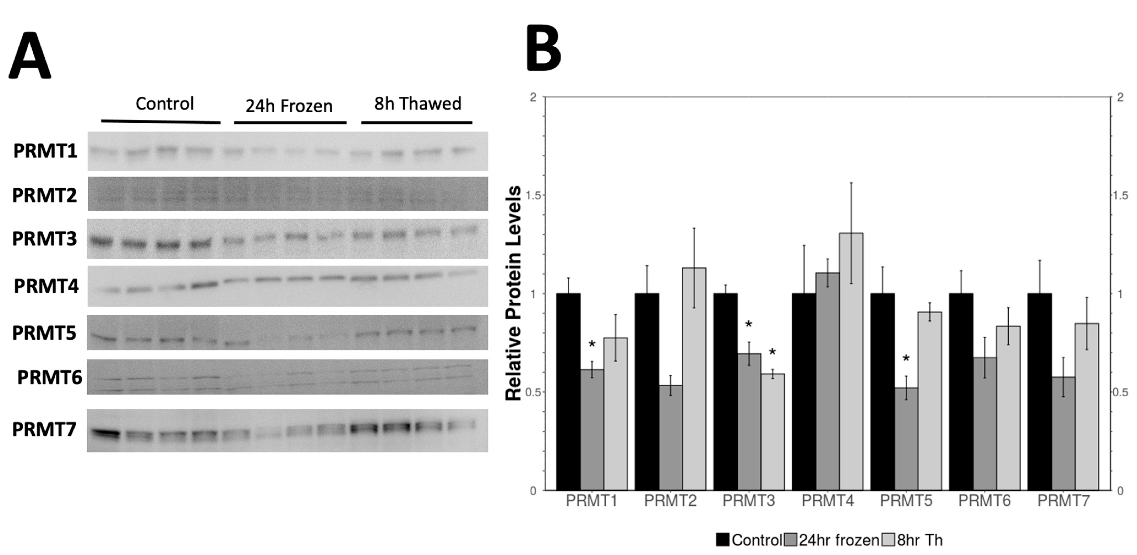
2.1. Expression of PRMTs in the Control, Frozen, and Thawed Samples

The relative protein levels of seven protein arginine methyltransferases (PRMT 1-7) were measured in the kidney of wood frogs, R. sylvatica, over the freeze–thaw cycle using Western blotting, comparing PRMT levels in kidneys from control frogs with those in 24 h frozen frogs and 8 h thawed frogs. PRMT1 was significantly downregulated after 24 h freezing (p < 0.05), falling to level of 0.60 ± 0.20 relative to control values whereas thawing led to an upward trend that was intermediate between control and frozen values. PRMT3 showed a significant downregulation after 24 h freezing compared to the control and remained downregulated after 8 h thawing. PRMT4 levels did not change over the freeze–thaw cycle but PRMT5 showed a significant downregulation after 24 h frozen as compared to control kidneys that was reversed after 8 h thawing. PRMT2, PRMT6, and PRMT7 showed a similar downward trend during freezing but the values did not meet statistical significance (Figure 1).

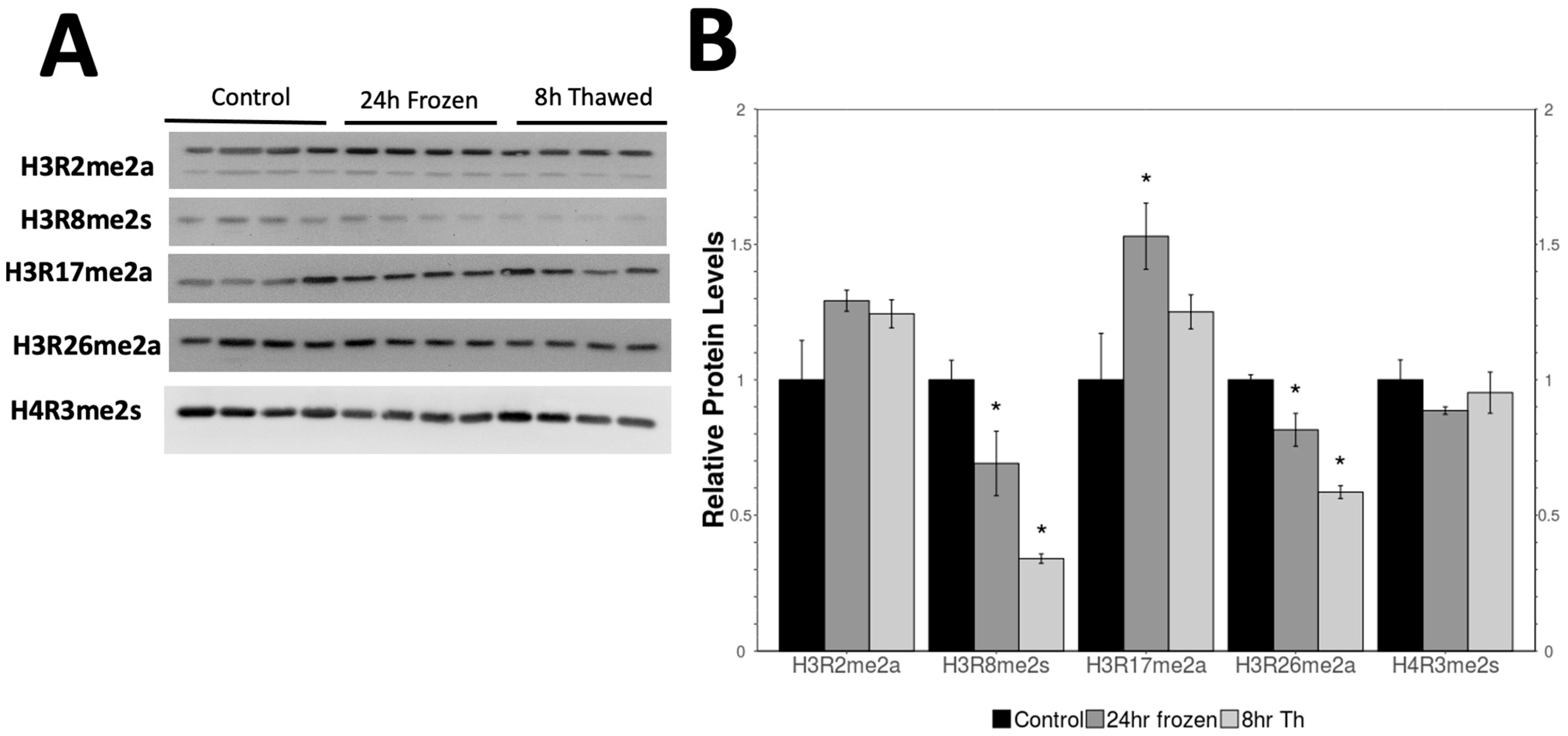
2.2. Analysis of Methylated Histone Levels over the Freeze–Thaw Cycle

Further investigation of the histone arginine methylation patterns in R. sylvatica kidney focused on the relative expression levels of various methylated histone marks: H3R2me2a, H3R8me2s, H3R17me2a, H3R26me2a, and H4R3me2s. Relative levels of five methylated histone marks (H3R2me2a, H3R8me2a, H3R17me2a, H3R26me2a, and H4R3me2s) on histone 3 and 4 proteins were assessed in histone protein extracts from kidney tissue under control, 24 h freezing, and 8 h thawing conditions. H3R8me2s showed no difference after 24 h freezing compared to control but was significantly reduced in 8 h thawed kidneys samples as compared to controls. H3R17me2a was significantly upregulated in 24 h frozen kidneys as compared to controls whereas the 8 h thawed kidneys showed no significant difference from either the control or frozen conditions. H3R26me2a levels showed a significant progressive decrease in 24 h frozen and 8 h thawed kidneys compared to the control. Both H3R2me2a and H4R3me2s showed no significant change across all conditions (Figure 2).

2.3. Expression of Readers and Demethylases

The relative expression levels of the methylated arginine readers (SMN1, SND1, TDRD3) and a demethylase (JMJD6) were also analyzed across the treatment conditions in the wood frog kidneys. SMN1 and TDRD3 showed no significant changes after freezing for 24 h or upon thawing. SND1 showed significant changes, where it was downregulated upon thawing, and JMJD6 was significantly downregulated upon freezing and stayed constant after thawing (Figure 3).

3. Discussion

Wood frogs (Rana sylvatica) are remarkable for their ability to survive whole-body freezing during harsh winter conditions, an adaptation that allows them to endure subzero temperatures. While frozen, the physiological activity of these frogs is drastically reduced (there is little or no activity in the brain, heart, or kidney). However, their survival is facilitated by the production of cryoprotectants, such as glucose and urea, which protect their organs during freezing [25,26]. During this period, the frogs rely entirely on internal energy reserves. Not only is foraging impossible, but blood circulation also halts due to intracellular ice formation, which prevents the distribution of metabolic fuels between organs. To survive, the frogs must carefully balance ATP generation and consumption, adjusting their metabolism to the duration of the freezing period.
Metabolic suppression is a key strategy employed by wood frogs during freezing. This involves downregulating non-essential processes, such as feeding, muscle movement, breathing, heartbeat, and neural activity, all of which are further suppressed by subzero temperatures [27,28,29]. At the molecular level, frogs utilize various epigenetic mechanisms to regulate these processes. Post-translational histone modifications, particularly methylation, play a crucial role in modulating gene expression and are integral to the regulation of metabolic machinery [7,19,30]. In this study, we aimed to examine the relative expression levels of histone arginine methylation, demethylation, and readers of arginine marks during the freeze–thaw cycle in wood frog kidneys, focusing on how these modifications contribute to the frogs’ adaptive response to freezing stress.
During freezing, extracellular ice formation disrupts inter-organ transport in wood frogs, including within the kidneys. Despite this, some intracellular processes, such as glycolysis, continue to provide limited metabolic support, maintaining critical functions. The kidneys, as vital organs responsible for filtration and waste excretion, experience compromised function due to the presence of extracellular ice. In response, the kidneys enter a hypometabolic state, selectively suppressing non-essential processes while preserving those necessary for tissue homeostasis [31]. The kidney’s ability to conserve energy through various cryoprotective mechanisms is essential for minimizing damage and ensuring survival.
Our immunoblotting data revealed that some PRMTs, as well as their associated methylated histone marks, were significantly altered during freezing and thawing. Specifically, PRMT1, PRMT3, and PRMT5 were downregulated after 24 h of freezing compared to control frogs, with PRMT3 remaining suppressed even after 8 h of thawing (Figure 1). These results suggest that the downregulation of these enzymes may serve to conserve energy during freezing, as they are involved in various cellular processes that require significant ATP consumption.
Further analysis of histone arginine methylation revealed differential regulation of specific methylation marks. For instance, H3R8me2s remained unchanged after 24 h of freezing but was significantly downregulated after 8 h of thawing. Conversely, H3R17me2a was upregulated after 24 h of freezing but returned to baseline levels after 8 h of thawing. H3R26me2a was significantly downregulated after both 24 h of freezing and 8 h of thawing (Figure 2). These changes in histone methylation suggest that different methyl marks may play distinct roles in gene regulation during freezing and thawing, with potential implications for the recovery of key metabolic pathways during thawing.

3.1. Role of Protein Arginine Methyltransferases (PRMTs) in Freezing Tolerance

PRMTs catalyze the methylation of arginine residues on histones and other proteins, and these modifications play key roles in cellular processes such as gene expression, RNA processing, and signal transduction [32]. In the context of freezing stress, the downregulation of PRMTs, including PRMT1, PRMT3, and PRMT5, suggests a strategy to conserve energy by suppressing transcriptional activity. PRMT1, the most studied of the PRMT family, catalyzes the formation of asymmetric and symmetric dimethylarginine, and has been implicated in regulating diverse cellular processes such as gene transcription, DNA repair, and signal transduction [32]. Similarly, PRMT2 and PRMT3 are involved in cell cycle regulation, cellular differentiation, and RNA splicing [33,34]. PRMT5, another important enzyme, plays a role in chromatin remodeling and RNA metabolism [35].
The downregulation of PRMTs in wood frog kidneys implicates that these enzymes’ effects in maintaining cellular function and gene expression are suppressed to conserve energy under freezing stress. This pattern is consistent with previous findings in other systems (liver, skeletal muscle, and brain), where lysine methyltransferases involved in active transcription are downregulated during metabolic depression, while those linked to gene silencing are maintained or upregulated [23]. The changes in PRMT expression observed here highlight the potential involvement of arginine methylation in freezing tolerance and metabolic reorganization, particularly in renal tissues. Given the importance of PRMTs in gene regulation, the downregulation of these enzymes may be part of a broader strategy to reduce metabolic activity during freezing while still maintaining the functions essential for survival. This is consistent with recent findings that show that histone methylation and demethylation in wood frog kidneys undergo significant changes during freezing and thawing, with key transcriptional repressors downregulated during freezing to conserve energy and facilitate survival [24].
Interestingly, PRMT1 has been shown to play a role in diabetic kidney injury, where it contributes to oxidative stress and inflammation [36,37] In the context of wood frog freezing, the downregulation of PRMT1 may serve to protect kidney cells from oxidative damage during the freezing process. Similarly, PRMT5, known for its role in regulating RNA splicing and gene expression, may contribute to the adaptation of kidney cells to freezing stress by modulating metabolic pathways [37].

3.2. Histone Arginine Methylation and Gene Regulation During Freezing Stress

Our study also focused on specific histone arginine methylation marks, which are key regulators of gene expression. The upregulation of H3R17me2a after 24 h of freezing suggests that this mark may be involved in the activation of genes critical for freezing survival. In contrast, the downregulation of H3R26me2a after both freezing and thawing indicates that this mark may be involved in silencing genes related to energy-consuming processes during freezing and reactivating them during thawing. The persistence of H3R8me2a after freezing, followed by its downregulation during thawing, suggests a potential role for this mark in recovery following freezing stress.
The dynamic regulation of histone arginine methylation is crucial for the control of gene expression under physiological stress, as shown in the freeze tolerance mechanisms of R. sylvatica. Among the various histone marks, H3R8me2s, H3R17me2a, and H3R26me2a serve as key markers of chromatin remodeling, influencing transcriptional activity in response to freezing stress. H3R8me2s, primarily catalyzed by PRMT1, PRMT2, and PRMT3, is typically associated with transcriptional repression [38]. This mark is known to promote a closed chromatin configuration, effectively reducing transcription factor accessibility and limiting the expression of genes not essential for survival during extreme stress. In contrast, H3R17me2a, predominantly mediated by PRMT1 and PRMT3, is linked to transcriptional activation [38]. This modification facilitates chromatin decompaction, enhancing accessibility for transcriptional machinery and promoting the expression of survival-related genes, particularly under stress conditions such as freezing. The upregulation of H3R17me2a observed during the freezing period in wood frog kidneys suggests its role in activating critical metabolic and stress response pathways. Conversely, H3R26me2a, also catalyzed by PRMT1 and PRMT5, is associated with transcriptional repression and the maintenance of a quiescent chromatin state [39]. The sustained downregulation of H3R26me2a observed after thawing in wood frogs may reflect an adaptive strategy to conserve energy by suppressing non-essential gene expression during recovery. Interestingly, the downregulation of SND1 during recovery is consistent with the fact that this enzyme is strongly linked to cancer [40] which is an ATP-expensive process. Once the frog starts to thaw, cellular processes must be rapidly restored, including the reactivation of transcription, translation, and other molecular processes. Downregulation of SND1 could also be an effort to minimize any leftover, unnecessary gene expression processes that might otherwise hinder the re-establishment of normal cellular function. It is therefore important that the presence of this protein be suppressed to further conserve energy. Collectively, these modifications highlight the importance of PRMT-mediated methylation in maintaining the balance between gene activation and repression during freezing stress. The methylation/demethylation cycles of these arginine marks facilitate a coordinated response to environmental stressors by fine-tuning the expression of genes involved in energy conservation, stress adaptation, and tissue protection, while minimizing the energy expenditure required for non-essential cellular processes. This regulation highlights the critical role histone arginine methylation plays in the epigenetic control of gene expression, particularly in organisms like wood frogs that rely on adaptive responses to extreme environmental challenges.

4. Materials and Methods

4.1. Ethics Statement

All animal protocols were approved by the Carleton University Animal Care Committee (Protocol #106935) within the guidelines of the Canadian Council on Animal Care.

4.2. Animal Collection

Male wood frogs (R. sylvatica) were collected during the spring breeding season from meltwater ponds around Ottawa, Ontario, Canada. Animals were conveyed to Carleton University on crushed ice or snow containers. Upon arrival, the frogs underwent a tetracycline bath before being placed in plastic containers with moist sphagnum moss. They were allowed to acclimate to laboratory conditions for at least one week in an incubator set at 5 °C, which closely matched the temperature of their natural pond habitat. After this acclimation period, the control frogs were euthanized and sampled. For freezing exposure, the remaining frogs were placed in sealed plastic basins with a damp paper towel lining. These frogs were then exposed to −4.0 °C for 45 min, a treatment that induced ice formation in their bodies. Afterward, the temperature was gradually raised to −2.5 °C and the frogs were kept at this temperature for 24 h. After this freezing period, half of the frogs were randomly selected for sampling. The other half was returned to the 5 °C incubator for 8 h to thaw and recover before being euthanized and sampled. Frogs from the control, 24 h frozen, and 8 h thawed groups were euthanized by pithing. Kidneys were quickly excised, frozen in liquid nitrogen, and stored at −80 °C until further analysis.

4.3. Total Protein Isolation

Total protein was extracted from approximately 50 mg of frozen kidney tissue from five individual frogs per condition (control, frozen, thawed) following the protocol outlined in previous studies [16,23]. The tissues were weighed and then pulverized using a mortar and pestle under liquid nitrogen. The resulting powdered samples were homogenized with a P10 homogenizer in an ice-cold buffer (20 mM HEPES, 200 mM NaCl, 0.1 mM EDTA, 10 mM NaF, 1 mM Na3VO4, and 10 mM β-glycerophosphate, pH 7.4) at a ratio of 1:5 w:v. The homogenization buffer also contained small amounts of the protease inhibitor phenylmethylsulfonyl fluoride (PMSF) and a protease inhibitor cocktail (AEBSF, aprotinin, bestatin, E64 & leupeptin; Catalogue # 808 PIC001.1, BioShop Canada Inc., Burlington, ON, Canada).
The tissue homogenates were centrifuged at 10,000× g for 15 min at 4 °C to separate the soluble proteins, which were collected from the supernatant. Protein concentration in each sample was quantified using the BioRad protein assay kit (Catalogue #500-0002; BioRad Laboratories, Hercules, CA, USA) by measuring absorbance at 595 nm on an MR5000 microplate reader (Dynatech Laboratories, Chantilly, VA, USA). After the determination of protein concentration, all samples were adjusted to 10 μg/μL by adding appropriate volumes of homogenization buffer.
For protein analysis, equal volumes of each sample were mixed with an equal volume of SDS-Tris buffer (100 mM Tris-base, 4% SDS (w/v), 20% glycerol (v/v), 0.2% bromophenol blue (w/v), and 10% 2-mercaptoethanol (v/v), pH 6.8) to achieve a final concentration of 5 μg/μL. The samples were then denatured by boiling for 10 min in a water bath. After cooling, the samples were stored at −40 °C until further use.

4.4. Histone Isolation

This protocol follows a method described by [41]. Briefly, Histone proteins were extracted from frozen kidney tissue samples (~100–200 mg) collected from control, 24 h frozen, and 8 h thawed wood frogs (n = 5 biological replicates per condition). After quickly weighing the tissues, the samples were homogenized using a Dounce homogenizer in the presence of 4 volumes of Triton Extraction Buffer (TEB), which consists of phosphate-buffered saline (PBS) containing 0.5% Triton X-100 (v/v), 0.02% sodium azide (w/v), 10 µL/mL protease inhibitor cocktail, 10 mM β-glycerophosphate, and 1 mM sodium orthovanadate. The homogenization was followed by brief vortexing and incubation on ice for 30 min.
The homogenates were then centrifuged at 6000 × g for 10 min at 4 °C, and the supernatant was discarded. The resulting pellet was washed with fresh TEB buffer and centrifuged again under the same conditions. After discarding the supernatant, the pellet containing nuclei was resuspended in 400 µL of 0.2 M H2SO4, vortexed, and incubated on ice for at least 30 min.
Following incubation, the samples were centrifuged at 12,000 rpm for 10 min at 4 °C, and the supernatant, containing histones, was carefully transferred to new tubes. To precipitate the histones, 132 µL of 100% trichloroacetic acid (TCA) was added to the supernatant to achieve a final concentration of 33%. The samples were then incubated on ice for 30 min, followed by centrifugation at 12,000 rpm for 10 min at 4 °C. The supernatant was discarded, and the histone pellet was washed with ice-cold acetone to remove excess acid without dissolving the pellet. This step was repeated by centrifuging at 12,000 rpm for 5 min at 4 °C, followed by careful removal of the supernatant. The pellets were air-dried for 20 min at room temperature.
The dried histone pellets were resuspended in 100 µL of deionized water (ddH2O) and sonicated to maximize histone protein yield. Histone concentrations were then determined, and the samples were analyzed by immunoblotting to confirm successful histone protein isolation.

4.5. Western Immunoblotting

Equal amounts of protein from kidney samples of control, 24 h frozen, and 8 h thawed Rana sylvatica frogs (15–40 μg/mL, depending on the target protein) were loaded onto 6–15% SDS-polyacrylamide gels. The percentage of acrylamide in the resolving gel was adjusted according to the molecular weight of the protein being analyzed. A molecular weight marker, 3 μL of the BLUeye prestained protein ladder (10–245 kDa; Catalogue #PM007-0500, FroggaBio, Toronto, ON, Canada), was included in separate lanes.
The upper stacking gel (pH 6.8) was composed of 5% acrylamide (v/v) in 1 M Tris buffer, with 0.1% SDS, 0.1% ammonium persulfate (APS), and 0.1% TEMED (N,N,N′,N′-tetramethylethane-1,2-diamine). The resolving gels (pH 8.8) consisted of 8–15% acrylamide (v/v) in 1.5 M Tris buffer, also containing 0.1% SDS, 0.1% APS, and 0.1% TEMED. The gels were run in a BioRad Mini Protean III system (BioRad Laboratories, Hercules, CA, USA) at 180 V for 30 to 180 min using running buffer (25 mM Tris-base, 190 mM glycine, 0.1% SDS, pH 7.6).
After electrophoresis, the proteins were transferred to 0.45 μm pore PVDF membranes by electroblotting at room temperature for 45 to 180 min at 160 mA, using 1X transfer buffer (25 mM Tris-base, 192 mM glycine, 10% methanol, pH 8.5). To reduce non-specific binding of antibodies, the membranes were incubated with either (a) 1–10% skimmed milk or (b) 1 mg/mL polyvinyl alcohol (PVA), MW 30,000–70,000, in 1X TBST (20 mM Tris-base, 140 mM NaCl, 0.05% Tween-20) at room temperature for 30 min.
Next, the membranes were incubated with primary antibodies (1:1000 dilution in 1X TBST) at 4 °C overnight. For detection, after primary antibody incubation, the membranes were washed three times with 1X TBST (5 min each) and then incubated with HRP-conjugated anti-rabbit secondary antibodies (1:8000 dilution in TBST; Catalogue #APA002P, BioShop Canada Inc., Burlington, ON, Canada) at room temperature for 30 min. The membranes were washed again with 1X TBST (3 × 5 min) and protein signals were visualized using chemiluminescence (1:1 v/v H2O2 and luminol) on a ChemiGenius Bio Imaging System (Syngene, Frederick, MD, USA).
Finally, to confirm equal loading, the membranes were stained with Coomassie brilliant blue (0.25% w/v, 7.5% acetic acid, 50% methanol) to visualize total protein bands.
The antibodies used in this study were purchased from commercial suppliers. The following is a list of suppliers and catalogue numbers: GeneTex, San Antonio, TX, USA (PRMT1, GTx128211; PRMT2, GTx103749; PRMT3, GTx116478; PRMT4, GTx116004), ABclonal Technology, Woburn, MA, USA (PRMT5, A2290; PRMT6, A7814; PRMT7, A12159; H3R2me2a, A2375; H3R17me2a, A2421; H3R26me2a, A22375; PADI4, A1906; JMJD6, A5840; TDRD3, A6000; SND1, A5874; SMN1, A16246), myBiosource, San Diego, CA, USA (H4R3me2s, MBS126222).

4.6. Statistical Analysis

The intensity of chemiluminescent protein bands on immunoblots was quantified using densitometry with the ChemiGenius Bio Imaging System and GeneTools Software (version 4.3.8.0, Syngene, Frederick, MD, USA). Band intensities were normalized to the combined intensity of Coomassie blue-stained bands from the same lane, which showed no differential expression between experimental conditions and were clearly separated from the target immunoblot bands. Data for each experimental group are presented as mean ± SEM, with n = 4 biological replicates per condition. Statistical analysis was performed using one-way ANOVA followed by Dunnett’s post hoc test, with a significance threshold set at p < 0.05. All statistical calculations were carried out using the RBioPlot software package [42].

5. Conclusions

In conclusion, this study provides new insights into the regulation of histone arginine methylation and PRMT expression in wood frog kidneys during freezing and thawing. The downregulation of PRMTs and the differential regulation of specific histone marks suggest that these modifications may play a key role in the frog’s ability to adapt to freezing stress. By regulating protein levels and conserving energy, these epigenetic mechanisms likely contribute to the survival of wood frogs in harsh winter environments. While this study focused on protein expression, further research is needed to fully understand the functional significance of these epigenetic modifications in freeze tolerance, particularly by exploring the downstream effects on cellular pathways. This work lays the foundation for future investigations into the role of protein-based epigenetic regulation in physiological adaptation to extreme environmental conditions, with potential implications for both biomedical research and conservation strategies.

Author Contributions

Conceptualization, O.O.T. and K.B.S.; methodology, O.O.T.; software, O.O.T.; validation, O.O.T.; formal analysis, O.O.T.; investigation, O.O.T.; resources, K.B.S.; data curation, O.O.T.; writing—original draft preparation, O.O.T.; writing—review and editing, O.O.T. and K.B.S.; visualization, O.O.T.; supervision, K.B.S.; project administration, K.B.S.; funding acquisition, K.B.S. All authors have read and agreed to the published version of the manuscript.

Funding

This study was funded by a NSERC Discovery Grant to KBS (RGPIN-2020-04733).

Institutional Review Board Statement

All animals were cared for in accordance with the guidelines of the Canadian Council on Animal Care and experimental procedures had the prior approval of the Carleton University Animal Care Committee (protocol #106935).

Data Availability Statement

The data that support the findings of this study are available from the corresponding author upon reasonable request.

Acknowledgments

We thank J.M. Storey for the editorial review.

Conflicts of Interest

The authors declare no competing interests.

References

  1. Gibney, E.R.; Nolan, C.M. Epigenetics and gene expression. Heredity 2010, 105, 4–13. [Google Scholar] [CrossRef] [PubMed]
  2. Berger, S.L. Histone modifications in transcriptional regulation. Curr. Opin. Genet. Dev. 2002, 12, 142–148. [Google Scholar] [CrossRef] [PubMed]
  3. Kouzarides, T. Histone methylation in transcriptional control. Curr. Opin. Genet. Dev. 2002, 12, 198–209. [Google Scholar] [CrossRef] [PubMed]
  4. Strahl, B.D.; Allis, C.D. The language of covalent histone modifications. Nature 2000, 403, 41–45. [Google Scholar] [CrossRef] [PubMed]
  5. Zhu, Q.; Wani, A.A. Histone Modifications: Crucial Elements for Damage Response and Chromatin Restoration. J. Cell. Physiol. 2010, 223, 283. [Google Scholar] [CrossRef]
  6. Martin, C.; Zhang, Y. The diverse functions of histone lysine methylation. Nat. Rev. Mol. Cell Biol. 2005, 6, 838–849. [Google Scholar] [CrossRef]
  7. Zhang, Y.; Sun, Z.; Jia, J.; Du, T.; Zhang, N.; Tang, Y.; Fang, Y.; Fang, D. Overview of Histone Modification. Adv. Exp. Med. Biol. 2021, 1283, 1–16. [Google Scholar] [CrossRef]
  8. Hyun, K.; Jeon, J.; Park, K.; Kim, J. Writing, erasing and reading histone lysine methylations. Exp. Mol. Med. 2017, 49, e324. [Google Scholar] [CrossRef]
  9. Miller, J.L.; Grant, P.A. The Role of DNA Methylation and Histone Modifications in Transcriptional Regulation in Humans. Sub-Cell. Biochem. 2013, 61, 289. [Google Scholar] [CrossRef]
  10. Zhang, J.; Jing, L.; Li, M.; He, L.; Guo, Z. Regulation of histone arginine methylation/demethylation by methylase and demethylase. Mol. Med. Rep. 2019, 19, 3963. [Google Scholar] [CrossRef]
  11. Wang, T.S.; Cheng, J.K.; Lei, Q.Y.; Wang, Y.P. A Switch for Transcriptional Activation and Repression: Histone Arginine Methylation. In RNA Technologies; Springer: Cham, Switzerland, 2019; pp. 521–541. [Google Scholar] [CrossRef]
  12. Lorton, B.M.; Shechter, D. Cellular consequences of arginine methylation. Cell. Mol. Life Sci. 2019, 76, 2933–2956. [Google Scholar] [CrossRef]
  13. Litt, M.; Qiu, Y.; Huang, S. Histone arginine methylations: Their roles in chromatin dynamics and transcriptional regulation. Biosci. Rep. 2009, 29, 131. [Google Scholar] [CrossRef] [PubMed]
  14. Hwang, J.W.; Cho, Y.; Bae, G.U.; Kim, S.N.; Kim, Y.K. Protein arginine methyltransferases: Promising targets for cancer therapy. Exp. Mol. Med. 2021, 53, 788–808. [Google Scholar] [CrossRef] [PubMed]
  15. Bannister, A.J.; Kouzarides, T. Regulation of chromatin by histone modifications. Cell Res. 2011, 21, 381–395. [Google Scholar] [CrossRef] [PubMed]
  16. Al-attar, R.; Storey, K.B. Lessons from nature: Leveraging the freeze-tolerant wood frog as a model to improve organ cryopreservation and biobanking. Comp. Biochem. Physiol. Part B Biochem. Mol. Biol. 2022, 261, 110747. [Google Scholar] [CrossRef]
  17. Luu, B.E.; Storey, K.B. Solving Donor Organ Shortage with Insights from Freeze Tolerance in Nature: Activating endogenous antioxidant systems with non-coding RNA to precondition donor organs. BioEssays News Rev. Mol. Cell. Dev. Biol. 2018, 40, 1800092. [Google Scholar] [CrossRef]
  18. Bedford, M.T.; Richard, S. Arginine methylation an emerging regulator of protein function. Mol. Cell 2005, 18, 263–272. [Google Scholar] [CrossRef]
  19. Di Lorenzo, A.; Bedford, M.T. Histone arginine methylation. FEBS Lett. 2011, 585, 2024–2031. [Google Scholar] [CrossRef]
  20. Chang, B.; Chen, Y.; Zhao, Y.; Bruick, R.K. JMJD6 is a histone arginine demethylase. Science 2007, 318, 444–447. [Google Scholar] [CrossRef]
  21. Wang, S.; Wang, Y. Peptidylarginine deiminases in citrullination, gene regulation, health and pathogenesis. Biochim. Biophys. Acta 2013, 1829, 1126–1135. [Google Scholar] [CrossRef]
  22. Walport, L.J.; Hopkinson, R.J.; Chowdhury, R.; Schiller, R.; Ge, W.; Kawamura, A.; Schofield, C.J. Arginine demethylation is catalysed by a subset of JmjC histone lysine demethylases. Nat. Commun. 2016, 7, 11974. [Google Scholar] [CrossRef]
  23. Hawkins, L.J.; Storey, K.B. Histone methylation in the freeze-tolerant wood frog (Rana sylvatica). J. Comp. Physiol. B Biochem. Syst. Environ. Physiol. 2018, 188, 113–125. [Google Scholar] [CrossRef] [PubMed]
  24. Taiwo, O.O.; Breedon, S.A.; Storey, K.B. Epigenetic Regulation by Histone Methylation and Demethylation in Freeze-Tolerant Frog Kidney. Cell Biochem. Funct. 2024, 42, e70036. [Google Scholar] [CrossRef] [PubMed]
  25. Costanzo, J.P.; Lee, R.E. Cryoprotection by urea in a terrestrially hibernating frog. J. Exp. Biol. 2005, 208 Pt 21, 4079–4089. [Google Scholar] [CrossRef] [PubMed]
  26. Storey, K.B. Glycolysis and the regulation of cryoprotectant synthesis in liver of the freeze tolerant wood frog. J. Comp. Physiol. B Biochem. Syst. Environ. Physiol. 1987, 157, 373–380. [Google Scholar] [CrossRef]
  27. Do Amaral, M.C.F.; Lee, R.E.; Costanzo, J.P. Enzymatic Regulation of Glycogenolysis in a Subarctic Population of the Wood Frog: Implications for Extreme Freeze Tolerance. PLoS ONE 2013, 8, e79169. [Google Scholar] [CrossRef]
  28. Sinclair, B.J.; Stinziano, J.R.; Williams, C.M.; MacMillan, H.A.; Marshall, K.E.; Storey, K.B. Real-time measurement of metabolic rate during freezing and thawing of the wood frog, Rana sylvatica: Implications for overwinter energy use. J. Exp. Biol. 2013, 216, 292–302. [Google Scholar] [CrossRef]
  29. Storey, K.B.; Storey, J.M. Molecular Physiology of Freeze Tolerance in Vertebrates. Physiol. Rev. 2017, 97, 623–665. [Google Scholar] [CrossRef]
  30. Sims, R.J.; Nishioka, K.; Reinberg, D. Histone lysine methylation: A signature for chromatin function. Trends Genet. 2003, 19, 629–639. [Google Scholar] [CrossRef]
  31. Gorr, T.A. Hypometabolism as the ultimate defence in stress response: How the comparative approach helps understanding of medically relevant questions. Acta Physiol. 2017, 219, 409–440. [Google Scholar] [CrossRef]
  32. Gui, S.; Gathiaka, S.; Li, J.; Qu, J.; Acevedo, O.; Hevel, J.M. A Remodeled Protein Arginine Methyltransferase 1 (PRMT1) Generates Symmetric Dimethylarginine. J. Biol. Chem. 2014, 289, 9320. [Google Scholar] [CrossRef] [PubMed]
  33. Bedford, M.T.; Clarke, S.G. Protein Arginine Methylation in Mammals: Who, What, and Why. Mol. Cell 2009, 33, 1–13. [Google Scholar] [CrossRef] [PubMed]
  34. Tewary, S.K.; Zheng, Y.G.; Ho, M.C. Protein arginine methyltransferases: Insights into the enzyme structure and mechanism at the atomic level. Cell. Mol. Life Sci. 2019, 76, 2917–2932. [Google Scholar] [CrossRef]
  35. Stopa, N.; Krebs, J.E.; Shechter, D. The PRMT5 arginine methyltransferase: Many roles in development, cancer and beyond. Cell. Mol. Life Sci. 2015, 72, 2041–2059. [Google Scholar] [CrossRef] [PubMed]
  36. Mei, X.; Zeng, J.; Liu, D.-F.; Zhao, Y.; Yang, H.-L.; Li, Y.; Qiu, P.; Tang, M.-W. Abnormalities of the PRMT1-ADMA-DDAH1 metabolism axis and probucol treatment in diabetic patients and diabetic rats. Ann. Palliat. Med. 2021, 10, 3343–3353. [Google Scholar] [CrossRef]
  37. Zhou, X.; Chen, H.; Li, J.; Shi, Y.; Zhuang, S.; Liu, N. The Role and Mechanism of Lysine Methyltransferase and Arginine Methyltransferase in Kidney Diseases. Front. Pharmacol. 2022, 13, 1488. [Google Scholar] [CrossRef]
  38. Qin, J.; Xu, J. Arginine methylation in the epithelial-to-mesenchymal transition. FEBS J. 2021, 289, 7292. [Google Scholar] [CrossRef]
  39. Xu, J.; Richard, S. Cellular Pathways Influenced by Protein Arginine Methylation: Implications for Cancer. Mol Cell 2021, 81, 4357. [Google Scholar] [CrossRef]
  40. Liu, S.; Hao, X.; Miao, D.; Zhang, Y. A Study on the Binding Mechanism and the Impact of Key Residue Mutations between SND1 and MTDH Peptide through Molecular Dynamics Simulations. J. Phys. Chem. B 2024, 128, 9074–9085. [Google Scholar] [CrossRef]
  41. Bloskie, T.; Taiwo, O.O.; Storey, K.B. Reversible Histone Modifications Contribute to the Frozen and Thawed Recovery States of Wood Frog Brains. Biomolecules 2024, 14, 839. [Google Scholar] [CrossRef]
  42. Zhang, J.; Storey, K.B. RBioplot: An easy-to-use R pipeline for automated statistical analysis and data visualization in molecular biology and biochemistry. PeerJ 2016, 4, e2436. [Google Scholar] [CrossRef] [PubMed]
Figure 1. Relative protein levels of arginine methyl transferase enzymes in R. sylvatica kidneys as determined by Western immunoblotting. (A) Representative Western blots for individual PRMTs under each experimental condition. Data were analyzed using a one-way ANOVA with Dunnett’s post hoc test. (B) Histogram showing mean (±SEM, n = 4) standardized expression levels of PRMT1, PRMT2, PRMT3, PRMT4, PRMT5, PRMT6, and PRMT7 under control, 24 h freezing, and 8 h thawing conditions. Data are mean ± SEM (n = 4 independent trials). Statistical significance for freezing and thawing values, relative to the standardized control, was determined using one-way analysis of variance (ANOVA) with Dunnett’s post hoc test (*—p < 0.05).
Kinasesphosphatases 03 00001 g001
Figure 2. Relative protein levels of methylated arginine residues in R. sylvatica kidneys as determined by Western immunoblotting. (A) Representative Western blots for individual histone marks under each experimental condition. Data were analyzed using a one-way ANOVA with Dunnett’s post hoc test. (B) Histogram showing mean (±SEM, n = 4) standardized expression levels of H3R2me2a, H3R8me2a, H3R17me2a, H3R26me2a, and H4R3me2s under control, 24 h freezing, and 8 h thawing conditions. For each histone mark, statistical significance for freezing and thawing values, relative to the standardized control, was determined using one-way ANOVA with Dunnett’s post hoc test (*—p < 0.05).
Kinasesphosphatases 03 00001 g002
Figure 3. Relative protein levels of methylated arginine readers and a demethylase enzyme present in R. sylvatica kidneys. (A) Representative Western blots for individual enzyme across each experimental condition. Data were analyzed using a one-way ANOVA with Dunnett’s post hoc test. a, b– values that share the same letter are not significantly different from one another. (B) Histogram showing mean standardized expression levels of SMN1, SND1, TDRD3, and JMJD6 under control, 24 h freezing, and 8 h thawing conditions where n = 4. For each enzyme, statistical significance for freezing and thawing values, relative to the standardized control, was determined using one-way ANOVA with Dunnett’s post hoc test (*—p < 0.05).
Kinasesphosphatases 03 00001 g003
Disclaimer/Publisher’s Note: The statements, opinions and data contained in all publications are solely those of the individual author(s) and contributor(s) and not of MDPI and/or the editor(s). MDPI and/or the editor(s) disclaim responsibility for any injury to people or property resulting from any ideas, methods, instructions or products referred to in the content.

