Abstract
Post-transplant lymphoproliferative disorder (PTLD) is the most common malignancy in adults who receive solid organ transplantation (SOT), apart from skin cancer. It is a serious and potentially fatal complication of chronic immunosuppression (ISI) in SOT recipients. This report describes a 20-year (2001–2021) clinicopathological experience with 59 PTLD patients at an urban center. The median time from transplant to PTLD was 8.5 years and the most common types of transplants were kidney (41%) and liver (31%). Epstein–Barr encoding region (EBER) was positive in 51% tumors, and 50% patients had Epstein–Barr virus (EBV) viremia at diagnosis. Overall survival (OS) at 1 year and 5 years was 78% and 64%, respectively. OS was significantly (p < 0.05) shorter in males (hazard ratio [HR] 3.7), certain organ transplants (lung HR 10.4; liver HR 3.9 relative to kidney), PTLD diagnosed within 12 months of transplant (HR 4.1), multi-organ involvement at diagnosis (HR 7.1), vitamin D deficiency at diagnosis (HR 4.5), and low serum albumin level at diagnosis (HR 3.6). Our study highlights the prognostic factors of PTLD and corroborates improved PTLD outcomes in the past 20 years.
1. Background
Post-transplant lymphoproliferative disease (PTLD) is a heterogeneous clinical and pathologic group of lymphoid disorders ranging from indolent polyclonal proliferation to aggressive monomorphic lymphoma that may complicate solid organ transplantation (SOT) or hematopoietic stem cell transplantation (HSCT) [1]. The spectrum of lymphoid proliferations ranges from Epstein–Barr virus (EBV)-positive or EBV-negative B cell lymphomas or T/NK cell lymphomas, as well as classic Hodgkin lymphoma. PTLD is the second most common malignancy in SOT recipients after non-melanoma skin cancers and is the most common cause of cancer-related death in SOT recipients [2,3]. While the overall incidence of lymphoproliferative disease is approximately 1% in the transplant population, the incidence varies dramatically with the type of allograft [4,5]. PTLD is seen in up to 10–15% of all SOT adult recipients, with the frequency being higher in multi-organ and intestinal transplantation (20%), followed by the lungs (5.7 per 1000 person-years), liver (2.4), heart (2.2), and kidney transplants (1.6), owing in part to the degree of immunosuppression (ISI) warranted in these cases [6,7].
Several risk factors have been elucidated over the past decade; further validation and analysis of these factors is needed to enrich the prediction of PTLD development. EBV pre-transplant status of the recipient and donor, along with age of the recipient, degree and duration of ISI, and type of organ transplanted are identified as strong risk factors for PTLD development [1,8,9]. CMV donor–recipient mismatch and other infections such as Hepatitis C virus (HCV) and Human Herpes Virus-8 (HHV-8) were also reported as risk factors, especially when they coincide with EBV infection [1,10,11].
About 55–65% of PTLD cases after SOT are associated with EBV infection, although this has not been associated with differences in response to therapy or survival, and these cases are treated similarly to EBV-negative (EBV–) PTLD patients [1,12,13,14]. It is postulated that ISI leads to depressed T cell function with associated lack of T cell control of B cell proliferation, resulting in uncontrolled proliferation of EBV-transformed B cells [1]. Several single-center analyses reported that pre-transplant EBV seronegativity can increase the incidence of PTLD by 10- to 76-fold when compared to EBV-seropositive (EBV+) patients due to primary EBV [12,13].
While the prognosis varies with clonality and the extent of the disease, historically, mortality rates in SOT-related PTLD were 50–70% and up to 70–90% in HSCT [12]. However, recent data suggest that outcomes have significantly improved [15,16]. Over 46,000 transplants were completed in the US in 2023, with transplantation numbers continuing to rise yearly after stagnation in the past decade [17,18]. Prognostication and optimization of the approach to PTLD is of importance because the prevalence of PTLD is expected to rise as transplant numbers continue to increase [12]. There is a paucity of information concerning the prognostic factors of patients with PTLD, and there are no validated prognostic risk stratification tools.
2. Results
2.1. Patient Demographics; Clinical and Transplant History
Our search identified 59 adult patients with PTLD who were evaluated at the UC Cancer Center from January 2001 to January 2021. The majority were males (66%) with a median age at transplant of 46.3 (9.3–73.1) years (Table 1). All patients had received SOT, mostly kidney (41%) or liver (31%) allografts. Diabetes was the most frequent indication for a transplant. Most donors were deceased males with a median age of 41.0 (21.0–69.0) years. Median follow-up was 17 (95% CI 10–19) years.

Table 1.
Baseline patient and donor demographics in relation to transplant. Continuous data are represented as medians (IQR). All categorical data are presented as numbers (percent). Percentages were rounded to nearest decimal point.
2.2. PTLD Characteristics
The median time from transplant to PTLD diagnosis was 8.5 (range 0.1–32.9) years, and the median age at diagnosis was 55.0 (range 10.0–82.0) years (Table 2). The majority had monomorphic histological type (80%), and two-thirds had extra nodal involvement, with the gastrointestinal (GI) tract (35%) being the most frequently involved site. The most common ISIs were CNI, antimetabolite agents, and steroids in varying combinations.

Table 2.
PTLD distribution and clinical manifestations at diagnosis. Continuous data are represented as medians (IQR). All categorical data are presented as numbers (percent). Percentages were rounded to the nearest decimal point.
Epstein–Barr encoding region in situ hybridization status (EBER-ISH) was positive in 51%, and 50% had EBV viremia at diagnosis (Table 2). Among 24 patients who had quantitative EBV viral load (via PCR) available, the median EBV viral load was 1486 (range 200–1,000,000) IU/mL, and 58% had viral load > 1000 IU/mL. Discordance was observed between tumor EBER-ISH status and whole blood EBV DNA in limited patients. There were three tumor EBER-ISH-positive cases that were serum EBV PCR-negative, and there were three EBER-ISH-negative cases that showed EBV PCR positivity.
2.3. Treatment and Outcomes
ISI was changed in 76% of patients after PTLD diagnosis (Table 3). Additional treatments included ones with rituximab alone (20%) or with chemotherapy (56%), with two-thirds (66%) achieving a CR or PR. The most common type of chemotherapy used with rituximab was a combination of cyclophosphamide, doxorubicin, and vincristine (R-CHOP), which was given at the same time as rituximab. At the time of the last follow-up, 34% had died. The median time from PTLD diagnosis to death was 5.3 months. Four patients were noted to have relapsed in the group at last follow-up.

Table 3.
Management of PTLD and patient outcomes. Continuous data are represented as medians (IQR). All categorical data are presented as numbers (percent). Percentages were rounded to the nearest decimal point.
2.4. Time from Transplant to PTLD
Lung transplant recipients had the shortest mean time from transplant to PTLD diagnosis (3.3 years), followed by liver (6.9 years) and kidney transplant recipients (11.3 years), p = 0.0581 (Table 4). Deceased donor (6.3 vs. 10.6 years) and donor–recipient EBV mismatch (1.5 vs. 6.5 years) were also noted to be associated with shorter time to PTLD, but with a p > 0.05.

Table 4.
Time from transplant to PTLD diagnosis in relationship with patient, donor, and transplant variables by univariate gamma regression models.
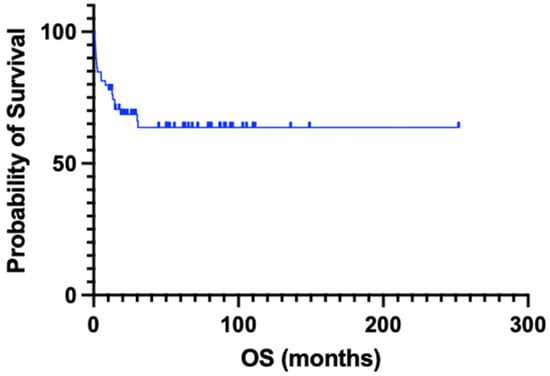
Median OS as measured by Kaplan–Meier analysis was not reached at data cutoff (December 2021). OS at 1 year was 77.9%, 68.6% at 2 years, and 63.7% at 5 years (Figure 1). When the data were available, the most common cause of death was disease, that is, PTLD (Table 3).

Figure 1.
Overall survival (OS) by Kaplan–Meier curves for the entire PTLD cohort. OS at 1 year, 2 years, and 5 years was 77.9%, 68.6%, and 63.7%, respectively.
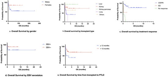
OS was significantly shorter in males (hazard ratio [HR] 3.7, p = 0.0401), for certain transplant types (lung, HR 10.4, p = 0.0013; liver, HR 3.9, p = 0.0236, when compared to kidney transplant), and in those with PTLD diagnosis within 12 months of transplant (HR 4.1, p = 0.0211), multiple organ involvement at diagnosis (HR 7.1, p = 0.0246), presentation with end-organ dysfunction (HR 3.0, p = 0.0319), vitamin D deficiency at diagnosis (HR 4.5, p = 0.0221), low serum albumin level at diagnosis (HR 3.6, p = 0.0002), and no response to treatment when compared with CR (HR 8.8, p = 0.0001), as seen in Table 5 and Figure 2.

Table 5.
Univariate analysis of overall survival by patient, transplant, and PTLD characteristics by univariate logistic regression models.

Figure 2.
Overall survival by gender (a), transplant type (b), treatment response (c), recipient EBV time from transplant to PTLD (e) using Kaplan–Meier curves. CR—complete response, PR—partial response, SD—stable disease.
Although EBV viral load > 1000 IU/mL had a trend towards lower survival (p > 0.05), tumor EBER-ISH did not show a similar pattern. Recipient EBV+ serostatus had a trend towards poos OS when compared with EBV−; however, this was not statistically significant.
Due to missing values, multivariate analysis was unable to be performed.
3. Discussion
In our study, the monomorphic type of PTLD was the most common type and male preponderance was common, which has also been noted in other studies [2,12,19,20]. Similar to prior analyses, we were able to demonstrate that gender had a significant association with survival, being shorter in males (HR 3.6, p = 0.0401) [21]. The majority of patients developed PTLD > 12 months from transplant (81%) and had better OS, when compared to those who developed PTLD diagnosis within 12 months of the transplant (HR 4.1, p = 0.0211). This is concordant with prior reports in the literature [20,22,23]. As expected, other indicators of high-risk disease such as multiple organ involvement at diagnosis (HR 7.1, p = 0.0246) and presentation with end-organ dysfunction (HR 3.0, p = 0.0319) also seemed to portend poor prognosis.
The median time from transplant to PTLD diagnosis in our study was 8.5 years. Comparing time to PTLD by organ transplanted, lung followed by liver transplant had the shortest time (3.3 and 6.9 years, respectively), while kidney transplant recipients had the longest time to PTLD (11.3 years). In contrast, two studies have reported overall median times from transplant to PTLD diagnosis of 5.2 and 5.5 years, the shortest of which were in liver transplants (0.49 years) and lung transplants (0.65 years) [2,12]. This difference in timing in the development of PTLD could be linked to a larger proportion of kidney transplants in our group, along with varying ISI durations used for different organs, that is, longer duration for the kidney compared to the liver. PTLD also occurs early and has higher cumulative incidence after lung transplantation, likely due to abundant transplanted lymphoid tissue [24]. Additionally, Lau et al. [12] also included infants and children and had more heart transplants in their study, which have been noted to have a longer time from transplant to PTLD. Whether these results should guide our screening strategies for different organ transplants remains an unclear area where more research is needed.
In our study, 50% of patients had EBV viremia at the time of PTLD diagnosis, with a median EBV viral load of 1486 (range 200–1,000,000) IU/mL. Some studies have reported presence of EBV viremia but not the quantitative viral load being associated with development of PTLD [14,25], while others have challenged this finding [26]. Presence of EBV viremia at diagnosis and EBV viral load > 1000 (IU/mL) in our study seemed to portend lower survival, but this was not statistically significant, likely due to the small number of patients with these data available. Lau et al. reported a median pre-treatment EBV viral load of 4393 copies/mL in their study, with longer median OS in patients with lower viral loads [12]. All these results confirm the association of EBV viremia with the diagnosis and outcome of PTLD. Thus, clinicians should have high suspicion of PTLD in post-SOT patients with high EBV viral load and concern for lymphoproliferative disorder. Additionally, EBV needs to be consistently monitored post-SOT in prospective studies to confirm this association.
EBV+ and EBV− PTLD have different genomic signatures and clinical presentations [27,28]. EBV+ PTLD typically occurs early and is most often polymorphic, nondestructive, or non-GCB monomorphic subtypes [27,28]. The role of EBV in pathogenesis is very well known, but its effect on survival is not entirely apparent. Our finding of no significant association of EBV serostatus with survival is concordant with some reports in the literature [14,25], while others have presented conflicting data [23,29]. This can be explained because most reports are retrospective and single-center, with heterogeneous patient cohorts, including age and transplant types. They also span decades during which immunosuppression regimens and diagnostic and management strategies have evolved.
Most of the patients (76%) in our group had changes in their ISI regimen either by adjusting the dose or switching to a different class following the diagnosis of PTLD. Additionally, 76% received rituximab-based therapy, alone or in combination with chemotherapy, and 54% achieved CR. Rituximab-based treatment was associated with a longer survival compared with chemotherapy alone, but this was not statistically significant (rituximab alone HR 2.2, p = 0.5367; rituximab with chemotherapy HR 1.5, p = 0.7026; chemotherapy alone HR 4.1, p = 0.2903). The National Cancer Comprehensive Network (NCCN) guidelines on PTLD have recommended the use of reduction in immunosuppression (RIS) for all patients, if possible, and the use of rituximab therapy with or without chemotherapy for systemic PTLD, while definitive local management can be employed for non-systemic PTLD [30]. Our findings are in line with Katz-Greenberg et al., in which the majority of patients (97%) were managed by RIS and 87% received chemotherapy alone or with rituximab resulting in CR in 67% of patients [2]. Further data are required to identify the outcomes of patients with various chemotherapy regimens alone or with rituximab. As expected, patients who did not respond to treatment had poor outcomes (HR 8.8, p = 0.0001) when compared to those who achieved CR [31]. This emphasizes that patients with no response to early treatment options should be managed aggressively.
In our cohort, median survival was not reached, which is longer than studies conducted in the past two decades [32,33]. The 1-year and 5-year OSs were 78% and 64%, respectively, which is similar to most of the published rates [2].
Among the serum biomarkers at diagnosis, vitamin D deficiency (HR 4.5, p = 0.0221) and low serum albumin level (HR 3.6, p = 0.0002) were associated with poor outcomes. Hypoalbuminemia has also been portrayed as a factor of OS in other studies [20,32], but it is not commonly evaluated by most. Vergote et al. noted hypoalbuminemia as a strong predictor of poor outcomes, with significant association to PTLD-related death and OS in multivariate analysis [20]. However, it is not included in the international prognostic index (IPI) risk scoring typically used for lymphomas including PTLD [34]. In contrast, vitamin D deficiency has not been commonly evaluated in PTLD, except in one study [35], although it has been frequently studied in multiple malignancies, including hematological cancers [36]. Further studies are needed to consolidate the role of serum biomarkers at diagnosis, including vitamin D deficiency and low serum albumin levels in prognostic stratification. These are not commonly included in risk stratification models but should be considered, as our study and Evens et al. demonstrated [32].
Our study is limited by the same inherent factors that affect studies that rely on retrospective review of data, such as missing data and lack of control over confounding factors. Due to missing values, multivariate analysis could not be performed. Some data regarding EBER-ISH, EBV serology, and EBV PCR were missing in a significant proportion of subjects, which we believe is related to the fact that most of these studies only came into practice in the past 20 years. Additionally, there is a recruitment bias with the absence of heart transplants and a high number of kidney transplants, which may have positively affected survival (Table 1). Due to multi-organ involvement of PTLD and multidisciplinary management, data collection is constrained, as diagnosis and management are often distributed over several institutions and subspecialties, including pathology, medical oncology, surgery, radiation oncology, and transplants. Although individual records exist, they need to be collected in unison.
4. Methods
4.1. Data Collection
All adult patients diagnosed with documented histopathologic diagnosis of PTLD who were evaluated at the University of Cincinnati (UC) Cancer Center from January 2001 to January 2021 were included. TriNetx version 5.1 and EMERSE (the Electronic Medical Record Search Engine) software was used to identify patients who satisfied the inclusion criteria, with the assistance of UC’s Center for Health Informatics. These software programs also have the capability to retrieve demographics from the institutional electronic health record (EHR) for selected patients. These data were validated and further details, including those related to cancer diagnosis and treatment, were extracted by the study members through chart review in the EHR. The UC performed more than 350 SOTs in 2022, which included majorly liver and kidney, followed by pancreas, lung, and heart.
Patient-related clinical characteristics reviewed and recorded for each subject included patient demographics, performance status, comorbidities, viral infections, transplant details, immunosuppressive regimen, donor demographics, EBV−, CMV−, and human leukocyte antigen (HLA) mismatch. Transplant-related characteristics included type of SOT, time from transplant to PTLD diagnosis, and type of ISI. PTLD-related characteristics included year of PTLD diagnosis, PTLD subtype according to WHO 2017 classification [37], Ann Arbor staging [38], organ involvement, extra nodal sites, laboratory parameters at diagnosis (albumin, vitamin D level, lactate dehydrogenase [39]), EBV viral load at diagnosis, tumor Epstein–Barr encoding region (EBER) in situ hybridization status (ISH), treatment and response, and date of last follow-up or date of death (if applicable, to calculate overall survival [OS]).
The data were collected and stored in a secured drive under Institutional Review Board (IRB) #2019-1249. IRB waived informed consent, and Health Insurance Portability and Accountability Act authorization was obtained given the retrospective nature of the study with minimal risk to the research patients.
4.2. Clinical Definitions
The higher stage of PTLD was considered as stage III or IV. Vitamin D level < 30 ng/mL was noted as deficient. Multi-organ transplant was defined as having received more than one SOT simultaneously or at a later time. For two patients, where only the year of transplant was known, 1 January of the respective year was included as the date of transplant for analysis. Treatment response was assessed by clinicians’ documented response according to the Lugano criteria [40] and defined as complete response (CR), partial response (PR), stable disease (SD), or no response.
Histologic subtypes of PTLD and Epstein–Barr encoding region in situ hybridization status (EBER-ISH) were obtained from pathology reports. Whole blood EBV DNA viral loads obtained via polymerase chain reaction (PCR) at the time of diagnosis were extracted from laboratory data. For our lab, the limit of detection (LoD) for EBV PCR assay is 18.8 IU/mL. Drugs used for ISI were classified as a calcineurin inhibitor (CNI), which was either tacrolimus or cyclosporine A; mTOR inhibitors sirolimus or everolimus; steroids; and an antimetabolite agent, either mycophenolate mofetil or azathioprine.
Time from transplant to PTLD diagnosis was calculated from date of transplant to the date of histologic diagnosis of PTLD. The time from PTLD diagnosis to death was calculated from the date of histologic diagnosis of PTLD to date of death. OS was calculated from the date of histologic diagnosis of PTLD to the date of death from any cause or censored at last follow-up. The date of the last follow-up was December 2021.
4.3. Data Analysis
An exploratory data analysis was used to identify variables and outcomes. For descriptive analysis, continuous variables such age, weight, height, and body mass index (BMI) were summarized and expressed as medians (interquartile range [IQR]), and categorical or binary variables such as sex, race, stage, and performance status (PS) scale were summarized and expressed as frequencies (%). Kaplan–Meier survival analysis with the log-rank test was used to estimate OS. All p-values were two-sided, and p ≤ 0.05 was considered statistically significant. Outcome estimates were given at specified time points with 95% confidence intervals (95% CIs) for the estimates. Gamma regression techniques were used to assess the relationship of patient and disease characteristics with time from transplant to PTLD diagnosis, while Cox proportional hazards regression was performed to assess the effects of patient and disease characteristics on OS. All statistical analyses were performed using the statistical package SAS 9.4 (SAS Institute, Cary, NC, USA). GraphPad Prism version 9 (Boston, MA, USA) was used for figures.
5. Conclusions
This study highlights the prognostic factors of PTLD that could be helpful in the management of patients; they may also guide future research. Moreover, clinical factors at diagnosis recognized patients with distinctly divergent outcomes. Our data highlight some less commonly sought factors such as vitamin D deficiency and hypoalbuminemia, which should be considered in the risk stratification prognostic models.
Author Contributions
Conceptualization, H.S.; methodology, H.S.; validation, H.S.; formal analysis, K.W.; investigation, Z.O., T.M. and H.S.; data curation, Z.O., T.M. and H.S.; writing—original draft preparation, Z.O. and H.S.; writing—review and editing, Z.O., T.M., H.S, S.A.S., K.W. and T.L.; visualization, H.S.; supervision, T.L.; project administration, H.S.; funding acquisition, T.L. All authors have read and agreed to the published version of the manuscript.
Funding
This research received no external funding.
Institutional Review Board Statement
The study was conducted in accordance with the Declaration of Helsinki, and approved by the Institutional Review Board (IRB) of the University of Cincinnati, procol #2019-1249. IRB waived informed consent, and Health Insurance Portability and Accountability Act authorization was obtained given the retrospective nature of the study with minimal risk to the research patients.
Informed Consent Statement
Patient consent was waived due to retrospective nature of the study with minimal risk to the research patients.
Data Availability Statement
No publicly archived datasets were used for this study.
Conflicts of Interest
The authors declare no relevant conflict of interest.
Abbreviations
| CI | Confidence interval |
| CMV | Cytomegalovirus |
| CR | Complete response |
| EBV | Epstein–Barr virus |
| EBER-ISH | Epstein–Barr-encoded RNA in situ hybridization |
| GI | Gastrointestinal |
| HLA | Human leukocyte antigen |
| HCV | Hepatitis C virus |
| HR | Hazard ratio |
| HSCT | Hematopoietic stem cell transplant |
| ISI | Immunosuppression |
| IU | International units |
| LDH | Lactate dehydrogenase |
| NS | Not significant |
| OS | Overall survival |
| PTLD | Post-transplant lymphoproliferative disorder |
| RIS | Reduction in immunosuppression |
| SOT | Solid organ transplantation |
References
- Al-Mansour, Z.; Nelson, B.P.; Evens, A.M. Post-transplant lymphoproliferative disease (PTLD): Risk factors, diagnosis, and current treatment strategies. Curr. Hematol. Malig. Rep. 2013, 8, 173–183. [Google Scholar] [CrossRef] [PubMed]
- Katz-Greenberg, G.; Ghimire, S.; Zhan, T.; Mallari, K.; Whitaker-Menezes, D.; Gong, J.; Uppal, G.; Martinez-Outschoorn, U.; Martinez Cantarin, M.P. Post-transplant lymphoproliferative disorders (PTLD)-from clinical to metabolic profiles-a single center experience and review of literature. Am. J. Cancer Res. 2021, 11, 4624–4637. [Google Scholar]
- Engels, E.A.; Pfeiffer, R.M.; Fraumeni, J.F., Jr.; Kasiske, B.L.; Israni, A.K.; Snyder, J.J.; Wolfe, R.A.; Goodrich, N.P.; Bayakly, A.R.; Clarke, C.A.; et al. Spectrum of cancer risk among US solid organ transplant recipients. JAMA 2011, 306, 1891–1901. [Google Scholar] [CrossRef] [PubMed]
- Andreone, P.; Gramenzi, A.; Lorenzini, S.; Biselli, M.; Cursaro, C.; Pileri, S.; Bernardi, M. Posttransplantation lymphoproliferative disorders. Arch. Intern. Med. 2003, 163, 1997–2004. [Google Scholar] [CrossRef] [PubMed]
- Cockfield, S. Identifying the patient at risk for post-transplant lymphoproliferative disorder. Transpl. Infect. Dis. 2001, 3, 70–78. [Google Scholar] [CrossRef] [PubMed]
- Dierickx, D.; De Rycke, A.; Vanderschueren, S.; Delannoy, A. New treatment options for immune-mediated hematological disorders. Eur. J. Intern. Med. 2008, 19, 579–586. [Google Scholar] [CrossRef] [PubMed]
- Sampaio, M.S.; Cho, Y.W.; Qazi, Y.; Bunnapradist, S.; Hutchinson, I.V.; Shah, T. Posttransplant Malignancies in Solid Organ Adult Recipients: An Analysis of the U.S. National Transplant Database. Transplantation 2012, 94, 990–998. [Google Scholar] [CrossRef]
- Doak, P.B.; Montgomerie, J.Z.; North, J.D.; Smith, F. Reticulum cell sarcoma after renal homotransplantation and azathioprine and prednisone therapy. Br. Med. J. 1968, 4, 746–748. [Google Scholar] [CrossRef]
- Riddler, S.A.; Breinig, M.C.; McKnight, J.L. Increased levels of circulating Epstein-Barr virus (EBV)-infected lymphocytes and decreased EBV nuclear antigen antibody responses are associated with the development of posttransplant lymphoproliferative disease in solid-organ transplant recipients. Blood 1994, 84, 972–984. [Google Scholar]
- Buda, A.; Caforio, A.; Calabrese, F.; Fagiuoli, S.; Pevere, S.; Livi, U.; Naccarato, R.; Burra, P. Lymphoproliferative disorders in heart transplant recipients: Role of hepatitis C virus (HCV) and Epstein-Barr virus (EBV) infection. Transpl. Int. 2000, 13 (Suppl. S1), S402–S405. [Google Scholar] [CrossRef]
- Mañez, R.; Breinig, M.C.; Linden, P.; Wilson, J.; Torre-Cisneros, J.; Kusne, S.; Dummer, S.; Ho, M. Posttransplant lymphoproliferative disease in primary Epstein-Barr virus infection after liver transplantation: The role of cytomegalovirus disease. J. Infect. Dis. 1997, 176, 1462–1467. [Google Scholar] [CrossRef] [PubMed]
- Lau, E.; Moyers, J.T.; Wang, B.C.; Jeong, I.S.D.; Lee, J.; Liu, L.; Kim, M.; Villicana, R.; Kim, B.; Mitchell, J.; et al. Analysis of Post-Transplant Lymphoproliferative Disorder (PTLD) Outcomes with Epstein-Barr Virus (EBV) Assessments-A Single Tertiary Referral Center Experience and Review of Literature. Cancers 2021, 13, 899. [Google Scholar] [CrossRef] [PubMed]
- Singavi, A.K.; Harrington, A.M.; Fenske, T.S. Post-transplant Lymphoproliferative Disorders. In Non-Hodgkin Lymphoma: Pathology, Imaging, and Current Therapy; Evens, A.M., Blum, K.A., Eds.; Springer International Publishing: Cham, Switzerland, 2015; pp. 305–327. [Google Scholar]
- Luskin, M.R.; Heil, D.S.; Tan, K.S.; Choi, S.; Stadtmauer, E.A.; Schuster, S.J.; Porter, D.L.; Vonderheide, R.H.; Bagg, A.; Heitjan, D.F.; et al. The Impact of EBV Status on Characteristics and Outcomes of Posttransplantation Lymphoproliferative Disorder. Am. J. Transplant. Off. J. Am. Soc. Transplant. Am. Soc. Transplant. Surg. 2015, 15, 2665–2673. [Google Scholar] [CrossRef]
- Orland, M.; Sheu, M.; Patel, M.; Adcock, B.; Ardila, V.; Shrivastava, G.; Garcia, S.M.; Majeed, A. Infectious Complications and Mortality in Post-Transplant Lymphoproliferative Disorder Following Solid Organ Transplantation: Experiences of 10 Years in a Single Center. Blood 2023, 142, 4404. [Google Scholar] [CrossRef]
- Ashrafi, F.; Shahidi, S.; Mehrzad, V.; Mortazavi, M.; Hosseini, S.F. Survival of Post-Transplant Lymphoproliferative Disorder after Kidney Transplantation in Patients under Rapamycin Treatment. Int. J. Hematol. Oncol. Stem Cell Res. 2021, 15, 239–248. [Google Scholar] [CrossRef]
- OPTN/SRTR 2019 Annual Data Report: Introduction. Am. J. Transplant. 2021, 21 (Suppl. S2), 11–20. [CrossRef]
- OPTN. Continued Increase in Organ Donation Drives New Records in 2023; New Milestones Exceeded. Available online: https://optn.transplant.hrsa.gov (accessed on 1 January 2024).
- Shaaban, S.; Jurdi, N.E.; Riad, S.; Terezakis, S.A. Clinical Outcomes of Post-Transplant Lymphoproliferative Disease (PTLD): Role of Radiation Therapy in Improvement of Disease Response. Int. J. Radiat. Oncol. Biol. Phys. 2021, 111, S107. [Google Scholar] [CrossRef]
- Vergote, V.K.J.; Deroose, C.M.; Fieuws, S.; Laleman, W.; Sprangers, B.; Uyttebroeck, A.; Van Cleemput, J.; Verhoef, G.; Vos, R.; Tousseyn, T.; et al. Characteristics and Outcome of Post-Transplant Lymphoproliferative Disorders After Solid Organ Transplantation: A Single Center Experience of 196 Patients Over 30 Years. Transplant. Int. 2022, 35, 10707. [Google Scholar] [CrossRef]
- Bishnoi, R.; Bajwa, R.; Franke, A.J.; Skelton, W.P.; Wang, Y.; Patel, N.M.; Slayton, W.B.; Zou, F.; Dang, N.H. Post-transplant lymphoproliferative disorder (PTLD): Single institutional experience of 141 patients. Exp. Hematol. Oncol. 2017, 6, 26. [Google Scholar] [CrossRef]
- Kim, S.H.; Kim, I.-c.; Rossano, J.W.; Lee, S.; Kim, H.; Kim, J.-j.; Jung, M.-H.; Cherikh, W.S.; Vece, G.; Stehlik, J.; et al. Abstract 12595: De Novo Lymphoproliferative Disorders in Heart Transplant Recipients: Predictors and Clinical Outcomes. Circulation 2022, 146, A12595. [Google Scholar] [CrossRef]
- King, R.L.; Khurana, A.; Mwangi, R.; Fama, A.; Ristow, K.M.; Maurer, M.J.; Macon, W.R.; Ansell, S.M.; Bennani, N.N.; Kudva, Y.C.; et al. Clinicopathologic Characteristics, Treatment, and Outcomes of Post-transplant Lymphoproliferative Disorders: A Single-institution Experience Using 2017 WHO Diagnostic Criteria. Hemasphere 2021, 5, e640. [Google Scholar] [CrossRef] [PubMed]
- Santarsieri, A.; Rudge, J.F.; Amin, I.; Gelson, W.; Parmar, J.; Pettit, S.; Sharkey, L.; Uttenthal, B.J.; Follows, G.A. Incidence and outcomes of post-transplant lymphoproliferative disease after 5365 solid-organ transplants over a 20-year period at two UK transplant centres. Br. J. Haematol. 2022, 197, 310–319. [Google Scholar] [CrossRef] [PubMed]
- Kim, A.J.; Goldberg, H.J.; Thaniyavarn, T.; Kennedy, J.C.; Coppolino, A.; Mallidi, H.R.; Mallidi, H.R.; Lee, S.F.; Joyce, M.-R.M.; Kovac, V.; et al. 209. Epstein-Barr Virus Surveillance in Lung Transplantation: Post-transplant Lymphoproliferative Disorder and Impact on Survival. Open Forum Infect. Dis. 2022, 9. [Google Scholar] [CrossRef]
- Cho, Y.U.; Chi, H.S.; Jang, S.; Park, S.H.; Park, C.J. Pattern analysis of Epstein-Barr virus viremia and its significance in the evaluation of organ transplant patients suspected of having posttransplant lymphoproliferative disorders. Am. J. Clin. Pathol. 2014, 141, 268–274. [Google Scholar] [CrossRef]
- Morscio, J.; Dierickx, D.; Ferreiro, J.F.; Herreman, A.; Van Loo, P.; Bittoun, E.; Verhoef, G.; Matthys, P.; Cools, J.; Wlodarska, I.; et al. Gene expression profiling reveals clear differences between EBV-positive and EBV-negative posttransplant lymphoproliferative disorders. Am. J. Transplant. 2013, 13, 1305–1316. [Google Scholar] [CrossRef] [PubMed]
- Ferla, V.; Rossi, F.G.; Goldaniga, M.C.; Baldini, L. Biological Difference Between Epstein-Barr Virus Positive and Negative Post-transplant Lymphoproliferative Disorders and Their Clinical Impact. Front. Oncol. 2020, 10, 506. [Google Scholar] [CrossRef]
- Jagadeesh, D.; Tsai, D.E.; Wei, W.; Alvarez Bustamante, J.; Wagner-Johnston, N.D.; Berg, S.; Kim, S.-H.; Reddy, N.M.; Sriram, D.; Portell, C.; et al. Post-transplant lymphoproliferative disorder (PTLD) after solid organ transplant (SOT): A multicenter real world analysis (RWA) of 877 patients (pts) treated in the modern era. J. Clin. Oncol. 2020, 38, e20026. [Google Scholar] [CrossRef]
- NCCN B-Cell Lymphoma. Available online: https://www.nccn.org/professionals/physician_gls/pdf/b-cell.pdf (accessed on 1 January 2024).
- Maecker, B.; Jack, T.; Zimmermann, M.; Abdul-Khaliq, H.; Burdelski, M.; Fuchs, A.; Hoyer, P.; Koepf, S.; Kraemer, U.; Laube, G.F.; et al. CNS or Bone Marrow Involvement As Risk Factors for Poor Survival in Post-Transplantation Lymphoproliferative Disorders in Children After Solid Organ Transplantation. J. Clin. Oncol. 2007, 25, 4902–4908. [Google Scholar] [CrossRef] [PubMed]
- Evens, A.M.; David, K.A.; Helenowski, I.; Nelson, B.; Kaufman, D.; Kircher, S.M.; Gimelfarb, A.; Hattersley, E.; Mauro, L.A.; Jovanovic, B.; et al. Multicenter analysis of 80 solid organ transplantation recipients with post-transplantation lymphoproliferative disease: Outcomes and prognostic factors in the modern era. J. Clin. Oncol. 2010, 28, 1038–1046. [Google Scholar] [CrossRef]
- Savage, P.; Waxman, J. Post-transplantation lymphoproliferative disease. QJM Mon. J. Assoc. Physicians 1997, 90, 497–503. [Google Scholar] [CrossRef][Green Version]
- Trappe, R.U.; Choquet, S.; Dierickx, D.; Mollee, P.; Zaucha, J.M.; Dreyling, M.H.; Dührsen, U.; Tarella, C.; Shpilberg, O.; Sender, M.; et al. International Prognostic Index, Type of Transplant and Response to Rituximab Are Key Parameters to Tailor Treatment in Adults With CD20-Positive B Cell PTLD: Clues From the PTLD-1 Trial. Am. J. Transplant. 2015, 15, 1091–1100. [Google Scholar] [CrossRef] [PubMed]
- McCulloch, A.; Massey, D.; Sharkey, L.; Middleton, S.; Green, J.; Russell, N.; Butler, A.; Woodward, J. Vitamin D Deficiency is Associated with an Increased Risk of Post-Transplant Lymphoproliferative Disorder Following Small Intestinal and Multivisceral Transplantation. Transplantation 2017, 101. [Google Scholar] [CrossRef]
- Kulling, P.M.; Olson, K.C.; Olson, T.L.; Feith, D.J.; Loughran, T.P., Jr. Vitamin D in hematological disorders and malignancies. Eur. J. Haematol. 2017, 98, 187–197. [Google Scholar] [CrossRef]
- Swerdlow, S.; Campo, E.; Harris, N.; Jaffe, E.; Pileri, S.; Stein, H.; Thiele, J. World Health Organization of Tumors-Tumors of the Haematopoietic and Lymphoid Tissues; 2017. Available online: https://publications.iarc.fr/Book-And-Report-Series/Who-Classification-Of-Tumours/WHO-Classification-Of-Tumours-Of-Haematopoietic-And-Lymphoid-Tissues-2017 (accessed on 1 January 2024).
- Rosenberg, S.A. Validity of the Ann Arbor staging classification for the non-Hodgkin’s lymphomas. Cancer Treat. Rep. 1977, 61, 1023–1027. [Google Scholar]
- Elbers, J.B.W.; Veldhuis, L.I.; Bhairosing, P.A.; Smeele, L.E.; Jozwiak, K.; van den Brekel, M.W.M.; Verheij, M.; Al-Mamgani, A.; Zuur, C.L. Salvage surgery for advanced stage head and neck squamous cell carcinoma following radiotherapy or chemoradiation. Eur. Arch Otorhinolaryngol. 2019, 276, 647–655. [Google Scholar] [CrossRef]
- Barrington, S.F.; Mikhaeel, N.G.; Kostakoglu, L.; Meignan, M.; Hutchings, M.; Müeller, S.P.; Schwartz, L.H.; Zucca, E.; Fisher, R.I.; Trotman, J.; et al. Role of imaging in the staging and response assessment of lymphoma: Consensus of the International Conference on Malignant Lymphomas Imaging Working Group. J. Clin. Oncol. 2014, 32, 3048–3058. [Google Scholar] [CrossRef]
Disclaimer/Publisher’s Note: The statements, opinions and data contained in all publications are solely those of the individual author(s) and contributor(s) and not of MDPI and/or the editor(s). MDPI and/or the editor(s) disclaim responsibility for any injury to people or property resulting from any ideas, methods, instructions or products referred to in the content. |
© 2025 by the authors. Licensee MDPI, Basel, Switzerland. This article is an open access article distributed under the terms and conditions of the Creative Commons Attribution (CC BY) license (https://creativecommons.org/licenses/by/4.0/).