Article

Discovery and Characterization of 7,8-Dihydropyrido[4,3-d]pyrimidines as SARS-CoV-2 Entry Inhibitors

1
UICentre: Drug Discovery, University of Illinois Chicago, Chicago, IL 60612, USA
2
Department of Pharmaceutical Sciences, University of Illinois Chicago, Chicago, IL 60612, USA
3
Department of Microbiology and Immunology, University of Illinois Chicago, Chicago, IL 60612, USA
4
Research Resources Center, Basic Sciences & Engineering Division, University of Illinois Chicago, Chicago, IL 60612, USA
5
Chicago BioSolutions Inc., Chicago, IL 60612, USA
*
Authors to whom correspondence should be addressed.
These authors contributed equally to this work.
Drugs Drug Candidates 2025, 4(4), 47; https://doi.org/10.3390/ddc4040047
Submission received: 24 September 2025 / Revised: 16 October 2025 / Accepted: 20 October 2025 / Published: 29 October 2025
(This article belongs to the Special Issue Fighting SARS-CoV-2 and Related Viruses)

Abstract

Background/Objectives: We have established a robust, cell-based high-throughput screening platform capable of identifying SARS-CoV-2 entry inhibitors within a BSL-2 facility. Methods: Using a curated compound library, we conducted a screening campaign that led to the discovery of potent viral entry inhibitors active in both pseudoviral and infectious SARS-CoV-2 inhibition assays. Results: Among those, Hit-1 exhibited submicromolar antiviral activity across all tested SARS-CoV-2 strains, including the highly transmissible Omicron subvariants. Biophysical binding assays confirmed that Hit-1 and related compounds directly engage the prefusion-stabilized SARS-CoV-2 spike proteins of both authentic WA1/2020 and Omicron viral strains. To elucidate potential binding orientations and interactions of the hit compounds with the SARS-CoV-2 spike protein, molecular docking studies were performed targeting two putative binding sites. Conclusions: Preliminary structure–activity relationship studies identified a promising subset of drug-like 7,8-dihydropyrido[4,3-d]pyrimidine-based inhibitors with potential for further development as novel therapeutic agents aimed at blocking viral entry and thereby preventing or mitigating SARS-CoV-2 infection. Among these, compound 13 stands out due to its superior in vitro potency and favorable pharmacokinetic properties, positioning it as a strong candidate for in vivo efficacy evaluation.

1. Introduction

Coronavirus disease 2019 (COVID-19), which is caused by severe acute respiratory syndrome coronavirus 2 (SARS-CoV-2), has had substantial economic and public health ramifications globally. SARS-CoV-2 initially emerged in December 2019, presenting cases of viral respiratory illness, then was officially declared a pandemic by the World Health Organization (WHO) on 11 March 2020 [1]. As a consequence of the highly contagious nature of this virus, there have been more than seven million deaths worldwide associated with COVID-19. Currently, there is a wide array of treatment options, with the most effective and widespread being vaccination. Other therapeutic areas include repurposed small molecule antivirals (remdesivir, ribavirin), novel antivirals (monoclonal antibodies, Paxlovid), and immunomodulators (corticosteroids, JAK inhibitors) [2]. Although many of these agents played a crucial role in treating COVID-19, they had numerous limitations, including high cost, immunosuppression, and low potency (due to low-specificity and/or emergence of resistance) [2]. It is paramount to continue developing therapeutics to combat SARS-CoV-2, as well as related viruses, in order to address future pandemics that are likely inevitable.
Coronaviruses (CoVs) are positive-sense, single-stranded viruses that belong to the Coronaviridae family. There are four genera of CoVs within the subfamily Orthocoronavirinae: Alpha, Beta, Gamma, and Delta [3], with the first two subtypes being clinically relevant as Gamma- and Deltacoronaviruses are not reported to cause infection in humans [4]. SARS-CoV-2 belongs to the genus Betacoronavirus (β-CoV), alongside SARS-CoV and MERS-CoV, which have previously triggered global epidemics [5]. In humans, zoonotic-origin β-CoVs are of the greatest importance. Currently, there are four lineages within the genus β-CoVs: lineage A (e.g., HCoV-OC43, HCoV-HKU1 implicated in 10–20% of common cold cases), lineage B (e.g., SARS-CoV, SARS-CoV-2), lineage C (e.g., MERS-CoV, Tylonycteris bat CoV HKU4), and lineage D (e.g., Rousettus bat CoV HKU9) [6]. Zoonotic β-CoVs remain the most consequential for human health, given their epidemic potential and broad pathogenicity. Clinical manifestations of β-CoV infections range from asymptomatic presentations to respiratory and enteric illnesses, encephalitis, and, in severe cases, death [7,8,9,10,11].
Throughout the duration of the COVID-19 pandemic, we witnessed the emergence of many variants of concern (VOCs), which enabled enhanced transmissibility, as well as host immune evasion and resistance to antiviral therapeutics [12,13,14]. These VOCs consist of Alpha, Beta, Gamma, Delta, and Omicron, which started to emerge at the end of 2020 and contain significant genetic mutations, placing an increased burden on public health. The Alpha VOC was detected in the United Kingdom and was the first to spread widely, with a transmissibility greater than 50% of the original SARS-CoV-2. The Delta VOC quickly replaced other variants in the US and around the globe through enhanced transmission and pathogenicity [15]. There are multiple subvariants of the substantially mutated Omicron variant (BA.1 through BA.5, BA.2.75.2, and BQ.1.1) that are able to partially evade the host immune response in previously vaccinated individuals [16]. Many of these variants possess mutations in the spike (S) protein, which plays a vital role in the ability of virus to infect host cells. The S protein binds to the angiotensin-converting enzyme 2 (ACE2) receptor in human cells, and it has been reported that many of the VOCs show increased binding to ACE2 [17]. The diversity of this virus is an immense challenge for those engaged in treatment modalities. Owing to the pivotal role that the S protein plays in SARS-CoV-2 infection, it has become a significant target for the development of antiviral therapeutics.
ACE2 is a transmembrane protein that is highly conserved across humans, dogs, cats, ferrets, and many other mammals [18,19], making the zoonotic transmission of SARS-CoV-2 an enormous concern. It is believed that the initial zoonotic spillover into humans originated from a viral lineage belonging to the subgenus Sarbecovirus, which circulated in bats [20]. The first instance of human-to-animal transmission occurred in mink farms in the Netherlands in April 2020, followed by similar outbreaks in Denmark in June 2020 [20]. Later that year, mink-to-human spillback events led to the dawn of a mink-derived SARS-CoV-2 lineage found in humans [21]. At the end of 2021, various studies emerged that reported reverse zoonotic transmission of SARS-CoV-2 from humans to white-tailed deer population in the United States [22,23,24]. Surveillance data indicated that approximately 30% of sampled deer in Ohio and Iowa tested positive for the virus [22,23], while serological analysis revealed a 40% seroprevalence among deer in Illinois, Michigan, Pennsylvania and New York [24]. These findings underscore the potential for sustained transmission within wildlife populations. In a separate line of research, genomic evidence suggests that the Omicron variant originated in humans, subsequently transmitted to mice, where it underwent rapid mutations, followed by spillback into humans [25]. Numerous animal reservoirs of SARS-CoV-2 contribute to the complexity of its transmission dynamics, including both zoonotic and reverse zoonotic pathways.
Many of the VOCs arose due to selective pressures, allowing for enhanced viral entry and transmissibility of SARS-CoV-2; most of these mutations are found in the S protein [26]. The S protein is composed of two subunits, S1 and S2, and is the major antigenic determinant of SARS-CoV-2 [27]. The S1 subunit is responsible for binding to ACE2, while the S2 subunit mediates viral membrane fusion with the host cell [27]. Most of the mutations to the spike protein are observed within the S1 subunit, as mutations in the S2 subunit typically decrease viral fitness [28]. Because of the significance of the S protein for viral entry, it is an evident target for SARS-CoV-2 intervention. The S protein has been successfully used as an antigen for mRNA vaccines [29], but there are currently no clinically approved small molecule inhibitors of the S protein. It is crucial to develop novel therapeutics that are effective entry inhibitors of SARS-CoV-2, as well as other related coronaviruses. Herein, we report the discovery of a series of SARS-CoV-2 entry inhibitors targeting the conserved S2 subunit, exhibiting broad-spectrum antiviral activity that position them as a promising foundation for pan-coronavirus therapeutic development.

2. Results and Discussion

2.1. High-Throughput Screening (HTS) Assay Development

To facilitate the identification of SARS-CoV-2 entry inhibitors, we developed a robust and safe cell-based HTS platform compatible with BSL-2 containment. Similar platforms have proven effective in identifying entry inhibitors targeting other highly pathogenic viruses, including Ebola, Marburg, and H5N1 avian influenza [30,31,32,33]. This pseudoviral system capitalizes on the established role of CoV S proteins in mediating host cell binding and membrane fusion and requires co-transfecting cells with a lentiviral packaging plasmid, a plasmid-expressing the S- protein to form a SARS-CoV-2 S-pseudotyped virus, and a luciferase-expressing reporter plasmid enabling quantitative assessment of spike-mediated viral entry by measuring luminescence. Therefore, to establish the SARS-CoV-2 HTS system, human embryonic kidney (HEK) 293T cells were transfected with an HIV-1 luciferase reporter vector [34] and plasmids encoding the SARS-CoV-2 spike proteins tagged with a C9 epitope [35]. Target cells were prepared as previously described [36], by transfecting 293T cells with plasmids encoding human ACE-2 and TMPRSS2, the latter being a host-derived transmembrane serine protease essential for proteolytic priming of the spike protein. To enhance pseudovirion release, the spike plasmids were engineered to remove the endoplasmic reticulum (ER) retention signal, resulting in a marked increase in pseudovirus production and luminescence signal (RLU > 105), thereby significantly improving the signal-to-noise (S/N) ratio (>300). The assay was further miniaturized and optimized in a 384-well format, achieving S/N ratios exceeding 400 and Z′-factor values > 0.5, indicative of a robust and reproducible screening platform suitable for large-scale compound evaluation.

2.2. Screening of a Small Molecule Library

A focused library of 10,000 compounds was curated from the 55K ChemDiv Smart™ Library. Compound selection prioritized molecules, specifically those ranging from 200 to 500 Da, aligning with drug-like physicochemical properties. Chemical filters, including Lipinski’s Rule of Five [37,38,39], were applied to exclude scaffolds associated with cytotoxicity or unfavorable pharmacological profiles, such as metal-containing compounds, highly conjugated systems, oxime esters, nitroso groups, and reactive Michael acceptors. In the primary screen conducted at a single 10 µM concentration, seventy seven compounds were identified that reduced luciferase activity by more than 70%, indicating potential inhibitory effects on viral entry. Of these, ten were excluded due to cytotoxicity, as determined by parallel viability assays. To assess target specificity, a counterscreen was performed using pseudovirions expressing heterologous viral fusion proteins. Notably, the three most potent hits (Table 1) selectively inhibited SARS-CoV and SARS-CoV-2 spike-mediated entry, while showing no activity (0% inhibition at 12.5 µM) against pseudovirions bearing Lassa virus glycoproteins (GP), vesicular stomatitis virus glycoprotein (VSV-G), or influenza fusion protein hemagglutinin (HA) subtypes H5 and H7.

2.3. HTS Hit Validation

A comprehensive panel of plasmids representing emerged SARS-CoV-2 variants with spike protein mutations was generated to support antiviral discovery efforts. The antiviral activities of Hits 1–3 were further validated through dose–response assays, demonstrating concentration-dependent inhibition of both pseudotyped and infectious SARS-CoV-2. In BHK-ACE2 cells, which are highly permissive to viral replication and are routinely employed in confirmatory assays with pseudotyped or authentic viruses, these compounds exhibited EC50 values of 0.31 µM for Hit-1 (M568-0084), 4.93 µM for Hit-2 (L094-3697), and 10.6 µM for Hit-3 (P058-0598), respectively, when tested against pseudotyped SARS-CoV-2 (USA-WA1/2020) (Table 1). Importantly, the identified hits retained potency across a panel of SARS-CoV-2 variant pseudoviruses. Specifically, Hit-1 maintained submicromolar activity against all tested strains, including the highly transmissible Omicron subvariant BA.5, which emerged in 2022 and posed considerable challenges to existing therapeutic strategies. Furthermore, when evaluated against pseudoviral SARS-CoV in BHK-ACE2 cells, the inhibitory trend was preserved, with Hit-1 again exhibiting the lowest EC50 value of 0.66 µM.
To validate antiviral efficacy in a physiologically relevant model, Hit-1 was evaluated in Calu-3 cells infected with authentic WA1/2020 and Omicron (B.1.1.529 BA.1) viruses. RT-qPCR analysis demonstrated a dose-dependent reduction in viral genome copy numbers following treatment with the inhibitor against both viruses (Figure 1). At 10 µM, the inhibitor induced a >3-log reduction in viral replication relative to the DMSO-treated control. Even at 2.5 µM, the inhibitor achieved measurable reductions of 0.4 log units for WA1/2020 and 1.1 log units for Omicron (Figure 1A,B). Such reductions are considered sufficient to lower the probability of resistance mutations arising. Furthermore, dose-dependence studies using a fluorescence-forming unit assay yielded EC50 values of 3.66 µM for WA1/2020 and 1.11 µM for B.1.1.529 BA.1, confirming potent antiviral activity in a human lung epithelial cell line.

2.4. Mechanism of Action

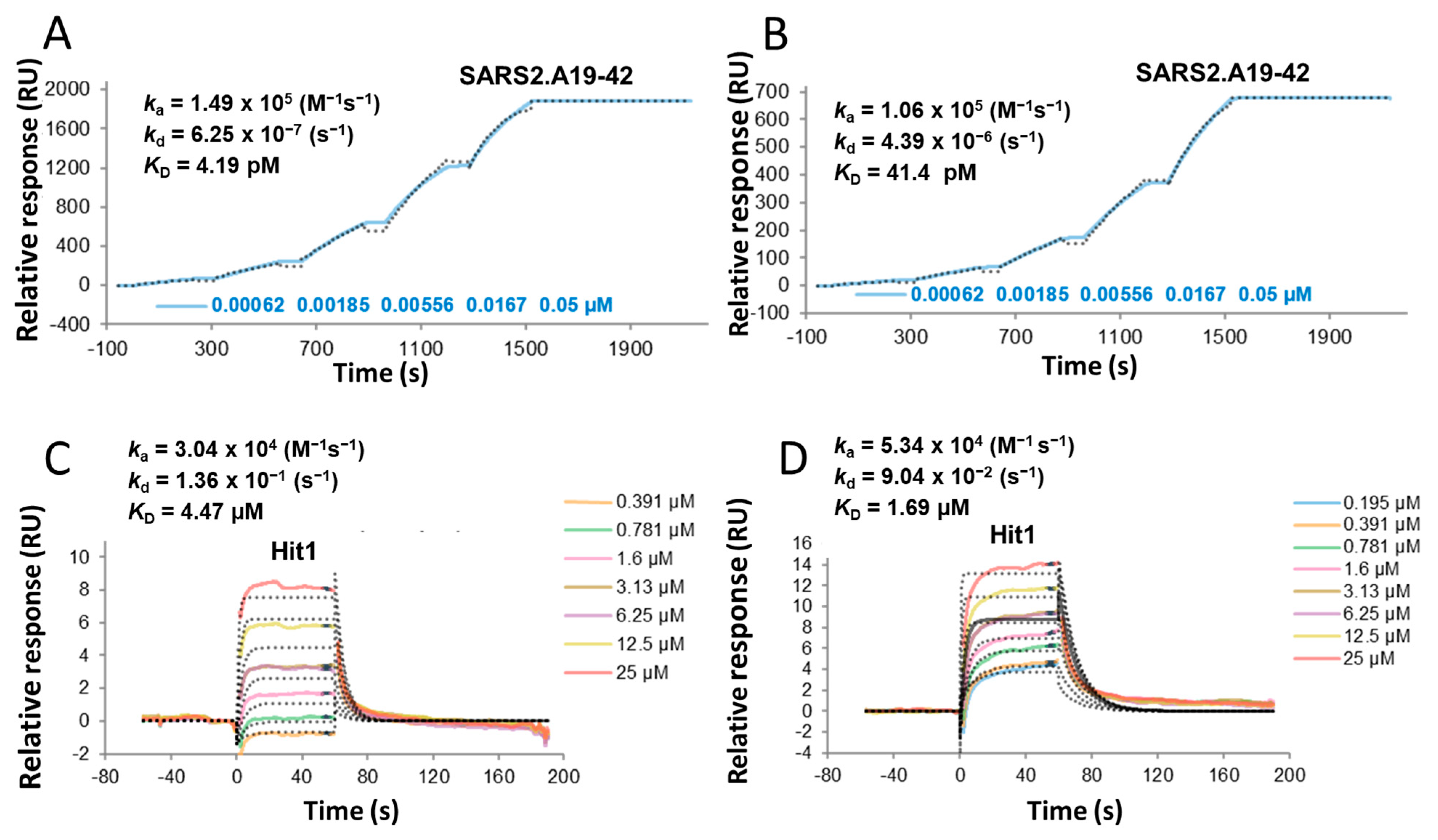
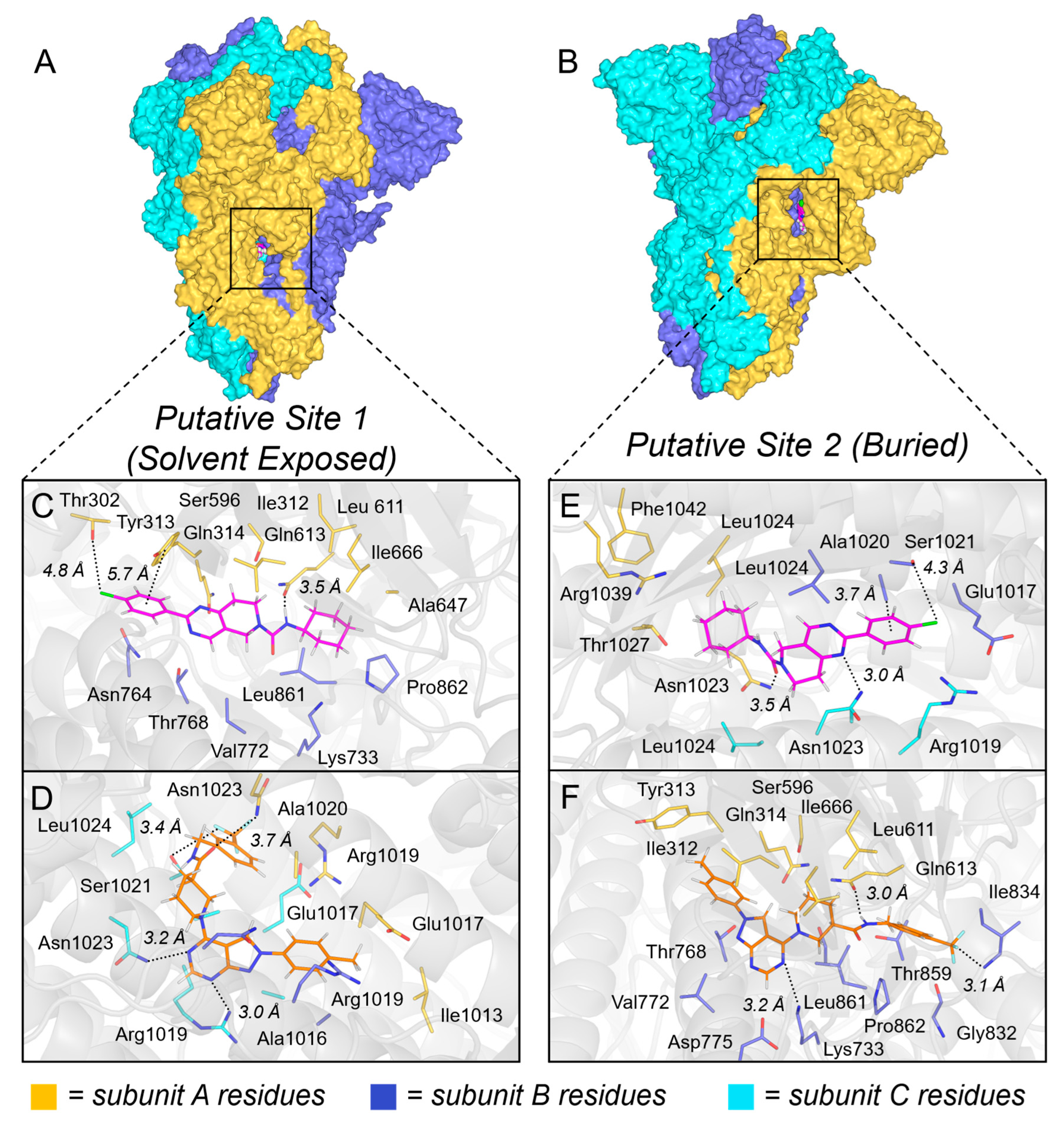
Using surface plasmon resonance (SPR) [40], we confirmed direct binding of the hit compounds to prefusion-stabilized SARS-CoV-2 spike proteins from both the WA1/2020 and B.1.1.529 (BA.1) variants [41]. The observed KD values ranged from 0.6 to 16.1 µM, consistent with expected affinities. Figure 2 displays representative sensorgrams for Hit-1 and the potent neutralizing antibody A19-42, which was included as a benchmark control [42]. The S2 subunit of the SARS-CoV-2 spike protein has historically been underutilized in vaccine development due to its pre-fusion instability in the absence of the S1 subunit. However, recent advances in protein engineering have enabled the stabilization of S2-only constructs, positioning it as a promising target for vaccine development. In parallel, the S2 subunit has gained attention in small molecule antiviral discovery due to its critical role in mediating membrane fusion and its interaction with the ACE2 receptor. Numerous in silico studies have reported strong binding affinities of phytochemicals (e.g., fisetin, quercetin, and kaempferol) [43] and repurposed drugs (e.g., clofazimine, toremifene, and arbidol) [44,45] to the S2 domain. Given its structural conservation and functional importance, S2-targeted therapeutics may offer enhanced efficacy against a broad spectrum of SARS-CoV-2 variants and related coronaviruses. To identify potential binding modes of the hit compounds on the SARS-CoV-2 spike protein, we performed molecular docking studies using Glide XP module (Schrödinger Suite, version 2023-4. New York, NY, USA) [46] on two putative binding sites identified by SiteMap [47]. The previously reported cryo-EM structure of the Omicron variant spike ectodomain in the closed conformation (PDB ID: 7TGY) [48] was used for docking studies. SiteMap initially predicted five potential ligand-binding pockets, which were narrowed down to two candidate sites based on structural relevance and accessibility. The two selected sites differed in solvent accessibility: Site 1 is solvent-exposed, while Site 2 is buried within the trimeric interface (Figure 3A,B). Notably, the pocket comprising Site 1 is formed exclusively by residues from the S2 domain and includes contributions from all three protomers.
The docking pose of Hit-1 in Site 1 (Figure 3C) reveals two key interactions with Asn1023. Mainly, the carbonyl oxygen atom of Hit-1 is within hydrogen-bonding distance to the amide nitrogen atom of Asn1023 side chain from subunit A (3.5 Å), while the pyrimidine nitrogen atom of the ligand is situated favorably within hydrogen-bonding distance to the side chain nitrogen atom of Asn1023 from subunit C (3.0 Å). A potential halogen-bond interaction is also observed between the chlorine atom of the ligand and the sidechain oxygen atom of Ser1021 from subunit B (4.3 Å). Furthermore, a hydrophobic interaction is formed between the 4-fluorophenyl group of Hit-1 and Ala1020 (3.7 Å from the Ala1020 carbon to the centroid of the aromatic ring). Similarly to Hit-1, the docking results of (R)-Hit-2 at Site 1 (Figure 3D) indicate that the ligand retains hydrogen-bond interactions with Asn1023 from both subunit A (3.5 Å) and subunit C (3.2 Å), reinforcing its function as a conserved anchoring residue within the S2 domain pocket. Furthermore, the pyrimidinyl nitrogen of the ligand is situated within hydrogen-bonding distance to the guanidine nitrogen of Arg1019. Also, the C-F fluorine atom of the ligand is within halogen-bonding distance to the sidechain oxygen atom of Ser1021 (subunit C). These contacts suggest a conserved binding mode across the hit series. Additionally, hydrophobic interactions of Hit-3 (Figure S1) with Leu1024 (present in all three subunits) and an electrostatic interaction with Thr1027 (subunit B) mirror the experimentally validated binding profile of Arbidol to the spike protein [44].
The binding pocket in Site 2 is composed of residues from two spike protein subunits, subunit A (S1 domain) and subunit B (S2 domain). Specifically, the S1 domain of subunit A contributes residues Thr302, Ile312, Tyr313, Glu314, Ser586, Leu611, Gln613, Ala647, and Ile666, while the S2 domain of subunit B includes Lys733, Asn764, Thr768, Val772, Asp775, Ile834, Leu861, Pro862, and Leu864. In Site 2 (Figure 3E), Hit-1 adopts a docking pose stabilized by a hydrogen-bonding interaction between the amide nitrogen atom of the ligand and the sidechain oxygen atom of Gln613 (3.6 Å). A potential π–π stacking interaction is observed between the 4-fluorophenyl moiety of the ligand and the phenol of Tyr313 (centroid-to-centroid distance of 5.7 Å). The ligand forms additional hydrophobic contacts with Ile312, Leu611, Ala647, Ile666, Val772, Leu861, and Pro862 in the pocket, which contribute to a well-anchored docking mode. The docking pose of (R)-Hit-2 (Figure 3F) reveals that the amide nitrogen atom of the ligand is within hydrogen-bonding distance to the sidechain carbonyl oxygen atom of Gln613 (3.0 Å) and forms new electrostatic contacts with Lys733 via a pyrimidine nitrogen atom (3.2 Å). The additional interactions include a halogen bond between a fluorine atom of the ligand with the backbone nitrogen atom of Ile834 (3.1 Å) and hydrophobic interactions with Ile312, Leu611, Ile666, Val772, Leu861, Pro862, and Leu864, further stabilizing the binding pose of Hit-2 within this buried pocket. While the docking studies presented here provide valuable insights into the potential ligand–protein interactions of these series of compounds, experimental structural elucidation of a ligand-protein complex would be instrumental in guiding rational scaffold optimization to enhance binding affinity and antiviral potency.

2.5. In Silico Predicted Parameters

Table 2 summarizes the physicochemical parameters and key ADME properties of the hit compounds, as predicted using the Percepta (Advanced Chemistry Development, Inc., Toronto, ON, Canada) and OSIRIS (Actelion Property Explorer software, version 05.05.00) [49]. These in silico assessments provide valuable insights into the potential pharmacokinetic properties of existing molecules and guide the rational design of new analogs for synthesis, where achieving an optimal balance between inhibitor potency and compound hydrophobicity is critical for druglikeness. Notably, the thieno[2,3-d]pyrimidine-based Hit-3 exhibited a relatively high cLogP value, suggesting increased lipophilicity. In contrast, Hit-1 (7,8-dihydropyrido[4,3-d]pyrimidine scaffold) and Hit-2 (pyrazolo[3,4-d]pyrimidine scaffold) displayed more favorable profiles with reduced cLogP values, aligning better with drug-like characteristics. Predicted water solubility remains a key consideration, as over 40% of the marketed drugs and approximately 80% of molecules in the discovery pipeline may encounter solubility challenges that can adversely affect oral bioavailability and therapeutic efficacy [50]. Among the three hits, Hit-1 demonstrated the most promising overall profile, with the highest predicted solubility, Caco-2 permeability, and oral bioavailability (Table 2). In our experience, relative overall drug scores of close to 50 are considered highly favorable. These scores incorporate structural criteria from Lipinski’s “rule of five” [37,38,51] alongside predicted toxicity risks, including mutagenic, tumorigenic, irritant, and reproductive effects. In addition to its relatively high drug score, Hit-1 exhibited submicromolar antiviral activity and favorable selectivity index (SI), making it a compelling lead for further optimization and synthesis of analogs with enhanced drug-like properties.
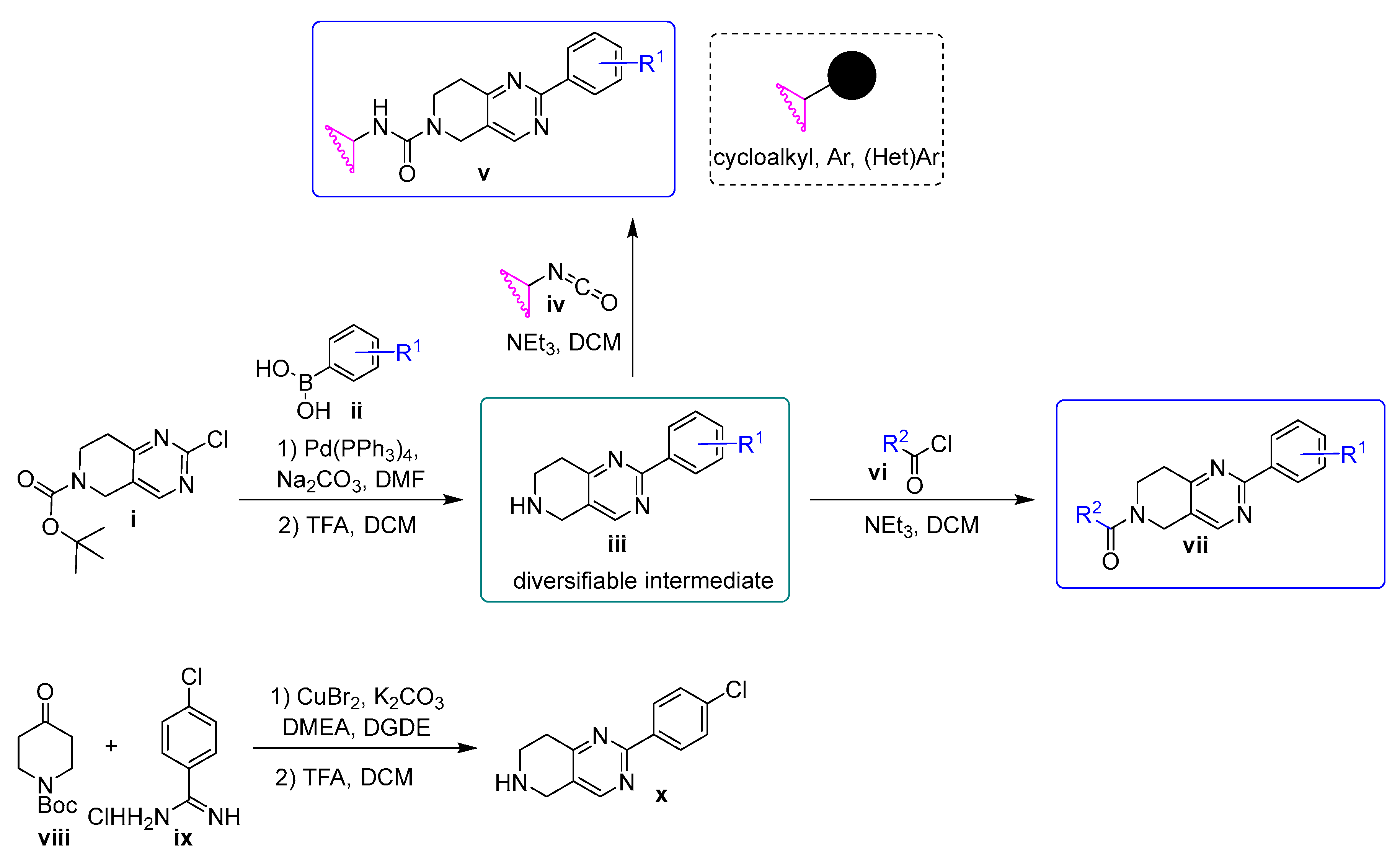
Synthesis. Hit-1 and analogs 27 and 1214 presented in Table 3 were prepared using the general procedure shown in Scheme 1, while a set of commercially available compounds (analogs 811) was acquired as a structure-activity relationship (SAR)-by-purchase approach to enhance chemical diversity.
The synthetic route commenced with tert-butyl 2-chloro-7,8-dihydropyrido[4,3-d]pyrimidine-6(5H)-carboxylate (i), a commercially available intermediate. This compound underwent palladium-catalyzed Suzuki coupling with various boronic acids (ii) to afford the corresponding aryl-substituted intermediates. Subsequent Boc deprotection using trifluoroacetic acid (TFA) yielded the free secondary amines (iii), which served as versatile intermediates for further functionalization. To expand the scope of analog development, intermediate iii was transformed into bioactive molecules bearing diverse functional groups. Treatment of iii with the appropriate isocyanates iv afforded a series of urea-containing derivatives (v, compounds 27), while reaction with acyl chlorides vi yielded amide-containing targets (vii, compounds 1214). For analogs incorporating a 4-chlorophenyl moiety, an alternative synthetic route was employed to circumvent potential cross-reactivity between reagents. Specifically, a copper-mediated coupling of N-Boc-4-piperidone (viii) with 4-chlorobenzamidine (ix), followed by Boc deprotection, furnished the key intermediate 2-(4-chlorophenyl)-5,6,7,8-tetrahydropyrido[4,3-d]pyrimidine (x). This intermediate was subsequently used in the synthesis of compound 1. Thus, the diversifiable nature of intermediate iii enables the rapid synthesis of analogs and facilitates the SAR of this scaffold. Encouragingly, none of the new analogs exhibit toxicity risks, and the majority of them, namely compounds 411 and 1314, show significantly improved predicted druglikeness and drug scores compared to Hit-1.
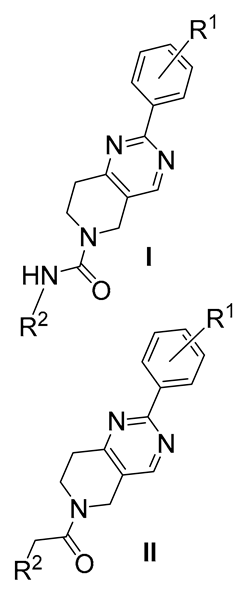
Table 3 summarizes the antiviral activities of Hit-1 (compound 1), which was resynthesized for hit validation purposes, along with new 7,8-dihydropyrido[4,3-d]pyrimidine series derivatives 214 that were tested in the pseudoviral SARS-CoV-2 inhibition assay to support SAR development. The compounds feature varied substituents on the aromatic ring, paired with either cyclohexyl or substituted benzyl moieties, and exhibit a range of potencies and SIs. The compounds presented in Table 3 constitute a relatively small yet focused set of compounds that were primarily designed to validate the antiviral potential of the 7,8-dihydropyrido[4,3-d]pyrimidine scaffold against CoV-2. Despite the limited scope, this preliminary dataset yields informative insights. Notably, select compounds from both the urea series (Type I) and amide series (Type II) exhibit desirable antiviral activity, supporting the rationale for development of expanded analog libraries for advanced hit-to-lead optimization. The most favorable substituent pattern on the aromatic ring was identified as 4-chloro and 3,4-di-fluoro for both types I and II. Specifically, these preferred substitution patterns are exemplified by Type I compounds, such as Hit-1 and compound 3 (EC50 value of 0.32 and 0.93 µM, respectively), and by Type II compounds 9, 13, and 14 (EC50 value of 0.47, 0.23, and 0.68 µM, respectively). Additionally, in series II, the presence of a 2-chlorophenyl group (compound 9) correlates with enhanced activity. Conversely, there may be a contraindication for a trifluoromethyl group at the 4-position of the pyrimidinyl phenyl ring in both series as indicated by the decreased antiviral efficacy of compounds 2 and 12. Overall, this work has led to the identification of five promising new SARS-CoV-2 inhibitors having EC50 values < 1 µM (Table 2). It is also noteworthy that none of the new 7,8-dihydropyrido[4,3-d]pyrimidines showed toxicity in A549, Calu-3, or HepG2 cells at the highest screening dose of 100 µM.

2.6. ADME/Tox Profile

To initiate metabolic profiling of the analogs, we selected six structurally diverse compounds that demonstrated promising in vitro antiviral activity for evaluation in plasma and liver microsome (LM) stability assays. Given the broad host range of SARS-CoV-2, LMs from multiple species, including human, mouse, dog, and monkey, were employed to obtain a more comprehensive assessment of metabolic stability of these analogs, with particular attention given to interspecies variability. Specifically, compounds 1, 3, and 7 (Type I) and compounds 10, 11, and 13 (Type II) were incubated with LM from each species (Table 4). Among the tested compounds, compound 7 emerged as the most metabolically stable across all species, with >65% of the parent compound remaining after 60 min of incubation as measured at both 10 and 2 µM final drug concentrations (Table 4). However, this analog exhibited relatively modest in vitro efficacy compared to others in the series. In contrast, compound 11 demonstrated the poorest metabolic stability, particularly in monkey LM, with only 1.8% remaining at 2 µM. Notably, compound 11 is the sole analog bearing a 3-OMe substituent at the R2 position, which may contribute to its rapid degradation. Encouragingly, all selected analogs displayed excellent plasma stability across species (Table 4).
We further investigated these compounds to assess their potential for CYP450 inhibition, a key factor in drug–drug interactions (DDIs). DDIs arise when one drug alters the pharmacokinetic profile or pharmacodynamic effect of another, often through interference with metabolic pathways. Inhibition of CYP450 enzymes is a common mechanism underlying such interactions, potentially leading to altered drug clearance and efficacy. Compounds 7 and Hit-1 exhibited strong inhibition of CYP2C9 and notably high inhibition of CYP3A4, at 81.20% and 68.77%, respectively (Table 5). In contrast, compound 13 showed reduced CYP3A4 inhibition (50.22%) relative to Hit-1. While this level of inhibition remains borderline with the desired threshold, the modest CYP2C9 inhibition and negligible activity against CYP1A2 and CYP2D6 further support compound 13 as a promising starting point for further structural refinement focused toward a reduced polypharmacology risk. The potential inhibitory effect of compound 13 on the human Ether-à-go-go related gene (hERG) channel was evaluated in transfected HEK293 cells by using a manual patch-clamp assay. Based on the results, this compound was ranked as a moderate inhibitor with an IC50 value of 6.9 µM (SI > 30). Also, compound 13 showed high permeability (−Log Pe = 4.99) in a PAMPA assay, indicating that it readily diffuses across the artificial lipid membrane and suggesting strong potential for passive absorption through biological barriers such as the gastrointestinal tract.
After a holistic analysis of the in vitro efficacy and ADME parameters, compound 13 was selected for progression to in vivo pharmacokinetic (PK) studies. PK analysis of compound 13 in BALB/c mice following intravenous (i.v.) and oral administration (p.o.) by oral gavage revealed that: (1) the maximal plasma levels are rapidly reached, with Tmax ranging from 5 to 110 min; (2) the half-life of this compound varied between 2.35 h (i.v) and 4.0 h (p.o.); (3) the oral bioavailability (F) of compound 13 is 68% (Table 6); and (4) no adverse clinical signs were observed for compound 13 in treated animals. Furthermore, in seven-day toxicity studies using a 50 mg/kg b.i.d dosing regimen in C57BL/6 mice, this compound was well-tolerated. The high plasma concentrations achieved via both administration routes, combined with the extended half-life and favorable bioavailability, support the progression of compound 13 to in vivo efficacy studies.

3. Materials and Methods

3.1. Chemistry

All solvents and reagents were purchased from commercial suppliers and used without further purification. 1H and 13C NMR spectra were recorded on Bruker DPX-400 (Billerica, MA, USA) or AVANCE-400 spectrometers (Billerica, MA, USA, at 400 MHz and 101 MHz, respectively. NMR chemical shifts were reported in δ (ppm) using residual solvent peaks as standards (CDCl3: 7.26 ppm (1H), 77.23 ppm (13C); CD3OD: 3.31 ppm (1H), 49.15 ppm (13C)). ESI mass spectra were recorded in the positive mode on Agilent 6230 TOF LC/MS. All key compounds possess a purity of at least 95% as assessed by analytical reversed phase HPLC using an ACE 3AQ C8 column (150 × 4.6 mm, particle size 3 μm, Mac-Mode Analytical, Chadds Ford, PA, USA) with detection at 254 and 280 nm on a Shimadzu SPD-20A VP detector (Columbia, MD, USA); flow rate =1.0 mL/min; gradient of 20−95% acetonitrile in water (both containing 0.1 vol % of FA) in 20 min.

3.2. General Method A: Suzuki Coupling

To a solution of aryl chloride (1 eq) in DMF (3 mL/mmol), boronic acid (1.2 eq) and an aqueous solution of Na2CO3 (10%, 2.1 eq) were added, followed by Pd(PPh3)4 (0.05 eq). The reaction mixture was stirred at 120 °C under Ar overnight. Upon completion, as indicated by TLC, the reaction mixture was cooled to room temperature (RT), diluted with DCM, and washed with water. The aqueous layer was extracted with DCM (2 × 50 mL), and the combined organic fractions were washed with brine, dried over Na2SO4, and concentrated under reduced pressure. The crude product was purified by flash chromatography (EtOAc—Hexane, 0–40%) to yield the corresponding cross-coupled product A, C, E or G (Supplemental Materials).

3.3. General Method B: Boc Deprotection

Boc-protected amine (1 eq) in a mixture of DCM-TFA (4:1, 3 mL/mmol) was allowed to stir at room temperature overnight. Upon completion, as indicated by TLC, the reaction mixture was concentrated under reduced pressure to yield the desired amine (B, D, F or H (Supplemental Materials)), which was used in the next step without further purification.

3.4. General Method C: Synthesis of Ureas

To a solution of amine iii (1 eq) in DCM (3 mL/mmol), NEt3 (5–10 eq) was added at 0 °C, followed by the addition of isocyanate (1 eq). The reaction mixture was allowed to reach RT and stirred overnight. Upon completion, the reaction mixture was diluted with DCM and washed with saturated aqueous NH4Cl and brine. The organic phase was dried over Na2SO4, filtered and concentrated under reduced pressure. The crude product was purified by flash chromatography (MeOH—DCM, 5%) or via prep TLC.

3.5. General Method D: Synthesis of Amides

To a solution of amine iii (1 eq) in DCM (3 mL/mmol) NEt3 (5–10 eq) was added at 0 °C, followed by the addition of acyl chloride (1 eq). The reaction mixture was allowed to reach RT and stirred overnight. Upon completion, the reaction mixture was diluted with DCM and washed with saturated aqueous NH4Cl and brine. The organic phase was dried over Na2SO4, filtered and concentrated under reduced pressure. The crude product was purified by flash chromatography (MeOH—DCM, 5%) or via prep TLC.
  • 2-(4-Chlorophenyl)-N-cyclohexyl-7,8-dihydropyrido[4,3-d]pyrimidine-6(5H)-carboxamide (1).
White amourphous solid. Yield: 10 mg, 71%. 1H NMR (400 MHz, CDCl3) δ 1.14–1.23 (m, 2H), 1.35–1.46 (m, 2H), 1.62–1.78 (m, 4H), 1.97–2.03 (m, 2H), 3.06 (t, J = 5.9 Hz, 2H), 3.74 (t, J = 5.9 Hz, 2H), 4.44 (d, J = 7.6 Hz, 1H), 4.61 (s, 2H), 7.46 (d, J = 8.6 Hz, 2H), 8.38 (d, J = 8.6 Hz, 2H), 8.55 (s, 1H). 13C NMR (101 MHz, CDCl3) δ 25.0, 25.6, 31.8, 34.0, 40.8, 42.7, 49.7, 124.8, 128.7, 129.4, 135.98, 136.7, 154.8, 156.6, 161.9, 163.8. HPLC Purity: 99.2%. HRMS (ESI) calculated for C20H23ClN4O ([M+H]+): 371.1638, found: 371.1649.
  • N-Cyclohexyl-2-(4-(trifluoromethyl)phenyl)-7,8-dihydropyrido[4,3-d]pyrimidine-6(5H)-carboxamide (2).
White amourphous solid. Yield: 12 mg (23%). 1H NMR (400 MHz, CDCl3) δ 1.18–1.21 (m, 2H), 1.39–1.43 (m, 2H), 1.63–1.70 (m, 2H), 1.72–1.76 (m, 2H), 1.99–2.03 (m, 2H), 3.10 (t, J = 5.9 Hz, 2H), 3.76 (t, J = 5.9 Hz, 2H), 4.45 (d, J = 7.6 Hz, 1H), 4.65 (s, 2H), 7.75 (d, J = 8.2 Hz, 2H), 8.56 (d, J = 8.2 Hz, 2H), 8.61 (s, 1H). 13C NMR (101 MHz, CDCl3) δ 25.1, 25.6, 31.9, 34.0, 40.9, 42.8, 49.7, 125.46, 125.48, 125.49, 125.5, 128.3, 131.9, 132.3, 140.7, 154.9, 156.6, 161.5, 164.0. HPLC Purity: 99.3%. HRMS (ESI) calculated for C21H23F3N4O ([M+H]+): 405.1902, found: 405.1914.
  • N-Cyclohexyl-2-(3,4-difluorophenyl)-7,8-dihydropyrido[4,3-d]pyrimidine-6(5H)-carboxamide (3).
White amourphous solid. Yield: 48 mg (55%). 1H NMR (400 MHz, CDCl3) δ 1.14–1.20 (m, 2H), 1.35–1.44 (m, 2H), 1.59–1.79 (m, 4H), 1.95–2.04 (m, 2H), 3.06 (t, J = 5.9 Hz, 2H), 3.66–3.77 (m, 3H), 4.62 (s, 2H), 7.21–7.27 (m, 1H), 8.19–8.23 (m, 1H), 8.27 (ddd, J = 11.9, 7.9, 2.1 Hz, 1H), 8.54 (s, 1H). 13C NMR (101 MHz, CDCl3) δ 25.0, 25.6, 31.8, 34.0, 40.8, 42.7, 49.7, 117.0, 117.1, 117.2, 117.3, 124.4, 124.2, 125.0, 134.6, 149.2, 149.3, 150.8, 150.9, 151.6, 151.8, 153.3, 153.5, 154.9, 156.6, 161.0, 163.9. HPLC Purity: 99.1%. HRMS (ESI) calculated for C20H22F2N4O ([M+H]+): 373.1840, found: 373.1853.
  • N-Cyclohexyl-2-phenyl-7,8-dihydropyrido[4,3-d]pyrimidine-6(5H)-carboxamide (4).
White amourphous solid. Yield: 28 mg (75.5%). 1H NMR (400 MHz, CDCl3) δ 1.14–1.21 (m, 2H), 1.36–1.45 (m, 2H), 1.63–1.78 (m, 2H), 1.98–2.03 (m, 2H), 3.08 (t, J = 5.9 Hz, 2H), 3.75 (t, J = 6.0 Hz, 2H), 4.46 (d, J = 7.6 Hz, 1H), 4.61 (s, 2H), 7.48–7.51 (m, 3H), 8.40–8.44 (m, 2H), 8.57 (s, 1H). 13C NMR (400 MHz, CDCl3) δ 25.0, 25.6, 31.9, 34.0, 40.8, 42.7, 49.7, 124.5, 128.0, 128.5, 130.5, 137.5, 154.8, 156.6, 163.0, 163.7. HPLC Purity: 99.3%. HRMS (ESI) calculated for C20H24N4O ([M+H]+): 337.2028, found: 337.2033.
  • N-(4-Chlorophenyl)-2-phenyl-7,8-dihydropyrido[4,3-d]pyrimidine-6(5H)-carboxamide (5).
White amourphous solid. Yield: 58 mg (86%). 1H NMR (400 MHz, MeOD) δ 3.13 (t, J = 5.9 Hz, 2H), 3.90 (t, J = 5.9 Hz, 2H), 4.75 (s, 2H), 7.23–7.27 (m, 2H), 7.35–7.40 (m, 3H), 7.45–7.53 (m, 3H), 8.35 (dd, J = 6.8, 3.0 Hz, 2H), 8.58 (s, 1H). 13C NMR (101 MHz, MeOD) δ 31.2, 40.6, 42.4, 121.4, 124.3, 127.5, 127.7, 128.1, 130.2, 136.7, 137.3, 154.3, 155.2, 162.6, 163.5. HPLC Purity: 98.1%. HRMS (ESI) calculated for C20H17ClN4O ([M+H]+): 365.1169, found: 365.1181.
  • N-(4-Fluorophenyl)-2-phenyl-7,8-dihydropyrido[4,3-d]pyrimidine-6(5H)-carboxamide (6).
White amourphous solid. Yield: 59 mg (90%). 1H NMR (400 MHz, CDCl3) δ 3.10 (t, J = 5.9 Hz, 2H), 3.85 (t, J = 5.9 Hz, 2H), 4.71 (s, 2H), 6.99 (t, J = 8.6Hz, 2H), 7.34 (m, 2H), 7.49 (m, 3H), 8.38 (dd, J = 6.6, 2.9, Hz, 2H), 8.55 (s, 1H). 13C NMR (400 MHz, CDCl3) δ = 31.7, 41.1, 42.9, 115.3, 115.5, 122.4, 122.5, 122.6, 124.4, 128.0, 128.6, 130.7, 134.6, 137.2, 154.8, 155.4, 157.9, 160.3, 163.1, 163.6. HPLC Purity: 99.3%. HRMS (ESI) calculated for C20H17FN4O ([M+H]+): 349.1464, found: 349.1490.
  • N-(Benzo[d][1,3]dioxol-5-yl)-2-phenyl-7,8-dihydropyrido[4,3-d]pyrimidine-6(5H)-carboxamide (7).
White amourphous solid. Yield: 13 mg (52%). 1H NMR (400 MHz, CDCl3) δ 3.13 (t, J = 5.9 Hz, 2H), 3.85 (t, J = 5.9 Hz, 2H), 4.71 (s, 2H), 5.95 (s, 2H), 6.55 (s, 1H), 6.68–6.75 (m, 2H), 7.05 (d, J = 1.9 Hz 1H), 7.49–7.56 (m, 3H), 8.44 (dd, J = 6.7, 3.0 Hz, 2H), 8.57 (s, 1H). 13C NMR (400 MHz, CDCl3) δ 31.9, 41.2, 43.0, 101.2, 104.0, 107.9, 114.1, 124.2, 128.1, 128.6, 130.6, 132.6, 137.4, 144.1, 147.9, 154.8, 155.3, 163.1, 163.4. HPLC Purity: 98.7%. HRMS (ESI) calculated for C21H18N4O3 ([M+H]+): 375.1457, found: 375.1461.
  • N-(3-Fluoro-4-methylphenyl)-2-(4-methoxyphenyl)-7,8-dihydropyrido[4,3-d]pyrimidine-6(5H)-carboxamide (8) was purchased from ChemDiv (Cat# M568-0042).
  • 2-(2-Chlorophenyl)-1-(2-(4-chlorophenyl)-7,8-dihydropyrido[4,3-d]pyrimidin-6(5H)-yl)ethan-1-one (9) was purchased from ChemDiv (Cat# M567-0228).
  • 2-(4-Chlorophenyl)-1-(2-(4-chlorophenyl)-7,8-dihydropyrido[4,3-d]pyrimidin-6(5H)-yl)ethan-1-one (10) was purchased from ChemDiv (Cat# M567-0223).
  • 1-(2-(4-Chlorophenyl)-7,8-dihydropyrido[4,3-d]pyrimidin-6(5H)-yl)-2-(3-methoxyphenyl)ethan-1-one (11) was purchased from ChemDiv (Cat# M567-0210).
  • 2-(2-Chlorophenyl)-1-(2-(4-(trifluoromethyl)phenyl)-7,8-dihydropyrido[4,3-d]pyrimidin-6(5H)-yl)ethan-1-one (12).
White amourphous solid. Yield: 28 mg (51%). 1H NMR (400 MHz, CDCl3) δ (characterized as a 2:1 mixture of rotamers. Major rotamer: δ 2.98 (t, J = 6.1 Hz, 2H), 3.88 (t, J = 5.9 Hz, 2H), 3.99 (s, 2H), 8.63 (s, 2H), 4.89 (s, 2H). Minor rotamer: 3.09 (t, J = 6.1 Hz, 1H), 3.97 (s, 1H), 4.05 (t, J = 6.0 Hz, 1H), 8.50 (s, 0.5H) 4.73 (s, 1H). Overlapping peaks between rotamers: 7.18–7.23 (m, 1H), 7.26 (dd, J = 5.9, 3.5 Hz, 2H), 7.33 (s, 2H), 7.42–7.45 (m, 1H), 7.75 (d, J = 8.2 Hz, 3H), 8.55 (d, J = 8.1 Hz, 3H). 13C NMR (101 MHz, CDCl3) Major rotamer: δ 32.2, 38.1, 41.4, 42.8. Minor rotamer: δ 31.4, 38.4, 39.2, 44.7. Overlapping peaks between rotamers: δ 125.4, 125.5, 127.2, 128.4, 128.7, 129.6, 130.6, 130.7, 132.7, 154.5, 155.4, 162.9, 169.4. HPLC Purity: 99.7%. HRMS (ESI) calculated for C21H23F3N4O ([M+H]+): 405.1902, found: 405.1914.
  • 2-(2-Chlorophenyl)-1-(2-(3,4-difluorophenyl)-7,8-dihydropyrido[4,3-d]pyrimidin-6(5H)-yl)ethan-1-one (13).
White amourphous solid. Yield: 55 mg (62%). 1H NMR (400 MHz, CDCl3) δ (characterized as a 2:1 mixture of rotamers). Major rotamer: δ 2.95 (t, J = 6.0 Hz, 2H), 3.87 (t, J = 6.0 Hz, 2H), 3.98 (t, J = 6.0 Hz, 2H), 4.87 (s, 2H), 8.57 (s, 1H). Minor rotamer: δ 3.06 (t, J = 6.2 Hz, 1H), 3.96 (s, 1H), 4.03 (t, J = 6.1 Hz, 1H), 4.71 (s, 1H), 8.45 (s, 0.5H) Overlapping peaks between rotamers: δ 7.18–7.27 (m, 9H), 7.30–7.36 (m, 3H), 7.39–7.46 (m, 3H), 8.16–8.23 (m, 3H), 8.25–8.31 (m, 3H). 13C NMR (101 MHz, CDCl3) Major rotamer: δ 32.2, 38.1, 41.3, 42.8. Minor rotamer: δ 31.4, 38.4, 39.2, 44.7. Overlapping peaks between rotamers: δ 117.1, 117.2, 117.3, 117.4, 124.1, 124.4, 124.9, 127.2, 128.7, 129.6, 130.6, 130.7, 132.8, 133.8, 134.5, 149.2, 149.4, 150.9, 151.0, 151.7, 151.8, 153.5, 154.5, 155.3, 161.0, 161.3, 162.8, 164.1, 169.1, 169.4. HPLC Purity: 99.7%. HRMS (ESI) calculated for C21H16ClF2N3O ([M+H]+): 400.1028, found: 400.1033.
  • 2-(4-Chlorophenyl)-1-(2-(3,4-difluorophenyl)-7,8-dihydropyrido[4,3-d]pyrimidin-6(5H)-yl)ethan-1-one (14).
White amourphous solid. Yield: 41 mg (66%). 1H NMR (400 MHz, CDCl3) δ (characterized as a 2:1 mixture of rotamers). Major rotamer: δ 2.88 (t, J = 5.9 Hz, 2H), 4.83 (s, 2 H), 8.56 (s, 1H). Minor rotamer: δ 3.04 (t, J = 6.0 Hz, 1H), 4.00 (t, J = 6.1 Hz, 1H), 4.65 (s, 1 H), 8.43 (s, 0.5H). Overlapping peaks between rotamers: δ 3.79–3.86 (m, 5H), 7.18–7.27 (m, 5 H), 7.31–7.35 (m, 2.5H), 8.17–8.21 (m, 1.5H), 8.24–8.30 (m, 1.5H). 13C NMR (101 MHz, CDCl3) Major rotamer: δ 32.1, 40.4, 41.3, 42.9. Minor rotamer: δ 31.4, 39.1, 40.6, 44.8. Overlapping peaks between rotamers: δ 117.1, 117.2, 117.3, 117.4, 124.5, 124.7, 129.1, 130.1, 132.9, 133.1, 134.5, 149.2, 149.4, 151.7, 151.8, 154.5, 155.3, 162.7, 169.7. HPLC Purity: 98.0%. HRMS (ESI) calculated for C21H16ClF2N3O ([M+H]+): 400.1028, found: 400.1033.

3.6. Cell Lines

Human embryonic kidney (HEK)293T cells transiently expressing the human angiotensin-converting enzyme 2 (ACE2) and TMPRSS2 receptor were cultured in DMEM supplemented with 10% fetal bovine serum (FBS), 1% penicillin-streptomycin, and maintained in a humidified incubator at 37 °C with 5% CO2. Calu-3 (ATCC HTB-55), cells were grown in MEM-high glucose (4500 mg/L) with 10% heat-inactivated fetal bovine serum (FBS), 2% L-Glutamine, 1% sodium pyruvate and 1% penicillin/streptomycin, and maintained in a humidified incubator at 37 °C with 5% CO2. Baby hamster kidney cells expressing ACE2 (BHK-hACE2-11) were grown in Dulbecco’s Modified Eagle’s Medium High glucose (4500 mg/L) with 10% fetal bovine serum (FBS) and 1% penicillin/streptomycin and maintained in a humidified incubator at 37 °C with 5% CO2.

3.7. Virus Stocks

Viral stocks of SARS-CoV-2 strains USA-WA1/2020 (A) and B.1.1.529 BA.1 (Omicron) were obtained from BEI Resources (Manassas, VA, USA).

3.8. Generation of Pseudoviruses

Pseudoviruses bearing spike protein from SARS-CoV-2 or SARS-CoV were generated as previously described [36] by using the pcDNA3.1(+)-SARS2-S or pcDNA3.1(+)-SARS-S plasmid, along with an HIV-1 vector expressing a luciferase gene (pNL4–3.Luc.R−E−, NIH AIDS Research and Reference Reagent Program). HEK293T cells were seeded in tissue culture dishes and co-transfected with the spike-expressing plasmid and the HIV-Luc plasmid using a polyethyleneimine (PEI)-based transfection protocol. Five hours post-transfection, DMEM media was added to each plate. Supernatants containing pseudoviruses were harvested 24 h after transfection, collected by filtration through a 0.45 μm pore-size filter, and stored at −80 °C until use.

3.9. Pseudovirus Neutralization Assay—HTS

HEK293T Cells were transfected with pcDNA3.1(+)-humanACE2 and pCSDest-TMPRSS2 plasmids for 5 h. The transfected cells in DMEM were then seeded (1 × 10⁵ cells/well) in 96- or 384-well plates and incubated at 37 °C and 5% CO2 overnight. SARS-CoV-2 and VSV pseudoviruses were incubated with the test samples at room temperature for 1 h and then added to the target cells. Plates were incubated for 48 h, and levels of viral entry were determined by luminescence using the Neolite Reporter Gene Assay System (Revvity, Waltham, MA, USA). Luminescence data were normalized to the untreated control to obtain percent inhibition. All samples were assayed in triplicate. Assay robustness was evaluated by calculating the signal-to-background ratio and Z′-factor. A Z′-factor > 0.5 was considered indicative of a high-quality assay suitable for HTS. Known entry inhibitors (e.g., camostat mesylate) were included as positive controls to validate assay performance.

3.10. Generation of SARS-CoV-2 Variant S Protein Plasmids

SARS-CoV-2 variants were generated from point mutations starting with the SARS-CoV-2 WA1/2020 S protein plasmid (pcDNA3.1+). Using the Agilent Technologies (Santa Clara, CA, USA) QuickChange Lightning Site-Directed Mutagenesis kit (Cat#210518). Site-specific nucleotide substitutions were introduced by PCR amplification using primers containing the desired mutations designed via Agilent’s online QuickChange Primer Design Program. Mutated plasmids were verified by Sanger sequencing to confirm successful incorporation of the intended changes.

3.11. Pseudovirus Inhibition and Cytotoxicity

BHK-hACE2 Cells (5 × 10³ cells/well) were seeded in 96-well white bottom plates and incubated at 37 °C and 5% CO2 for 24 h. Cells were transduced with the pseudovirions with or without compounds at 1% DMSO final concentration. To obtain EC50 and CC50 values, the compounds were serially diluted, and the plates were incubated for 48 h. The luciferase activity was measured using the Neolite Reporter Gene Assay System (PerkinElmer, Waltham, MA, USA), while cytotoxicity was measured using CellTiter-Glo Luminescent Cell Viability Assay (Promega, Madison, WI, USA). EC50 and CC50 values were obtained by analyzing the dose−response data using four-parameter nonlinear logistic regression analysis in GraphPad Prism (Version 10.6.0).

3.12. Live Virus Neutralization Assay

48 h prior to treatment, Calu-3 cells were seeded at 3.0 × 10⁵ cells/well in 24-well plates. Following the previously described protocol [52], cells were infected with clinical isolates of SARS-CoV-2 (USA-WA1/2020 or SARS-CoV-2B.1.1.529 BA.1 (Omicron)) (BEI Resources, (Manassas, VA, USA). Three non-treatment controls and two-fold serial dilutions of the test compound Hit-1 (final concentrations 10 µM to 0.6 µM) were mixed with an equal volume of virus to achieve a final multiplicity of infection (MOI) of 0.01. The mixtures were added to the monolayer of cells and incubated at 37 °C and 5% CO2 for 1 h. After incubation, the inoculum was removed, cells were washed with PBS, and monolayers overlayed with infection media (DMEM supplemented with 2% FBS and 1% P/S). After 48 h, cell supernatants were collected, and 250 µL aliquots were mixed with 750 µL of TRIzol. RNA was isolated using PureLink RNA Mini Kits (Invitrogen, Carlsbad, CA, USA) according to the manufacturer’s protocol. RT-qPCR was carried out using 5 µL of RNA template in TaqMan Fast Virus 1-Step Master Mix using primers and probes for the N gene (N1 primers, CDC, IDT cat# 10006713). A standard curve was generated using dilution of synthetic SARS-CoV-2 RNA (BEI Resources, NR-52358). All experiments prior to RNA isolation were conducted in a BSL-3 facility.

3.13. Statistical Analysis

EC50 and CC50 values are represented as means ± standard deviation (SD). These were calculated by analyzing the dose–response data using a four-parameter nonlinear logistic regression analysis in GraphPad Prism (Version 10.6.1). Statistical analysis was also performed with GraphPad Prism. Comparisons between multiple groups were performed using an Ordinary one-way analysis of variance (ANOVA), followed by Dunnett’s multiple comparisons test, which evaluates each treatment group against the untreated (DMSO only) control. The p-values are defined as follows: * p ≤ 0.05, ** p ≤ 0.01, *** p ≤ 0.001, **** p ≤ 0.0001, ns p > 0.05 (not significant).

3.14. Assessment of the Binding Properties by SPR

The recombinant S proteins used in these studies were provided by Dr. Fang Li (University of Minnesota). Direct binding kinetics [association rate constants (ka), and dissociation rate constants (kd)] and binding affinity (KD) of the new inhibitors to the SARS-CoV-2 GP were obtained on Biacore T200 (Uppsala, Sweden) and Biacore 8K SPR instrument (Uppsala, Sweden) as previously described [53]. Briefly, prefusion-stabilized SARS-CoV-2 spike (S) protein [41] was immobilized on a CM5 chip by standard amine coupling. A reference flow channel was prepared in parallel by blank immobilization, activation/deactivation without protein. Hit compounds were prepared as a 2-fold dilution series (0.195–25 µM) in SPR binding buffer (20 mM Phosphate buffer (pH 7.4), 137 mM NaCl, 2.7 mM KCl, 0.05% Tween20, 2% DMSO) and injected using a multi-cycle kinetics format at a 30 µL/min flow rate. The antibody was injected over the same immobilized surface in a single-cycle kinetics format as a 3-fold dilution series (0.62–50 nM). Sensorgrams were double-referenced (reference channel and zero concentration response subtraction) and fitted with a 1 to 1 Langmuir kinetic model using Biacore Insight evaluation software version 5.0.18.22102 to obtain two rate constants (ka and kd). The equilibrium dissociation constants (KD) were calculated as KD = kd/ka. Solvent correction for DMSO was applied according to the instrument manufacturer’s recommendations.

3.15. Liver Microsome Stability Assay

Samples at 2 and 10 µM concentrations were incubated with liver microsomes from human (Gibco HMMCPL, Lot no. PL0501. Waltham, MA, USA), mouse (Xenotech M3000, Lot no. 2210125. Kansas City, KS, USA), dog (Xenotech D1000, Lot no. 2310325. Kansas City, KS, USA), or monkey (Xenotech P2000, Lot no. 1310274. Kansas City, KS, USA) at a final protein concentration of 0.5 mg protein/mL. Incubations were carried out in 100 mM phosphate buffer containing 20 mM NADPH as a co-factor, in a 37 °C water bath. Reactions were initiated by the addition of the test compounds and terminated by quenching 50 µL aliquots at designated time points (0, 30 and 60 min) with 200 µL of cold acetonitrile containing 0.6 µM Reserpine as an internal standard. Samples were vortexed for 30 s and centrifuged at 12,000 rpm for 15 min at 4 °C. Following centrifugation, 100 µL of the supernatant was transferred to an autosampler tray for LC-MS/MS analysis. LC-MS/MS analysis was performed using a Shimadzu LC-MS-8050 triple quadrupole system (Kyoto, Japan) equipped with a turbo spray electrospray ionization (ESI) source. Chromatographic separation was achieved using a Waters XBridge BEH C18 column (2.5 µm, 2.1 × 50 mm, Milford, MA, USA). The mobile phases consisted of Milli-Q water with 0.1% formic acid (mobile phase A) and 30% acetonitrile with 0.1% formic acid (mobile phase B). A gradient from 30% to 90% mobile phase B was applied at a flow rate of 0.25 mL/min. Multiple reaction monitoring (MRM) was used for quantification. Experimental controls included verapamil (Lot no. LRAD0939) tested under identical conditions, incubation of 2 µM test compounds without NADPH, and a matrix control containing all components except the test compounds. All samples were assayed in duplicate.

3.16. Plasma Stability Assay

The test articles (2 µM) were incubated with human (BioIVT HUMANPLK2-0000291, Lot no. HMN1218024. Westbury, NY, USA), mouse (BioIVT MSE02PLK2-0101314, Lot no. MSE518590. Westbury, NY, USA), dog (BioIVT CAN00PLK2-0000103, Lot no. BGL164082. Westbury, NY, USA), or monkey (Innovative Research IGMNCYPLAK2E, Lot no. 53394. Novi, MI, USA) plasmas in a 37 °C water bath. Reactions were initiated by the addition of the test compounds and terminated by quenching 50 µL aliquots at designated time points (0, 30 and 60 min) with 200 µL of cold acetonitrile containing 0.6 µM Reserpine as an internal standard. Samples were vortexed for 30 s and centrifuged at 12,000 rpm for 15 min at 4 °C. Following centrifugation, 100 µL of the supernatant was transferred to an autosampler tray for LC-MS/MS analysis. LC-MS/MS analysis was conducted as described above. Experimental controls included a reference standard (Verapamil, Lot no. LRAD0939. Purchased from Sigma-Aldrich: St. Louis, MO, USA) tested under the same conditions as the test compounds. All samples were assayed in duplicate.

3.17. CYP Inhibition Assay

The assay was conducted as previously described [54,55]. A master solution was prepared containing phosphate buffer, ultra-pure water, human liver microsomes (Corning Microsomes Cat. 452117; Lot. 38298. Corning, NY, USA), and the test compound at a final concentration of 10 μM. Specific probe substrates were added to the master solution as follows: Tolbutamide (CYP2C9) at 200 μM, Dextromethorphan (CYP2D6) at 10 μM, Midazolam (CYP3A4) at 5 μM, and Phenacetin (CYP1A2) at 40 μM. The mixture was pre-incubated at 37 °C for 5 min and the reaction was initiated by adding NADPH solution (10 mM) to achieve a final concentration of 1 mM. After 20 min, the reaction was quenched with 200 μL of cold acetonitrile containing 10 μM Reserpine as internal standard. Experiments were performed in duplicate. Samples were vortexed for 30 s and centrifuged at 12,000 rpm for 15 min at 4 °C. Following centrifugation, 100 µL of the supernatant was transferred to an autosampler tray for LC-MS analysis. LC-MS analysis was performed using an Agilent Single Quad GG125B (Santa Clara, CA, USA) equipped with a turbo spray electrospray ionization (ESI) source. Chromatographic separation was achieved on a Waters XBridge BEH C18 (2.5 µm, 2.1 × 50 mm, Milford, MA, USA) column. Samples were eluted using a gradient of mobile phase A (MilliQ Water with 0.1% of formic acid, v/v) and mobile phase B (30% acetonitrile with 0.1% of formic acid, v/v), going from 35% to 95% mobile phase B at a flow rate of 0.35 mL/min. Percent inhibition values were calculated based on the average ratios of test compound signal relative to vehicle control. Tolbutamide and MilliQ Water were purchased from Sigma-Aldrich (St. Louis, MO, USA). Dextromethorphan, Phenacetin, Midazolam and Reserpine were all purchased from Cayman Chemical Company (Ann Arbor, MI, USA).

3.18. Molecular Docking

All docking calculations were performed using the Schrödinger Maestro Suite (Release 2023-4). Molecular models of the three ligands were generated using LigPrep. The SARS-CoV-2 spike protein (PDB ID: 7TGY) was prepared for simulation using the Protein Preparation Workflow [56], with missing residues added via Prime [57] and residue protonation states generated with Epik [58]. Models of all three ligands were docked to a SARS-CoV-2 spike protein putative binding sites identified with SiteMap [47] using Glide [46] in extra-precision (XP) mode. Docking grids were centered at coordinates X, Y, Z (183, 151, 162) for the buried site (Site 1) and (169, 167, 147) for the solvent-exposed site (Site 2). The grid box size was expanded to accommodate ligands up to 30 Å in length, allowing for extended conformers. Three-dimensional molecular figures were rendered using PyMOL (Schrodinger, LLC. 2010. The PyMOL Molecular Graphics System, Version 2.5.0. New York, NY, USA).

4. Conclusions

We have developed a versatile, cell-based HTS platform compatible with BSL-2 laboratories to enable the discovery of SARS-CoV-2 entry inhibitors. Our lead compounds demonstrated high specificity for the SARS-CoV-2 spike protein and effectively inhibited viral entry across multiple pseudotyped variants, including VOCs, and infectious WA1/2020 and Omicron BA.1 variants, by targeting a conserved epitope. The Hit-1 series inhibitors further demonstrated favorable ADME/Tox and PK characteristics. These compounds hold strong potential for development as antiviral agents, either as standalone therapies or in synergistic combinations, with relevance to both pandemic response and pan-coronavirus strategies.

Supplementary Materials

The following supporting information can be downloaded at: https://www.mdpi.com/article/10.3390/ddc4040047/s1. Supplementary Table S1 (List of possible interacting residues of SARS-CoV-2 spike protein with docked ligands); Figure S1 (In silico prediction of putative binding sites at the protein-protein of SARS-CoV-2 Spike protein trimer); characterization of intermediates used in the synthesis of the final compounds; and Figures S2–S7 (NMR, HPLC, and HRMS traces of key compounds 1 and 13).

Author Contributions

S.P.B.: Data curation, Investigation, Visualization, Writing—original draft, Writing—review and editing. J.M.G.A.: Data curation, Investigation, Visualization, Writing—review and editing. L.C.: Data curation, Investigation, Visualization, Writing—original draft. M.D.A.: Investigation, Writing—review and editing. H.C.: Data curation, Investigation, Validation. R.B.: Data curation, Validation. C.A.Z.: Data curation, Formal analysis, Investigation. A.M.G.: Conceptualization, Formal analysis, Supervision. L.T.J.: Formal analysis, Investigation, Writing—original draft. J.A.V.: Methodology, Supervision. H.L.: Investigation, Methodology, Writing—review and editing. K.R.: Investigation, Supervision, Writing—review and editing. N.P.P.: Conceptualization, Supervision, Writing—review and editing. L.R.: Conceptualization, Methodology, Supervision, Writing—review and editing. I.N.G.: Conceptualization, Investigation, Methodology, Supervision, Writing—original draft, Writing—review and editing. All authors have read and agreed to the published version of the manuscript.

Funding

This research received no external funding.

Institutional Review Board Statement

Not applicable.

Informed Consent Statement

Not applicable.

Data Availability Statement

The original contributions presented in this study are included in the article/Supplementary Material. Further inquiries can be directed to the corresponding authors.

Acknowledgments

SPR services were provided by the Research Resources Center—Structural Biology Core at the University of Illinois Chicago, established with the support of the Vice Chancellor of Research. We thank F. Li (University of Minnesota) for providing the recombinant S proteins.

Conflicts of Interest

L.R. is the owner of Chicago BioSolutions, Inc. N.P.P. and I.N.G. were employed by Chicago BioSolutions, Inc. The company was not involved in the study design, data collection, analysis, interpretation, writing of the article, or the decision to submit it for publication. The remaining authors declare that the research was conducted in the absence of any commercial or financial relationships that could be construed as a potential conflict of interest.

Abbreviations

ACE2, angiotensin-converting enzyme 2; ADME, absorption, distribution, metabolism and excretion; AUC, area under the curve; BSL, biosafety level; CCS, cosmic calf serum; COVID-19, coronavirus disease 2019; CoVs, coronaviruses, DAAs, direct-acting antivirals; DCM, dichloromethane; DMEM, Dulbecco’s Modified Eagle Medium; ESI, electrospray ionization; FBS, fetal bovine serum; HIV-1, human immunodeficiency virus-1; HLM, human liver microsome; HPLC, high performance liquid chromatography; HRMS, high resolution mass spectrometry; LCMS, liquid chromatography mass spectrometry; HTS, high-throughput screening; MERS-CoV, Middle East respiratory syndrome coronavirus; MLM, mouse liver microsome; MOA, mechanism of action; MOI, multiplicity of infection; MRT, mean residence time; NADPH, nicotinamide adenine dinucleotide phosphate; NMP, N-methyl-2-pyrrolidone; NMR, nuclear magnetic resonance; PCR, Polymerase chain reaction; PEG, polyethylene glycol; PK, pharmacokinetics; RLU, relative light units; RT-qPCR, reverse transcriptase quantitative PCR; SAR, structure-activity relationship; SARS-CoV-2, severe acute respiratory syndrome coronavirus 2; SI, selectivity index; TMPRSS2, transmembrane protease, serine 2; TLC, thin layer chromatography; VOC, variant of concern; WHO, World Health Organization.

References

  1. Sharma, A.; Ahmad Farouk, I.; Lal, S.K. COVID-19: A Review on the Novel Coronavirus Disease Evolution, Transmission, Detection, Control and Prevention. Viruses 2021, 13, 202. [Google Scholar] [CrossRef] [PubMed]
  2. Chan, J.F.-W.; Yuan, S.; Chu, H.; Sridhar, S.; Yuen, K.-Y. COVID-19 drug discovery and treatment options. Nat. Rev. Microbiol. 2024, 22, 391–407. [Google Scholar] [CrossRef] [PubMed]
  3. Zmasek, C.M.; Lefkowitz, E.J.; Niewiadomska, A.; Scheuermann, R.H. Genomic evolution of the Coronaviridae family. Virology 2022, 570, 123–133. [Google Scholar] [CrossRef]
  4. Kesheh, M.M.; Hosseini, P.; Soltani, S.; Zandi, M. An overview on the seven pathogenic human coronaviruses. Rev. Med. Virol. 2022, 32, e2282. [Google Scholar] [CrossRef]
  5. Chan, J.F.; Kok, K.H.; Zhu, Z.; Chu, H.; To, K.K.; Yuan, S.; Yuen, K.Y. Genomic characterization of the 2019 novel human-pathogenic coronavirus isolated from a patient with atypical pneumonia after visiting Wuhan. Emerg. Microbes Infect. 2020, 9, 221–236. [Google Scholar] [CrossRef]
  6. Wong, A.C.P.; Li, X.; Lau, S.K.P.; Woo, P.C.Y. Global Epidemiology of Bat Coronaviruses. Viruses 2019, 11, 174. [Google Scholar] [CrossRef]
  7. Zhou, P.; Yang, X.L.; Wang, X.G.; Hu, B.; Zhang, L.; Zhang, W.; Si, H.R.; Zhu, Y.; Li, B.; Huang, C.L.; et al. A pneumonia outbreak associated with a new coronavirus of probable bat origin. Nature 2020, 579, 270–273. [Google Scholar] [CrossRef]
  8. Chen, N.; Zhou, M.; Dong, X.; Qu, J.; Gong, F.; Han, Y.; Qiu, Y.; Wang, J.; Liu, Y.; Wei, Y.; et al. Epidemiological and clinical characteristics of 99 cases of 2019 novel coronavirus pneumonia in Wuhan, China: A descriptive study. Lancet 2020, 395, 507–513. [Google Scholar] [CrossRef]
  9. Huang, C.; Wang, Y.; Li, X.; Ren, L.; Zhao, J.; Hu, Y.; Zhang, L.; Fan, G.; Xu, J.; Gu, X.; et al. Clinical features of patients infected with 2019 novel coronavirus in Wuhan, China. Lancet 2020, 395, 497–506. [Google Scholar] [CrossRef]
  10. Li, Q.; Guan, X.; Wu, P.; Wang, X.; Zhou, L.; Tong, Y.; Ren, R.; Leung, K.S.M.; Lau, E.H.Y.; Wong, J.Y.; et al. Early Transmission Dynamics in Wuhan, China, of Novel Coronavirus-Infected Pneumonia. N. Engl. J. Med. 2020, 382, 1199–1207. [Google Scholar] [CrossRef] [PubMed]
  11. Chan, J.F.; Yuan, S.; Kok, K.H.; To, K.K.; Chu, H.; Yang, J.; Xing, F.; Liu, J.; Yip, C.C.; Poon, R.W.; et al. A familial cluster of pneumonia associated with the 2019 novel coronavirus indicating person-to-person transmission: A study of a family cluster. Lancet 2020, 395, 514–523. [Google Scholar] [CrossRef]
  12. Prévost, J.; Finzi, A. The great escape? SARS-CoV-2 variants evading neutralizing responses. Cell Host Microbe 2021, 29, 322–324. [Google Scholar] [CrossRef]
  13. Zhao, Y.; Huang, J.; Zhang, L.; Chen, S.; Gao, J.; Jiao, H. The global transmission of new coronavirus variants. Env. Res. 2022, 206, 112240. [Google Scholar] [CrossRef] [PubMed]
  14. Kustin, T.; Harel, N.; Finkel, U.; Perchik, S.; Harari, S.; Tahor, M.; Caspi, I.; Levy, R.; Leshchinsky, M.; Ken Dror, S.; et al. Evidence for increased breakthrough rates of SARS-CoV-2 variants of concern in BNT162b2-mRNA-vaccinated individuals. Nat. Med. 2021, 27, 1379–1384. [Google Scholar] [CrossRef] [PubMed]
  15. Mlcochova, P.; Kemp, S.A.; Dhar, M.S.; Papa, G.; Meng, B.; Ferreira, I.; Datir, R.; Collier, D.A.; Albecka, A.; Singh, S.; et al. SARS-CoV-2 B.1.617.2 Delta variant replication and immune evasion. Nature 2021, 599, 114–119. [Google Scholar] [CrossRef] [PubMed]
  16. Cao, Y.; Wang, J.; Jian, F.; Xiao, T.; Song, W.; Yisimayi, A.; Huang, W.; Li, Q.; Wang, P.; An, R.; et al. Omicron escapes the majority of existing SARS-CoV-2 neutralizing antibodies. Nature 2022, 602, 657–663. [Google Scholar] [CrossRef]
  17. Santos, T.M.; Lisboa, A.B.P.; Rodrigues, W.; Gomes, H.; Abrahão, J.; Del-Bem, L.-E. Human variation in the protein receptor ACE2 affects its binding affinity to SARS-CoV-2 in a variant-dependent manner. J. Biomol. Struct. Dyn. 2023, 41, 2947–2955. [Google Scholar] [CrossRef]
  18. Zaki, A.M.; van Boheemen, S.; Bestebroer, T.M.; Osterhaus, A.D.; Fouchier, R.A. Isolation of a novel coronavirus from a man with pneumonia in Saudi Arabia. N. Engl. J. Med. 2012, 367, 1814–1820. [Google Scholar] [CrossRef]
  19. Santaniello, A.; Perruolo, G.; Cristiano, S.; Agognon, A.L.; Cabaro, S.; Amato, A.; Dipineto, L.; Borrelli, L.; Formisano, P.; Fioretti, A.; et al. SARS-CoV-2 Affects Both Humans and Animals: What Is the Potential Transmission Risk? A Literature Review. Microorganisms 2023, 11, 514. [Google Scholar] [CrossRef]
  20. Tan, C.C.S.; Lam, S.D.; Richard, D.; Owen, C.J.; Berchtold, D.; Orengo, C.; Nair, M.S.; Kuchipudi, S.V.; Kapur, V.; van Dorp, L.; et al. Transmission of SARS-CoV-2 from humans to animals and potential host adaptation. Nat. Commun. 2022, 13, 2988. [Google Scholar] [CrossRef]
  21. Oude Munnink, B.B.; Sikkema, R.S.; Nieuwenhuijse, D.F.; Molenaar, R.J.; Munger, E.; Molenkamp, R.; van der Spek, A.; Tolsma, P.; Rietveld, A.; Brouwer, M.; et al. Transmission of SARS-CoV-2 on mink farms between humans and mink and back to humans. Science 2021, 371, 172–177. [Google Scholar] [CrossRef]
  22. Hale, V.L.; Dennis, P.M.; McBride, D.S.; Nolting, J.M.; Madden, C.; Huey, D.; Ehrlich, M.; Grieser, J.; Winston, J.; Lombardi, D.; et al. SARS-CoV-2 infection in free-ranging white-tailed deer. Nature 2022, 602, 481–486. [Google Scholar] [CrossRef]
  23. Kuchipudi, S.V.; Surendran-Nair, M.; Ruden, R.M.; Yon, M.; Nissly, R.H.; Vandegrift, K.J.; Nelli, R.K.; Li, L.; Jayarao, B.M.; Maranas, C.D.; et al. Multiple spillovers from humans and onward transmission of SARS-CoV-2 in white-tailed deer. Proc. Natl. Acad. Sci. USA 2022, 119, e2121644119. [Google Scholar] [CrossRef]
  24. Chandler, J.C.; Bevins, S.N.; Ellis, J.W.; Linder, T.J.; Tell, R.M.; Jenkins-Moore, M.; Root, J.J.; Lenoch, J.B.; Robbe-Austerman, S.; DeLiberto, T.J.; et al. SARS-CoV-2 exposure in wild white-tailed deer (Odocoileus virginianus). Proc. Natl. Acad. Sci. USA 2021, 118, e2114828118. [Google Scholar] [CrossRef] [PubMed]
  25. Wei, C.; Shan, K.-J.; Wang, W.; Zhang, S.; Huan, Q.; Qian, W. Evidence for a mouse origin of the SARS-CoV-2 Omicron variant. J. Genet. Genom. 2021, 48, 1111–1121. [Google Scholar] [CrossRef]
  26. Farhud, D.D.; Mojahed, N. SARS-CoV-2 Notable Mutations and Variants: A Review Article. Iran. J. Public Health 2022, 51, 1494–1501. [Google Scholar] [CrossRef]
  27. Kumar, R.; Srivastava, Y.; Muthuramalingam, P.; Singh, S.K.; Verma, G.; Tiwari, S.; Tandel, N.; Beura, S.K.; Panigrahi, A.R.; Maji, S.; et al. Understanding Mutations in Human SARS-CoV-2 Spike Glycoprotein: A Systematic Review & Meta-Analysis. Viruses 2023, 15, 856. [Google Scholar] [CrossRef]
  28. Magazine, N.; Zhang, T.; Wu, Y.; McGee, M.C.; Veggiani, G.; Huang, W. Mutations and Evolution of the SARS-CoV-2 Spike Protein. Viruses 2022, 14, 640. [Google Scholar] [CrossRef] [PubMed]
  29. Zieneldien, T.; Kim, J.; Cao, J.; Cao, C. COVID-19 Vaccines: Current Conditions and Future Prospects. Biology 2021, 10, 960. [Google Scholar] [CrossRef] [PubMed]
  30. Manicassamy, B.; Wang, J.; Jiang, H.; Rong, L. Comprehensive Analysis of Ebola Virus GP1 in Viral Entry. J. Virol. 2005, 79, 4793–4805. [Google Scholar] [CrossRef]
  31. Guo, Y.; Tisoncik, J.; McReynolds, S.; Farzan, M.; Prabhakar, B.S.; Gallagher, T.; Rong, L.; Caffrey, M. Identification of a new region of SARS-CoV S protein critical for viral entry. J. Mol. Biol. 2009, 394, 600–605. [Google Scholar] [CrossRef]
  32. Wang, J.; Cheng, H.; Ratia, K.; Varhegyi, E.; Hendrickson, W.G.; Li, J.; Rong, L. A comparative high-throughput screening protocol to identify entry inhibitors of enveloped viruses. J. Biomol. Screen. 2014, 19, 100–107. [Google Scholar] [CrossRef]
  33. Rumschlag-Booms, E.; Zhang, H.; Soejarto, D.D.; Fong, H.H.; Rong, L. Development of an antiviral screening protocol: One-stone-two-birds. J. Antivir. Antiretrovir. 2011, 3, 8–10. [Google Scholar] [CrossRef]
  34. He, J.; Choe, S.; Walker, R.; Di Marzio, P.; Morgan, D.O.; Landau, N.R. Human immunodeficiency virus type 1 viral protein R (Vpr) arrests cells in the G2 phase of the cell cycle by inhibiting p34cdc2 activity. J. Virol. 1995, 69, 6705–6711. [Google Scholar] [CrossRef]
  35. Xia, S.; Liu, M.; Wang, C.; Xu, W.; Lan, Q.; Feng, S.; Qi, F.; Bao, L.; Du, L.; Liu, S.; et al. Inhibition of SARS-CoV-2 (previously 2019-nCoV) infection by a highly potent pan-coronavirus fusion inhibitor targeting its spike protein that harbors a high capacity to mediate membrane fusion. Cell Res. 2020, 30, 343–355. [Google Scholar] [CrossRef] [PubMed]
  36. Iwanaga, N.; Cooper, L.; Rong, L.; Maness, N.J.; Beddingfield, B.; Qin, Z.; Crabtree, J.; Tripp, R.A.; Yang, H.; Blair, R.; et al. ACE2-IgG1 fusions with improved in vitro and in vivo activity against SARS-CoV-2. iScience 2022, 25, 103670. [Google Scholar] [CrossRef]
  37. Lipinski, C.A. Lead- and drug-like compounds: The rule-of-five revolution. Drug Discov. Today Technol. 2004, 1, 337–341. [Google Scholar] [CrossRef]
  38. Lipinski, C.A.; Lombardo, F.; Dominy, B.W.; Feeney, P.J. Experimental and computational approaches to estimate solubility and permeability in drug discovery and development settings. Adv. Drug Deliv. Rev. 2001, 46, 3–26. [Google Scholar] [CrossRef]
  39. Lipinski, C.A. Rule of five in 2015 and beyond: Target and ligand structural limitations, ligand chemistry structure and drug discovery project decisions. Adv. Drug Deliv. Rev. 2016, 101, 34–41. [Google Scholar] [CrossRef]
  40. Schafer, A.; Xiong, R.; Cooper, L.; Nowar, R.; Lee, H.; Li, Y.; Ramirez, B.E.; Peet, N.P.; Caffrey, M.; Thatcher, G.R.J.; et al. Evidence for distinct mechanisms of small molecule inhibitors of filovirus entry. PLoS Pathog. 2021, 17, e1009312. [Google Scholar] [CrossRef]
  41. Hsieh, C.L.; Goldsmith, J.A.; Schaub, J.M.; DiVenere, A.M.; Kuo, H.C.; Javanmardi, K.; Le, K.C.; Wrapp, D.; Lee, A.G.; Liu, Y.; et al. Structure-based design of prefusion-stabilized SARS-CoV-2 spikes. Science 2020, 369, 1501–1505. [Google Scholar] [CrossRef]
  42. Wang, L.; Zhou, T.; Zhang, Y.; Yang, E.S.; Schramm, C.A.; Shi, W.; Pegu, A.; Oloniniyi, O.K.; Henry, A.R.; Darko, S.; et al. Ultrapotent antibodies against diverse and highly transmissible SARS-CoV-2 variants. Science 2021, 373, eabh1766. [Google Scholar] [CrossRef]
  43. Sharanya, C.S.; Sabu, A.; Haridas, M. Potent phytochemicals against COVID-19 infection from phyto-materials used as antivirals in complementary medicines: A review. Future J. Pharm. Sci. 2021, 7, 113. [Google Scholar] [CrossRef]
  44. Shuster, A.; Pechalrieu, D.; Jackson, C.B.; Abegg, D.; Choe, H.; Adibekian, A. Clinical Antiviral Drug Arbidol Inhibits Infection by SARS-CoV-2 and Variants through Direct Binding to the Spike Protein. ACS Chem. Biol. 2021, 16, 2845–2851. [Google Scholar] [CrossRef] [PubMed]
  45. Freidel, M.R.; Vakhariya, P.A.; Sardarni, S.K.; Armen, R.S. The Dual-Targeted Fusion Inhibitor Clofazimine Binds to the S2 Segment of the SARS-CoV-2 Spike Protein. Viruses 2024, 16, 640. [Google Scholar] [CrossRef] [PubMed]
  46. Friesner, R.A.; Banks, J.L.; Murphy, R.B.; Halgren, T.A.; Klicic, J.J.; Mainz, D.T.; Repasky, M.P.; Knoll, E.H.; Shelley, M.; Perry, J.K.; et al. Glide: A new approach for rapid, accurate docking and scoring. 1. Method and assessment of docking accuracy. J. Med. Chem. 2004, 47, 1739–1749. [Google Scholar] [CrossRef]
  47. Halgren, T. New method for fast and accurate binding-site identification and analysis. Chem. Biol. Drug Des. 2007, 69, 146–148. [Google Scholar] [CrossRef]
  48. Ye, G.; Liu, B.; Li, F. Cryo-EM structure of a SARS-CoV-2 omicron spike protein ectodomain. Nat. Commun. 2022, 13, 1214. [Google Scholar] [CrossRef] [PubMed]
  49. Sander, T.; Freyss, J.; von Korff, M.; Reich, J.R.; Rufener, C. OSIRIS, an entirely in-house developed drug discovery informatics system. J. Chem. Inf. Model. 2009, 49, 232–246. [Google Scholar] [CrossRef]
  50. Salunke, S.; O’Brien, F.; Cheng Thiam Tan, D.; Harris, D.; Math, M.-C.; Ariën, T.; Klein, S.; Timpe, C. Oral drug delivery strategies for development of poorly water soluble drugs in paediatric patient population. Adv. Drug Deliv. Rev. 2022, 190, 114507. [Google Scholar] [CrossRef]
  51. Lipinski, C.A. Drug-like properties and the causes of poor solubility and poor permeability. J. Pharmacol. Toxicol. Methods 2000, 44, 235–249. [Google Scholar] [CrossRef]
  52. Zhang, L.; Narayanan, K.K.; Cooper, L.; Chan, K.K.; Skeeters, S.S.; Devlin, C.A.; Aguhob, A.; Shirley, K.; Rong, L.; Rehman, J.; et al. An ACE2 decoy can be administered by inhalation and potently targets omicron variants of SARS-CoV-2. EMBO Mol. Med. 2022, 14, e16109. [Google Scholar] [CrossRef]
  53. Markgren, P.O.; Hamalainen, M.; Danielson, U.H. Kinetic analysis of the interaction between HIV-1 protease and inhibitors using optical biosensor technology. Anal. Biochem. 2000, 279, 71–78. [Google Scholar] [CrossRef]
  54. Argade, M.D.; Anirudhan, V.; Bradley, S.P.; Tomorowicz, L.; Bott, R.; Sownthirarajan, B.; Zielinski, C.A.; Sloan, J.P.; Nikolic, D.S.; Gaisin, A.M.; et al. Refinement of imidazo[2,1-a]pyrimidines in pursuit of potential drug candidates against group 2 influenza A viruses. Eur. J. Med. Chem. 2025, 292, 117679. [Google Scholar] [CrossRef] [PubMed]
  55. Argade, M.D.; Achi, J.G.; Bott, R.; Morsheimer, K.M.; Owen, C.D.; Zielinski, C.A.; Gaisin, A.M.; Alvarez, M.; Moore, T.W.; Bu, F.; et al. Guardians at the Gate: Optimization of Small Molecule Entry Inhibitors of Ebola and Marburg Viruses. J. Med. Chem. 2025, 68, 135–155. [Google Scholar] [CrossRef] [PubMed]
  56. Sastry, G.M.; Adzhigirey, M.; Day, T.; Annabhimoju, R.; Sherman, W. Protein and ligand preparation: Parameters, protocols, and influence on virtual screening enrichments. J. Comput. Aided Mol. Des. 2013, 27, 221–234. [Google Scholar] [CrossRef] [PubMed]
  57. Jacobson, M.P.; Pincus, D.L.; Rapp, C.S.; Day, T.J.; Honig, B.; Shaw, D.E.; Friesner, R.A. A hierarchical approach to all-atom protein loop prediction. Proteins 2004, 55, 351–367. [Google Scholar] [CrossRef]
  58. Johnston, R.C.; Yao, K.; Kaplan, Z.; Chelliah, M.; Leswing, K.; Seekins, S.; Watts, S.; Calkins, D.; Chief Elk, J.; Jerome, S.V.; et al. Epik: pK(a) and Protonation State Prediction through Machine Learning. J. Chem. Theory Comput. 2023, 19, 2380–2388. [Google Scholar] [CrossRef]
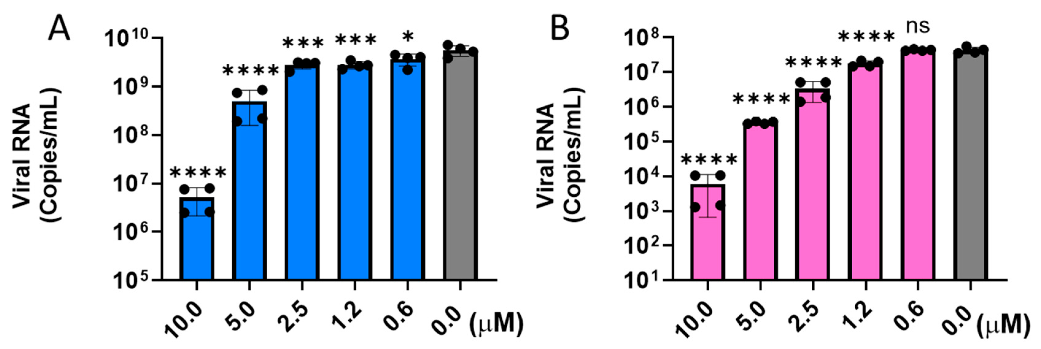
Figure 1. Hit-1 reduces replication of live SARS-CoV-2 in Calu-3 cells as quantified by RT-qPCR for USA-WA1/2020 (A) and Omicron (B.1.1.529 BA.1) (B) variants. Ordinary one-way ANOVA (Dunnett’s test) was used to evaluate the statistical significance between variables and the DMSO control. * p ≤ 0.05, *** p ≤ 0.001, **** p ≤ 0.0001, ns p > 0.05 (not significant).
Ddc 04 00047 g001
Figure 2. Representative SPR binding sensorgrams. Binding of neutralizing antibody SARS2.A.19-42 to SARS-CoV-2 spike proteins from WA1/2020 (A) and B.1.1.529 BA.1 (B) variants analyzed using single-cycle kinetics. Binding of Hit-1 to SARS-CoV-2 spike proteins from WA1/2020 (C) and B.1.1.529 BA.1 (D) variants analyzed using multi-cycle kinetics. Black dotted lines represent fitted curves generated using a 1:1 Langmuir kinetic model.
Ddc 04 00047 g002
Figure 3. In silico prediction of Hit-1 (magenta) and Hit-2 (orange) putative sites at the protein-protein interface of SARS-CoV-2 Spike protein trimer (PDB ID: 7TGY): solvent exposed Site 1 (A); and buried Site 2 (B). Close-up view of S protein interaction with Hit-1 (C,E) and Hit-2 (D,F). All electrostatic interaction distances shown are from donor heteroatom to acceptor heteroatom. All aromatic interaction distances shown are relative to the centroid of the aromatic system.
Ddc 04 00047 g003
Scheme 1. Synthesis of 7,8-dihydropyrido[4,3-d]pyrimidine-based analogs.
Ddc 04 00047 sch001
Table 1. Inhibition of SARS-CoV-2 pseudovirus variants by the HTS hits (in BHK-ACE2 cells).
Ddc 04 00047 i001
VariantHit-1Hit-2Hit-3
EC50 (µM) aSI bEC50 (µM) aSI bEC50 (µM) aSI b
A (Wuhan)0.31 ± 0.092104.93 ± 0.472010.63 ± 0.699
B.1.1.7 (alpha)0.54 ± 0.071204.27 ± 0.532312.69 ± 1.038
B.1.351 (beta)0.32 ± 0.052033.18 ± 1.043113.03 ± 1.658
P.1 (gamma)0.35 ± 0.041863.11 ± 0.413215.53 ± 0.916
B.1.617.2 (Delta)0.21 ± 0.013105.36 ± 0.051910.75 ± 0.359
B.1.1.529 BA.1 (Omicron)0.98 ± 0.07667.23 ± 0.461429.53 ± 0.843
B.1.1.529 BA.20.43 ± 0.012327.63 ± 0.35 13ND-
B.1.1.529 BA.4/50.23 ± 0.034347.31 ± 1.0214ND-
SARS-CoV0.66 ± 0.029915.14 ± 0.5378.89 ± 0.3011
a Values are represented as means ± SD. b Selectivity index is the ratio of CC50/EC50.
Table 2. Physicochemical parameters and predicted ADME properties for Hits 1–3 a.
CompoundMWcLogPSolubilityCaco-2F %Drug Score b
µg/mL× 10−6 cm/s
Hit-13703.46801019843
Hit-24943.64962326
Hit-33844.25401375216
a Determined by Percepta (ACD Labs). Caco-2 permeability: Good > 20, Mod > 10, Bad < 10. Aqueous Solubility (Sw): Good > 60 µg/mL, at pH = 7.4, Mod 10–60 µg/mL, Low < 10 µg/mL. b Determined using Actelion OSIRIS software v.05.05.00 (×100).
Table 3. Inhibition of pseudoviral SARS-CoV-2 by the new 7,8-dihydropyrido[4,3-d]pyrimidine based compounds in BHK-ACE2 cells.
Ddc 04 00047 i002CompoundR1R2EC50 (µM) aSI b
Hit-1I4-Clcyclohexyl-0.32 ± 0.03210
2I4-CF3cyclohexyl-5.20 ± 0.2219
3I3,4-di-Fcyclohexyl-0.93 ± 0.09108
4IHcyclohexyl-6.14 ± 0.3316
5IH4-Cl-phenyl-2.53 ± 0.1040
6IH4-F-phenyl-3.20 ± 0.1331
7IH3,4-methylene
dioxyphenyl-
6.07 ± 0.2516
8I4-OMe3-F-4-Me-phenyl2.11 ± 0.3347
9II4-Cl2-Cl-phenyl-0.47 ± 0.02 207
10II4-Cl4-Cl-phenyl-1.44 ± 0.1469
11II4-Cl3-MeO-phenyl-2.67 ± 0.1628
12II4-CF32-Cl-phenyl-7.13 ± 0.3514
13II3,4-di-F2-Cl-phenyl-0.23 ± 0.07223
14II3,4-di-F4-Cl-phenyl-0.68 ± 0.05147
a Values are represented as means ± SD. b Selectivity index is the ratio of CC50/EC50.
Table 4. Metabolic stability of the selected 7,8-dihydropyrido[4,3-d]pyrimidines.
CompoundMetabolic Stability a
PlasmaLiver Microsome
2 µM2 µM10 µMNo NADPH
HumanHuman
199.578.796.6100
310079.498.1100
710067.3100100
1010031.184.576.5
1110023.172.5100
1310028.374.2100
Verapamil 17.864.1
MouseMouse
110050.688.4100
399.165.989.6100
710081.395.1100
1010028.995.1100
1110016.463.6100
1310023.045.9100
Verapamil 17.959.1
DogDog
110081.498.0100
310085.598.9100
710094.6100100
1010071.557.9100
1110031.168.1100
1310072.782.1100
Verapamil 21.169.9
MonkeyMonkey
110054.494.9100
310070.597.797.9
710072.691.895.4
1010031.194.1100
111001.845.995.6
131005.453.7100
Verapamil 0.227.8
a % of compound remaining after 60 min related to t0 (measured using 2 or 10 µM concentrations of the test compounds). Results are from three replicates.
Table 5. Inhibition of CYP P450 by selected 7,8-dihydropyrido[4,3-d] pyrimidines.
CompoundCYP Inhibition *
CYP1A2CYP2C9CYP2D6CYP3A4
% Inhibition
113.8278.50−0.5768.77
32.5873.0210.0953.56
732.0365.443.2281.20
109.3144.2350.6863.14
1133.05−5.8948.4881.24
13−0.1551.66−1.7450.22
* % Inhibition at 10 µM; phenacetin, tolbutamide, dextromethorphan, and midazolam were used as positive controls in the CYP inhibition assays. Results are from three replicates.
Table 6. PK parameters of compound 13 in BALB/c mice.
Parameter aUniti.v.
(5 mg/kg) b
p.o.
(50 mg/kg) c
T1/2h2.354.0
Tmaxh0.081.83
Cmaxng/mL26774440
AUC(inf)ng·h/mL536636,675
MRT(inf)h3.865.40
Vz/FmL/kg31724260
ClmL/h/kg9401376
F%-68
a The values presented are the mean values from three animals. b Formulation: 5%NMP, 20% PEG400 and 75% of 20% HP-β-CD in water. c Formulation: 10% NMP and 90% sesame oil. T1/2, terminal half-life; Cmax, the maximum concentration that a drug achieves after dosing; AUC, area under curve; MRT, mean residence time; Vz/F, apparent volume of distribution during the terminal phase; Cl, apparent total body clearance. F%, bioavailability. Tested at Pharmaron, Inc. (Beijing, China).
Disclaimer/Publisher’s Note: The statements, opinions and data contained in all publications are solely those of the individual author(s) and contributor(s) and not of MDPI and/or the editor(s). MDPI and/or the editor(s) disclaim responsibility for any injury to people or property resulting from any ideas, methods, instructions or products referred to in the content.