Article

Antimicrobial and Anti-Inflammatory Potentials of Silver Tungstate Nanoparticles, Cytotoxicity and Interference on the Activity of Antimicrobial Drugs

by
Washington de Souza Leal
1,2,†,
Juliane Zacour Marinho
2,3,†,
Isabela Penna Ceravolo
4,
Lucas Leão Nascimento
5,
Antonio Otávio de Toledo Patrocínio
5 and
Marcus Vinícius Dias-Souza
2,6,*
1
Única University Center, Ipatinga 35164-779, MG, Brazil
2
Integrated Pharmacology and Drug Interactions Research Group (GPqFAR), Ipatinga, MG, Brazil
3
Inorganic Chemistry Department, Chemistry Institute, State University of Campinas, Campinas 13083-970, SP, Brazil
4
René Rachou Institute, FIOCRUZ Minas, Belo Horizonte 30190-009, MG, Brazil
5
Chemistry Institute, Federal University of Uberlândia, Uberlândia 38400-902, MG, Brazil
6
Chemistry Laboratory, Anhanguera College, Ipatinga 35160-036, MG, Brazil
*
Author to whom correspondence should be addressed.
These authors contributed equally to this work.
Drugs Drug Candidates 2025, 4(3), 30; https://doi.org/10.3390/ddc4030030
Submission received: 28 May 2025 / Revised: 14 June 2025 / Accepted: 19 June 2025 / Published: 23 June 2025
(This article belongs to the Section Medicinal Chemistry and Preliminary Screening)

Abstract

Background: Bacterial resistance to antimicrobial drugs is a critical phenomenon that is hampering clinical treatments, raising the need for promising compounds that can be explored as pharmaceutical products. This study investigated the antimicrobial potential of α-Ag2WO4–alpha phase, orthorhombic structure silver tungstate nanoparticles (STN), against clinical isolates of Staphylococcus aureus, Pseudomonas aeruginosa and Escherichia coli, alone and combined to clinically relevant antimicrobial drugs. Methods: We used classical methods (MIC/checkerboard) to investigate the antimicrobial activity of STN. We characterized STN using X-ray diffraction, photoluminescence and scanning electron microscopy. We also performed cytotoxicity tests on BGM cells and anti-inflammatory tests in vitro. Results: STN was effective at 128 µg/mL for S. aureus and at 256 µg/mL for E. coli, but was not effective against P. aeruginosa. When combined with antimicrobials, STN decreased their MIC values, and its anti-inflammatory potential was confirmed. CC50 of STN was of 16.23 ± 1.09 μg/mL against BGM cells. Conclusions: Our data open doors for further studies in animal models to investigate the effects on STN in infectious diseases.

1. Introduction

Semiconductors are nanostructured crystals of electronic behavior between insulators and conductors, being generally classified as extrinsic (with doping agents) or intrinsic (no doping agent), and crystalline or amorphous [1,2]. Semiconductors have been explored in automotive and computing industries, and in technologies such as power supply [3,4]. In recent years, they have been explored as nanoparticles in health sciences, mostly as new materials for diagnostics, non-surgical and surgical treatments, and for drug delivery [5,6].
In this study, we investigated the antimicrobial potential of silver tungstate (Ag2WO4) nanoparticles (STN), a thermostable semiconductor of technological interest due to its optical and electromagnetic properties [7,8]. STN are effective in releasing silver, which is widely known for its antimicrobial properties [9,10], which becomes even more relevant considering the complex scenario of bacterial resistance to antimicrobial drugs (BRAD). BRAD is a critical issue in public health, as it decreases the effectiveness of pharmacological treatments [11]. Throughout intricate molecular pathways, bacteria evade the mechanism of action of the drugs and keep the clinical course of infectious diseases. Clinically, it becomes necessary to increase the amount and/or number of drugs during the treatment [11]. There is a need for alternative therapies, but the development of new drugs and their approval by regulatory institutions are not happening at the speed necessary to properly deal with the BRAD scenario, which is expected to become even more complex in the forthcoming years.
Here we show that STN can be effective against clinical isolates of Gram-positive and Gram-negative bacterial species, alone and combined to clinically relevant antimicrobial drugs. The nanoparticles were synthesized using a hydrothermal reactor and were structurally investigated by X-ray diffraction (XRD) confirming the crystal structure of STN, photoluminescence and scanning electron microscopy (SEM). We also tested STN for cytotoxicity, and for its anti-inflammatory potential in vitro. Given the scarcity of studies on STN for antimicrobial activity and the potential for clinical use, our data become even more relevant.

2. Results

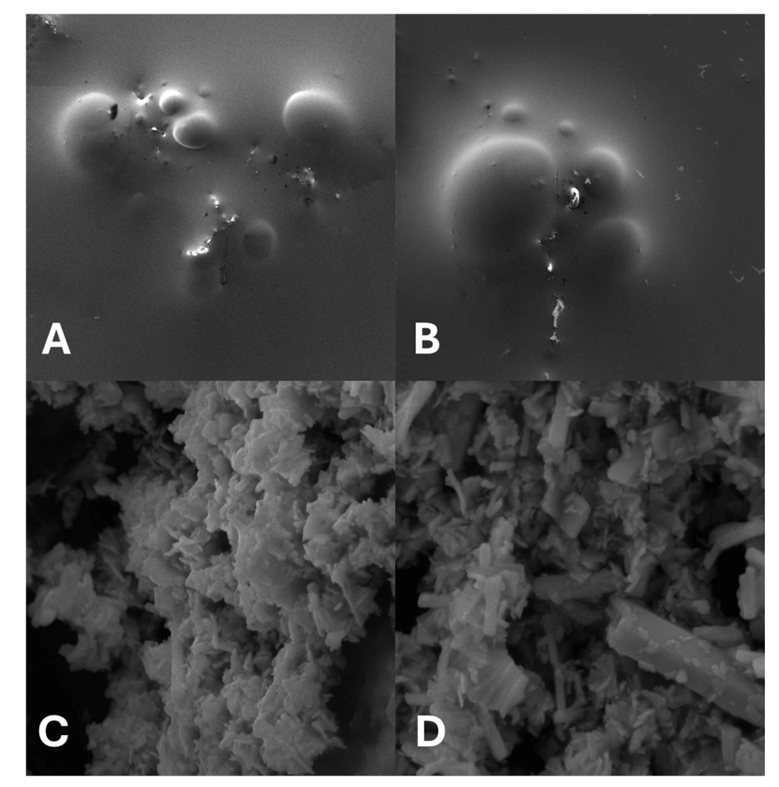
2.1. Structural, Optical and Morphological Study of the Nanoparticles

Figure 1A shows the XRD pattern of the synthesized α-Ag2WO4 nanostructures for the STN, indicating that the sample have an α-orthorhombic structure with a space group of Pn2n and a C2v10 symmetry (JCDPS n° 34-0061) [12], without any secondary phases or impurities. The diffraction peaks are narrow and well defined, indicating good long-range structural ordering in the lattice, and they are all in agreement with the standard crystallographic data. α-Ag2WO4 has photoluminescence emission at room temperature with a characteristic broadband, as observed in Figure 1B. The emission band has a maximum intensity in the range of 500−550 nm. This peak may be associated with the presence of silver or oxygen vacancies [12].
The SEM images of STN with bacteria were obtained with gold coating, and the images without bacteria were obtained from the short period synthesis without surfactant under 90 °C (Figure 2). The nanoparticles are micro-sized rod-like shaped, with wide size distribution.

2.2. Antimicrobial Activity of STN

STN was effective against S. aureus and E. coli isolates, but no activity was seen against P. aeruginosa isolates (Table 1). The antimicrobial drugs were tested following the indication for clinical use, and the MIC values were significantly lower than those obtained for STN (p < 0.01).
Given these results, we performed the checkerboard assay for S. aureus strains only (Table 2). The combination of STN and the drugs resulted in significant decrease in MIC values (p < 0.05), with a FICi value of 0.156 for the combination of STN and sulfamethoxazole, and 0.5 for the combination of STN and clindamycin. STN displayed synergism with both drugs, with more interesting results for sulfamethoxazole.

2.3. Cytotoxicity and Anti-Inflammatory Potential of STN

The CC50 of STN was of 16.23 ± 1.09 μg/mL against BGM cells. Concerning the anti-inflammatory test, STN was effective in preventing BSA denaturation (Figure 3) at the concentrations of 128 μg/mL (MIC for S. aureus) and at 1024 μg/mL, but there was no significant difference between their effects. Tenoxicam at 1 mg/mL was more effective than STN in both concentrations (p < 0.05).

3. Discussion

This study provides evidence on the antimicrobial potential of STN being more effective to S. aureus than to Gram-negative pathogens. When combined to antimicrobials, the MIC of STN decreased significantly, and its anti-inflammatory potential was observed in vitro. Although the MIC of STN was higher than clinically relevant antimicrobial drugs and lacked activity against P. aeruginosa, they remain of interest as an antimicrobial due to important properties related to possible mechanisms of action. STN ferroelasticity (i.e., reversible mechanical deformation) can provide different patterns of interaction with bacterial membranes [13], which can cause direct damage to bacterial membranes. Its photoluminescence is linked to the formation of electron–hole pairs upon excitation [14], as expected for semiconductors, which leads to the generation of radical species with antimicrobial activity [13,14,15]. The presence of silver increases the generation of radical species, and thus, the antimicrobial potential of STN [15].
The STN used in this study are of the α-Ag2WO4 polymorph type, which has photoluminescence emission at room temperature with a characteristic broadband, as observed in Figure 1B. This is typical of materials in which the relaxation step occurs in several paths, with the participation of several energy states within the band gap, according to the broadband model [13,14,15,16,17]. This broadband is generally formed by blue, green and yellow light components, which indicates a high level of surface irregularities [18], and can potentially contribute to mechanical mechanisms of action such as pore formation [15].
Our data are in agreement with the observation of others concerning the antimicrobial activity of STN. However, few studies used standardized methods of antimicrobial susceptibility tests, such as CLSI protocols. In this context, a study synthesized Ag2WO4 microcrystals using a microwave hydrothermal method and tested them against an ATCC strain of methicillin-resistant Staphylococcus aureus (MRSA) [19]. The microcrystals were divided into two groups: with and without irradiation by an electron beam. The MIC value of irradiation-treated microcrystals was 31 μg/mL, whereas the nonirradiated ones were effective at 125 μg/mL [19], in similarity to our results.
A study tested Ag2WO4 against different pathogenic bacterial species [20]. Among E. coli strains, the microcrystals were effective at concentrations ranging from 7.81 to 62.5 μg/mL. The highest MIC value (250 μg/mL) was observed for Yersinia enterocolítica, Salmonella typhimurium and Proteus mirabilis, followed by Klebisiella pneumoniae (125 μg/mL), Pseudomonas aeruginosa (62.5 μg/mL) and Acinetobacter baumanii (31.25 μg/mL). The microcrystals were not effective against Hafnia alvei, Salmonella typhi and Serratia marcescens in both microdilution and agar well-diffusion tests. A commercial combination of imipenem and sodium cilastatin was more effective than the microcrystals in all tested strains, as observed in this study for the clinically relevant antimicrobial drugs and STN.
A MIC value of 62.5 μg/mL was obtained for Ag2WO4 microcrystals of rod-like elongated shape with around 1 μm length, 140 nm width and 120 nm thickness. The same value was obtained in minimal fungicidal concentration tests, indicating its rather fungicidal (than fungistatic) character [21]. Also, α-Ag2WO4 microcrystals were effective against Candida albicans and Trichophyton rubrum, with MIC values ranging from 0.5 to 2 µg/mL. The results for C. albicans strains were superior to ketoconazole [22].
To the best of our knowledge, this is the first time that STN are combined to antimicrobial drugs against clinical isolates of S. aureus. Synergism was confirmed using the classical checkerboard method. The combined mechanisms might be explored in further in vivo studies. Recently, our group used a similar approach to investigate the effects of combining zinc oxide nanoparticles to antimicrobial drugs, and a significant decrease in MIC values was also observed [23].
Concerning the anti-inflammatory potential of STN, it was effective at 128 μg/mL, despite being not statistically superior to tenoxicam at 1 mg/mL. Few studies have investigated the effects of silver tungstate on the immune system. A recent investigation described that silver tungstate microcrystals could decrease the production of pro-inflammatory cytokines in macrophage-like cells, but they could remain unchanged as well [24]. Here we used an in vitro model that explores preventing BSA denaturation as an indicator of anti-inflammatory potential. Our group explored this approach with zinc oxide nanoparticles and Moringa oleifera extract [23,25], and their anti-inflammatory potential was confirmed. In vivo studies are necessary to investigate the effects on immunoregulation and confirm the anti-inflammatory activity.
Regarding the cytotoxicity of STN, CC50 was of 16.23 ± 1.09 μg/mL, which is superior to other silver nanoparticles thresholds described elsewhere [26,27]. The MIC of STN exceeds the CC50 value observed in BGM cells, thus, structural modifications or even encapsulation strategies such as liposomes or cyclodextrins need to be explored in further studies to overcome this limitation for direct therapeutic use. However, the observed toxicity may not fully reflect STN behavior in more complex biological systems. Also, several structural variations are possible for silver tungstate, and thus, this can influence cytotoxicity, beyond the concentration and size of the nanoparticles, which open doors for more cytotoxicity studies. In this context, α-Ag2WO4 microcrystals of hexagonal long shape measuring approximately 100 nm were cytotoxic to fibroblast cells at the concentration of 7.81 μg/mL, causing DNA degradation but no significant effect on cell viability [28].

4. Materials and Methods

4.1. Synthesis and Characterization of Silver Tungstate Nanoparticles (STN)

In a typical synthesis procedure of Ag2WO4 crystals, appropriate quantities of AgNO3 and Na2WO4·2H2O were dissolved separately into 50 mL of deionized water. After 5 min of stirring, to ensure complete dissolution, the AgNO3 solution was then added to the Na2WO4·2H2O solution with continuous stirring. The chosen ratio for the precursor concentrations was 1:1 AgNO3/Na2WO4·2H2O. The pH of the solution was adjusted to 9 with 1 mol/L NH4OH solution, and then it was transferred to a hydrothermal reactor and kept heated at 180 °C for 2 h. The sample was washed several times using deionized water, ethanol and then dried in an oven at 70 °C, 12 h.
We carried out X-ray diffraction (XRD) measurements using a Shimadzu XRD-6000 diffractometer with CuK radiation (λ = 1.5406 Å), scanning over a 2θ range of 10° to 80° at a rate of 0.05°/min. Photoluminescence measurements were performed on a Fluoromax 4C spectrofluorometer in front-face geometry.

4.2. Cytotoxicity Assay

We evaluated the cytotoxic potential of STN using BGM cell cultures.. Cells were maintained in RPMI medium (Gibco, Thermo Scientific, Waltham, MA, USA), supplemented with 2 mM glutamine, penicillin, streptomycin and 10% fetal bovine serum (Gibco), as detailed in earlier studies from our group [29]. For the assay, 1 × 10⁴ cells were seeded per well in 180 μL of medium. After a 24 h incubation period, cells were treated with seven concentrations of the test samples (20 μL, ranging from 250 to 3.9 μg/mL). Following 3–4 h of incubation, lysosomal membrane integrity was assessed using a neutral red uptake assay (50 µg/mL). Cytotoxicity was quantified by measuring absorbance at 540 nm on a Synergy H4 Hybrid Reader spectrophotometer (BioTek Instruments, Winooski, VT, USA). Untreated cells cultured in RPMI served as the negative control. The concentration required to reduce cell viability by 50% (CC50) was calculated based on results from at least two independent experiments, in triplicate, using Origin 8.0 software (OriginLab Corporation, Northampton, MA, USA).

4.3. Bacterial Isolates

We used a total of 30 bacterial isolates, including 10 strains each of Escherichia coli, Pseudomonas aeruginosa and Staphylococcus aureus. E. coli samples were obtained from urinary tract infections, P. aeruginosa from tracheal aspirates and S. aureus from catheter tips. All the isolates were sourced from the microorganism collection of Anhanguera College. Species identification was performed using the VITEK 2 system, version R04.02 (bioMérieux, Marcy-l’Étoile, France). Their identification was considered valid if the similarity index reached 90% or greater values.

4.4. Minimal Inhibitory Concentration (MIC) Assay

We determined the minimum inhibitory concentration (MIC) of STN using the microdilution assay in sterile, untreated 96-well polystyrene microplates, in accordance with CLSI guidelines [30]. A 4 mg/mL stock solution of STN was prepared in a sterilized blend of propylene glycol, glycerin and water (4:4:2 v/v/v), and subsequently diluted in sterile water to achieve a final volume of 100 µL/well. The bacterial inoculum was initially adjusted to a 0.5 McFarland standard (approximately 1.5 × 10⁸ CFU/mL) in 0.9% sterile saline, and then further diluted in freshly prepared sterile Mueller-Hinton broth at double concentration (Difco, Becton Dickinson, Franklin Lakes, NJ, USA) to reach a final concentration of 1 × 10⁵ CFU/mL. The final STN concentrations ranged from 1024 to 8 µg/mL, with a bacterial load of 5 × 10⁴ CFU/mL in each well (final volume of 200 µL). MIC values were defined as the lowest STN concentrations at which resazurin dye (0.1 g/L, 50 μL) did not change color from blue to pink, indicating complete inhibition of bacterial growth. The negative control consisted in an STN solution at 1000 µg/mL.
This procedure was also performed to determine the MIC of azithromycin, gentamicin, clindamycin and sulfamethoxazole (all from Sigma, St. Louis, MO, USA), for each species. Stock solutions of the drugs (4 mg/mL) were prepared in sterile deionized water, reaching a final concentration ranging from 1024 to 8 µg/mL, with a final concentration of 5 × 10⁴ CFU/mL for the bacterial suspensions (final volume of the wells: 200 μL). Resazurine staining (0.1 g/L, 50 μL) was used as described.

4.5. Drug Interactions Assay

We used the checkerboard assay to evaluate the interaction between STN and clinically relevant antimicrobial drugs. The antimicrobials were horizontally diluted in a two-fold series to obtain final concentrations ranging from 1024 to 0.5 µg/mL. STN was serially diluted vertically in the same plate, with final concentrations ranging from 1024 to 8 µg/mL. For each combination, the fractional inhibitory concentration (FIC) of both agents was determined, and the FIC index (FICi) was calculated. The interactions were classified as synergistic when FICi ≤ 0.5, additive when 0.5 < FICi ≤ 4 and antagonistic when FICi > 4 [31].

4.6. Anti-Inflammatory Potential of STN

The anti-inflammatory potential of STN was assessed using the bovine serum albumin (BSA) denaturation assay, as described by our group [25]. BSA (Thermo Fisher, Waltham, MA, USA) was prepared as a 5% aqueous solution and exposed to heat in a water bath (70 °C, 15 min) with the addition of STN at 1024 μg/mL and at the lowest MIC value found in the experiments. Tenoxicam (Sigma, St. Louis, MO, USA) at 1 mg/mL was used as a reference anti-inflammatory drug. Pure BSA and tenoxicam-added BSA were used as controls.

4.7. Scanning Electron Microscopy (SEM)

We prepared sterile glass slides with overnight cultures of S. aureus and STN at the concentration of 1024 μg/mL (1:1 v/v), and a 5% formaldehyde solution was used as a fixative (overnight exposure at room temperature). Following, the slides were air dried, gold-coated under deep vacuum and fixed to metallic supports. The analysis was then carried out in a VEGA 3 LMU scanning electron microscope (Tescan, Czech Republic) at 5 kV, using the equipment software, following the manufacturer’s instructions.

4.8. Statistics

Results were analyzed using Shapiro–Wilk test (normality) and Chi-square (antimicrobial activity data) or ANOVA followed by post hoc Tukey test (anti-inflammatory potential data). Homoscedasticity was checked using the Bartlett test. Significance was set as p < 0.05. The analysis was carried out using Bioestat 5.3 for Windows.

5. Conclusions

This study highlights the potential of STN as an antimicrobial product, and despite its higher MIC compared to clinically relevant drugs, its properties such as ferroelasticity, photoluminescence and anti-inflammatory activity, contribute to make it of interest for further studies, especially if combined to antimicrobial drugs. The structural and surface characteristics, beyond the generation of radical species, align with its antimicrobial mechanism. More studies are necessary to decrease its cytotoxicity and possibly improve the efficacy on Gram-negative pathogens, as well as chemical and physical properties, making it safe enough for in vivo tests using animal models. Furthermore, long-term biocompatibility studies with biological tissues are necessary to take them for clinical studies. Such limitations, however, do not impair our assessment of the antimicrobial potential of STN against bacterial pathogens.

Author Contributions

W.d.S.L.: Investigation, formal analysis, writing—original draft; J.Z.M.: Investigation, formal analysis, writing—original draft; I.P.C.: Investigation, formal analysis; L.L.N.: Investigation, formal analysis: A.O.d.T.P.: Supervision, methodology, validation, resources; M.V.D.-S.: Conceptualization, supervision, methodology, validation, formal analysis, resources, writing—final version, project administration. All authors have read and agreed to the published version of the manuscript.

Funding

This research received no external funding.

Institutional Review Board Statement

Not applicable.

Informed Consent Statement

Not applicable.

Data Availability Statement

All data supporting results can be found in the manuscript.

Acknowledgments

We are thankful to Renan Martins dos Santos (Unica University Center, MG, Brazil), for all the assistance with the biological experiments; to Murilo Neia Thomaz da Silva, for the assistance with SEM analysis; to Rodrigo Emanoel Feliciano Ramos (Federal University of Juiz de Fora, MG, Brazil), for the discussions about the paper; and to Miriam Chaves Schultz (Federal University of Minas Gerais, MG, Brazil), for kindly providing the BGM cells used in this study.

Conflicts of Interest

The authors declare no conflicts of interest.

Abbreviations

The following abbreviations are used in this manuscript:
BRADbacterial resistance to antimicrobial drugs
BSAbovine serum albumin
CFUcolony forming units
FICfractional inhibitory concentration
FICifractional inhibitory concentration index
MICminimal inhibitory concentration
STNsilver tungstate nanoparticles
XRDX-ray diffraction

References

  1. Huang, A.Q. Power semiconductor devices for smart grid and renewable energy systems. In Power Electronics in Renewable Energy Systems and Smart Grid: Technology and Applications; Wiley: Hoboken, NJ, USA, 2019; pp. 85–152. [Google Scholar] [CrossRef]
  2. Zhang, Y.; Dong, D.; Li, Q.; Zhang, R.; Udrea, F.; Wang, H. Wide-bandgap semiconductors and power electronics as pathways to carbon neutrality. Nat. Rev. Electr. Eng. 2025, 2, 155–172. [Google Scholar] [CrossRef]
  3. Maximenko, S.I.; Moore, J.E.; Affouda, C.A.; Jenkins, P.P. Optimal Semiconductors for 3H and 63Ni Betavoltaics. Sci. Rep. 2019, 9, 10892. [Google Scholar] [CrossRef] [PubMed]
  4. Lu, H.; Vardeny, Z.V.; Beard, M.C. Control of light, spin and charge with chiral metal halide semiconductors. Nat. Rev. Chem. 2022, 6, 470–485. [Google Scholar] [CrossRef]
  5. Maya-Vetencourt, J.F.; Manfredi, G.; Mete, M.; Colombo, E.; Bramini, M.; Di Marco, S.; Shmal, D.; Mantero, G.; Dipalo, M.; Rocchi, A.; et al. Subretinally injected semiconducting polymer nanoparticles rescue vision in a rat model of retinal dystrophy. Nat. Nanotechnol. 2020, 15, 698–708. [Google Scholar] [CrossRef]
  6. Tan, Y.; Xiong, M.; Liu, Q.; Yin, Y.; Yin, X.; Liao, S.; Wang, Y.; Hu, L.; Zhang, X.-B. Precisely controlling the cellular internalization of DNA-decorated semiconductor polymer nanoparticles for drug delivery. RSC Adv. 2022, 12, 31173–31179. [Google Scholar] [CrossRef] [PubMed]
  7. Farsi, M.; Nezamzadeh-Ejhieh, A. A coupled Cobalt(II) oxide-Silver Tungstate nano-photocatalyst: Moderate characterization and evaluation of the photocatalysis kinetics towards methylene blue in aqueous solution. Polyhedron 2022, 219, 115823. [Google Scholar] [CrossRef]
  8. Rakshitha, R.; Gurupadayya, B.; Devi, S.H.K.; Pallavi, N. Coprecipitation aided synthesis of bimetallic silver tungstate: A response surface simulation of sunlight-driven photocatalytic removal of 2,4-dichlorophenol. Environ. Sci. Pollut. Res. 2022, 29, 59433–59443. [Google Scholar] [CrossRef]
  9. de Abreu, C.B.; Gebara, R.C.; dos Reis, L.L.; Rocha, G.S.; Alho, L.d.O.G.; Alvarenga, L.M.; Virtuoso, L.S.; Assis, M.; Mansano, A.d.S.; Longo, E.; et al. Toxicity of α-Ag2WO4 microcrystals to freshwater microalga Raphidocelis subcapitata at cellular and population levels. Chemosphere 2022, 288 Pt 2, 132536. [Google Scholar] [CrossRef]
  10. Lansdown, A.B. Silver in health care: Antimicrobial effects and safety in use. Curr. Probl. Dermatol. 2006, 33, 17–34. [Google Scholar]
  11. Dias-Souza, M.V.; Perpétuo, A.A.; dos Santos, G.S.; Machado, L.F.C.; dos Santos, R.M. Natural products in drug discovery: Meeting the urgency for new antimicrobials for human and veterinary use. AIMS Mol. Sci. 2023, 10, 11–21. [Google Scholar] [CrossRef]
  12. Macedo, N.G.; Gouveia, A.F.; Roca, R.A.; Assis, M.; Gracia, L.; Andrés, J.; Leite, E.R.; Longo, E. Surfactant-mediated morphology and photocatalytic activity of α-Ag2WO4 material. J. Phys. Chem. C 2018, 122, 8667–8679. [Google Scholar] [CrossRef]
  13. Isupov, V.A. ferroelectric and ferroelastic phase transitions in molybdates and tungstates of monovalent and bivalent elements. Ferroelectrics 2005, 322, 83–114. [Google Scholar] [CrossRef]
  14. Cavalcante, L.S.; Almeida, M.A.P.; Avansi, W.; Tranquilin, R.L.; Longo, E.; Batista, N.C.; Mastelaro, V.R.; Li, M.S. Cluster coordination and photoluminescence properties of α-Ag2WO4 microcrystals. Inorg. Chem. 2012, 51, 10675–10687. [Google Scholar] [CrossRef]
  15. Alvarez-Roca, R.; Gouveia, A.F.; de Foggi, C.C.; Lemos, P.S.; Gracia, L.; da Silva, L.F.; Vergani, C.E.; San-Miguel, M.; Longo, E.; Andrés, J. Selective synthesis of α-, β-, and γ-Ag2WO4 polymorphs: Promising platforms for photocatalytic and antibacterial materials. Inorg. Chem. 2021, 61, 1245–1258. [Google Scholar] [CrossRef]
  16. Goldmann, D.; Rajan, S.; Udayakumar, K. Preventing and Controlling Global Antimicrobial Resistance—Implementing a Whole-System Approach. N. Engl. J. Med. 2024, 391, 681–685. [Google Scholar] [CrossRef]
  17. Kokilavani, S.; Al-Farraj, S.A.; Thomas, A.M.; El-Serehy, H.A.; Raju, L.L.; Khan, S.S. Enhanced visible light driven photocatalytic and antibacterial activities of Ag2WO4 decorated ZnS nanocomposite. Ceram. Int. 2021, 47, 12997–13006. [Google Scholar] [CrossRef]
  18. Longo, E.; Volanti, D.P.; Longo, V.M.; Gracia, L.; Nogueira, I.C.; Almeida, M.A.P.; Pinheiro, A.N.; Ferrer, M.M.; Cavalcante, L.S.; Andrés, J. Toward an understanding of the growth of Ag filaments on α-Ag2WO4 and their photoluminescent properties: A combined experimental and theoretical study. J. Phys. Chem. C 2014, 118, 1229–1239. [Google Scholar] [CrossRef]
  19. Foggi, C.C.; De Oliveira, R.C.; Fabbro, M.T.; Vergani, C.E.; Andres, J.; Longo, E.; Machado, A.L. Tuning the morphological, optical, and antimicrobial properties of α-Ag2WO4 microcrystals using different solvents. Cryst. Growth Des. 2017, 17, 6239–6246. [Google Scholar] [CrossRef]
  20. Bastos, I.S.; Nobre, F.X.; da Silva, E.R.; Orlandi, P.P.; Lima, D.C.; da Cunha Mendes, O.; Manzato, L.; Pereira, M.L.R.D.; Leyet, Y.; Couceiro, P.R.C.; et al. Silver tungstate microcrystals and their performance over several clinical multidrug resistant microorganisms. Colloids Surf. A Physicochem. Eng. Asp. 2022, 647, 129132. [Google Scholar] [CrossRef]
  21. Foggi, C.C.; Fabbro, M.T.; Santos, L.P.; de Santana, Y.V.; Vergani, C.E.; Machado, A.L.; Cordoncillo, E.; Andrés, J.; Longo, E. Synthesis and evaluation of α-Ag2WO4 as novel antifungal agent. Chem. Phys. Lett. 2017, 674, 125–129. [Google Scholar] [CrossRef]
  22. Nobre, F.X.; Bastos, I.S.; Fontenelle, R.O.d.S.; Júnior, E.A.A.; Takeno, M.L.; Manzato, L.; de Matos, J.M.E.; Orlandi, P.P.; Mendes, J.d.F.S.; Brito, W.R.; et al. Antimicrobial properties of α-Ag2WO4 rod-like microcrystals synthesized by sonochemistry and sonochemistry followed by hydrothermal conventional method. Ultrason. Sonochemistry 2019, 58, 104620. [Google Scholar] [CrossRef] [PubMed]
  23. Machado, L.F.C.; Marinho, J.Z.; de Lima, R.C.; Ceravolo, I.P.; Dias-Souza, M.V. Rapidly synthesized zinc oxide nanoparticles can increase the activity of antimicrobial drugs against clinical isolates of Pseudomonas aeruginosa and Escherichia coli. Acta Pharm. Sci. 2023, 61, 414. [Google Scholar] [CrossRef]
  24. Pimentel, B.N.A.d.S.; De Annunzio, S.R.; Assis, M.; Barbugli, P.A.; Longo, E.; Vergani, C.E. Biocompatibility and inflammatory response of silver tungstate, silver molybdate, and silver vanadate microcrystals. Front. Bioeng. Biotechnol. 2023, 11, 1215438. [Google Scholar] [CrossRef]
  25. Alcântara, L.K.S.; Machado, L.F.C.; Ceravolo, I.P.; Martins, R.; Souza, M.V.D. Antibiofilm effect of moringa oleifera leaf extract against staphylococcus aureus, cytotoxicity, biochemical aspects, anti-inflammatory potential, and interference on the activity of antimicrobial drugs. Fabad J. Pharm. Sci. 2023, 48, 241–254. [Google Scholar] [CrossRef]
  26. Arora, S.; Jain, J.; Rajwade, J.M.; Paknikar, K.M. Cellular responses induced by silver nanoparticles: In vitro studies. Toxicol. Lett. 2008, 179, 93–100. [Google Scholar] [CrossRef]
  27. Kim, S.; Choi, J.E.; Choi, J.; Chung, K.H.; Park, K.; Yi, J.; Ryu, D.Y. Oxidative stress-dependent toxicity of silver nanoparticles in human hepato-ma cells. Toxicol. Vitr. 2009, 23, 1076–1084. [Google Scholar] [CrossRef] [PubMed]
  28. Chávez, N.L.H.; de Avila, E.D.; Barbugli, P.A.; de Oliveira, R.C.; de Foggi, C.C.; Longo, E.; Vergani, C.E. Promising effects of silver tungstate microcrystals on fibroblast human cells and three dimensional collagen matrix models: A novel non-cytotoxic material to fight oral disease. Colloids Surf. B Biointerfaces 2018, 170, 505–513. [Google Scholar] [CrossRef]
  29. Dias-Souza, M.V.; dos Santos, R.M.; Cerávolo, I.P.; Cosenza, G.; Marçal, P.H.F.; Figueiredo, F.J.B. Euterpe oleracea pulp extract: Chemical analyses, antibiofilm activity against Staphylococcus aureus, cytotoxicity and interference on the activity of antimicrobial drugs. Microb. Pathog. 2018, 114, 29–35. [Google Scholar] [CrossRef]
  30. CLSI Standard M07; CLSI. Methods for Dilution Antimicrobial Susceptibility Tests for Bacteria That Grow Aerobically, 12th ed. Clinical and Laboratory Standards Institute: Wayne, PA, USA, 2018.
  31. Osman, M.E.; Abo-Elnasr, A.A.; Mohamed, E.T. Therapeutic potential activity of quercetin complexes against Streptococcus pneumoniae. Sci. Rep. 2024, 14, 12876. [Google Scholar] [CrossRef]
Figure 1. Structural and morphological characterization of α-Ag2WO4 nanostructures. (A) X-ray diffractogram, Miller index values in blue. (B) Photoluminescence spectrum.
Ddc 04 00030 g001
Figure 2. SEM images of STN with S. aureus strain ((A,B) magnification of 1 Kx) and alone ((C,D) magnification of 15 Kx).
Ddc 04 00030 g002
Figure 3. Anti-inflammatory potential of STN compared to untreated control and tenoxicam. BSA: bovine serum albumin, MIC: minimal inhibitory concentration. *: Statistically significant.
Ddc 04 00030 g003
Table 1. Results on the antimicrobial activity of STN and clinically relevant drugs against the bacterial isolates.
Bacterial SpeciesMIC Values (μg/mL)
STNAzithromycinGentamycinSulfamethoxazoleClindamycin
S. aureus128NTNT168
P. aeruginosaNE88NTNT
E. coli25688NTNT
MIC: minimal inhibitory concentration. NE: not effective. NT: not tested. STN: silver tungstate nanoparticles. Results are referent to all tested isolates for each species.
Table 2. Results on the antimicrobial activity of STN combined to clinically relevant drugs against the bacterial isolates.
ParameterSulfamethoxazoleClindamycinSTN
MIC alone16 μg/mL8 μg/mL128 μg/mL
MIC in combination0.5 μg/mL2 μg/mL16 μg/mL α
32 μg/mL β
FIC0.031250.250.125 γ
0.25 δ
FIC: fractional inhibitory concentration. STN: silver tungstate nanoparticles. α: MIC of STN combined to sulfamethoxazole. β: MIC of STN combined to clindamycin. γ: FIC of STN combined to sulfamethoxazole. δ: FIC of STN combined to clindamycin.
Disclaimer/Publisher’s Note: The statements, opinions and data contained in all publications are solely those of the individual author(s) and contributor(s) and not of MDPI and/or the editor(s). MDPI and/or the editor(s) disclaim responsibility for any injury to people or property resulting from any ideas, methods, instructions or products referred to in the content.

