Abstract
Background/Objectives: This systematic review explores the utilization of electrospinning for the incorporation of natural compounds, focusing on their pharmacological applications. Methods: This systematic review focused on studies investigating the incorporation of natural bioactive compounds into nanofibers produced via the electrospinning technique for pharmacological applications. The search was conducted for English-language articles published online between 1 January 2013 and 10 December 2023. The review followed a structured approach, excluding review articles, clinical studies, and gray literature such as unpublished works, non-peer-reviewed journals, theses, and industry data. Results: The review of 99 articles highlighted the advantages of electrospun nanofibers in tissue regeneration, infection control, and controlled drug release, with notable potential in oncology for targeted antitumor drug delivery. It discussed the influence of polymers and solvents on fiber characteristics and identified a significant gap in cosmetic applications, emphasizing the technique’s potential for prolonged release and improved ingredient stability. Additionally, this review noted the underutilization of marine-derived substances, which possess rich bioactive properties that could benefit biomedical and cosmetic fields. Conclusions: This systematic review highlights the advantages of electrospinning for pharmacological applications, including tissue regeneration, infection control, and controlled drug release, with promising potential in oncology. However, gaps were identified in the cosmetic field and the use of marine-derived substances. Future advancements in electrospinning technology and interdisciplinary collaboration are essential to unlocking its full potential in medicine and cosmetics.
1. Introduction
Electrospinning is a versatile polymer processing technique that has garnered increasing attention in recent decades due to its ability to produce ultrafine fibers with diameters ranging from nanometers to micrometers [1,2]. This technique relies on the application of an electric field to a polymeric solution or melt, which is extruded through a needle or nozzle (Figure 1). The electric field induces electrostatic charge in the solution, resulting in the formation of a Taylor cone and, ultimately, the ejection of very fine fibers towards a collector, where the fibers are deposited to form a fibrous material [3].

Figure 1.
Schematic diagram of the electrospinning setup, depicting the key components: syringe pump, spinneret (needle), high-voltage source, and collector. The diagram emphasizes the formation of the polymer jet and its subsequent solidification into fibers.
The fibers produced by electrospinning exhibit a high surface-to-volume ratio and a distinctive porous structure, making these highly desirable for a wide range of applications [4,5]. In the biomedical field, electrospinning has been explored for the ability to produce scaffolds (three-dimensional structures) that mimic the natural extracellular matrix, providing a conducive three-dimensional environment for cell growth, proliferation, and differentiation [6]. Additionally, electrospun fibers produced by can be loaded with therapeutic agents such as drugs, genes, or growth factors for drug delivery or tissue engineering applications [6].
Recently, there has been a growing interest in the incorporation of natural products into electrospun materials, owing to the therapeutic and bioactive properties of these compounds. Natural products derived from plants, marine organisms, and other biological sources are rich in bioactive compounds such as polyphenols, flavonoids, terpenes, and alkaloids, which exhibit a variety of beneficial biological effects, including antioxidant, anti-inflammatory, antimicrobial, and wound-healing activity [7].
The incorporation of these natural products into electrospun materials can confer additional functionalities to the resulting products, making them suitable for an even wider range of biomedical applications. The combination of the unique properties obtained by electrospun materials, with the therapeutic features of natural products, offers a promising opportunity for the development of advanced materials with applications in tissue engineering, regenerative medicine, biomimetic dressings, and more [4].
In this systematic review, we address the current state or the art of the research on incorporation of natural products into fibrous materials through electrospinning, highlighting the pharmacological applications and recent advances in this area. Additionally, we discuss the challenges and opportunities associated with this approach, as well as the prospects for the development of advanced nanofiber materials based on electrospinning and natural products.
2. Results and Discussion
A total of 2391 articles were found, of which 99 were chosen to compose this systematic review (Supplementary Materials). A total of 2292 articles were excluded, of which 38 were reviews and 115 were duplicates (Figure 2).
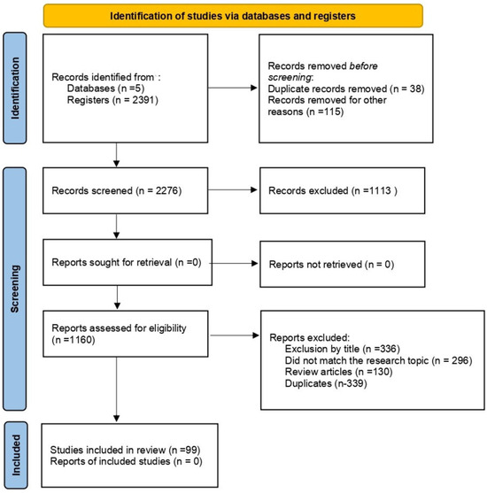
Figure 2.
PRISMA flowchart on article screening: This diagram represents the step-by-step process of identifying, screening, and selecting articles for inclusion in the study. It details the number of records retrieved from databases, the screening criteria applied, the exclusions made, and the final number of studies included in the analysis, ensuring transparency and reproducibility in systematic reviews.
2.1. General Findings
The use of natural substances in pharmacology dates back to ancient times and has evolved over the centuries. Recently, the development of new material fabrication techniques, such as electrospinning, has opened up new possibilities by allowing the incorporation of these substances into fibrous materials, widening their pharmacological applications. This method has gained prominence due to its ability to produce ultrafine fibers with high surface area, facilitating the controlled release of drugs and bioactive substances.
An analysis of the geographical distribution of articles reveals a significant diversity of contributions worldwide (Figure 3). Countries in Asia and the Middle East stand out as leaders in this area, with a considerable number of studies published. For example, Iran stands out with an impressive number of 21 articles, exploring the potential of substances derived from saffron, cinnamon, cod liver oil, propolis extract, and various traditional medicinal herbs found in the region, such as Satureja mutica Fisch. and C.A.Mey. and Zataria multiflora Boiss. This prolific scientific output reflects the rich cultural and medicinal heritage of the region.

Figure 3.
Geographical distribution of the studies included in the review.
China boasts a long and rich tradition in herbal medicine, which is evident in its significant contributions to research on electrospinning with natural substances. Traditional Chinese medicine (TCM) principles are deeply integrated into this field of study. For instance, equisetin, a compound derived from a deep-sea fungus, has been investigated for its potential in treating topical MRSA infections [8]. Other Asian countries, such as South Korea and India, also play a notable role in this research area, with seven and nine articles published, respectively.
Although fewer in number, countries in Europe and the Americas also contribute to the development of this science. Germany, Bulgaria, Spain, Italy, and Portugal represent Europe in this context, while Brazil, Mexico, and the USA are the main contributors from the Americas. Although these countries have a smaller number of articles published compared to the regions mentioned earlier, their contributions offer valuable perspectives on the use of natural substances in electrospinning for pharmacological applications. In the case of Brazil, research has focused on the use of propolis extract, Sedum dendroideum Moc. and Sessé ex DC. (known as balsam), Pterodon pubescens (Benth.) Benth. (sucupira), and Arrabidaea chica (Bonpl.) Verl. (crajiru) [9,10].
2.1.1. Natural Sample Type
After examining the 99 articles, we identified a diversity of samples used. In Figure 4, we highlight the types and quantities employed in the studies, categorized as extracts, pure compounds, oils, and others. The latter may include compounds of commercial origin or not specified in the article. We can observe that most of the samples of natural compounds used were extracts, accounting for 57.58% of the articles found.
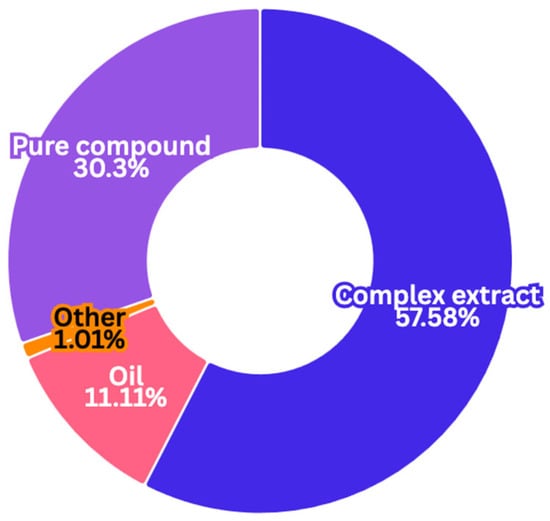
Figure 4.
Types of samples used in each study, categorized by extracts, oils, and pure substances (commercially available natural substances).
The articles that used gel as a sample were those that prepared nanofibers incorporating Aloe vera (L.) Burm.f. [11]. This is because Aloe vera extract is a gel obtained directly from its leaves, containing various bioactive compounds such as polysaccharides, vitamins A, C, thiamine, niacin, and B12, as well as choline and folic acid [7].
Among the articles explored, 11% investigated various essential oils, including clove oil, which is recognized for its broad pharmacological properties, such as analgesic, antibacterial, antifungal, antiallergic, anti-inflammatory, anticarcinogenic, and antimutagenic effects. Eugenol, the main phenolic component of clove oil, is primarily responsible for these biological activities [12]. In an experimental study on diabetic wound healing in male rats, Khazaeli et al. (2020) [13] investigated lavender oil, known for its antibacterial properties and wound-healing promotion, and cod liver oil, rich in omega-3 fatty acids, which may modulate diseases like diabetes mellitus and improve vasodilator properties.
Other essential oils studied include Zataria multiflora Boiss, a thyme species native to Southeast Asia and commonly found in Iran, Pakistan, and Afghanistan. Known for its medicinal properties, it is traditionally used to treat cramps, muscle pains, and infections. The essential oil extracted from Zataria multiflora Boiss has shown effectiveness against bacterial infections and holds promise for wound healing applications [14].
Marine Origin
Among the 99 studies analyzed herein, 4 focused on marine-derived sources. The incorporation of marine-derived compounds through electrospinning is generating significant interest in the pharmaceutical field, fostering the development of novel pharmaceutical forms. These compounds, derived from marine sources like algae, fungi, and marine invertebrates, offer a diverse range of bioactive properties that can be leveraged for various therapeutic applications.
Kikiones et al. (2023) [15] developed electrospun nanofibers incorporating echinochrome A, derived from sea urchins, which demonstrated enhanced stability and controlled release, making them promising candidates for drug delivery applications. Similarly, Kwak et al. (2014) [16] fabricated gelatin nanofiber mats enriched with Phaeodactylum tricornutum extract, showcasing antimicrobial properties suitable for wound care. Luo et al. (2018) [1,8] in turn, encapsulated equisetin, isolated from Fusarium sp., within nanofibers, achieving significant antibacterial activity and promoting wound healing. Additionally, Thamer et al. (2021) [17,18,19] engineered biohybrid nanofibers incorporating a marine Streptomyces sp. extract, demonstrating potential for combating drug-resistant infections and enabling diverse industrial applications.
Together, these studies illustrate the promising applications of electrospinning with marine-derived compounds. This technique enables the creation of nanofibers with high encapsulation efficiency and controlled release, offering advanced therapeutic solutions for a range of medical conditions, from infections to chronic diseases. The continued exploration and development of marine bioactive compounds in electrospinning are poised to drive innovations in pharmaceutical and biomedical fields.
2.1.2. Polymers
There is a wide variety of polymers that can be used in the electrospinning technique, capable of creating nanofibers within the submicron range and can be used in a variety of applications. Nanofibers can be manufactured using synthetic polymers, natural polymers, or a combination of both, including proteins, nucleic acids, and even polysaccharides. In the Figure 5, it is possible to see which are the most commonly used polymers [18,19].

Figure 5.
Most used polymers, according to the articles reviewed, for incorporating natural products through the electrospinning technique.
Synthetic polymers, such as PCL, PLA, PVA, and PVP, are widely used due to their ease of mass production, dimensional uniformity, and durability. However, these polymers generally lack biological signals that promote cell viability. This limitation can be addressed by combining them with natural polymers, resulting in hybrid nanofibers that merge the benefits of both types of materials [20].
Polycaprolactone (PCL) stands out in this context for its excellent biocompatibility, low toxicity, and degradability—essential characteristics for pharmacological applications. Additionally, its versatility favors the incorporation of natural substances during the electrospinning process, enriching the properties of the produced scaffolds. PCL also has good processing properties, enabling the creation of controlled structures and morphologies, which are crucial in electrospinning [21].
Biopolymers such as gelatin, chitosan, and collagen are also commonly used due to their biodegradability and biocompatibility. Although they decompose more quickly than synthetic polymers, this characteristic makes them ideal for ecological and biomedical applications, despite the structural challenges they may face [22].
Examples of biopolymers include chitin, chitosan, collagen, and proteins such as gelatin, albumin, and fibroin, which are widely used in various industries. Despite structural challenges, nanobiomaterials offer enhanced properties such as high porosity and mechanical strength, making them promising for biomedical applications [23] Table 1 lists the biopolymers used in the articles found, along with their types and sources.

Table 1.
Biopolymers used in the reviewed articles, along with their sources of origin.
Electrospinning synthetic polymers is relatively straightforward due to their solubility and molecular structure. However, electrospinning biopolymers encounters obstacles like low solubility in organic solvents and hydrogel formation. To overcome these limitations, it is common to blend biopolymers with synthetic or water-soluble polymers, enabling fiber production. This blending practice is prevalent, as most polymers found in articles are combined, either between synthetic polymers or with biopolymers [20,24]. Figure 6 shows the most commonly used polymers combined with synthetic or biopolymers. Polycaprolactone (PCL) is frequently paired with other polymers, thanks to its unique characteristics, which makes it well-suited for combining with natural substances in electrospinning and broadening its potential applications.

Figure 6.
Polymer combinations mentioned in the articles highlighting polycaprolactone as one of the most commonly used polymers, both in combinations with other synthetic polymers and biopolymers.
2.1.3. Effects of Various Parameters on Electrospinning
Solvents Used in Electrospinning
The electrospinning process is influenced by various factors, including solution parameters such as viscosity, conductivity, molecular weight, and surface tension, as well as process parameters like voltage, tip-to-collector distance, and flow rate. Higher viscosity generally increases fiber diameter and reduces bead formation, while higher conductivity tends to decrease fiber diameter. Solvent choice is critical as it affects polymer compatibility and fiber quality. Solvents with high vapor pressure can accelerate fiber solidification but may lead to defects if evaporation occurs too quickly. Additionally, surface tension and viscosity play key roles in fiber morphology, being crucial for avoiding undesirable bead formation [20,25].
The diameter of electrospun fibers is directly influenced by solvent properties like surface tension, dielectric constant, and boiling point. These factors must be carefully considered when choosing materials for the electrospinning process. Figure 7 displays the most commonly used solvents and their frequencies according to the articles found.
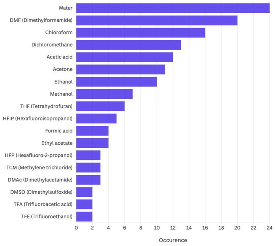
Figure 7.
Common solvents in the analyzed articles.
Dimethylformamide (DMF) is an important solvent in electrospinning due to its high dielectric constant and dipole moment, which improve solution conductivity and fiber uniformity. Nissi et al. (2023) [26,27] found that DMF enhanced the uniformity of PHBV fibers with Morinda. Tinctoria Roxb. extract, aiding in wound healing. Other solvents like chloroform, distilled water, dichloromethane, acetic acid, and ethanol are also used. Dichloromethane with polylactic acid and Sedum dendroideum extract caused undesired spherical structures, while acetic acid effectively dissolved gelatin and Moringa oleifera extracts, reducing bead formation. Blending solvents, like mixing DMF with DMAc, can optimize fiber quality and uniformity by adjusting solution properties.
Other Parameters
The molecular weight of the polymer plays a critical role in electrospinning, influencing viscosity, surface tension, conductivity, and dielectric strength. High-molecular-weight polymers, like HM-PLLA, are favored for their ability to maintain higher viscosity, reduce bead formation, and produce uniform, larger-diameter fibers. Research shows that higher-molecular-weight polymers result in better fiber quality, while strong intermolecular interactions can sometimes allow for the use of lower-molecular-weight oligomers [9,12].
Viscosity is essential for uniform fiber production, as both excessively low and high viscosities can cause irregularities. Conductivity also impacts fiber quality, with higher conductivity resulting in finer fibers. The addition of substances such as ions, gold nanoparticles, or complex mixtures like Garcinia mangostana L. (mangosteen) extracts, as reported by Sriyanti et al. (2018) [28], has been shown to enhance both the solution’s conductivity and the uniformity of the produced fibers [29,30].
Surface tension, influenced by solvent composition, affects bead formation and jet stability. A balance is necessary to avoid defects and ensure optimal fiber formation. The right choice of polymer molecular weight, viscosity, and surface tension is crucial for producing high-quality electrospun fibers, particularly for biomedical applications [2,14,27].
2.1.4. Pharmacological Applications
Polymeric nanofibers excel in various biomedical applications, including the manufacturing of scaffolds for tissue engineering (TE), wound dressings, and drug delivery systems (DDS), among others [1] The chart in Figure 8 illustrates the primary applications of electrospinning using natural substances. As depicted, the majority of the applications are focused on drug delivery, which accounts for over 46%, indicating a strong interest in using electrospun materials for controlled and targeted drug release. Wound healing follows closely with 40%, highlighting the significant role of electrospun fibers in creating wound dressings that promote tissue regeneration and recovery. Lastly, tissue engineering represents 12%, showing a smaller, yet notable, interest in using electrospinning for developing scaffolds that support tissue growth and repair. These statistics reflect the growing relevance of electrospinning techniques in medical and biomedical fields [3].

Figure 8.
Main applications of electrospinning for the incorporation of natural products found in the analyzed articles.
Research continues to evolve, with new materials and formulations enhancing dressing effectiveness [5]. In tissue engineering, electrospun scaffolds mimic the extracellular matrix, aiding tissue and organ regeneration [10,18]. Nanofibers also show promise for drug delivery, particularly for unstable or toxic drugs [1].
In the upcoming sections, we will delve into the subcategories within each application of electrospinning using natural substances. For instance, in wound healing, we will cover areas such as antimicrobial effects, tissue regeneration, and the incorporation of plant extracts to accelerate healing. In the realm of drug delivery, we will explore subtopics like controlled release systems, targeted drug delivery, and the use of bioactive compounds. When discussing tissue engineering, we will focus on scaffold fabrication, cell culture techniques, and the role of natural polymers in promoting tissue regeneration. These subcategories will be explored in detail in their respective sections. Notably, the materials produced by electrospinning with natural products hold significant potential for applications in cancer treatment and as antimicrobials. In cancer therapy, these fibers act as controlled-release systems, delivering bioactive compounds (such as curcumin, prodigiosin, or doxorrubicin) directly to tumors, reducing side effects and promoting antiangiogenic and pro-apoptotic effects. They also enable combination therapies, pairing chemotherapeutic agents with natural antioxidants. As antimicrobials, nanofibers serve as bioactive barriers, incorporating natural compounds with antibacterial, antifungal, or antiviral properties, such as essential oils and tannins. The sustained release of these agents prolongs effectiveness, while combinations of various bioactives enhance activity against pathogens. These fibers offer innovative and versatile solutions for healthcare applications.
2.2. Specifc Findings
2.2.1. Drug Delivery
Controlled Release
Various strategies for creating electrospun nanofibers to enable controlled or rapid release of bioactive compounds were discussed throughout the articles, focusing on their potential biomedical applications. Ravichandran et al. (2019) investigated the use of Clerodendrum phlomidis L.f. leaf extract-infused polycaprolactone (PCL) nanofibers for in vitro biological applications [31]. The researchers found that the nanofibers provided a controlled release of bioactive compounds, suggesting their potential for use wound healing and antimicrobial treatments. Similarly, a study on a coaxial alginate–PCL nanofiber dressing developed a system for the controlled release of Spirulina extract, showing promise as a wound healing material with sustained antioxidant properties [32].
In another study, hybrid electrospun films were created for the fast and convenient delivery of active herb extracts. These films demonstrated rapid release, making them suitable for treatments requiring quick bioavailability, while also offering ease of application. Similarly, emulsion-based polylactic acid (PLA) and Apocynum venetum A.DC nanocellulose membranes were designed with a controlled release mechanism for Hippophae rhamnoides L. extract, which showed both therapeutic and protective effects on the skin [33].
Yakub et al. (2016) [34] also developed polylactide-based materials for curcumin release, investigating the photostability, antimicrobial activity, and anticoagulant effects of the nanofibers. The study confirmed that curcumin-loaded nanofibers exhibited controlled release properties, offering a promising approach for anti-inflammatory and antimicrobial therapies. Likewise, another study focused on polyvinyl alcohol (PVA) nanofibrous membranes loaded with Lappaconitine trifluoroacetate, revealing their potential for transdermal applications, including analgesic and anti-inflammatory treatments [35].
L-menthol-loaded electrospun fibers of PMVEMA anhydride were created for topical administration, demonstrating controlled release, and their potential for use in skin therapies [36]. Another interesting study embedded Garcinia Mangostana Linn extract in electrospun PVP nanofibers, revealing the physicochemical properties and release mechanisms of α-mangostin, a compound known for its antioxidant and anti-inflammatory activities [30].
Several studies also explored the use of electrospun gelatin nanofibers for encapsulating bioactive extracts. Estrella-Osuna et al. (2022) [37] developed a novel delivery system using electrospinning to encapsulate bioactive compounds from Solanum melongena L. Eggplant peel is known for its rich content of antioxidants, such as flavonoids and phenolic compounds, which can be beneficial in skincare and wound healing due to their anti-inflammatory and antimicrobial properties. The study also focused on the in vitro release profile of the encapsulated eggplant peel extract. It was found that the nanofibers provided a controlled and sustained release of the bioactive compounds, which is ideal for therapeutic applications like wound healing. The controlled release ensures that the bioactive agents are gradually delivered over time, promoting longer-lasting therapeutic effects.
Additionally, Terminalia bellirica (Gaertn.) Roxb.-loaded PCL nanofibrous mats were developed with controlled release profiles that could benefit various therapeutic areas. A study on the effect of molecular weight and content of PEG (polyethylene glycol) in PLA/PEG nanofibers demonstrated its impact on the release of curcumin, offering insights into the optimization of drug delivery systems for specific treatments [38].
Lastly, a study by Sugumaran and Rathinam (2021) [39] on Siddha drug-incorporated electrospun nanofibrous mats with controlled drug release highlighted the versatility of electrospinning in integrating traditional medicine with modern drug delivery technologies. By using the electrospinning technique, nanofibers were created to encapsulate Siddha drugs, which could be herbal extracts or minerals known for their medicinal properties. One of the key findings was that these nanofibrous mats not only enhanced the stability and bioavailability of the Siddha drug but also allowed for its controlled release, which could be beneficial for treating a range of diseases, such as skin disorders, inflammatory conditions, and infections. The electrospun nanofibers act as both a carrier and a barrier, providing a localized and sustained release of the therapeutic agents at the site of action.
These studies highlight the significant potential of electrospun nanofibers in creating advanced drug delivery systems. By enabling the controlled or rapid release of bioactive compounds, these materials offer versatile solutions for wound healing, antimicrobial therapy, and other biomedical applications. The findings from these works point to the growing promise of electrospinning as a technique for developing efficient, customizable, and biocompatible delivery systems for a range of therapeutic agents.
Cancer Therapy
Recent advancements in nanotechnology, particularly the development of electrospun nanofibers, have significantly impacted the field of medicine, offering innovative methods for drug delivery, especially in cancer treatment. Electrospun nanofibers provide a promising approach for controlled and targeted drug release, which is crucial for effective cancer therapy. This review consolidates findings from various studies on the use of electrospun nanofibers for enhancing drug delivery in cancer treatment.
Nam et al. (2017) [40] investigated the use of electrospun nanofibers loaded with Angelica gigas Nakai extract (AGN) in a polyvinyl alcohol (PVA) and Soluplus (SP) matrix. These nanofibers, with an average diameter of 170 nm and encapsulation efficiency over 80%, demonstrated rapid dissolution and effective oral delivery of AGN. The transformation of AGN from a crystalline to an amorphous form facilitated improved drug release. In vitro tests revealed that AGN-loaded nanofibers had superior antiproliferative activity compared to AGN in suspension, with promising safety profiles in mice, indicating their potential as an oral cancer treatment.
Several studies have focused on doxorubicin-loaded nanofibers for cancer therapy. PCL nanofibers incorporated with doxorubicin and berberine showed sustained drug release and high antioxidant activity. These nanofibers effectively prevented local recurrence of breast cancer and adapted to the acidic tumor microenvironment [41,42,43].
Akpan et al. (2020) [44] investigated electrospun nanofibers loaded with prodigiosin, a natural red pigment with anticancer properties. The study aimed to improve prodigiosin’s bioavailability and safety by using a composite of poly (D,L-lactic-co-glycolic acid) (PLGA), gelatin, and pluronic F127. The resulting nanofibers exhibited sustained drug release, while in vitro studies demonstrated that these nanofibers promoted cell proliferation and viability in breast cancer cell lines.
In another study, Akpan et al. (2021) [45] developed electrospun nanofibers loaded with Annona muricata L. (soursop) extract, showing efficacy in localized breast cancer treatment. The nanofibers effectively released the extract and inhibited tumor growth in vitro, providing a targeted and efficient approach for cancer therapy. Wen et al. (2019) [46] focused on the targeted delivery of phycocyanin, a pigment with antioxidant and anti-inflammatory properties, for colon cancer prevention. Electrospun nanofibers achieved controlled release of phycocyanin, demonstrating efficacy in preventing tumor progression in experimental models and highlighting their potential in preventive cancer therapies.
Additional studies have explored various bioactive compounds in electrospun nanofibers. Arbade et al. (2019) [47] investigated PCL nanofibers loaded with Emblica officinalis Gaertn. extract, showing antibacterial and anticancer properties. Kyritsi et al. (2021) [48] used nanofibrous patches with Pinus halepensis Mill bark extract to manage acute radiodermatitis in non-melanoma skin cancer patients, proving effective in symptom relief and skin restoration.
Other Findings in Drug Delivery
Recent studies highlight the growing potential of electrospinning in the development of advanced materials with a range of therapeutic applications, including antibiotic production, anti-inflammatory and analgesic treatments, and antioxidant properties.
In the field of anti-inflammatory and analgesic treatments, several studies have utilized electrospun nanofibers to incorporate plant extracts and essential oils with therapeutic properties [49]. One study developed Acanthus ebracteatus Vahl. extract-loaded cellulose acetate ultrafine fibers for controlled-release applications, showing that the nanofibers effectively delivered anti-inflammatory compounds to targeted areas [50]. Another innovation involved clove essential oil nanoemulgel designed using Taguchi’s model and scaffold-based nanofibers, demonstrating its potential as a cyclooxygenase-2 (COX-2) inhibitor for treating external inflammation [12].
Electrospun nanofibers also show great promise in antioxidant applications. For example, an antioxidative nanofiber-based film made with pullulan, sodium hyaluronate, and Ganoderma lucidum fermentation products was developed for enhanced skincare applications, demonstrating significant antioxidant activity [51]. Additionally, a dual biocompatible platform for immobilizing genistin, a bioactive compound, showed potential for antioxidant and anti-inflammatory therapies [52]. Other studies focused on encapsulating Moringa oleifera Lam bioactive extract in gelatin nanofibers, providing sustained release of powerful antioxidants, which can aid skin regeneration and wound healing [53]. Similarly, a novel antioxidant fibrous material was created using extracts from Portulaca oleracea L. and polylactide, demonstrating substantial antioxidant properties for biomedical use. Rheum ribes L. extract-loaded PLA/PEG nanofibers also showed potent antioxidant activity, further supporting the potential of electrospun materials in combating oxidative stress [54,55].
These studies collectively underscore the versatility of electrospinning in producing nanofibers that integrate various bioactive compounds for controlled release, offering wide and significant therapeutic benefits. Whether it is enhancing antibiotic production, providing localized anti-inflammatory and analgesic effects, or delivering antioxidants for skin and wound care, electrospun nanofibers present a promising platform for developing advanced, effective, and sustainable medical solutions
2.2.2. Wound Healing Dressing and Skin Tissue Regeneration
The process of wound healing is a complex biological event that involves multiple stages: inflammation, proliferation, and remodeling. Successful healing requires not only the formation of new tissue but also the restoration of function and structure of the injured area. Electrospun nanofibers have emerged as a key technology in wound healing, offering an ideal platform to mimic the extracellular matrix, which supports cellular attachment, migration, and proliferation. In this section, various applications of electrospun nanofibers in wound healing will be explored, focusing on the integration of natural substances and bioactive compounds to enhance therapeutic outcomes. The applications are divided into subcategories, each targeting a specific aspect of the wound healing process, including antimicrobial, antioxidant, and tissue regeneration properties [45].
Applications in Wound Healing
One of the most promising approaches in wound healing involves the use of natural extracts incorporated into electrospun nanofibers. These extracts provide therapeutic properties such as anti-inflammatory, antimicrobial, and regenerative effects. For example, zein/MMT nanocomposites loaded with Hypericum perforatum L. (St. John’s Wort) oil have been shown to promote faster wound healing due to anti-inflammatory and antimicrobial properties of this extract [56]. Similarly, Artemisia annua L. (sweet wormwood) extract has been investigated for its ability to accelerate tissue regeneration, reduce oxidative stress, and promote faster recovery, making it a valuable addition to wound care treatments [57]. Vitamin E (α-tocopherol acetate), incorporated into bilayered membranes, is another example; α-tocopherol acetate is known for its antioxidant properties and its role in reducing oxidative damage at the wound site, which accelerates tissue regeneration and minimizes scarring [58]. These bioactive compounds, when encapsulated within nanofibers, are released gradually, providing sustained therapeutic effects.
Scaffolds for Chronic Wound Management
Chronic wounds, such as diabetic ulcers and pressure sores, present significant clinical challenges due to impaired healing, often accompanied by recurrent infections. These wounds are particularly difficult to treat because the tissue regeneration process is disrupted or delayed, leading to severe complications for patients. However, advancements in biomaterials, particularly with the use of electrospun nanofibers, offer promising new approaches for treating these conditions.
A notable example is the use of poly(L-lactic acid) (PLLA) nanofibers incorporated with Centella asiatica extract. This plant, known for its regenerative and healing properties, stimulates the production of collagen, a critical protein for wound closure. Collagen formation is essential not only for skin integrity but also for the repair of underlying structures. Additionally, C. asiatica (L.) Urb. also enhances skin elasticity, which facilitates the stretching and movement of healed areas, promoting more functional and long-lasting healing [59].
Another example is the use of chitosan-polyvinyl alcohol (PVA) nanofiber dressings loaded with ursolic acid, a bioactive compound known for its anti-inflammatory and wound-healing properties. Studies have shown that ursolic acid accelerates the healing of diabetic wounds by promoting collagen synthesis and reducing oxidative stress. These nanofibers not only provide mechanical support but also enhance the healing process in diabetic patients, who often face prolonged healing times due to poor blood circulation and immune response [60].
Electrospun nanofibers made from biodegradable polymers such as polylactic acid (PLA) and polycaprolactone (PCL) have proven effective as scaffolds for cell growth. These scaffolds not only provide structural support for the cells but can also be designed to release bioactive compounds in a controlled manner. A key example is the incorporation of propolis, a natural compound with well-established antimicrobial and anti-inflammatory properties [61,62,63,64]. Propolis helps prevent bacterial infections in wounds, a common issue with chronic wounds, and also reduces inflammation, aiding the tissue regeneration process. The controlled release of these compounds through nanofibers improves treatment efficacy by ensuring that therapeutic agents are delivered continuously and over an extended period at the wound site, optimizing healing [61].
In addition to their therapeutic properties, these PCL and PLA nanofibers are biocompatible and biodegradable, meaning they break down in the body once the wound has healed, without causing adverse effects. The combination of these materials with natural compounds, such as C. asiatica (L.) Urb. extract and propolis, offers a highly effective treatment, promoting faster and more efficient chronic wound healing while minimizing the risk of infection and supporting long-term tissue regeneration [65].
Antimicrobial Properties
Electrospun nanofibers have gained significant attention in the field of wound healing due to their unique properties, such as high surface area, porosity, and biocompatibility. These properties make them ideal for mimicking the extracellular matrix, offering structural support for cell attachment, migration, and proliferation, all of which are essential for tissue regeneration [21]. One of the most promising approaches in wound healing involves integrating natural antimicrobial agents into electrospun nanofibers, enhancing both their therapeutic and protective properties. Several studies have explored this strategy, creating multifunctional nanofiber-based wound dressings that promote healing while preventing infection, a common barrier in chronic wounds [5].
In one study of Khan et al. (2023) [66], electrospun PVA/MMT nanofibers were loaded with the root extract of Berberis lycium Royle, a plant known for its antimicrobial and wound-healing properties. The nanofibers, made from polyvinyl alcohol (PVA) and montmorillonite (MMT) clay, provided a strong mechanical structure while allowing for the controlled release of the antimicrobial extract. The researchers demonstrated that the nanofibers effectively inhibited bacterial growth while also supporting cell proliferation, suggesting that these nanofibers could serve as a promising dual-function wound dressing for both infection prevention and tissue regeneration.
Another study explored the integration of Chelidonium majus L. (greater celandine) extract into emulsion electrospun nanofibers composed of polycaprolactone (PCL), PVA, and polyethylene glycol (PEC). The study showed that the nanofibers not only exhibited significant antimicrobial activity against common wound pathogens but also promoted wound healing by reducing inflammation. The electrospun mesh demonstrated both antimicrobial and anti-inflammatory effects, making it an excellent candidate for wound dressings [67].
In another investigation, co-electrospinning was used to create nanofibrous mats loaded with Momordica charantia Descourt. (bitter gourd) extract, which is rich in antioxidants, anti-inflammatory, and antibacterial compounds. The PVA and PCL blend allowed for the even distribution of the bitter gourd extract, which enhanced the antimicrobial activity of the nanofibers. The researchers conducted both in vitro and in vivo studies, showing that the nanofibers significantly reduced bacterial load on wound sites and accelerated wound closure. These results confirm the potential of bitter gourd-loaded nanofibers as a highly effective treatment for chronic wounds, especially those prone to infection [68].
Another promising approach by Barzegar et al. (2021) [69] involved the development of core–shell nanofibers made from chitosan and PVA that were loaded with essential oils from Satureja mutica Fisch. and C.A.Mey. and Oliveria decumbens Vent. These essential oils have strong antimicrobial properties and were encapsulated in the core of the nanofibers, allowing for controlled release. The outer shell provided structural integrity and stability. The study showed that the nanofibers had significant antimicrobial activity against Staphylococcus aureus and Pseudomonas aeruginosa and were effective in reducing wound infection while promoting cell growth and tissue regeneration.
The integration of marine-derived compounds into electrospun nanofibers has also been explored. One study focused on gelatin nanofibers loaded with extract from Phaeodactylum tricornutum, a marine microalga with antimicrobial properties [16]. These nanofibrous mats were developed for applications in wound healing and antibacterial action. The nanofibers, fabricated via electrospinning, exhibited a structure suitable for promoting tissue regeneration, creating an optimal environment for wound repair. The P. tricornutum extract, rich in bioactive compounds, endowed the nanofibers with effective antimicrobial activity against pathogens such as Staphylococcus aureus and Escherichia coli. Additionally, the controlled release of the extract ensured prolonged protection against infections, contributing to a faster and safer healing process. This innovative approach combines antibacterial and regenerative properties, making it a promising solution for advanced wound dressings [16].
Antioxidant Properties
Oxidative stress significantly impairs the healing process by damaging cells and delaying tissue repair. Antioxidants incorporated into electrospun nanofibers can reduce oxidative damage and support faster healing. Curcumin-loaded PCL/PVA nanofibers and silk fibroin nanofibers loaded with Periplaneta americana extract have shown strong antioxidant properties, protecting cells at the wound site and promoting tissue regeneration. The studies explored the application of nanofibers in advanced wound dressings, combining wound healing properties with antioxidant activity, utilizing the electrospinning technique [70].
In the first study by Saeed et al. (2017) [71], multilayered structures were developed using a combination of PCL (polycaprolactone) and PVA (polyvinyl alcohol) loaded with curcumin, a compound known for its anti-inflammatory and antioxidant properties. The electrospinning process enabled the fabrication of individual layers with distinct functions: the PCL layer provided mechanical durability and biocompatibility, while the PVA layer incorporated with curcumin offered controlled release of the bioactive compound. The resulting structure effectively promoted tissue repair, mitigated inflammation, and protected against oxidative stress.
In the second study, silk fibroin was used as the base material to produce nanofibers incorporating Periplaneta americana extract. The electrospinning technique allowed for the creation of a homogeneous matrix that retained and gradually released the bioactive extract. The resulting material demonstrated potent antioxidant activity, neutralizing reactive oxygen species (ROS) and creating an environment conducive to tissue regeneration. Additionally, the natural properties of silk fibroin contributed to excellent biocompatibility and mechanical performance of the dressing [70]. Both studies exemplify the innovative use of electrospinning in creating multifunctional dressings, combining controlled release of bioactive compounds, support for wound healing, and protection against oxidative damage. These advancements highlight the potential of the technique in the development of advanced therapeutic solutions.
2.2.3. Tissue Engineering
Tissue regeneration is a crucial component of tissue engineering, an interdisciplinary field that integrates biology, engineering, and medicine to restore or enhance the function of damaged or lost tissues. The goal is to develop innovative methods using a combination of cells, biomaterials, and bioactive factors for effective tissue regeneration.
Tissue regeneration has diverse medical applications, including skin, bone, cartilage, blood vessels, and internal organs. For example, nanofiber scaffolds loaded with natural extracts have demonstrated promising results in diabetic wound healing. In bone regeneration, bioactive materials like calcium phosphate scaffolds are employed to promote new bone tissue formation. For cartilage regeneration, hydrogels and composite biomaterials are utilized. In cardiovascular tissue engineering, the formation of new blood vessels is of paramount importance [72].
Several recent studies have explored the potential of electrospinning in tissue engineering, focusing on the development of nanofibrous scaffolds for regenerative applications. One such study investigated Aloe vera gel-blended PHBV nanofibrous scaffolds for bone tissue engineering. The study highlighted the scaffold’s potential to mimic natural bone architecture, providing an ideal environment for bone cell growth and improving healing processes [11]. Another significant study assessed Spirulina-PCL nanofibers for regenerating dermal fibroblast layers. The results indicated that Spirulina-infused nanofibers could enhance fibroblast proliferation, showing promise for wound healing and skin regeneration applications [73]. Additionally, Sadia et al. (2023) [74] studied a Moringa oleifera Lam infused biodegradable polycaprolactone (PCL) nanofiber for their hemocompatibility improvement, which is crucial for biomedical applications involving blood contact, such as vascular implants.
Furthermore, the development of Emu oil-loaded PCL/collagen bioactive nanofibers has shown potential in promoting the proliferation and stemness preservation of human adipose-derived stem cells. This is particularly relevant for regenerative medicine, where maintaining stem cell functionality is vital for tissue regeneration [75]. In a similar vein, PCL-based nanofibers loaded with extracts from Arrabidaea chica Verlot and Pterodon pubescens Benth demonstrated a synergistic effect in fibroblast formation, further supporting their use in skin tissue engineering [10]. Other notable studies have focused on the enhancement of skin regeneration using herbal extract-coated poly-L-lactic acid (PLLA) nanofibrous scaffolds. The incorporation of grape seed extract into silk fibroin nanofibers has shown excellent cytocompatibility and antioxidant properties, further enhancing their potential in skin tissue regeneration [71]. Hybrid PCL/chitosan-PEO scaffolds incorporated with A. euchroma extract were also developed for skin tissue engineering, demonstrating their ability to support cell growth and promote healing [76].
The use of curcumin-loaded electrospun filaments has also been investigated for soft tissue repair applications, highlighting curcumin’s anti-inflammatory and antioxidant properties in enhancing tissue healing [77,78,79]. Similarly, Lycium barbarum Mill. polysaccharides encapsulated in polylactic-co-glycolic acid (PLGA) nanofibers have been proposed as a cost-effective herbal medicine for peripheral nerve tissue engineering, showing potential in nerve regeneration [80]. Finally, the development of a multicompartment vascular implant using electrospun wintergreen oil (Gaultheria procumbens L.)/polycaprolactone fibers coated with poly(ethylene oxide) has been explored by Eldurini et al. (2020) [81], showing promise in improving vascular tissue regeneration. The preparation of PLA/curcumin composite membranes further exemplifies the versatility of electrospun materials in supporting tissue engineering applications, providing not only structural support but also bioactive properties for enhanced healing. These studies demonstrate the vast potential of electrospun nanofibers in tissue engineering, offering versatile and biocompatible scaffolds that can promote cell growth, tissue regeneration, and overall healing across a range of applications.
2.2.4. Miscellaneous Applications
Plasma-modified PLA electrospun membranes were used by Scafaro et al. (2017) [82] to optimize the production of the antibiotic by Streptomyces coelicolor in the microbial cultivation environment. The modified membranes provided a stable platform for microbial growth and continuous antibiotic production, increasing antibiotic yield by 5 to 10-fold, highlighting the potential of electrospun materials in enhancing microbial production of bioactive secondary metabolites and improving bioprocesses.
2.2.5. Bioavailability of Natural Compounds in Nanofibers
The therapeutic potential of natural compounds is often limited by their poor bioavailability, characterized by low solubility, rapid degradation, and limited permeability. Nanotechnology, particularly the use of electrospun nanofibers, has emerged as a promising strategy to overcome these limitations by providing controlled release and targeted delivery. This section will explore the mechanisms by which nanofibers enhance bioavailability, the critical factors influencing this process, and specific examples of successful applications [83].
The bioavailability of natural compounds incorporated into electrospun nanofibers is a key factor in their pharmacological effectiveness, directly influencing the stability, controlled release, and absorption of bioactive substances by the body. The choice of polymers and solvents used in the electrospinning process plays a crucial role in the formulation of these nanofibers, as it can modify the interaction between active compounds and the biological environment, affecting solubility, diffusion, and biocompatibility [84].
Electrospun nanofibers improve the bioavailability of natural compounds through several key mechanisms. The nanofiber matrix acts as a protective barrier, shielding the encapsulated compounds from enzymatic degradation, pH variations, and oxidation. This is particularly important for labile molecules. By manipulating polymer selection, solvent systems, and fiber morphology, researchers can control the release kinetics of encapsulated compounds [85]. Biodegradable polymers, such as polycaprolactone (PCL), allow for sustained release, while water-soluble polymers, like polyvinyl alcohol (PVA), enable rapid release. Additionally, the high surface area and small size of nanofibers promote interaction with biological tissues, enhancing the permeation of compounds through barriers like the skin and intestinal epithelium. Nanofiber encapsulation can also improve the dispersion of hydrophobic compounds, increasing their solubility in aqueous media and, consequently, their absorption [86].
Functionalization with ligands enables targeted delivery to specific tissues or cells, thereby enhancing therapeutic efficacy. To fully assess how nanofibers impact bioavailability, it is crucial to conduct comprehensive physicochemical and biological analyses (as shown in Table 2).
Table 2.
Physicochemical and biological analyses.
By integrating data from these analyses, researchers can optimize nanofiber design to maximize the bioavailability of natural compounds. This includes selecting the appropriate polymer, controlling fiber morphology, achieving high encapsulation efficiency, optimizing release kinetics, and ensuring biocompatibility. Studies have shown that different polymers impart specific characteristics to nanofibers, modulating the release of bioactive compounds. Poly(ε-caprolactone) (PCL), for instance, is widely used due to its biocompatibility, biodegradability, and ability to form porous structures that enable sustained drug release over time. De Figueiredo et al. (2022) [62] reported that incorporating propolis into PCL nanofibers resulted in a sustained release profile, increasing the bioactive’s contact time with the target tissue and optimizing its therapeutic effect. Furthermore, polymers such as poly(lactic-co-glycolic acid) (PLGA) and polyvinylpyrrolidone (PVP) have also been explored due to their ability to form hydrophilic or hydrophobic polymer matrices, modulating interactions with natural compounds of varying polarities.
The choice of solvent directly influences fiber morphology and the efficiency of bioactive incorporation. Solvents such as acetic acid, dimethylformamide (DMF), chloroform, and ethanol are frequently used, depending on their compatibility with both the polymer and the bioactive compound. Mirbehbahani et al. (2020) [57] demonstrated that fiber morphology can be significantly altered by the type of solvent used, which directly affects degradation rates and drug release profiles. Additionally, polymer blending allows for the creation of hybrid structures, as observed by Hussein et al. (2021) [29], who used PVA and chitosan nanofibers enriched with gold nanoparticles and Punica granatum extract, resulting in a formulation with enhanced antimicrobial properties and superior biocompatibility.
A study by Aman et al. (2020) [12] evaluated topical formulations based on clove essential oil (CEO), including nanoemulsions (NEG) and nanofibers (NFs), focusing on physicochemical characterization, skin permeation, anti-inflammatory efficacy, and bioavailability. Structural analyses (TEM, FT-IR, and DSC) confirmed homogeneous dispersion and stability of the components. Ex vivo skin permeation tests showed that the CEO-NE-based NEG had the highest efficiency, followed by the CEO-NE-based NFs, while pure CEO exhibited the lowest absorption. In vivo studies using a croton oil-induced skin inflammation model demonstrated that the CEO-NE-based NEG (two applications) and CEO-NE-based NFs (one application) significantly reduced inflammation and COX-2 expression, confirming their anti-inflammatory efficacy. The formulations remained stable for six months under refrigeration, with CEO-NE-based NFs retaining 97.74% of the drug. Skin irritation tests indicated safety for topical use.
Regarding bioavailability, the CEO-NE-based NEG showed higher skin penetration efficiency due to its polymeric gel matrix, while the CEO-NE-based NFs provided sustained drug release, maintaining effective concentrations for a longer duration. Kinetic analysis revealed that the CEO-NE-based NEG followed a super case II transport mechanism, associated with polymeric matrix swelling and controlled release, whereas the CEO-NE-based NFs followed a Fickian diffusion model, ensuring gradual and sustained absorption. These formulations enhanced the bioavailability of CEO in the skin, optimizing its anti-inflammatory action with fewer applications, making them promising alternatives for controlled drug delivery systems.
Another study investigated hybrid nanofibrous scaffolds of PCL/chitosan-PEO incorporated with A. euchroma extract for skin tissue engineering. SEM characterization indicated uniform fiber distribution and homogeneous extract incorporation. Contact angle analysis revealed that increasing A. euchroma concentration reduced scaffold hydrophilicity due to its hydrophobic nature. Mechanical properties showed a decrease in stiffness and strength with extract addition, though they remained suitable for tissue regeneration applications. Swelling tests indicated that formulations with up to 15% A. euchroma exhibited good exudate absorption capacity, whereas higher concentrations reduced this property. Water vapor permeability remained stable, ensuring a moist environment for wound healing. Biodegradation was accelerated in scaffolds with higher extract content, suggesting interaction with lysozyme and rapid degradation [76]. Biocompatibility was confirmed through cytotoxicity assays, demonstrating safety and fibroblast proliferation stimulation, particularly at 15% and 20% A. euchroma concentrations. The extract contributed to cell adhesion and extracellular matrix formation, promoting wound healing through anti-inflammatory and angiogenic effects. The in vitro release study showed an initial rapid release followed by controlled extract release over 96 h, influenced by the hydrophobic nature of the compound.
Antibacterial activity was significant against E. coli and S. aureus, with higher efficacy against E. coli. Microscopy revealed bacterial morphology alterations after exposure to the scaffolds, indicating potential applications in antimicrobial wound dressings. Thus, the PCL/Cs-PEO/A. euchroma hybrid scaffolds demonstrated promising properties for skin regeneration, combining biocompatibility, controlled drug release, and antimicrobial activity [84].
The study by M. Luo et al. (2018) [8] investigated the local delivery of equisetin (EQ), derived from the marine fungus Fusarium sp. 152, encapsulated in polyvinylpyrrolidone (PVP) nanofibers for anti-MRSA activity. The study showed that EQ/NPs nanofibers exhibited strong anti-MRSA activity and accelerated wound healing in rats infected with MRSA. This study highlights the potential of marine-derived natural compounds in developing new strategies against hospital-acquired MRSA infections.
Based on this evidence, the bioavailability of natural compounds in electrospun nanofibers must be considered a fundamental aspect in the development of new pharmaceutical formulations. Optimizing electrospinning parameters, along with selecting appropriate polymers and solvents, can enhance the therapeutic effects of bioactive compounds, ensuring their efficient and safe release.
2.2.6. Challenges in Electrospinning: Standardization and Scalability
Despite advances in the application of electrospun nanofibers for incorporating natural compounds, transitioning this process from laboratory research to industrial-scale production still faces significant challenges. The reproducibility of the technique and the standardization of electrospinning conditions are critical factors influencing the final quality of nanofibers and their viability for biomedical applications [87].
One of the main challenges lies in the need for precise adjustments in operational parameters such as applied voltage, solution flow rate, and needle-to-collector distance. Small variations in these parameters can lead to substantial changes in fiber morphology and mechanical properties, directly impacting their functionality. Additionally, environmental factors such as humidity and temperature also play a crucial role in fiber formation and must be strictly controlled to ensure product consistency [88].
The scalability of electrospinning for large-scale production remains limited by processing time constraints and the cost of materials used. High-purity biodegradable polymers, such as PCL and PLGA, have high costs, making the process economically unfeasible for some applications. Furthermore, commonly used electrospinning solvents, such as DMF and chloroform, are toxic and environmentally restricted, necessitating the search for safer and more sustainable alternatives. Hussein et al. (2021) [29] highlighted that replacing toxic solvents with aqueous systems could be a viable solution for making the electrospinning process safer and more environmentally friendly.
Another fundamental aspect for the commercial feasibility of electrospun nanofibers is regulatory approval and acceptance by agencies such as the Food and Drug Administration (FDA) and the European Medicines Agency (EMA). Most electrospun nanofiber studies remain in the preclinical phase, with limited progress in clinical trials to confirm their safety and efficacy for human use. The approval of new materials for medical applications requires rigorous studies on biocompatibility, long-term stability, and potential adverse effects, making the regulatory process one of the main challenges for the industrial implementation of electrospinning [89].
Therefore, for electrospinning to become a viable technology for producing pharmaceutical and biomedical systems, it is essential to invest in research focused on process standardization, material optimization, and evaluation of economic and regulatory feasibility. Collaboration among researchers in materials engineering, biotechnology, and the pharmaceutical industry can significantly contribute to overcoming these challenges and enable the widespread application of electrospun nanofibers in regenerative medicine and the development of new controlled drug delivery systems.
3. Potential of Electrospinning for Cosmetic Applications
Electrospinning is widely studied for biomedical applications, but its use in the cosmetics industry remains underexplored. During the literature review, only one article addressing this application was identified, suggesting a significant gap in the development of cosmetic products based on nanofibers. However, considering the advances in incorporating bioactive natural compounds into electrospun systems, this technology could represent an innovation in the sector, offering new approaches for cosmetic formulations with controlled release of active ingredients.
The use of electrospun nanofibers in cosmetics could bring benefits such as increased stability of sensitive compounds, controlled release of active ingredients, and better adhesion to the skin. Liu et al. (2023) [51] demonstrated that incorporating Ganoderma lucidum into pullulan nanofibers resulted in a formulation with high antioxidant activity, suggesting its potential for anti-aging products and protection against oxidative skin damage. Likewise, bioactives commonly used in the cosmetics industry, such as polyphenols, flavonoids, and terpenes, could be incorporated into nanofibers to enhance their effects and ensure longer action on the skin.
However, the adoption of electrospinning in the cosmetics industry still faces challenges related to regulation and production costs. Unlike pharmaceutical applications, where controlled drug release is a critical factor, the cosmetics industry seeks formulations with pleasant textures and easy application. Integrating electrospinning with other technologies, such as nanoencapsulation and hybrid release systems, could be an alternative to enable the use of nanofibers in cosmetics.
Considering the potential of electrospun systems to optimize the absorption and effectiveness of natural actives, more research is needed to assess the applicability of this technology in the cosmetics industry. Expanding studies in this field could open new opportunities for the development of innovative products, offering benefits to both consumers and the industry by enabling more stable, efficient, and sustainable formulations.
4. Marine Natural Compound Exploration
A particularly underexplored area in the electrospinning field is the incorporation of marine-derived natural compounds, which represent a vast and largely untapped resource of bioactive molecules with unique properties. Marine organisms, such as algae, sponges, and marine fungi, produce bioactive compounds with antimicrobial, antioxidant, anticancer, and anti-inflammatory properties that could be highly beneficial for biomedical applications. Luo et al. (2018) [8] investigated the incorporation of equisetin, a secondary metabolite derived from deep-sea fungi, into PVP-based nanofibers and demonstrated significant antibacterial activity against MRSA strains, suggesting its potential for treating resistant infections. Similarly, Kikionis et al. (2023) [15] explored the development of electrospun nanofibers loaded with Echinochrome A, a marine-derived bioactive pigment, highlighting its potential for alternative drug administration routes.
The study by Akpan et al. (2020) [44] investigated the development of electrospun nanofiber scaffolds loaded with prodigiosin for the localized treatment of triple-negative breast cancer. Prodigiosin is a bioactive compound that has shown potential in cancer treatments due to its cytotoxic and antitumor properties.
While the study does not focus directly on “marine origin” materials, it is important to note that prodigiosin, the compound used in this study, is of marine origin. Prodigiosin is a natural molecule produced by certain marine bacteria, such as Serratia marcescens. This compound has shown effectiveness in combating cancer cells, and its integration into controlled release systems like electrospun nanofibers can enhance its targeted delivery and therapeutic efficacy.
In the context of study, the use of electrospun nanofibers made from polyesters (such as PCL, PLGA, and others) allows prodigiosin to be released in a controlled manner at the tumor site, increasing the treatment’s efficacy by directly targeting cancer cells. The electrospinning method enables the creation of nanofibers with large surface areas and very small dimensions, which are advantageous for enhancing interaction with tumor cells and improving permeation.
This study reinforces the idea that natural compounds from marine sources, like prodigiosin, can be enhanced by technologies such as electrospinning, providing the advantage of enabling more targeted and less invasive treatments. Thus, combining prodigiosin with controlled-release systems made from nanofibers offers a promising approach for treating cancer, particularly aggressive types like triple-negative breast cancer.
Given these findings, optimizing the bioavailability of natural compounds in electrospun nanofibers requires careful selection of polymer type, solvent system, and processing parameters. The ability to modulate degradation rates, enhance solubility, and achieve controlled release kinetics through nanofiber engineering highlights the versatility of electrospinning as a platform for improving the therapeutic efficacy of natural compounds. Further research into polymer-drug interactions, in vitro and in vivo bioavailability studies, and clinical translation efforts will be essential to fully realize the potential of electrospun nanofibers in pharmaceutical and biomedical applications. Expanding the investigation of marine-derived bioactives within electrospun systems represents an exciting frontier with potential applications in wound healing, antimicrobial therapies, and drug delivery systems.
5. Materials and Methods
5.1. Selection and Exclusion Criteria
Eligible studies included those investigating the incorporation of natural bioactive compounds into nanofibers produced through the electrospinning technique, intended for pharmacological applications. The search was limited to English articles published between 1 January 2013 and 10 December 2023 and available online. Review articles, clinical studies, and any form of gray literature (such as unpublished, non-peer-reviewed journals, theses, and industry data) were excluded from the analysis.
5.2. Databases
The databases of the indexers PubMed (https://pubmed.ncbi.nlm.nih.gov, accessed on 30 November 2024), Scopus (https://scopus.com), Web of Science, Google Scholar (https://scholar.google.com), and Springer Link (https://link.springer.com/) were accessed for article retrieval between November 2022 and May 2023.
5.2.1. Keywords
The electronic search was conducted using the following search terms: “electrospinning” AND “natural products” AND “drug applications”; “electrospinning” AND “Bioactive extract” AND “drug application”; “electrospinning” AND “marine natural compound” AND “drug application”; “electrospinning” AND “marine bioactive compound” AND “drug application”. Additionally, the following terms were applied to refine the search: NOT “food applications”, NOT “engineering applications”, NOT “biomedical”. Once the electrospinning technique can also be called electrospun, the same keywords as before were used, only changing “electrospinning” to “electrospun”.
5.2.2. Selection of Articles and Application of Inclusion and Exclusion Criteria
This study employs inclusion and exclusion criteria to ensure the relevance, quality, and consistency of the analyzed studies. Inclusion criteria were based on the analysis of titles, keywords, and the topics covered in the research articles. Exclusion criteria involved titles and keywords related to areas such as food, engineering, and chemistry, as the focus was on the use of electrospinning for incorporating natural substances with pharmacological properties into polymers. Additionally, reviews, books, undergraduate theses, dissertations, and doctoral theses were also excluded.
The protocol for this systematic review was retrospectively registered in the Open Science Framework (OSF). The registration can be accessed through the following link and DOI: 10.17605/OSF.IO/2X3GE; Short link: https://bit.ly/ElectrospunBioactiveNaturalCompounds, accessed on 30 November 2024.
6. Concluding Remarks
The use of electrospinning to incorporate natural compounds is emerging as a promising field, particularly in health and biomedical applications. This technology has been extensively explored for wound healing and dressing development, offering significant advantages such as tissue regeneration and protection against infections. Additionally, electrospinning has proven effective in controlled drug delivery, enabling sustained and targeted administration of medications, which can enhance therapeutic efficacy and reduce side effects. Particularly in the field of oncology, the application of electrospinning for the delivery of antitumor drugs is an innovative strategy with significant potential for further growth. The ability to incorporate chemotherapeutic agents into nanofibers and release these substances in a controlled manner has the potential to revolutionize cancer treatment by improving drug efficacy and minimizing adverse effects.
Despite these promising advancements, the broader application of electrospinning still faces critical challenges, particularly regarding cost-effectiveness and regulatory approval. While electrospun nanofibers have demonstrated substantial therapeutic potential, the transition from laboratory research to large-scale production remains limited due to the high cost of materials and the need for process standardization. Additionally, regulatory requirements for the approval of new nanofiber-based pharmaceutical formulations must be thoroughly addressed to ensure safety, efficacy, and compliance with international standards. The economic feasibility of electrospun formulations is also highly dependent on the choice of polymers, solvents, and bioactive compounds, reinforcing the need for further studies to evaluate the cost-benefit ratio of these materials in real-world applications.
Another significant aspect is the limited exploration of electrospinning in the cosmetic sector. Although this technique could transform the formulation of cosmetic products by enabling prolonged release of active ingredients and enhancing the stability of sensitive components, the scarcity of studies in this area suggests a gap in research and development. The incorporation of bioactive compounds into nanofibers could provide innovative solutions for skincare and dermatological applications, particularly for antioxidants, anti-inflammatory agents, and natural moisturizers. However, challenges such as regulatory approval, production costs, and market acceptance still need to be addressed before electrospun nanofibers can be widely adopted in the cosmetic industry.
Additionally, this review highlights the underutilization of marine-derived substances in electrospinning applications. Marine resources provide a rich and diverse source of bioactive compounds, many of which exhibit cytotoxic, antimicrobial, antioxidant, and anti-inflammatory properties that could be highly beneficial for biomedical and cosmetic applications. Despite their potential, only a few studies have explored the incorporation of marine-derived compounds into electrospun nanofibers. Expanding research into these bioactive materials could open new frontiers in tissue engineering, regenerative medicine, and pharmaceutical development, offering novel approaches for treating chronic wounds, infections, and other medical conditions.
This systematic review on literature concerning the use of electrospinning to incorporate natural substances highlights how this technology enables the creation of nanofibers with specific properties tailored for various substances and therapeutic goals. The incorporation of natural compounds into nanofibers has been shown to enhance their efficacy and stability, optimizing their therapeutic and cosmetic benefits. Furthermore, the findings emphasize the need for further investigation into the scalability of electrospinning, the cost-effectiveness of formulations, and regulatory considerations.
Future opportunities in this field include the investigation of new natural materials, particularly those of marine origin, and the development of complementary technologies to enhance the effectiveness of electrospinning. The interdisciplinary collaboration between material science, biology, and pharmacology will be crucial for fully exploring the potential of this technique. Such partnerships can drive innovations in the application of natural compounds in medicine, expanding the possibilities of electrospun nanofibers beyond their current applications.
Looking ahead, research and development in the field of electrospinning and natural products will likely continue to advance, expanding application possibilities and providing more effective and sustainable solutions to current challenges in pharmacology and biomaterials. By addressing existing limitations and fostering interdisciplinary collaboration, electrospinning technology has the potential to play a transformative role in the future of drug delivery, wound healing, and beyond.
Supplementary Materials
The following supporting information can be downloaded at: https://www.mdpi.com/article/10.3390/ddc4010008/s1.
Author Contributions
Conceptualization, R.F.F. and P.C.J.; methodology, R.F.F. (PRISMA); validation, R.F.F. and P.C.J.; formal analysis, R.F.F.; investigation, R.F.F.; resources, R.F.F.; data curation, R.F.F. and P.C.J.; writing—original draft preparation, R.F.F.; writing—review and editing, P.C.J.; visualization R.F.F.; supervision, P.C.J.; project administration, P.C.J.; funding acquisition, P.C.J. (São Paulo Research Foundation–FAPESP). All authors have read and agreed to the published version of the manuscript.
Funding
This research was funded by the São Paulo Research Foundation, project number 2022/12654-4.
Institutional Review Board Statement
Not applicable.
Informed Consent Statement
Not applicable.
Data Availability Statement
No new data were created or analyzed in this study. Data supporting the findings of this review can be found in the publicly available datasets of the studies reviewed.
Conflicts of Interest
The authors declare no conflicts of interest.
References
- Luraghi, A.; Peri, F.; Moroni, L. Electrospinning for drug delivery applications: A review. J. Control. Release 2021, 334, 463–484. [Google Scholar] [CrossRef] [PubMed]
- Bonakdar, M.A.; Rodrigue, D. Electrospinning: Processes, Structures, and Materials. Macromol 2024, 4, 58–103. [Google Scholar] [CrossRef]
- Xue, J.; Wu, T.; Dai, Y.; Xia, Y. Electrospinning and electrospun nanofibers: Methods, materials, and applications. Chem. Rev. 2019, 119, 5298–5415. [Google Scholar] [CrossRef] [PubMed]
- Shi, S.; Si, Y.; Han, Y.; Wu, T.; Iqbal, M.I.; Fei, B.; Li, R.K.Y.; Hu, J.; Qu, J. Recent Progress in Protective Membranes Fabricated via Electrospinning: Advanced Materials, Biomimetic Structures, and Functional Applications. Adv. Mater. 2022, 34, 2107938. [Google Scholar] [CrossRef]
- Dziemidowicz, K.; Sang, Q.; Wu, J.; Zhang, Z.; Zhou, F.; Lagaron, J.M.; Mo, X.; Parker, G.J.M.; Yu, D.G.; Zhu, L.M.; et al. Electrospinning for healthcare: Recent advancements. J. Mater. Chem. B 2021, 9, 939–951. [Google Scholar] [CrossRef]
- Han, W.H.; Wang, Q.Y.; Kang, Y.Y.; Shi, L.R.; Long, Y.; Zhou, X.; Hao, C.C. Cross-linking electrospinning. Nanoscale 2023, 15, 15513–15551. [Google Scholar] [CrossRef]
- Jouybar, A.; Seyedjafari, E.; Ardeshirylajimi, A.; Zandi-Karimi, A.; Feizi, N.; Khani, M.; Pousti, I. Enhanced Skin Regeneration by Herbal Extract-Coated Poly-L-Lactic Acid Nanofibrous Scaffold. Artif. Organs 2017, 41, E296–E307. [Google Scholar] [CrossRef]
- Luo, M.; Ming, Y.; Wang, L.; Li, Y.; Li, B.; Chen, J.; Shi, S. Local delivery of deep marine fungus-derived equisetin from polyvinylpyrrolidone (PVP) nanofibers for anti-MRSA activity. Chem. Eng. J. 2018, 350, 157–163. [Google Scholar] [CrossRef]
- Binotto, J.P.; Mendes, L.G.; de Gaspari Gaspi, F.O.; Esquisatto, M.A.M.; de Andrade, T.A.M.; Mendonça, F.A.S.; Santos, G.M.T. Poly(Lactic Acid) membrane and Sedum dendroideum extract favors the repair of burns in rats. Acta Cir. Bras. 2020, 35, e202000302. [Google Scholar] [CrossRef]
- Salles, T.H.C.; Volpe-Zanutto, F.; de Oliveira Sousa, I.M.; Machado, D.; Zanatta, A.C.; Vilegas, W.; Lancellotti, M.; Foglio, M.A.; D’ávila, M.A. Electrospun PCL-based nanofibers Arrabidaea chica Verlot—Pterodon pubescens Benth loaded: Synergic effect in fibroblast formation. Biomed. Mater. 2020, 15, 065001. [Google Scholar] [CrossRef]
- Tahmasebi, A.; Moghadam, A.S.; Enderami, S.E.; Islami, M.; Kaabi, M.; Saburi, E.; Farshchi, A.D.; Soleimanifar, F.; Mansouri, V. Aloe Vera–Derived Gel-Blended PHBV Nanofibrous Scaffold for Bone Tissue Engineering. ASAIO J. 2020, 66, 966–973. [Google Scholar] [CrossRef] [PubMed]
- Aman, R.M.; Abu Hashim, I.I.; Meshali, M.M. Novel Clove Essential Oil Nanoemulgel Tailored by Taguchi’s Model and Scaffold-Based Nanofibers: Phytopharmaceuticals with Promising Potential as Cyclooxygenase-2 Inhibitors in External Inflammation. Int. J. Nanomed. 2020, 15, 2171–2195. [Google Scholar] [CrossRef] [PubMed]
- Khazaeli, P.; Alaei, M.; Khaksarihadad, M.; Ranjbar, M. Preparation of PLA/chitosan nanoscaffolds containing cod liver oil and experimental diabetic wound healing in male rats study. J. Nanobiotechnol. 2020, 18, 176. [Google Scholar] [CrossRef] [PubMed]
- Farahani, H.; Barati, A.; Arjomandzadegan, M.; Vatankhah, E. Nanofibrous cellulose acetate/gelatin wound dressing endowed with antibacterial and healing efficacy using nanoemulsion of Zataria multiflora. Int. J. Biol. Macromol. 2020, 162, 762–773. [Google Scholar] [CrossRef] [PubMed]
- Kikionis, S.; Papakyriakopoulou, P.; Mavrogiorgis, P.; Vasileva, E.A.; Mishchenko, N.P.; Fedoreyev, S.A.; Valsami, G.; Ioannou, E.; Roussis, V. Development of Novel Pharmaceutical Forms of the Marine Bioactive Pigment Echinochrome A Enabling Alternative Routes of Administration. Mar. Drugs 2023, 21, 250. [Google Scholar] [CrossRef]
- Kwak, H.W.; Kang, M.J.; Bae, J.H.; Hur, S.B.; Kim, I.-S.; Park, Y.H.; Lee, K.H. Fabrication of Phaeodactylum tricornutum extract-loaded gelatin nanofibrous mats exhibiting antimicrobial activity. Int. J. Biol. Macromol. 2014, 63, 198–204. [Google Scholar] [CrossRef]
- Thamer, B.M.; Esmail, G.A.; Al-Dhabi, N.A.; Moydeen, M.; Arasu, M.V.; Al-Enizi, A.M.; El-Newehy, M.H. Fabrication of biohybrid electrospun nanofibers for the eradication of wound infection and drug-resistant pathogens. Colloids Surfaces A Physicochem. Eng. Asp. 2021, 609, 125691. [Google Scholar] [CrossRef]
- Jiang, H.; Fang, D.; Hsiao, B.S.; Chu, B.; Chen, W. Optimization and Characterization of Dextran Membranes Prepared by Electrospinning. Biomacromolecules 2004, 5, 326–333. [Google Scholar] [CrossRef]
- Ohgo, K.; Zhao, C.; Kobayashi, M.; Asakura, T. Preparation of non-woven nanofibers of Bombyx mori silk, Samia cynthia ricini silk and recombinant hybrid silk with electrospinning method. Polymer 2013, 44, 841–846. [Google Scholar] [CrossRef]
- Abdulhussain, R.; Adebisi, A.; Conway, B.R.; Asare-Addo, K. Electrospun nanofibers: Exploring process parameters, polymer selection, and recent applications in pharmaceuticals and drug delivery. J. Drug Deliv. Sci. Technol. 2023, 90, 105156. [Google Scholar] [CrossRef]
- Xing, J.; Zhang, M.; Liu, X.; Wang, C.; Xu, N.; Xing, D. Multi-material electrospinning: From methods to biomedical applications. Mater. Today Bio 2023, 21, 100710. [Google Scholar] [CrossRef] [PubMed]
- Wróblewska-Krepsztul, J.; Rydzkowski, T.; Michalska-Pożoga, I.; Thakur, V.K. Biopolymers for Biomedical and Pharmaceutical Applications: Recent Advances and Overview of Alginate Electrospinning. Nanomaterials 2019, 9, 404. [Google Scholar] [CrossRef] [PubMed]
- Schiffman, J.D.; Schauer, C.L. A Review: Electrospinning of Biopolymer Nanofibers and their Applications. Polym. Rev. 2008, 48, 317–352. [Google Scholar] [CrossRef]
- Al-Abduljabbar, A.; Farooq, I. Electrospun Polymer Nanofibers: Processing, Properties, and Applications. Polymers 2022, 15, 65. [Google Scholar] [CrossRef]
- Liu, Y.; Wang, Y.; Lee, C.-H.; Kan, C.-W.; Lu, X. Influence of Electrospinning Parameters on the Morphology of Electrospun Poly(3-hydroxybutyrate-co-3-hydroxyvalerate) Fibrous Membranes and Their Application as Potential Air Filtration Materials. Polymers 2024, 16, 154. [Google Scholar] [CrossRef]
- Nissi, J.S.; Vyaishnavi, S.; Sivaranjanee, R.; Sekar, M.P.; Sundaramurthi, D.; Vadivel, V. Development and characterization of Morinda tinctoria incorporated electrospun PHBV fiber mat for wound healing application. Macromol. Res. 2023, 31, 393–405. [Google Scholar] [CrossRef]
- Wang, X.-X.; Yu, G.-F.; Zhang, J.; Yu, M.; Ramakrishna, S.; Long, Y.-Z. Conductive polymer ultrafine fibers via electrospinning: Preparation, physical properties and applications. Prog. Mater. Sci. 2021, 115, 100704. [Google Scholar] [CrossRef]
- Sriyanti, I.; Edikresnha, D.; Rahma, A.; Munir, M.M.; Rachmawati, H.; Khairurrijal, K. Mangosteen pericarp extract embedded in electrospun PVP nanofiber mats: Physicochemical properties and release mechanism of α-mangostin. Int. J. Nanomed. 2018, 13, 4927–4941. [Google Scholar] [CrossRef]
- Hussein, M.A.M.; Ulag, S.; Dena, A.S.A.; Sahin, A.; Grinholc, M.; Gunduz, O.; El-Sherbiny, I.; Megahed, M. Chitosan/Gold Hybrid Nanoparticles Enriched Electrospun PVA Nanofibrous Mats for the Topical Delivery of Punica granatum L. Extract: Synthesis, Characterization, Biocompatibility and Antibacterial Properties. Int. J. Nanomed. 2021, 16, 5133–5151. [Google Scholar] [CrossRef]
- Suwantong, O.; Pankongadisak, P.; Deachathai, S.; Supaphol, P. Electrospun poly(l-lactic acid) fiber mats containing crude Garcinia mangostana extracts for use as wound dressings. Polym. Bull. 2014, 71, 925–949. [Google Scholar] [CrossRef]
- Ravichandran, S.; Radhakrishnan, J.; Jayabal, P.; Venkatasubbu, G.D. Antibacterial screening studies of electrospun Polycaprolactone nano fibrous mat containing Clerodendrum phlomidis leaves extract. Appl. Surf. Sci. 2019, 484, 676–687. [Google Scholar] [CrossRef]
- Kim, M.S.; Kim, H.J.; Jang, J.Y.; Shin, H.S. Development of coaxial alginate-PCL nanofibrous dressing for controlled release of Spirulina extract. J. Biomater. Sci. Polym. Ed. 2018, 29, 1389–1400. [Google Scholar] [CrossRef] [PubMed]
- Wang, L.; Wang, C.; Wang, L.; Zhang, Q.; Wang, Y.; Xia, X. Emulsion electrospun polylactic acid/Apocynum venetum nanocellulose nanofiber membranes with controlled sea buckthorn extract release as a drug delivery system. Text. Res. J. 2020, 91, 1046–1055. [Google Scholar] [CrossRef]
- Yakub, G.; Toncheva, A.; Manolova, N.; Rashkov, I.; Danchev, D.; Kussovski, V. Electrospun polylactide-based materials for curcumin release: Photostability, antimicrobial activity, and anticoagulant effect. J. Appl. Polym. Sci. 2016, 133. [Google Scholar] [CrossRef]
- Teng, G.; Zhang, X.; Zhang, C.; Chen, L.; Sun, W.; Qiu, T.; Zhang, J. Lappaconitine trifluoroacetate contained polyvinyl alcohol nanofibrous membranes: Characterization, biological activities and transdermal application. Mater. Sci. Eng. C 2019, 108, 110515. [Google Scholar] [CrossRef]
- Mira, A.; Rubio-Camacho, M.; Alarcón, D.; Rodríguez-Cañas, E.; Fernández-Carvajal, A.; Falco, A.; Mallavia, R. L-Menthol-Loadable Electrospun Fibers of PMVEMA Anhydride for Topical Administration. Pharmaceutics 2021, 13, 1845. [Google Scholar] [CrossRef]
- Estrella-Osuna, D.E.; Tapia-Hernández, J.A.; Ruíz-Cruz, S.; Márquez-Ríos, E.; de Jesús Ornelas-Paz, J.; Del-Toro-Sánchez, C.L.; Ocaño-Higuera, V.M.; Rodríguez-Félix, F.; Estrada-Alvarado, M.I.; Cira-Chávez, L.A. Nanoencapsulation of Eggplant (Solanum melongena L.) Peel Extract in Electrospun Gelatin Nanofiber: Preparation, Characterization, and In Vitro Release. Nanomaterials 2022, 12, 2303. [Google Scholar] [CrossRef]
- Ramamoorthy, R.; Andiappan, M.; Muthalagu, M. Preparation and characterization of Terminalia bellerica loaded PCL nanofibrous mats for biomedical applications. Mater. Today Proc. 2021, 45, 7247–7252. [Google Scholar] [CrossRef]
- Sugumaran, D.; Rathinam, R. Siddha drug incorporated electrospun nanofibrous mat with controlled drug release. Mater. Lett. 2021, 302, 130365. [Google Scholar] [CrossRef]
- Nam, S.; Lee, J.-J.; Lee, S.Y.; Jeong, J.Y.; Kang, W.-S.; Cho, H.-J. Angelica gigas Nakai extract-loaded fast-dissolving nanofiber based on poly(vinyl alcohol) and Soluplus for oral cancer therapy. Int. J. Pharm. 2017, 526, 225–234. [Google Scholar] [CrossRef]
- Mozaffari, S.; Seyedabadi, S.; Alemzadeh, E. Anticancer efficiency of doxorubicin and berberine-loaded PCL nanofibers in preventing local breast cancer recurrence. J. Drug Deliv. Sci. Technol. 2021, 67, 102984. [Google Scholar] [CrossRef]
- Kiliç, E.; Yakar, A.; Bayramgil, N.P. Preparation of electrospun polyurethane nanofiber mats for the release of doxorubicine. J. Mater. Sci. Mater. Med. 2018, 29, 8. [Google Scholar] [CrossRef] [PubMed]
- Radmansouri, M.; Bahmani, E.; Sarikhani, E.; Rahmani, K.; Sharifianjazi, F.; Irani, M. Doxorubicin hydrochloride–Loaded electrospun chitosan/cobalt ferrite/titanium oxide nanofibers for hyperthermic tumor cell treatment and controlled drug release. Int. J. Biol. Macromol. 2018, 116, 378–384. [Google Scholar] [CrossRef]
- Akpan, U.; Pellegrini, M.; Obayemi, J.; Ezenwafor, T.; Browl, D.; Ani, C.; Yiporo, D.; Salifu, A.; Dozie-Nwachukwu, S.; Odusanya, S.; et al. Prodigiosin-loaded electrospun nanofibers scaffold for localized treatment of triple negative breast cancer. Mater. Sci. Eng. C 2020, 114, 110976. [Google Scholar] [CrossRef]
- Akpan, U.M.; Pellegrini, M.; Salifu, A.A.; Obayemi, J.D.; Ezenwafor, T.; Browe, D.; Ani, C.J.; Danyuo, Y.; Dozie-Nwachukwu, S.; Odusanya, O.S.; et al. In vitro studies of Annona muricata L. extract-loaded electrospun scaffolds for localized treatment of breast cancer. J. Biomed. Mater. Res. Part B Appl. Biomater. 2021, 109, 2041–2056. [Google Scholar] [CrossRef]
- Wen, P.; Hu, T.-G.; Wen, Y.; Linhardt, R.J.; Zong, M.-H.; Zou, Y.-X.; Wu, H. Targeted delivery of phycocyanin for the prevention of colon cancer using electrospun fibers. Food Funct. 2019, 10, 1816–1825. [Google Scholar] [CrossRef]
- Arbade, G.K.; Kumar, V.; Tripathi, V.; Menon, A.; Bose, S.; Patro, T.U. Emblica officinalis-loaded poly(ε-caprolactone) electrospun nanofiber scaffold as potential antibacterial and anticancer deployable patch. New J. Chem. 2019, 43, 7427–7440. [Google Scholar] [CrossRef]
- Kyritsi, A.; Kikionis, S.; Tagka, A.; Koliarakis, N.; Evangelatou, A.; Papagiannis, P.; Stratigos, A.; Karalis, V.; Dallas, P.; Vitsos, A.; et al. Management of Acute Radiodermatitis in Non-Melanoma Skin Cancer Patients Using Electrospun Nanofibrous Patches Loaded with Pinus halepensis Bark Extract. Cancers 2021, 13, 2596. [Google Scholar] [CrossRef]
- Hadisi, Z.; Nourmohammadi, J.; Nassiri, S.M. The antibacterial and anti-inflammatory investigation of Lawsonia Inermis-gelatin-starch nano-fibrous dressing in burn wound. Int. J. Biol. Macromol. 2018, 107, 2008–2019. [Google Scholar] [CrossRef]
- Vongsetskul, T.; Phurayar, P.; Chutimasakul, T.; Tuchinda, P.; Uamsiri, S.; Kumkate, S.; Pearngam, P.; Jitpibull, J.; Samphaongern, C.; Tangboriboonrat, P. Acanthus ebracteatus Vahl. extract-loaded cellulose acetate ultrafine fibers as a topical carrier for controlled-release applications. Polym. Bull. 2016, 73, 3319–3331. [Google Scholar] [CrossRef]
- Liu, J.; Xu, H.; Liang, H.; Zhang, J.; Yuan, H.; Zhao, D.; Wang, C. An antioxidative, green and safe nanofibers-based film containing pullulan, sodium hyaluronate and Ganoderma lucidum fermentation for enhanced skincare. Int. J. Biol. Macromol. 2023, 253, 127047. [Google Scholar] [CrossRef] [PubMed]
- Esmaeilneia, S.; Dehkharghani, R.A.; Benisi, S.Z. Architecture of a dual biocompatible platform to immobilize genistin: Fabrication with physio-chemical and in vitro evaluation. Sci. Rep. 2023, 13, 22439. [Google Scholar] [CrossRef] [PubMed]
- Hani, N.M.; E Torkamani, A.; Azarian, M.H.; Mahmood, K.W.; Ngalim, S.H. Characterisation of electrospun gelatine nanofibres encapsulated with Moringa oleifera bioactive extract. J. Sci. Food Agric. 2017, 97, 3348–3358. [Google Scholar] [CrossRef]
- Spasova, M.; Stoyanova, N.; Manolova, N.; Rashkov, I.; Taneva, S.; Momchilova, S.; Georgieva, A. Facile preparation of novel antioxidant fibrous material based on natural plant extract from Portulaca oleracea and polylactide by electrospinning for biomedical applications. Polym. Int. 2022, 71, 689–696. [Google Scholar] [CrossRef]
- Seyhan, S.A.; DEMIREL, A.B.; Cesur, S.; Alkaya, D.B. Production, characterization, and antioxidant activity evaluation of Rheum Ribes L. extract-loaded PLA/PEG nanofibers. J. Res. Pharm. 2023, 27, 146–156. [Google Scholar] [CrossRef]
- Gunes, S.; Tamburaci, S.; Tihminlioglu, F. A novel bilayer zein/MMT nanocomposite incorporated with H. perforatum oil for wound healing. J. Mater. Sci. Mater. Med. 2020, 31, 1–19. [Google Scholar] [CrossRef]
- Mirbehbahani, F.S.; Hejazi, F.; Najmoddin, N.; Asefnejad, A. Artemisia annua L. as a promising medicinal plant for powerful wound healing applications. Prog. Biomater. 2020, 9, 139–151. [Google Scholar] [CrossRef]
- Zahid, S.; Khalid, H.; Ikram, F.; Iqbal, H.; Samie, M.; Shahzadi, L.; Shah, A.T.; Yar, M.; Chaudhry, A.A.; Awan, S.J.; et al. Bi-layered α-tocopherol acetate loaded membranes for potential wound healing and skin regeneration. Mater. Sci. Eng. C 2019, 101, 438–447. [Google Scholar] [CrossRef]
- Isoglu, I.A.; Koc, N. Centella asiatica Extract Containing Bilayered Electrospun Wound Dressing. Fibers Polym. 2020, 21, 1453–1465. [Google Scholar] [CrossRef]
- Lv, H.; Zhao, M.; Li, Y.; Li, K.; Chen, S.; Zhao, W.; Wu, S.; Han, Y. Electrospun Chitosan–Polyvinyl Alcohol Nanofiber Dressings Loaded with Bioactive Ursolic Acid Promoting Diabetic Wound Healing. Nanomaterials 2022, 12, 2933. [Google Scholar] [CrossRef]
- Khoshnevisan, K.; Maleki, H.; Samadian, H.; Doostan, M.; Khorramizadeh, M.R. Antibacterial and antioxidant assessment of cellulose acetate/polycaprolactone nanofibrous mats impregnated with propolis. Int. J. Biol. Macromol. 2019, 140, 1260–1268. [Google Scholar] [CrossRef] [PubMed]
- de Figueiredo, A.C.; Anaya-Mancipe, J.M.; da Silva de Barros, A.O.; Santos-Oliveira, R.; Dias, M.L.; da Silva Moreira Thiré, R.M. Nanostructured Electrospun Polycaprolactone—Propolis Mats Composed of Different Morphologies for Potential Use in Wound Healing. Molecules 2022, 27, 5351. [Google Scholar] [CrossRef] [PubMed]
- Abdi, M.; Zakeri-Milani, P.; Ghorbani, M. Designing and Evaluating pH-Responsive Electrospun Eudragit® L-100/Hydroxypropyl Methyl Cellulose Composite Mats for Release of Propolis as a Novel Wound Dressing. J. Polym. Environ. 2023, 31, 3215–3229. [Google Scholar] [CrossRef]
- Behyari, M.; Imani, R.; Keshvari, H. Evaluation of Silk Fibroin Nanofibrous Dressing Incorporating Niosomal Propolis, for Potential Use in Wound Healing. Fibers Polym. 2021, 22, 2090–2101. [Google Scholar] [CrossRef]
- Ulag, S.; Ilhan, E.; Demirhan, R.; Sahin, A.; Yilmaz, B.K.; Aksu, B.; Sengor, M.; Ficai, D.; Titu, A.M.; Ficai, A.; et al. Propolis-Based Nanofiber Patches to Repair Corneal Microbial Keratitis. Molecules 2021, 26, 2577. [Google Scholar] [CrossRef]
- Khan, A.K.; Kaleem, S.; Pervaiz, F.; Sherazi, T.A.; Khan, S.A.; Khan, F.A.; Jamshaid, T.; Umar, M.I.; Hassan, W.; Ijaz, M.; et al. Antibacterial and wound healing potential of electrospun PVA/MMT nanofibers containing root extract of Berberis lycium. J. Drug Deliv. Sci. Technol. 2023, 79, 103987. [Google Scholar] [CrossRef]
- Mouro, C.; Gomes, A.P.; Ahonen, M.; Fangueiro, R.; Gouveia, I.C. Chelidoniummajus L. Incorporated Emulsion Electrospun PCL/PVA_PEC Nanofibrous Meshes for Antibacterial Wound Dressing Applications. Nanomaterials 2021, 11, 1785. [Google Scholar] [CrossRef]
- Salami, M.S.; Bahrami, G.; Arkan, E.; Izadi, Z.; Miraghaee, S.; Samadian, H. Co-electrospun nanofibrous mats loaded with bitter gourd (Momordica charantia) extract as the wound dressing materials: In vitro and in vivo study. BMC Complement. Med. Ther. 2021, 21, 111. [Google Scholar] [CrossRef]
- Barzegar, S.; Zare, M.R.; Shojaei, F.; Zareshahrabadi, Z.; Koohi-Hosseinabadi, O.; Saharkhiz, M.J.; Iraji, A.; Zomorodian, K.; Khorram, M. Core-shell chitosan/PVA-based nanofibrous scaffolds loaded with Satureja mutica or Oliveria decumbens essential oils as enhanced antimicrobial wound dressing. Int. J. Pharm. 2021, 597, 120288. [Google Scholar] [CrossRef]
- Gao, S.; Xiong, X.; Xie, H.; Zha, X.; Li, P.; Kong, F.; Fan, Y.; Meng, S.; Yuan, J.; Jiang, Q. Periplaneta americana extract incorporated silk fibroin nanofiber mat: A potential antioxidant dressing for wound healing. Process. Biochem. 2023, 134, 218–231. [Google Scholar] [CrossRef]
- Lin, S.; Chen, M.; Jiang, H.; Fan, L.; Sun, B.; Yu, F.; Yang, X.; Lou, X.; He, C.; Wang, H. Green electrospun grape seed extract-loaded silk fibroin nanofibrous mats with excellent cytocompatibility and antioxidant effect. Colloids Surfaces B Biointerfaces 2016, 139, 156–163. [Google Scholar] [CrossRef]
- Han, F.; Wang, J.; Ding, L.; Hu, Y.; Li, W.; Yuan, Z.; Guo, Q.; Zhu, C.; Yu, L.; Wang, H.; et al. Tissue Engineering and Regenerative Medicine: Achievements, Future, and Sustainability in Asia. Front. Bioeng. Biotechnol. 2020, 8, 83. [Google Scholar] [CrossRef]
- Jung, S.-M.; Kim, D.S.; Ju, J.H.; Shin, H.S. Assessment of Spirulina-PCL nanofiber for the regeneration of dermal fibroblast layers. Vitr. Cell. Dev. Biol. Anim. 2013, 49, 27–33. [Google Scholar] [CrossRef]
- Sadia, M.; Mohd Zaki, M.A.; Jaganathan, S.K.; Md Shakhih, M.F.; Kamarozaman, A.S.; Ab’lah, N.; Saidin, S. Blending of Moringa oleifera into Biodegradable Polycaprolactone/Silver Electrospun Membrane for Hemocompatibility Improvement. Arab. J. Sci. Eng. 2023, 48, 7323–7336. [Google Scholar] [CrossRef]
- Nejati-Koshki, K.; Pilehvar-Soltanahmadi, Y.; Alizadeh, E.; Ebrahimi-Kalan, A.; Mortazavi, Y.; Zarghami, N. Development of Emu oil-loaded PCL/collagen bioactive nanofibers for proliferation and stemness preservation of human adipose-derived stem cells: Possible application in regenerative medicine. Drug Dev. Ind. Pharm. 2017, 43, 1978–1988. [Google Scholar] [CrossRef]
- Asghari, F.; Faradonbeh, D.R.; Malekshahi, Z.V.; Nekounam, H.; Ghaemi, B.; Yousefpoor, Y.; Ghanbari, H.; Faridi-Majidi, R. Hybrid PCL/chitosan-PEO nanofibrous scaffolds incorporated with A. euchroma extract for skin tissue engineering application. Carbohydr. Polym. 2022, 278, 118926. [Google Scholar] [CrossRef]
- Mouthuy, P.-A.; Škoc, M.S.; Gašparović, A.Č.; Milković, L.; Carr, A.J.; Žarković, N. Investigating the use of curcumin-loaded electrospun filaments for soft tissue repair applications. Int. J. Nanomed. 2017, 12, 3977–3991. [Google Scholar] [CrossRef]
- Gaydhane, M.K.; Kanuganti, J.S.; Sharma, C.S. Honey and curcumin loaded multilayered polyvinylalcohol/cellulose acetate electrospun nanofibrous mat for wound healing. J. Mater. Res. 2020, 35, 600–609. [Google Scholar] [CrossRef]
- Moradkhannejhad, L.; Abdouss, M.; Nikfarjam, N.; Shahriari, M.H.; Heidary, V. The effect of molecular weight and content of PEG on in vitro drug release of electrospun curcumin loaded PLA/PEG nanofibers. J. Drug Deliv. Sci. Technol. 2020, 56, 101554. [Google Scholar] [CrossRef]
- Wang, J.; Tian, L.; He, L.; Chen, N.; Ramakrishna, S.; So, K.-F.; Mo, X. Lycium barbarum polysaccharide encapsulated Poly lactic-co-glycolic acid Nanofibers: Cost effective herbal medicine for potential application in peripheral nerve tissue engineering. Sci. Rep. 2018, 8, 8669. [Google Scholar] [CrossRef]
- Eldurini, S.; El-Hady, B.M.A.; Shafaa, M.W.; Gad, A.A.M.; Tolba, E. A multicompartment vascular implant of electrospun wintergreen oil/polycaprolactone fibers coated with poly(ethylene oxide). Biomed. J. 2020, 44, 589–597. [Google Scholar] [CrossRef]
- Scaffaro, R.; Lopresti, F.; Sutera, A.; Botta, L.; Fontana, R.M.; Gallo, G. Plasma modified PLA electrospun membranes for actinorhodin production intensification in Streptomyces coelicolor immobilized-cell cultivations. Colloids Surfaces B Biointerfaces 2017, 157, 233–241. [Google Scholar] [CrossRef]
- Khadka, P.; Ro, J.; Kim, H.; Kim, I.; Kim, J.T.; Kim, H.; Cho, J.M.; Yun, G.; Lee, J. Pharmaceutical particle technologies: An approach to improve drug solubility, dissolution and bioavailability. Asian J. Pharm. Sci. 2014, 9, 304–316. [Google Scholar] [CrossRef]
- Hodaei, H.; Esmaeili, Z.; Erfani, Y.; Esnaashari, S.S.; Geravand, M.; Adabi, M. Preparation of biocompatible Zein/Gelatin/Chitosan/PVA based nanofibers loaded with vitamin E-TPGS via dual-opposite electrospinning method. Sci. Rep. 2024, 14, 23796. [Google Scholar] [CrossRef]
- Duan, X.; Chen, H.-L.; Guo, C. Polymeric Nanofibers for Drug Delivery Applications: A Recent Review. J. Mater. Sci. Mater. Med. 2022, 33, 78. [Google Scholar] [CrossRef]
- Mwiiri, F.K.; Brandner, J.M.; Daniels, R. Electrospun Bioactive Wound Dressing Containing Colloidal Dispersions of Birch Bark Dry Extract. Pharmaceutics 2020, 12, 770. [Google Scholar] [CrossRef]
- Maduna, L.; Patnaik, A. Challenges Associated with the Production of Nanofibers. Processes 2024, 12, 2100. [Google Scholar] [CrossRef]
- Kannan, B.; Cha, H.; Hosie, I.C. Electrospinning—Commercial applications, challenges and opportunities. In Nano-Size Polymers: Preparation, Properties, Applications; Springer: Cham, Switzerland, 2016; pp. 309–342. [Google Scholar] [CrossRef]
- Hameed, A.; Rehman, T.U.; Rehan, Z.A.; Noreen, R.; Iqbal, S.; Batool, S.; Qayyum, M.A.; Ahmed, T.; Farooq, T. Development of polymeric nanofibers blended with extract of neem (Azadirachta indica), for potential biomedical applications. Front. Mater. 2022, 9, 1042304. [Google Scholar] [CrossRef]
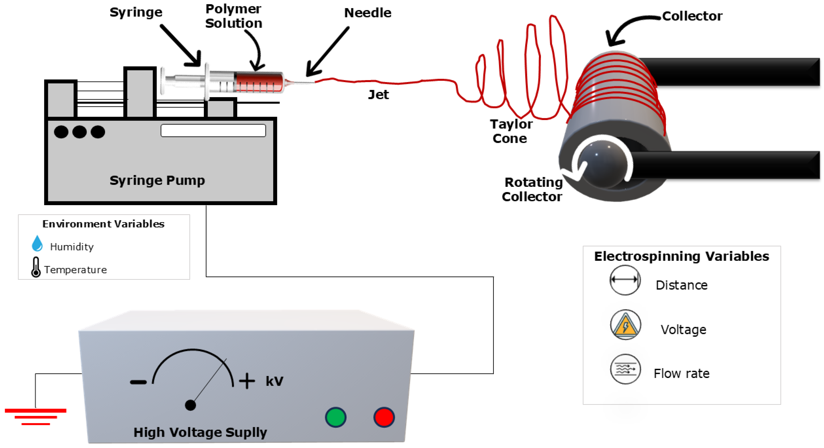
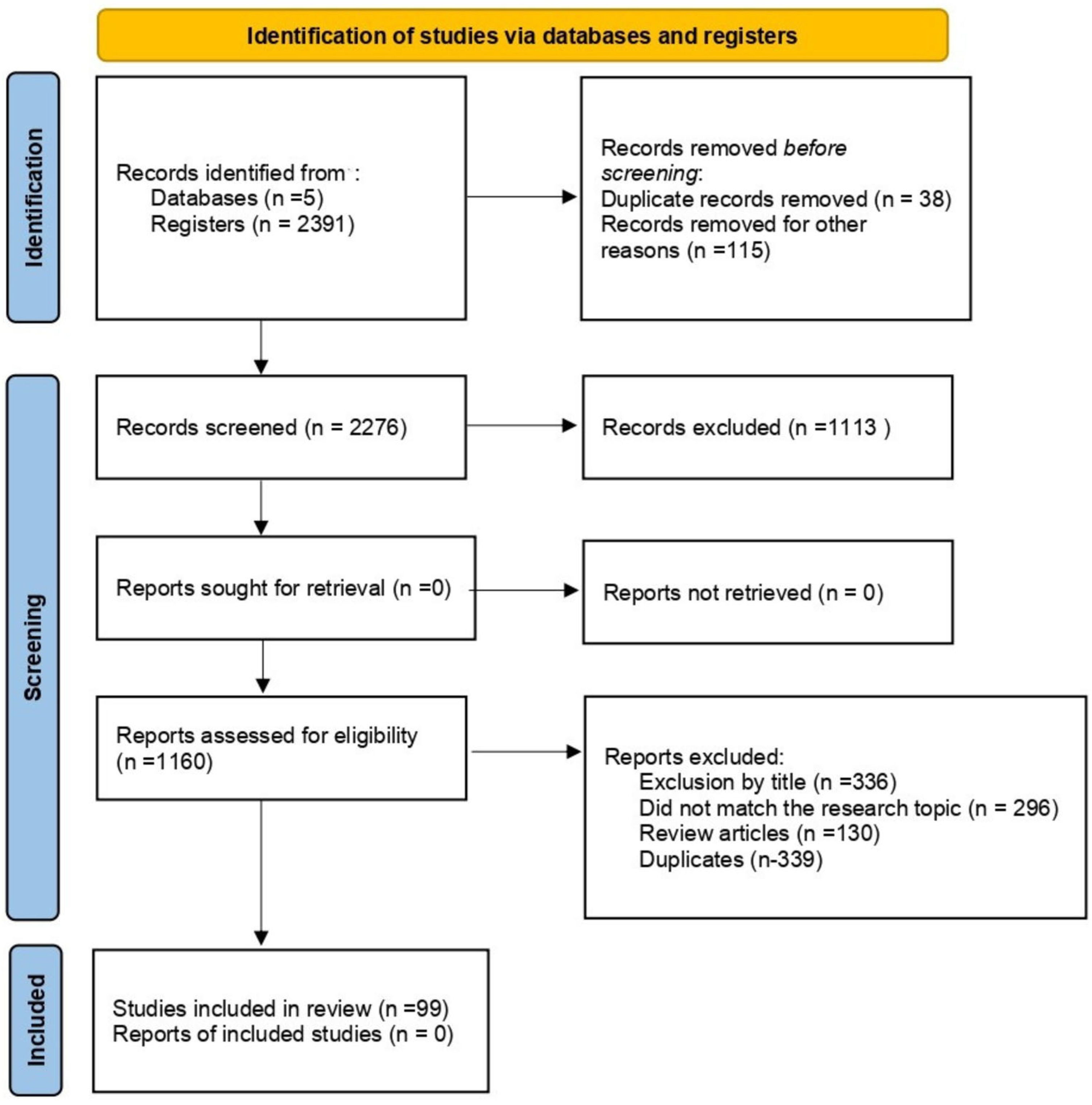
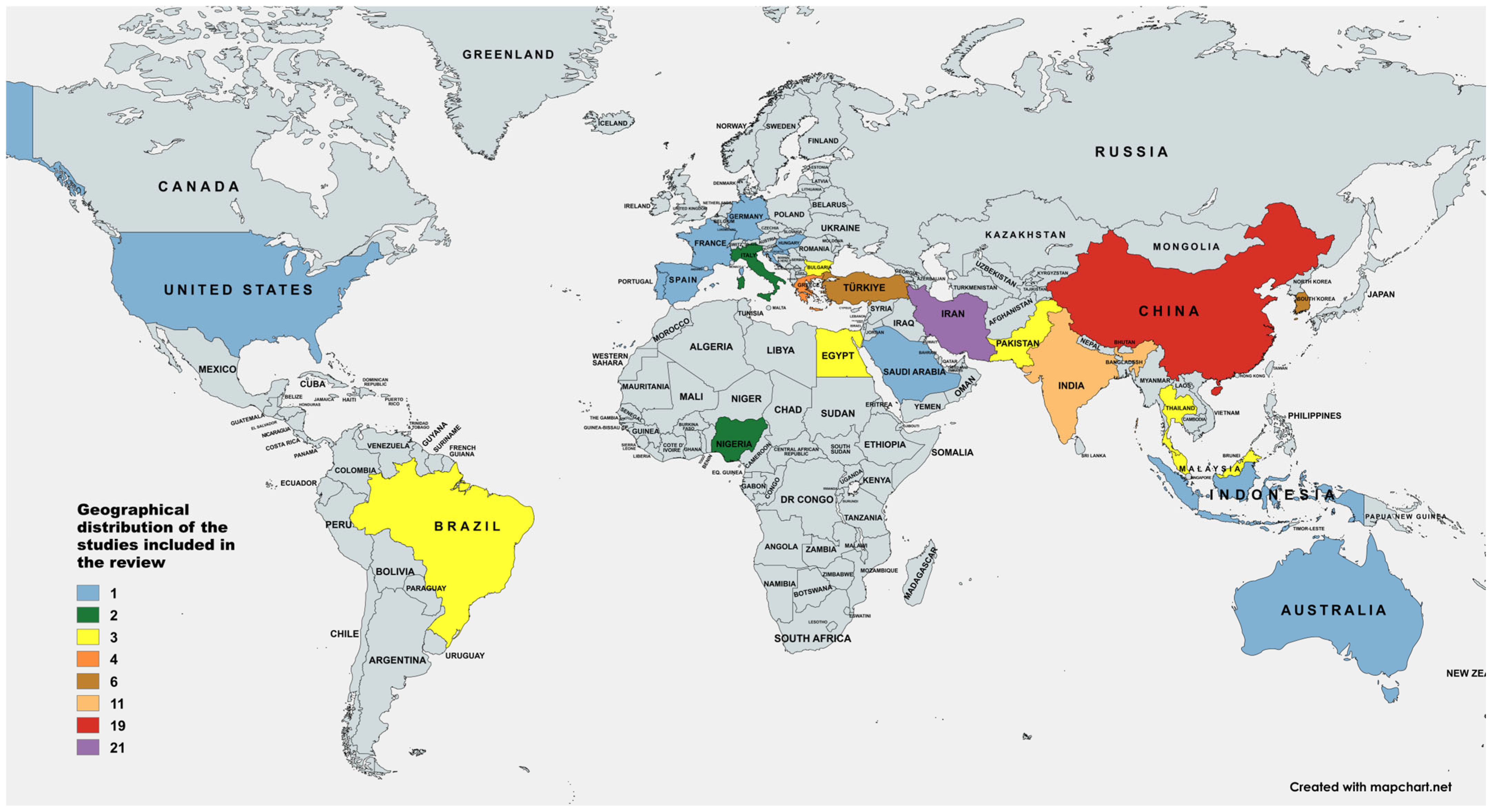
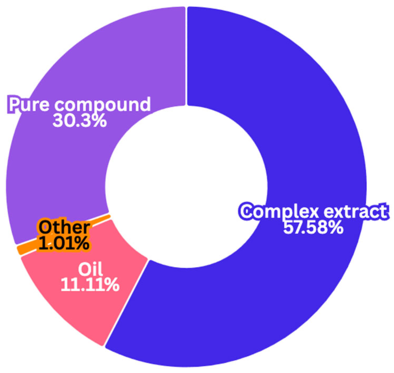
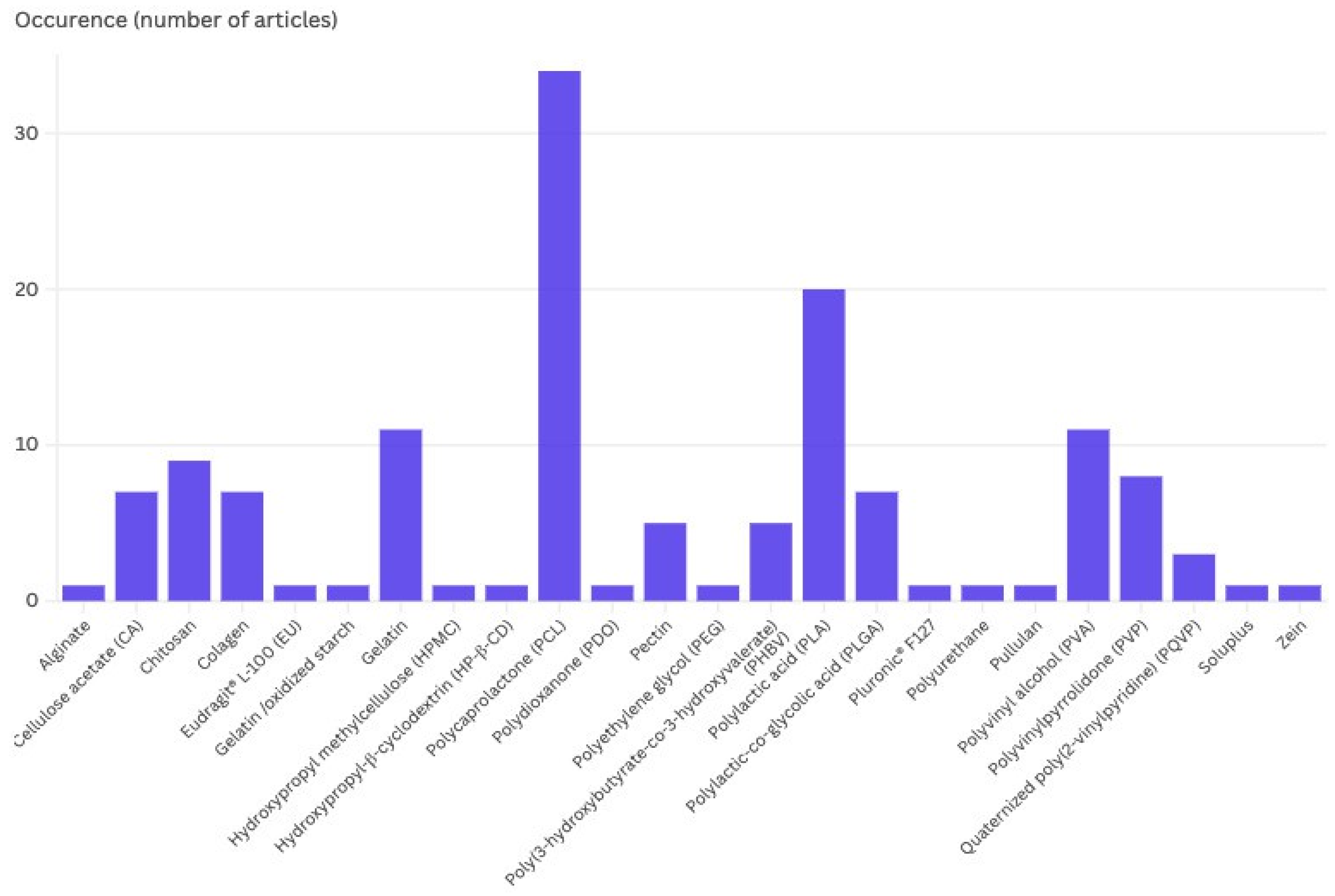
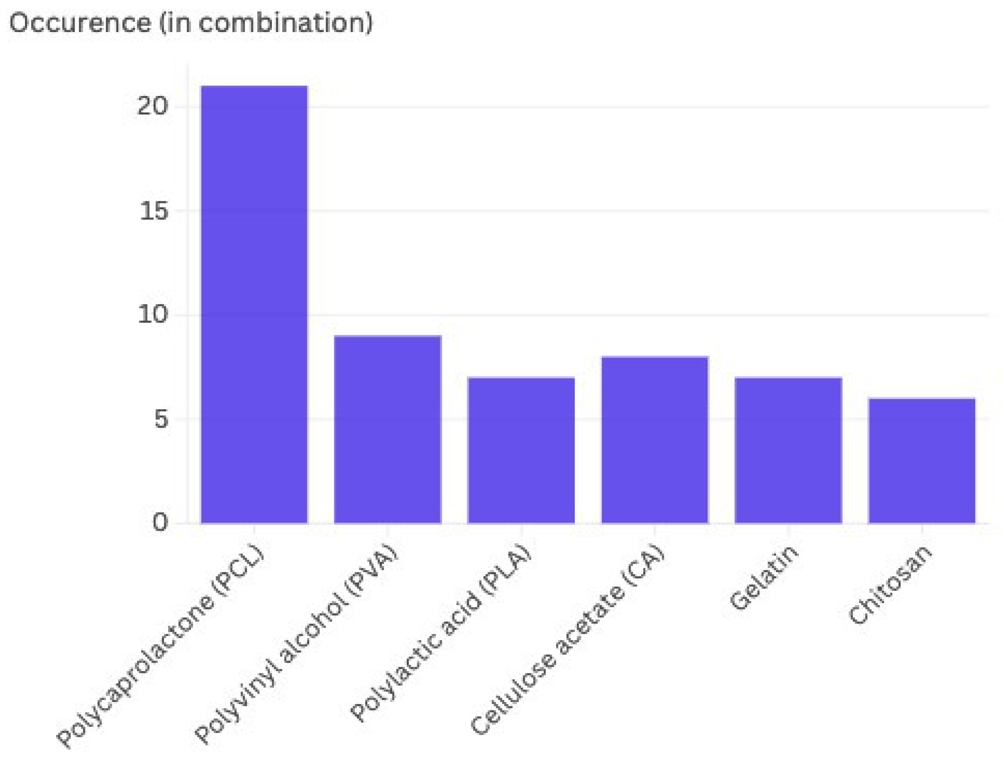

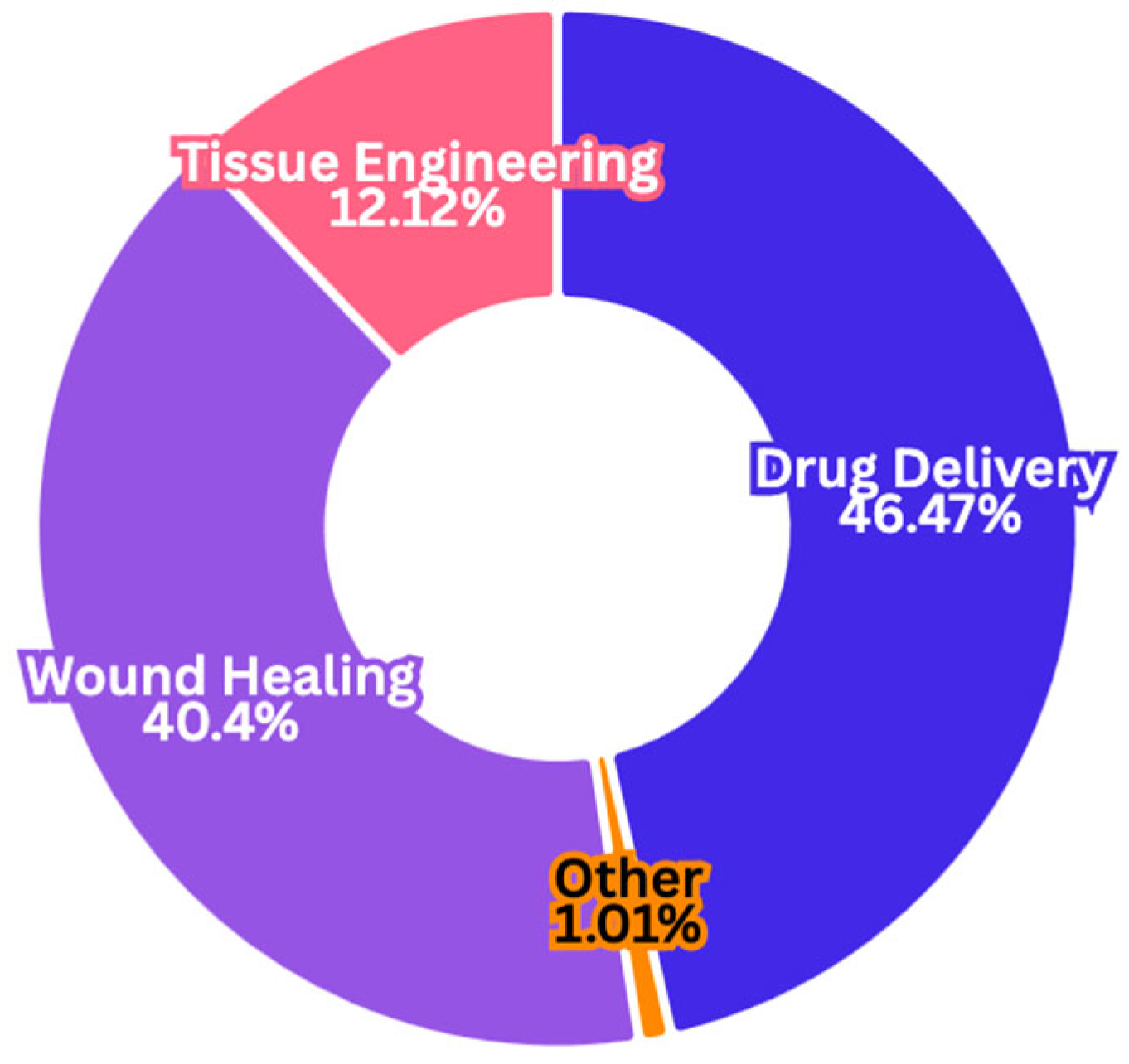
Disclaimer/Publisher’s Note: The statements, opinions and data contained in all publications are solely those of the individual author(s) and contributor(s) and not of MDPI and/or the editor(s). MDPI and/or the editor(s) disclaim responsibility for any injury to people or property resulting from any ideas, methods, instructions or products referred to in the content. |
© 2025 by the authors. Licensee MDPI, Basel, Switzerland. This article is an open access article distributed under the terms and conditions of the Creative Commons Attribution (CC BY) license (https://creativecommons.org/licenses/by/4.0/).