Article

Regulation of Hippocampal GABAergic Transmission by Fluoxetine and Its Metabolite Norfluoxetine

by
Elizabeth Vázquez-Gómez
,
Andy Hernández-Abrego
,
Jassiel Mejía-Piedras
and
Jesús García-Colunga
*
Departamento de Neurobiología Celular y Molecular, Instituto de Neurobiología, Universidad Nacional Autónoma de México, Campus Juriquilla, Boulevard Juriquilla 3001, Juriquilla 76230, Querétaro, Mexico
*
Author to whom correspondence should be addressed.
These authors contributed equally to this work.
Receptors 2024, 3(1), 1-12; https://doi.org/10.3390/receptors3010001
Submission received: 7 October 2023 / Revised: 15 November 2023 / Accepted: 28 December 2023 / Published: 4 January 2024

Abstract

:
Major depression is related to dysfunction of the GABAergic pathway. Interestingly, the antidepressant fluoxetine modifies GABAergic neurotransmission in human and animal models of depression. However, the effects of norfluoxetine (the main metabolite of fluoxetine) on GABAergic neurotransmission have not yet been studied. Therefore, we explored whether fluoxetine and/or norfluoxetine may regulate GABAergic transmission and whether these substances interact with GABAA receptors in hippocampal CA1 stratum radiatum interneurons. For these purposes, we recorded the firing profile, GABAergic spontaneous inhibitory postsynaptic currents (sIPSCs), and currents induced by GABA puffs in stratum radiatum interneurons using both whole-cell current- and voltage-clamp techniques. Interneurons were selected according with their high firing profile. We found that both fluoxetine and norfluoxetine (at 20 µM) significantly decreased the frequency of sIPSCs without modifying their amplitude and decreased the amplitude of GABA-induced currents. These results indicate that fluoxetine and norfluoxetine decrease GABA release from neurons contacting stratum radiatum interneurons and negatively modulate GABAA receptors in these interneurons, resulting in their disinhibition, which in turn may contribute to increasing the inhibition of hippocampal CA1 pyramidal neurons.

1. Introduction

Glutamate and γ-aminobutyric acid (GABA) are the primary neurotransmitters of the central nervous system, both of which are tightly regulated to ensure a balance between excitatory and inhibitory neurotransmission in the brain [1,2]. Interestingly, alterations in the balance of these neurotransmitters are relevant in the pathophysiology of mood disorders [2,3,4,5].
In the hippocampus, excitation/inhibition balance is mediated by a wide variety of interneurons, the main function of which is to inhibit the pyramidal neurons [6,7,8]. In this context, neurogenesis defects, altered excitation/inhibition balance, synaptic disinhibition of hippocampal CA1 neurons, and alterations of the GABA neurotransmitter system have been associated with depression [9,10,11,12]. Thus, the GABAergic hypothesis of depression is supported by dysregulation of GABA levels, a reduction in GABA receptor binding sites, and altered expression of GABA receptor subunit genes [13,14,15,16,17,18].
In turn, a deficit in GABA receptors alters monoaminergic activity, since GABAergic and monoaminergic neurons are interconnected [16,19,20]. In this context, the administration of antidepressants changes the functioning of the GABAergic system. For instance, chronic administration of fluoxetine (a selective serotonin reuptake inhibitor) prevented a reduction in parvalbumin cells in the hippocampus and prelimbic prefrontal cortex of socially isolated rats [21,22]. Furthermore, the administration of fluoxetine or norfluoxetine (its main metabolite) increased the levels of allopregnanolone (a potent positive allosteric modulator of the GABAA receptor) in a socially isolated mouse [23]. Fluoxetine also reduces the basal extracellular levels of GABA in the visual cortex [24], decreases the unitary IPSCs in the parvalbumin-positive fast-spiking interneurons in the hippocampus [25], and increases the firing rate of fast-spiking interneurons in the prefrontal cortex [26].
The GABAA receptors are membrane proteins belonging to the superfamily of Cys-loop ligand-gated ion channels that include serotonin (5-HT3), GABAC, nicotinic acetylcholine, and glycine receptors [27,28]. They are pentameric structures composed by the combination of different subunits (α1–α6, β1–β3, γ1–γ3, δ, ρ1–ρ3, ε, θ, and π) conferring different physiological, pharmacological, and biophysical properties [28,29,30]. These GABA receptors are located at the synapse (containing α1–α3 and γ subunits) and extrasynaptic regions (containing α4–α6 and δ subunits), mediating phasic and tonic inhibition, respectively [27,28,29].
The functioning of GABAA receptors is modulated by a wide diversity of substances, including benzodiazepines, antipsychotics, antiepileptics, anesthetics, cations, anxiolytics, and antidepressants, among others [28,31,32,33,34]. Regarding the modulation of GABAA receptors by antidepressants, there are differential effects between tricyclic antidepressants (imipramine and nortriptyline) on the one hand and fluoxetine and norfluoxetine on the other hand concerning modulation of synaptic and/or extrasynaptic GABAA receptors. The former inhibit α5-containing GABAA receptors located extrasynaptically, thus decreasing tonic GABAergic inhibition [31,35,36]. In contrast, the latter positively modulate GABAA receptors containing α1–α4, α6, γ, and δ subunits located at the synapse (α1- and γ-containing receptors) and extrasynaptically (α4-, α5-, α6-, and δ-containing receptors), increasing in this way fast and tonic inhibition, respectively [34,35,36].
It is also known that both fluoxetine and norfluoxetine inhibit the functioning and decrease the expression of a variety of receptors and channels related to Ca2+ permeability. In this context, fluoxetine inhibits N-methyl-D-aspartate (NMDA) and Ca2+-permeable α-amino-3-hydroxy-5-methylisoxazole-4-propionate (AMPA) glutamate receptors in the neurons of the hippocampal CA1 area [37] and decreases the expression of the GluA4 AMPA receptor subunit in the GABAergic parvalbumin-positive interneurons of the prefrontal cortex [38]. Furthermore, fluoxetine and norfluoxetine inhibit, in a non-competitive manner, nicotinic acetylcholine receptors, including α7 nicotinic receptors, which are highly permeable to Ca2+ [39,40,41]. Likewise, fluoxetine and norfluoxetine also inhibit T-type Ca2+ channels [42,43]. Thus, the inhibition by fluoxetine and norfluoxetine of all of these membrane proteins, when located presynaptically in GABAergic interneurons, results in a decrease in Ca2+ influx with the consequent reduction in GABA release.
Since the effects of fluoxetine on GABAergic synaptic transmission are not completely understood, and norfluoxetine has not been studied on this neurotransmitter system, the aim of the present work was to study how both fluoxetine and norfluoxetine modify GABAergic synaptic neurotransmission, particularly spontaneous inhibitory postsynaptic currents (sIPSCs) and GABAA receptors in the interneurons of the hippocampal CA1 stratum radiatum.

2. Materials and Methods

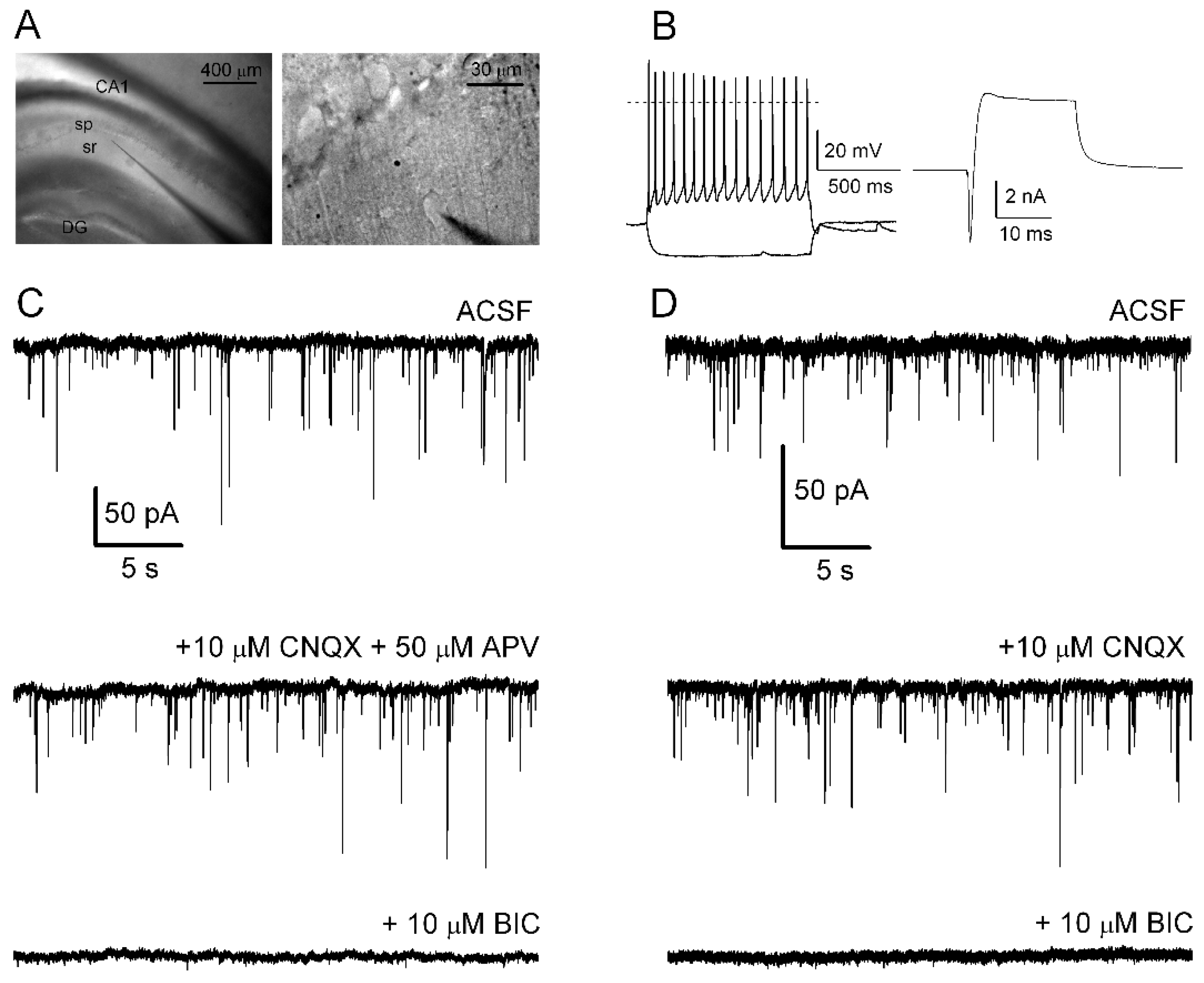
2.1. Hippocampal Brain Slices

All experimental procedures were carried out in accordance with the National Institutes of Health’s Guide for the Care and Use of Laboratory Animals and were approved by the Institutional Animal Care Committee of the Universidad Nacional Autónoma de México, with effort made to minimize the number of animals used and their suffering.
The experiments were performed as previously described [44]. Sprague Dawley rats, on postnatal days 13–16, were deeply anesthetized with isoflurane, perfused with ice cold (4 °C) solution containing (in mM) 250 sucrose, 2.5 KCl, 1.2 NaH2PO4, 5 MgCl2, 0.5 CaCl2, 26 NaHCO3, and 10 glucose (pH 7.4), and then decapitated. Their brains were quickly removed (~3 min) and placed in the same cold solution. Coronal slices (350 µm thick) containing the hippocampal CA1 area were cut from individual rats, in ~5 min, using a Vibratome Leica VT 1000 and submerged in artificial cerebrospinal fluid (ACSF) containing (in mM): 125 NaCl, 2.5 KCl, 1.23 NaH2PO4, 1 MgCl2, 2 CaCl2, 26 NaHCO3, and 10 glucose (pH 7.4). The slices were stabilized in this solution for at least 1 h before electrical recording while being continuously bubbled with 95% O2 and 5% CO2 at room temperature (20–23 °C).

2.2. Single-Cell Electrophysiological Recordings

One slice was transferred to the recording chamber and superfused during the entire experiment with ACSF at a rate of ~2 mL/min at room temperature. Cells were visualized using an infrared-differential interference contrast microscope (Olympus BX51W1) endowed with an 80× water immersion objective. During recording, an infrared-sensitive CCD camera (Hitachi, KP-M2R) displayed the slice images on a monitor. Whole-cell voltage- and current-clamp recordings [45] were performed with a PC-ONE Patch/Whole Cell Clamp (Dagan Corporation, MN, USA), using the Digidata 1440A acquisition system (Molecular Devices, CA, USA). Patch-clamp pipettes made with borosilicate glass (Sutter Instrument, CA, USA) had a resistance of 3–7 MΩ when filled with the internal solution (in mM): 140 KCl, 5 NaCl, 1 MgCl2, 10 HEPES, and 10 EGTA (pH 7.4). Recorded cells were located in the hippocampal CA1 striatum radiatum (Figure 1A) and were maintained at a membrane potential of −70 mV. The recorded cells often had fusiform-shaped soma with a size between 15 and 25 µm. They were interneurons as judged from the firing of action potentials in response to current pulses in current-clamp mode (Figure 1B, left trace), and/or inward and outward currents generated by a depolarized pulse from −70 to 20 mV in voltage-clamp mode (Figure 1B, right trace). Data were stored on a PC using a Digidata 1440A AD converter, at a sampling rate of 10 kHz.

2.2.1. Recording of sIPSCs

For recording spontaneous inhibitory postsynaptic currents (sIPSCs), 6-cyano-2,3-dihydroxy-7-nitroquinoxaline (CNQX; 10 µM) and/or DL-2-amino-5-phosponovaleric acid (APV; 50 µM) were added to the bath solution; they inhibited glutamatergic transmission mediated by AMPA and NMDA glutamate receptors, respectively. After recording control sIPSCs for at least 10 min, fluoxetine or norfluoxetine was added to the bath solution for ~10 min, and then it was washed out from the perfusing solution for an additional 10 min.

2.2.2. Recording of GABA-Induced Currents

Another way to explore the effects of both fluoxetine and norfluoxetine was previously described for other substances [44,46,47]. GABA puffs (10 µM, 2–5 psi, 500 ms) were applied to cells through a fine-tip glass micropipette placed ~10 µm from the recorded cell using a pneumatic picopump (PV830, WPI, FL, USA). Thus, GABA puffs were applied every 5 min before, during, and after fluoxetine or norfluoxetine was added to the bath solution for ~10 min. This was sufficient time to obtain the effect of a substance with a perfusion rate of ~2 mL/min because the perfusion solution was completely replaced in the recording chamber after ~2 min [44,46,47]. The amplitude of the GABA-induced current was measured as a function of the recording time.

2.3. Data Analysis and Statistics

Mini Analysis software (Synaptosoft, Decatur, GA, USA) was used to detect and evaluate both the frequency and amplitude of sIPSCs, with a threshold set at 8 pA. These spontaneous synaptic events were detected and measured automatically and then visually confirmed. For a recorded interneuron, both the mean frequency and amplitude of sIPSCs were calculated for 2–4 min under each experimental condition. Changes in the frequency and amplitude of sIPSCs due to fluoxetine or norfluoxetine were expressed as relative values, that is, the ratio between the frequency or amplitude of sIPSCs in the presence of the applied substance and its corresponding control parameter. pClamp 10 software was used to measure the GABA-induced current amplitude in the absence or presence of the applied substance. Origin 7.0 software (Microcal Software, Piscataway, MA, USA) was used to analyze and graph the results. Data are presented as the mean ± standard error. Comparison of the two population means of the relative frequency and amplitude of the sIPSCs before and during fluoxetine or norfluoxetine application was performed by paired Student’s t-test. p < 0.05 was considered statistically significant.

3. Results

3.1. Spontaneous GABAergic Synaptic Activity in CA1 Interneurons

Hippocampal CA1 stratum radiatum interneurons are relevant for the integration of inhibitory circuits and excitation/inhibition balance in this brain region, which is associated with processing of memories, emotions, and affective behavior [7,48,49].
Therefore, for recording, we selected cells located in the hippocampal CA1 stratum radiatum, a region abundant in inhibitory interneurons (see Figure 1A). Under current-clamp conditions, we applied hyperpolarizing (−80 pA) and depolarizing (160 pA) current pulses to recording cells; in the first case, the cells responded with a passive hyperpolarizing membrane potential, while in the second case they responded with a high frequency action potential that showed overshoot (Figure 1B, left traces). On the other hand, under voltage-clamp conditions, the cells responded to a change in the depolarizing voltage pulse from the holding potential of −70 to 20 mV, with a fast inward current followed by a slowly decaying outward current (Figure 1B, right trace). The high firing profile was attributed to interneurons [7,25]. Cells that did not respond with this profile were discarded for the study.
First, the cells were superfused with ACSF as a control solution, and then we recorded the spontaneous postsynaptic currents (Figure 1C, upper trace). To avoid the contribution of spontaneous excitatory postsynaptic currents (sEPSCs), 10 µM CNQX plus 50 µM APV (AMPA and NMDA receptor antagonists, respectively) were added to the bath solution (Figure 1C, middle trace). The spontaneous synaptic activity was eliminated by adding 10 µM bicuculline (antagonist of GABAA receptors) to the bath solution for 15 min (Figure 1C, bottom trace), indicating that these synaptic events corresponded to sIPSCs due to the activation of GABAA receptors. Similar results were obtained when only 10 µM CNQX was added to the bath ACSF (Figure 1D).
Therefore, to obtain sIPSCs, all the following experiments were performed by superfusing brain slices with ACSF plus 10 µM CNQX.

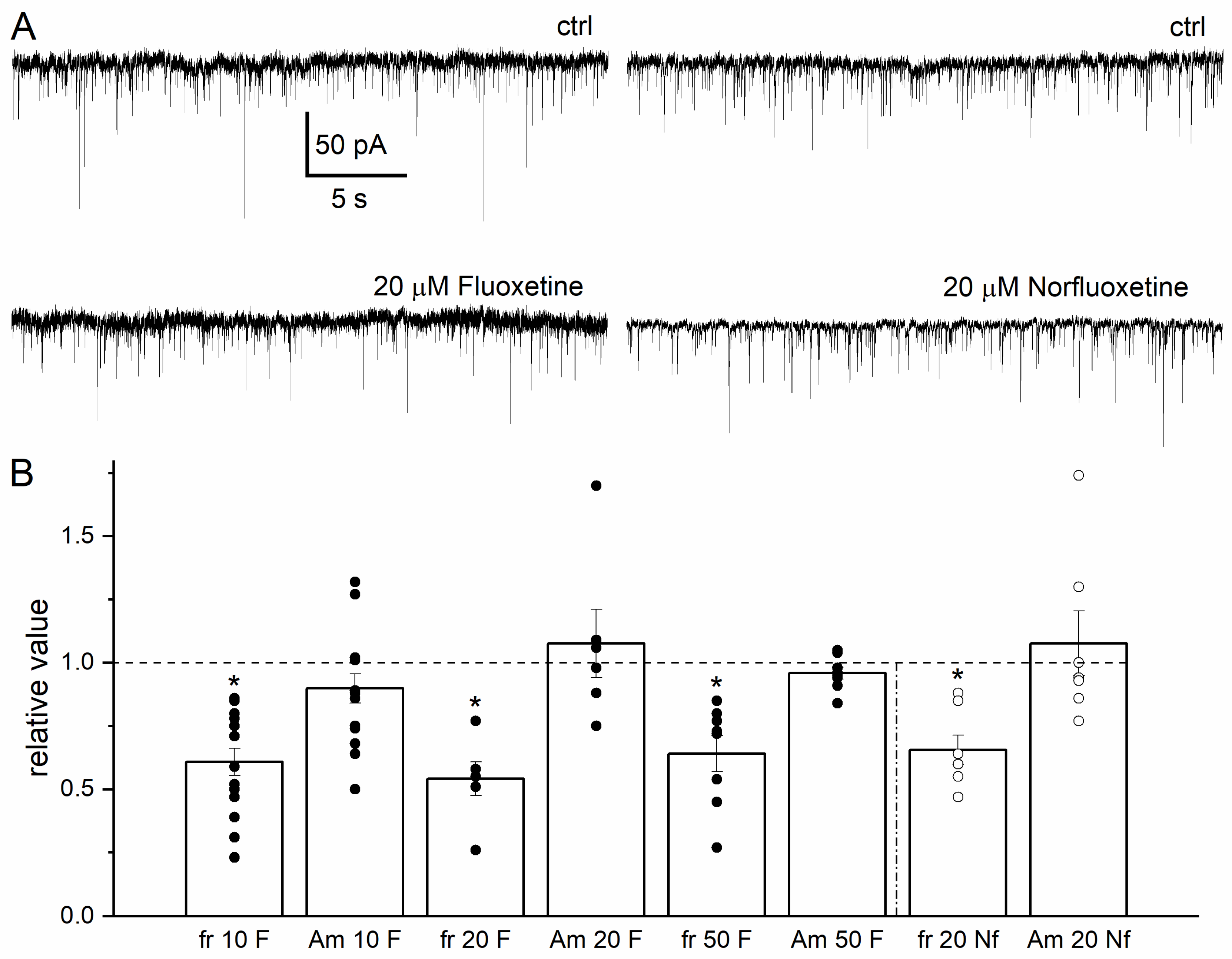
3.2. Effects of Fluoxetine and Norfluoxetine on sIPSCs in Hippocampal Interneurons

Malfunctioning of the inhibitory networks alters the excitation/inhibition balance and, in consequence, the excitability of principal neurons. These alterations have been associated with mood disorders such anxiety and depression disorders [12,14,50]. In addition, the antidepressant fluoxetine has been shown to modulate GABAergic neurotransmission in the hippocampus and amygdala [25,51,52].
In this regard, and once spontaneous GABAergic synaptic activity was pharmacologically isolated, the effects of both fluoxetine and its principal metabolite norfluoxetine were explored on sIPSCs in hippocampal CA1 stratum radiatum interneurons.
Initially, control sIPSCs were recorded for at least 10 min (Figure 2A, upper traces). Then, fluoxetine at 10, 20, and 50 µM or norfluoxetine at 20 µM was added to the bath ACSF for 10 min while sIPSCs were continuously recorded (Figure 2A, bottom traces are sample records at 20 µM of each substance). The effects of fluoxetine and norfluoxetine on the frequency and amplitude of sIPSCs were evaluated and normalized with respect to their own control values. The frequency of sIPSCs decreased with the application of fluoxetine or norfluoxetine (Figure 2B). At 10 µM fluoxetine, the relative frequency of sIPSCs was 0.6 ± 0.05 compared to its control; at 20 µM, this relative frequency was 0.54 ± 0.06, and at 50 µM, it was 0.64 ± 0.07 (Figure 2B). On the other hand, the relative sIPSC amplitude had no statistical significance at any fluoxetine concentration, ranging between 0.95 and 1.07. Finally, 20 µM norfluoxetine also resulted in a decrease in the relative frequency of sIPSCs to 0.65 ± 0.05, without affecting the amplitude of sIPSCs (Figure 2B, right columns, open circles). The actions of both fluoxetine and norfluoxetine on sIPSCs were compared at 20 µM, a concentration reached in the rat brain after acute administration of fluoxetine [53], and because the extent of the decrease in the frequency of sIPSCs was similar with 10, 20 and 50 µM fluoxetine.

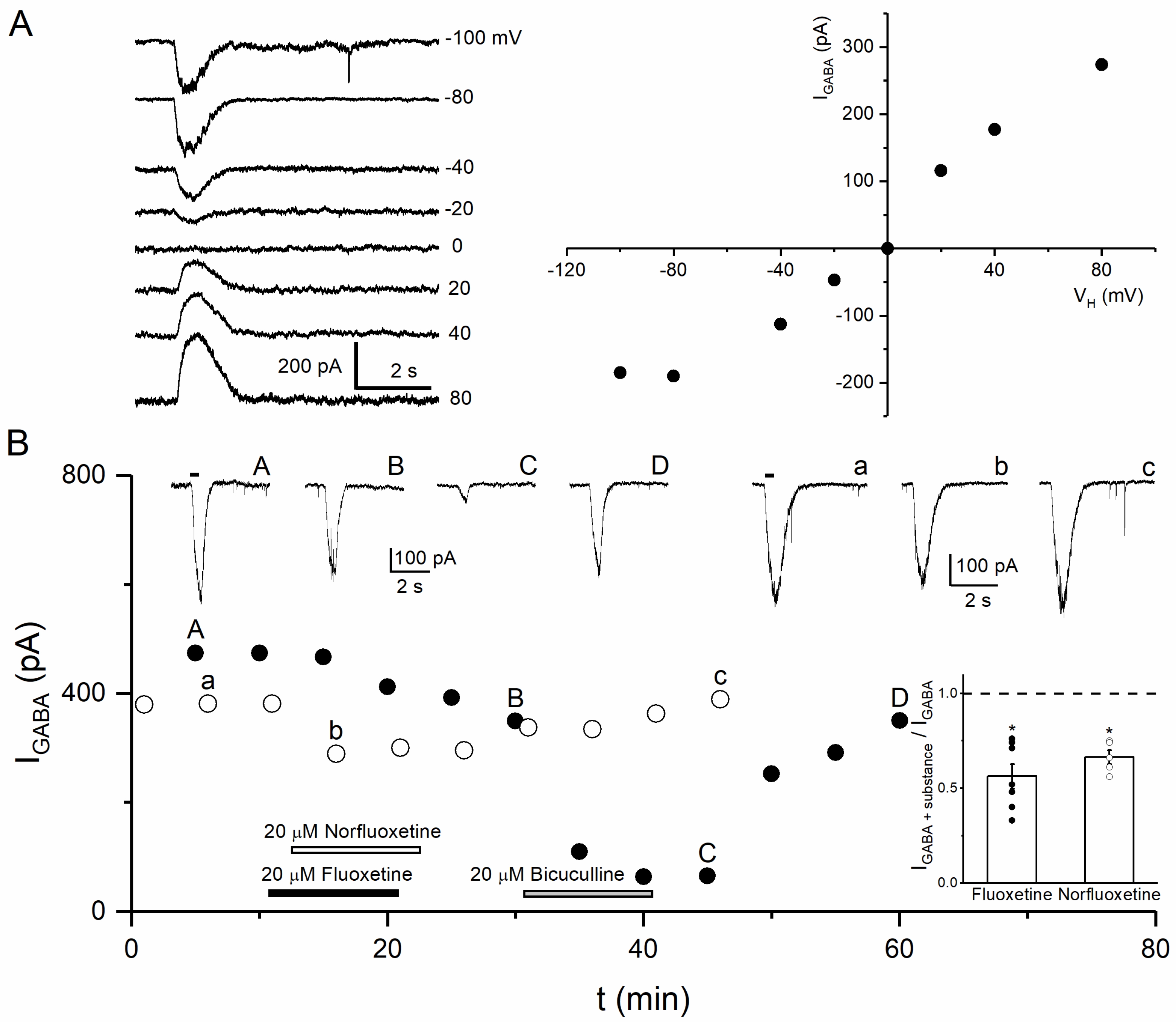
3.3. Effects of Fluoxetine and Norfluoxetine on GABA-Induced Currents

Since the functioning and expression of the GABAA receptors are affected during depression [17,54], and these receptors are modulated by a wide variety of substances, including different classes of antidepressants [31,34], the next step was to determine whether fluoxetine and norfluoxetine interacted directly with GABAA receptors, potentiating or inhibiting their function [34,55,56].
Firstly, activation of native GABAA receptors in stratum radiatum interneurons was achieved by applying local puffs of 10 µM GABA ~10 µm near the recorded interneuron. The holding potential was changed from −100 to 80 mV. As expected, the amplitude and direction of the GABA-induced current depended on the potential: GABA puffs resulted in transient inward GABA-induced currents for negative holding potentials and outward GABA-induced currents for positive potentials (Figure 3A, left traces). The relationship between the amplitude of GABA-induced currents as a function of holding potential showed that the reversal potential was near 0 mV, according to the equilibrium potential calculated using the Nernst equation for ion solutions: ECl = 2.4 mV (Figure 3A, right plot; sample of n = 3).
Then, we explored the actions of both fluoxetine and norfluoxetine on the GABA-induced currents in hippocampal CA1 stratum radiatum interneurons. As control responses, GABA puffs were applied at 5 min intervals; the amplitude of the GABA-induced currents remained constant for up to 60 min (n = 3, data not shown), indicating no rundown of the response. Subsequently, 20 µM fluoxetine or norfluoxetine was added to the bath solution for ~10 min. The GABA-induced current amplitude was measured as a function of the recording time before, during, and after application of the substance (Figure 3B). Under these conditions, both fluoxetine and norfluoxetine decreased the GABA-induced currents (Figure 3B filled circles, records A, B; open circles, records a, b, respectively). Thus, the ratio between the GABA-induced currents in the presence of fluoxetine or norfluoxetine and the corresponding control GABA-induced currents was 0.56 ± 0.07 (n = 7) or 0.67 ± 0.037 (n = 5), respectively (Figure 3B, right inset). Note that after fluoxetine was removed from the bath solution, ~10 min later 20 µM bicuculline was added to the bath for ~10 min, resulting in strong inhibition of the GABA-induced currents to ~0.13 of the control currents (Figure 3B filled circles, records C). Then, bicuculline was washed out from the bath and the GABA-induced currents were partially recovered (Figure 3B filled circles, records D, n = 3).

4. Discussion

It is known that the antidepressant fluoxetine modifies GABAergic neurotransmission [24,25,57]; however, its mechanisms of action are not completely understood and the effects of norfluoxetine have not yet been explored. Therefore, in the present study, we showed that different concentrations of fluoxetine and norfluoxetine decrease the frequency of sIPSCs in hippocampal CA1 stratum radiatum interneurons, without changing their amplitude. In addition, 20 µM of these substances decreases the GABA-induced currents.
In the hippocampal CA1 stratum radiatum area, the most abundant type of cells are interneurons, the main function of which is to inhibit the dendrites of pyramidal neurons [7,8]. In this area, the inhibition of inhibitory circuits results in disinhibition of CA1 neurons and consequently despair-like behaviors [12]. In this context, fluoxetine increases sIPSCs in the pyramidal neurons of the hippocampus, resulting in inhibition of these cells [25,58,59]. The decrease in the frequency of sIPSCs of interneurons due to fluoxetine or norfluoxetine together with a reduction in the GABA-induced currents by these substances disinhibit these interneurons, resulting in increased inhibition of CA1 pyramidal neurons. In this regard, the disinhibition of GABA interneurons by fluoxetine and norfluoxetine could be necessary for the antidepressant response [60].
Assuming that both fluoxetine and norfluoxetine modulate different targets in addition to serotonin transport, another feasible way to explain the decrease in the frequency of sIPSCs in hippocampal stratum radiatum interneurons may be through their interaction with nicotinic acetylcholine receptors, Ca2+-permeable AMPA receptors, and/or T-type calcium channels [37,38,42,43]. It is known that both fluoxetine and norfluoxetine inhibit nicotinic acetylcholine receptors in a non-competitive manner [39,40,41]; in this context, interneurons of the hippocampal CA1 stratum radiatum selectively express the α7 nicotinic receptor, which is highly permeable to Ca2+ [61,62]. Therefore, the possible inhibition of all of these membrane proteins by fluoxetine and norfluoxetine in cells that establish synapses with stratum radiatum interneurons would lead to a decrease in GABA release, with the consequent disinhibition of stratum radiatum interneurons.
Another way to explain the actions of fluoxetine and norfluoxetine may be related to the inhibition of potassium channels. In this regard, it has been shown that these two substances inhibit TREK-1 and TREK-2 potassium channels [63,64], which are expressed in both GABAergic neurons and hippocampal cells [65,66]. The activation of these channels decreases neuron excitability by increasing potassium efflux [67,68]. Therefore, another way by which norfluoxetine decreases sIPSCs may be mediated by TREK inhibition, accordingly increasing the excitability of GABAergic recorded cells.
Furthermore, we found that both fluoxetine and norfluoxetine inhibited the GABA-induced currents, similar to imipramine and nortriptyline [31], possibly due to the interaction of these antidepressants with extrasynaptic α5-containing GABAA receptors, contributing to tonic inhibition [35,36]. Interestingly, the α5 subunit is abundantly expressed in hippocampal CA1 interneurons of the stratum radiatum, oriens, and lacunosum molecular [69,70,71]. The same substances, fluoxetine and norfluoxetine, positively modulate GABAA receptors containing the α1–α4 and α6, β1–β3, γ1–γ3, and δ subunits [34], interacting with synaptic and extrasynaptic GABAA receptors and participating in fast and tonic inhibition [35,36]. Thus, interestingly, antidepressant substances may impact the regulation of hippocampal cellular excitability, which depends on the phasic and tonic inhibition mediated by synaptic and extrasynaptic GABAA receptors, respectively [27,28,29]. All of this together indicates different targets and mechanisms of fluoxetine and norfluoxetine that may be associated with their therapeutic actions.
In particular, mice subjected to social defeat or social isolation were susceptible to stress, and α5 subunit-containing GABAA receptors and their mRNA levels were increased in the hippocampus and prefrontal cortex, respectively [72,73]. In this regard, the overfunctioning and/or overexpression of this receptor could be counteracted by inhibition of the α5-containing GABAA receptor due to its interaction with fluoxetine and norfluoxetine, as we assumed above.
Moreover, following a single oral dose of fluoxetine and norfluoxetine, the half-life times were approximately 2–4 and 7–9 days, respectively [74,75,76], whereas the therapeutic effects of several antidepressants, including fluoxetine, were detectable after at least 4 to 8 weeks, improving cognition and other symptoms of depression [76,77,78]. However, the acute application (in the range of minutes) of fluoxetine or norfluoxetine modified sIPSCs and the GABA-induced currents, as reported in the present work, consistent with the GABAergic hypothesis of depression [13,14,15,16,17,18].
In this regard, when fluoxetine was administered to rats, the brain concentration of fluoxetine and its principal metabolite norfluoxetine ranged between 1.5 and 25 µM [53,79]. Interestingly, the lowest concentration of fluoxetine tested here (10 µM), which is very likely reached during the treatment of depressive patients, affected the sIPSCs. Moreover, the antidepressant effects of both substances could be attributable to their interaction with different targets: serotonin transporters [80], nicotinic receptors [39,40,41], potassium channels [63], Ca2+-permeable AMPA receptors and/or T-type calcium channels [37,38,42,43], and GABAA receptors ([34], this work), among others.

5. Conclusions

The results presented here allow us to conclude that fluoxetine and norfluoxetine, at the presynaptic level, mainly decrease GABA release from cells impinging stratum radiatum interneurons, possibly by inhibiting presynaptic Ca2+-permeable membrane proteins, and that both substances probably interact with extrasynaptic α5-containing GABAA receptors, increasing tonic inhibition. These results may help to better understand how antidepressants and their metabolites could be relevant in the regulation of hippocampal inhibitory circuits, as well as for the treatment of depressive patients.

Author Contributions

J.G.-C. conceived and designed the experiments; E.V.-G., A.H.-A. and J.M.-P. performed the experiments; E.V.-G., A.H.-A., J.M.-P. and J.G.-C. analyzed the data, wrote the manuscript, and approved the final manuscript. All authors have read and agreed to the published version of the manuscript.

Funding

This work was supported by Dirección General de Asuntos del Personal Académico, UNAM (DGAPA, PAPIIT grants IN202420 and IN206423).

Institutional Review Board Statement

The animal study protocol was carried out in accordance with the National Institutes of Health’s Guide for the Care and Use of Laboratory Animals and were approved by the Animal Care Committee of the Instituto de Neurobiología, Universidad Nacional Autónoma de México, protocol code 055, approved on 28 June 2012.

Data Availability Statement

The data presented in this study are available upon request from the corresponding author.

Acknowledgments

We thank Martín García-Servín for his assistance in taking care of the rats and Benito Ordaz Sánchez for help with pictures of brain slices.

Conflicts of Interest

The authors declare that they have no conflicts of interest.

References

  1. Iascone, D.M.; Li, Y.; Sümbül, U.; Doron, M.; Chen, H.; Andreu, V.; Goudy, F.; Blockus, H.; Abbott, L.F.; Segev, I.; et al. Whole-neuron synaptic mapping reveals spatially precise excitatory/inhibitory balance limiting dendritic and somatic spiking. Neuron 2020, 106, 566–578. [Google Scholar] [CrossRef] [PubMed]
  2. Selten, M.; Van Bokhoven, H.; Nadif Kasri, N. Inhibitory control of the excitatory/inhibitory balance in psychiatric disorders. F1000Research 2018, 7, 23. [Google Scholar] [CrossRef] [PubMed]
  3. Duman, R.S.; Aghajanian, G.K. Synaptic dysfunction in depression: Potential therapeutic targets. Science 2012, 338, 68–72. [Google Scholar] [CrossRef] [PubMed]
  4. Sohal, V.S.; Rubenstein, J.L.R. Excitation-inhibition balance as a framework for investigating mechanisms in neuropsychiatric disorders. Mol. Psychiatry 2019, 24, 1248–1257. [Google Scholar] [CrossRef]
  5. Wang, H.L.; Sun, Y.X.; Liu, X.; Wang, H.; Ma, Y.N.; Su, Y.A.; Li, J.T.; Si, T.M. Adolescent stress increases depression-like behaviors and alters the excitatory-inhibitory balance in aged mice. Chin. Med. J. 2019, 132, 1689–1699. [Google Scholar] [CrossRef] [PubMed]
  6. Freund, T.F.; Katona, I. Perisomatic inhibition. Neuron 2007, 56, 33–42. [Google Scholar] [CrossRef]
  7. Pelkey, K.A.; Chittajallu, R.; Craig, M.T.; Tricoire, L.; Wester, J.C.; McBain, C.J. Hippocampal GABAergic inhibitory interneurons. Physiol. Rev. 2017, 97, 1619–1747. [Google Scholar] [CrossRef] [PubMed]
  8. Udakis, M.; Pedrosa, V.; Chamberlain, S.E.L.; Clopath, C.; Mellor, J.R. Interneuron-specific plasticity at parvalbumin and somatostatin inhibitory synapses onto CA1 pyramidal neurons shapes hippocampal output. Nat. Commun. 2020, 11, 4395. [Google Scholar] [CrossRef]
  9. Bielau, H.; Steiner, J.; Mawrin, C.; Trübner, K.; Brisch, R.; Meyer-Lotz, G.; Brodhun, M.; Dobrowolny, H.; Baumann, B.; Gos, T.; et al. Dysregulation of GABAergic neurotransmission in mood disorders: A postmortem study. Ann. N. Y. Acad. Sci. 2007, 1096, 157–169. [Google Scholar] [CrossRef]
  10. Brambilla, P.; Perez, J.; Barale, F.; Schettini, G.; Soares, J.C. GABAergic dysfunction in mood disorders. Mol. Psychiatry 2003, 8, 721–737. [Google Scholar] [CrossRef]
  11. Kim, I.B.; Park, S.C. Neural circuitry-neurogenesis coupling model of depression. Int. J. Mol. Sci. 2021, 22, 2468. [Google Scholar] [CrossRef] [PubMed]
  12. Yoon, S.H.; Chung, G.; Song, W.S.; Oh, S.P.; Kim, J.; Kim, S.J.; Kim, M.H. Altered GABAergic inhibition in CA1 pyramidal neurons modifies despair-like behavior in mice. bioRxiv 2021. [Google Scholar] [CrossRef]
  13. Czéh, B.; Vardya, I.; Varga, Z.; Febbraro, F.; Csabai, D.; Martis, L.S.; Højgaard, K.; Henningsen, K.; Bouzinova, E.V.; Miseta, A.; et al. Long-term stress disrupts the structural and functional integrity of GABAergic neuronal networks in the medial prefrontal cortex of rats. Front. Cell. Neurosci. 2018, 12, 148. [Google Scholar] [CrossRef] [PubMed]
  14. Dósa, Z.; Nieto-Gonzalez, J.; Elfving, B.; Hougaard, K.; Holm, M.; Wegener, G.; Jensen, K. Reduction in hippocampal GABAergic transmission in a low birth weight rat model of depression. Acta Neuropsychiatr. 2023, 35, 315–327. [Google Scholar] [CrossRef] [PubMed]
  15. Fogaça, M.V.; Duman, R.S. Cortical GABAergic dysfunction in stress and depression: New insights for therapeutic interventions. Front. Cell. Neurosci. 2019, 13, 87. [Google Scholar] [CrossRef]
  16. Luscher, B.; Shen, Q.; Sahir, N. The GABAergic deficit hypothesis of major depressive disorder. Mol. Psychiatry 2011, 16, 383–406. [Google Scholar] [CrossRef]
  17. Merali, Z.; Du, L.; Hrdina, P.; Palkovits, M.; Faludi, G.; Poulter, M.O.; Anisman, H. Dysregulation in the suicide brain: mRNA expression of corticotropin-releasing hormone receptors and GABA(A) receptor subunits in frontal cortical brain region. J. Neurosci. 2004, 24, 1478–1485. [Google Scholar] [CrossRef]
  18. Zhong, H.; Rong, J.; Zhu, C.; Liang, M.; Li, Y.; Zhou, R. Epigenetic modifications of GABAergic interneurons contribute to deficits in adult hippocampus neurogenesis and depression-like behavior in prenatally stressed mice. Int. J. Neuro-Psychopharmacol. 2020, 23, 274–285. [Google Scholar] [CrossRef]
  19. Judge, S.J.; Ingram, C.D.; Gartside, S.E. GABA receptor modulation of 5-HT neuronal firing: Characterization and effect of moderate in vivo variations in glucocorticoid levels. Neurochem. Int. 2004, 45, 1057–1065. [Google Scholar] [CrossRef]
  20. Lladó-Pelfort, L.; Santana, N.; Ghisi, V.; Artigas, F.; Celada, P. 5-HT1A receptor gonists enhance pyramidal cell firing in prefrontal cortex through a preferential action on GABA interneurons. Cereb. Cortex. 2012, 22, 1487–1497. [Google Scholar] [CrossRef]
  21. Filipović, D.; Stanisavljević, A.; Jasnić, N.; Bernardi, R.E.; Inta, D.; Perić, I.; Gass, P. Chronic treatment with fluoxetine or clozapine of socially isolated rats prevents subsector-specific reduction of parvalbumin immunoreactive cells in the hippocampus. Neuroscience 2018, 371, 384–394. [Google Scholar] [CrossRef] [PubMed]
  22. Todorović, N.; Mićić, B.; Schwirtlich, M.; Stevanović, M.; Filipović, D. Subregion-specific protective effects of fluoxetine and clozapine on parvalbumin expression in medial prefrontal cortex of chronically isolated rats. Neuroscience 2019, 396, 24–35. [Google Scholar] [CrossRef] [PubMed]
  23. Pinna, G.; Costa, E.; Guidotti, A. Fluoxetine and norfluoxetine stereospecifically and selectively increase brain neurosteroid content at doses that are inactive on 5-HT reuptake. Psychopharmacology 2006, 186, 362–372. [Google Scholar] [CrossRef] [PubMed]
  24. Maya Vetencourt, J.F.; Sale, A.; Viegi, A.; Baroncelli, L.; De, P.R.; O’Leary, O.F.; Castren, E.; Maffei, L. The antidepressant fluoxetine restores plasticity in the adult visual cortex. Science 2008, 320, 385–388. [Google Scholar] [CrossRef] [PubMed]
  25. Méndez, P.; Pazienti, A.; Szabó, G.; Bacci, A. Direct alteration of a specific inhibitory circuit of the hippocampus by antidepressants. J. Neurosci. 2013, 32, 16616–16628. [Google Scholar] [CrossRef] [PubMed]
  26. Zhong, P.; Yan, Z. Differential regulation of the excitability of prefrontal cortical fast-spiking interneurons and pyramidal neurons by serotonin and fluoxetine. PLoS ONE 2011, 6, e16970. [Google Scholar] [CrossRef] [PubMed]
  27. Hannan, S.; Minere, M.; Harris, J.; Izquierdo, P.; Thomas, P.; Tench, B.; Smart, T.G. GABAAR isoform and subunit structural motifs determine synaptic and extrasynaptic receptor localisation. Neuropharmacology 2020, 169, 107540. [Google Scholar] [CrossRef] [PubMed]
  28. Kasaragod, V.B.; Mortensen, M.; Hardwick, S.W.; Wahid, A.A.; Dorovykh, V.; Chirgadze, D.Y.; Smart, T.G.; Miller, P.S. Mechanisms of inhibition and activation of extrasynaptic alpha beta GABAA receptors. Nature 2022, 602, 529–533. [Google Scholar] [CrossRef]
  29. Luscher, B.; Maguire, J.L.; Rudolph, U.; Sibille, E. GABAA receptors as targets for treating affective and cognitive symptoms of depression. Trends Pharmacol. Sci. 2023, 44, 586–600. [Google Scholar] [CrossRef]
  30. Thompson, S.M. Modulators of GABAA receptor-mediated inhibition in the treatment of neuropsychiatric disorders: Past, present, and future. Neuropharmacology 2023, 49, 83–95. [Google Scholar] [CrossRef]
  31. Bampali, K.; Koniuszewski, F.; Silva, L.L.; Rehman, S.; Vogel, F.D.; Seidel, T.; Scholze, P.; Zirpel, F.; Garon, A.; Langer, T.; et al. Tricyclic antipsychotics and antidepressants can inhibit α5-containing GABAA receptors by two distinct mechanisms. Br. J. Pharmacol. 2022, 179, 3675–3692. [Google Scholar] [CrossRef] [PubMed]
  32. Goldschen-Ohm, M.P. Benzodiazepine modulation of GABAA receptors: A mechanistic perspective. Biomolecules 2022, 12, 1784. [Google Scholar] [CrossRef] [PubMed]
  33. Luo, Y.; Balle, T. GABAA receptors as targets for anaesthetics and analgesics and promising candidates to help treat coronavirus infections: A mini-review. Basic Clin. Pharmacol. Toxicol. 2022, 131, 443–451. [Google Scholar] [CrossRef] [PubMed]
  34. Robinson, R.T.; Drafts, B.C.; Fisher, J.L. Fluoxetine increases GABA(A) receptor activity through a novel modulatory site. J. Pharmacol. Exp. Ther. 2003, 304, 978–984. [Google Scholar] [CrossRef] [PubMed]
  35. Carver, C.M.; Reddy, D.S. Neurosteroid interactions with synaptic and extrasynaptic GABA(A) receptors: Regulation of subunit plasticity, phasic and tonic inhibition, and neuronal network excitability. Psychopharmacology 2013, 230, 151–188. [Google Scholar] [CrossRef] [PubMed]
  36. Reddy, D.S.; Mbilinyi, R.H.; Estes, E. Preclinical and clinical pharmacology of brexanolone (allopregnanolone) for postpartum depression: A landmark journey from concept to clinic in neurosteroid replacement therapy. Psychopharmacology 2023, 240, 1841–1863. [Google Scholar] [CrossRef] [PubMed]
  37. Barygin, O.I.; Nagaeva, E.I.; Tikhonov, D.B.; Belinskaya, D.A.; Vanchakova, N.P.; Shestakova, N.N. Inhibition of the NMDA and AMPA receptor channels by antidepressants and antipsychotics. Brain Res. 2017, 1660, 58–66. [Google Scholar] [CrossRef]
  38. Heinrich, I.A.; Freitas, A.E.; Wolin, I.A.V.; Nascimento, A.P.M.; Walz, R.; Rodrigues, A.L.S.; Leal, R.B. Neuronal activity regulated pentraxin (narp) and GluA4 subunit of AMPA receptor may be targets for fluoxetine modulation. Metab. Brain. Dis. 2021, 36, 711–722. [Google Scholar] [CrossRef]
  39. Duarte, Y.; Rojas, M.; Canan, J.; Pérez, E.G.; González-Nilo, F.; García-Colunga, J. Different classes of antidepressants inhibit the rat α7 nicotinic acetylcholine receptor by interacting within the ion channel: A functional and structural study. Molecules 2021, 26, 998. [Google Scholar] [CrossRef]
  40. García-Colunga, J.; Vázquez-Gómez, E.; Miledi, R. Combined actions of zinc and fluoxetine on nicotinic acetylcholine receptors. Pharmacogenomics J. 2004, 4, 388–393. [Google Scholar] [CrossRef]
  41. López-Valdés, H.E.; García-Colunga, J. Antagonism of nicotinic acetylcholine receptors by inhibitors of monoamine uptake. Mol. Psychiatry 2001, 6, 511–519. [Google Scholar] [CrossRef] [PubMed]
  42. Deák, F.; Lasztóczi, B.; Pacher, P.; Petheö, G.L.; Kecskeméti, V.; Spät, A. Inhibition of voltage-gated calcium channels by fluoxetine in rat hippocampal pyramidal cells. Neuropharmacology 2000, 39, 1029–1036. [Google Scholar] [CrossRef] [PubMed]
  43. Traboulsie, A.; Chemin, J.; Kupfer, E.; Nargeot, J.; Lory, P. T-type calcium channels are inhibited by fluoxetine and its metabolite norfluoxetine. Mol. Pharmacol. 2006, 69, 1963–1968. [Google Scholar] [CrossRef] [PubMed]
  44. Hernández-Abrego, A.; Vázquez-Gómez, E.; García-Colunga, J. Effects of the antidepressant mirtazapine and zinc on nicotinic acetylcholine receptors. Neurosci. Lett. 2018, 665, 246–251. [Google Scholar] [CrossRef] [PubMed]
  45. Hamill, O.P.; Marty, A.; Neher, E.; Sakmann, B.; Sigworth, F.J. Improved patch-clamp techniques for high-resolution current recording from cells and cell-free membrane patches. Pflug. Arch. 1981, 391, 85–100. [Google Scholar] [CrossRef] [PubMed]
  46. López, J.J.; Pérez, E.G.; García-Colunga, J. Dual effects of a 2-benzylquinuclidinium derivative on alpha-7-containing nicotinic acetylcholine receptors in rat hippocampal interneurons. Neurosci. Lett. 2015, 607, 35–39. [Google Scholar] [CrossRef]
  47. Vázquez-Gómez, E.; Arias, H.R.; Feuerbach, D.; Miranda-Morales, M.; Mihailescu, S.; Targowska-Duda, K.M.; Jozwiak, K.; García-Colunga, J. Bupropion-induced inhibition of alpha-7 nicotinic acetylcholine receptors expressed in heterologous cells and neurons from dorsal raphe nucleus and hippocampus. Eur. J. Pharmacol. 2014, 740, 103–111. [Google Scholar] [CrossRef]
  48. Singh, S.; Topolnik, L. Inhibitory circuits in fear memory and fear-related disorders. Front. Neural Circuits 2023, 17, 1122314. [Google Scholar] [CrossRef]
  49. Tzilivaki, A.; Tukker, J.J.; Maier, N.; Poirazi, P.; Sammons, R.P.; Schmitz, D. Hippocampal GABAergic interneurons and memory. Neuron 2023, 111, 3154–3175. [Google Scholar] [CrossRef]
  50. Hu, Y.T.; Tan, Z.L.; Hirjak, D.; Northoff, G. Brain-wide changes in excitation-inhibition balance of major depressive disorder: A systematic review of topographic patterns of GABA- and glutamatergic alterations. Mol. Psychiatry 2023, 28, 3257–3266. [Google Scholar] [CrossRef]
  51. Carceller, H.; Perez-Rando, M.; Castren, E.; Nacher, J.; Guirado, R. Effects of the antidepressant fluoxetine on the somatostatin interneurons in the basolateral amygdala. Neuroscience 2018, 386, 205–213. [Google Scholar] [CrossRef] [PubMed]
  52. Lazarevic, V.; Mantas, I.; Flais, I.; Svenningsson, P. Fluoxetine suppresses glutamate- and GABA-mediated neurotransmission by altering SNARE complex. Int. J. Mol. Sci. 2019, 20, 4247. [Google Scholar] [CrossRef] [PubMed]
  53. Qu, Y.; Aluisio, L.; Lord, B.; Boggs, J.; Hoey, K.; Mazur, C.; Lovenberg, T. Pharmacokinetics and pharmacodynamics of norfluoxetine in rats: Increasing extracellular serotonin level in the frontal cortex. Pharmacol. Biochem. Behav. 2009, 92, 469–473. [Google Scholar] [CrossRef] [PubMed]
  54. Mahmoodkhani, M.; Ghasemi, M.; Derafshpour, L.; Amini, M.; Mehranfard, N. Long-term decreases in the expression of calcineurin and GABAA receptors induced by early maternal separation are associated with increased anxiety-like behavior in adult male rats. Dev. Neurosci. 2020, 42, 135–144. [Google Scholar] [CrossRef] [PubMed]
  55. Derry, J.M.; Paulsen, I.M.; Davies, M.; Dunn, S.M. A single point mutation of the GABA(A) receptor alpha5-subunit confers fluoxetine sensitivity. Neuropharmacology 2007, 52, 497–505. [Google Scholar] [CrossRef] [PubMed]
  56. Tunnicliff, G.; Schindler, N.L.; Crites, G.J.; Goldenberg, R.; Yochum, A.; Malatynska, E. The GABA(A) receptor complex as a target for fluoxetine action. Neurochem. Res. 1999, 24, 1271–1276. [Google Scholar] [CrossRef] [PubMed]
  57. Caiati, M.D.; Cherubini, E. Fluoxetine impairs GABAergic signaling in hippocampal slices from neonatal rats. Front. Cell. Neurosci. 2013, 7, 63. [Google Scholar] [CrossRef] [PubMed]
  58. Shannonhouse, J.L.; DuBois, D.W.; Fincher, A.S.; Vela, A.M.; Henry, M.M.; Wellman, P.J.; Frye, G.D.; Morgan, C. Fluoxetine disrupts motivation and GABAergic signaling in adolescent female hamsters. Prog. Neuro-Psychopharmacol. Biol. Psychiatry 2016, 69, 19–30. [Google Scholar] [CrossRef]
  59. Ye, Z.Y.; Zhou, K.Q.; Xu, T.L.; Zhou, J.N. Fluoxetine potentiates GABAergic IPSCs in rat hippocampal neurons. Neurosci. Lett. 2008, 442, 24–29. [Google Scholar] [CrossRef]
  60. Fuchs, T.; Jefferson, S.J.; Hooper, A.; Yee, P.H.; Maguire, J.; Luscher, B. Disinhibition of somatostatin-positive GABAergic interneurons results in an anxiolytic and antidepressant-like brain state. Mol. Psychiatry 2017, 22, 920–930. [Google Scholar] [CrossRef]
  61. Buhler, A.V.; Dunwiddie, T.V. Alpha7 nicotinic acetylcholine receptors on GABAergic interneurons evoke dendritic and somatic inhibition of hippocampal neurons. J. Neurophysiol. 2002, 87, 548–557. [Google Scholar] [CrossRef]
  62. Massey, K.A.; Zago, W.M.; Berg, D.K. BDNF up-regulates alpha7 nicotinic acetylcholine receptor levels on subpopulations of hippocampal interneurons. Mol. Cell Neurosci. 2006, 33, 381–388. [Google Scholar] [CrossRef]
  63. Kennard, L.E.; Chumbley, J.R.; Ranatunga, K.M.; Armstrong, S.J.; Veale, E.L.; Mathie, A. Inhibition of the human two-pore domain potassium channel, TREK-1, by fluoxetine and its metabolite norfluoxetine. Br. J. Pharmacol. 2005, 144, 821–829. [Google Scholar] [CrossRef]
  64. Proks, P.; Schewe, M.; Conrad, L.J.; Rao, S.; Rathje, K.; Rödström, K.E.J.; Carpenter, E.P.; Baukrowitz, T.; Tucker, S.J. Norfluoxetine inhibits TREK-2 K2P channels by multiple mechanisms including state-independent effects on the selectivity filter gate. J. Gen. Physiol. 2021, 153, e202012812. [Google Scholar] [CrossRef]
  65. Hervieu, G.J.; Cluderay, J.E.; Gray, C.W.; Green, P.J.; Ranson, J.L.; Randall, A.D.; Meadows, H.J. Distribution and expression of TREK-1, a two-pore-domain potassium channel, in the adult rat CNS. Neuroscience 2001, 103, 899–919. [Google Scholar] [CrossRef]
  66. Kim, E.J.; Kwon, O.S.; Hur, C.G.; Nyiramana, M.M.; Lee, D.K.; Hong, S.G.; Han, J.; Kang, D. Muscimol directly activates the TREK-2 channel expressed in GABAergic neurons through its N-terminus. Int. J. Mol. Sci. 2021, 22, 9320. [Google Scholar] [CrossRef]
  67. Djillani, A.; Mazella, J.; Heurteaux, C.; Borsotto, M. Roles of TREK-1 in health and disease, focus on the central nervous system. Front. Pharmacol. 2019, 10, 379. [Google Scholar] [CrossRef]
  68. Noël, J.; Sandoz, G.; Lesage, F. Molecular regulations governing TREK and TRAAK channel functions. Channels 2011, 5, 402–409. [Google Scholar] [CrossRef]
  69. Pirker, S.; Schwarzer, C.; Wieselthaler, A.; Sieghart, W.; Sperk, G. GABA(A) receptors: Immunocytochemical distribution of 13 subunits in the adult rat brain. Neuroscience 2000, 101, 815–850. [Google Scholar] [CrossRef]
  70. Sperk, G.; Schwarzer, C.; Tsunashima, K.; Fuchs, K.; Sieghart, W. GABA(A) receptor subunits in the rat hippocampus I: Immunocytochemical distribution of 13 subunits. Neuroscience 1997, 80, 987–1000. [Google Scholar] [CrossRef]
  71. Wisden, W.; Laurie, D.J.; Monyer, H.; Seeburg, P.H. The distribution of 13 GABAA receptor subunit mRNAs in the rat brain. I. Telencephalon, diencephalon, mesencephalon. J. Neurosci. 1992, 12, 1040–1062. [Google Scholar] [CrossRef]
  72. Matsumoto, K.; Puia, G.; Dong, E.; Pinna, G. GABA(A) receptor neurotransmission dysfunction in a mouse model of social isolation-induced stress: Possible insights into a non-serotonergic mechanism of action of SSRIs in mood and anxiety disorders. Stress 2007, 10, 3–12. [Google Scholar] [CrossRef]
  73. Xiong, Z.; Zhang, K.; Ishima, T.; Ren, Q.; Chang, L.; Chen, J.; Hashimoto, K. Comparison of rapid and long-lasting antidepressant effects of negative modulators of α5-containing GABAA receptors and (R)-ketamine in a chronic social defeat stress model. Pharmacol. Biochem. Behav. 2018, 175, 139–145. [Google Scholar] [CrossRef]
  74. Brunswick, D.J.; Amsterdam, J.D.; Fawcett, J.; Quitkin, F.M.; Reimherr, F.W.; Rosenbaum, J.F.; Beasley, C.M. Fluoxetine and norfluoxetine plasma levels after discontinuing fluoxetine therapy. J. Clin. Psychopharmacol. 2001, 21, 616–618. [Google Scholar] [CrossRef]
  75. Deodhar, M.; Rihani, S.B.A.; Darakjian, L.; Turgeon, J.; Michaud, V. Assessing the mechanism of fluoxetine-mediated CYP2D6 inhibition. Pharmaceutics 2021, 13, 148. [Google Scholar] [CrossRef]
  76. Sagahón-Azúa, J.; Medellín-Garibay, S.E.; Chávez-Castillo, C.E.; González-Salinas, C.G.; Milán-Segovia, R.D.C.; Romano-Moreno, S. Factors associated with fluoxetine and norfluoxetine plasma concentrations and clinical response in Mexican patients with mental disorders. Pharmacol. Res. Perspect. 2021, 9, e00864. [Google Scholar] [CrossRef]
  77. Blumberg, M.J.; Vaccarino, S.R.; McInerney, S.J. Procognitive effects of antidepressants and other therapeutic agents in major depressive disorder: A systematic review. J. Clin. Psychiatry 2020, 81, 19r13200. [Google Scholar] [CrossRef]
  78. Ostuzzi, G.; Matcham, F.; Dauchy, S.; Barbui, C.; Hotopf, M. Antidepressants for the treatment of depression in people with cancer. Cochrane Database Syst. Rev. 2018, 4, CD011006. [Google Scholar] [CrossRef]
  79. An, H.; Fang, J.; Wang, M.; Lin, H.; Sun, Y.; Hu, B.; He, Z.; Ge, Z.; Wei, Y. Stereoselective study of fluoxetine and norfluoxetine across the blood-brain barrier mediated by organic cation transporter 1/3 in rats using an enantioselective UPLC-MS/MS method. Chirality 2023, 35, 983–992. [Google Scholar] [CrossRef]
  80. Yan, Z.; Chen, Y.; Zhang, X.; Lu, G. The metabolites could not be ignored: A comparative study of the metabolite norfluoxetine with its parent fluoxetine on zebrafish (Danio rerio). Aquat. Toxicol. 2023, 257, 106467. [Google Scholar] [CrossRef]
Figure 1. Electrical activity of CA1 stratum radiatum interneurons. (A) Light micrograph of the hippocampal CA1 region at two magnifications. The recording pipette is located on a stratum radiatum (sr) cell. DG, dentate gyrus; sp, stratum pyramidale. (B) Action potentials in response to current pulses of −80 and 160 pA (left), dashed line corresponds to 0 mV. Inward and outward current in response to a voltage pulse from −70 to 20 mV (right). (C) Spontaneous postsynaptic currents were recorded in a stratum radiatum cell with ACSF in the bath perfusion (upper trace), after applying CNQX and APV (middle trace), and after adding bicuculline (BIC, bottom trace; n = 5). (D) Similar to (C), in which only CNQX was added to the bath perfusion; n = 4. Unless otherwise stated, cells were at a holding potential of −70 mV.
Receptors 03 00001 g001
Figure 2. Effects of fluoxetine and norfluoxetine on hippocampal CA1 stratum radiatum cells. (A) Samples of sIPSCs in the presence of 10 µM CNQX, before (upper records) and during application of fluoxetine (lower left record) or norfluoxetine (lower right record). (B) The heights of the columns correspond to the relative frequency (indicated as fr) and amplitude (indicated as Am) for 10, 20, and 50 µM fluoxetine (F), and dots correspond to individual relative values (n = 15, 6, and 8, respectively) or 20 µM norfluoxetine (Nf) (n = 7). Data correspond to the mean ± standard error of at least six interneurons. Comparison of experimental data means to their own control (dashed line) was performed by paired Student’s t-test, * p < 0.05 was considered statistically significant.
Receptors 03 00001 g002
Figure 3. Effects of fluoxetine and norfluoxetine on postsynaptic GABA receptors. (A) GABA-induced currents at the indicated holding potentials, and the current-to-voltage relationship (representative of n = 3 experiments). (B) GABA-induced currents (IGABA) inhibited by bath application of fluoxetine (upper left records and filled circles, n = 7) or norfluoxetine (upper right records and open circles, n = 5) as a function of time. Data labeled as A–D for fluoxetine or a–c for norfluoxetine correspond to upper records; A, a: control IGABA; B, b: maximal inhibition; C: inhibition by bicuculline; D, c: recovered IGABA. Ion currents were elicited by 10 µM GABA puff (500 ms, 5 psi, lines above records A and a). The timing of fluoxetine, bicuculine, and norfluoxetine application is indicated by filled, gray, and open bars, respectively. Inset: The heights of the columns correspond to the ratio between IGABA in the presence of the substance (IGABA + substance) and control IGABA. Comparison of experimental data means with their own control (dashed line) was performed by paired Student´s t-test, * p < 0.05 was considered statistically significant.
Receptors 03 00001 g003
Disclaimer/Publisher’s Note: The statements, opinions and data contained in all publications are solely those of the individual author(s) and contributor(s) and not of MDPI and/or the editor(s). MDPI and/or the editor(s) disclaim responsibility for any injury to people or property resulting from any ideas, methods, instructions or products referred to in the content.

