Article

The HEART-FGF Study: Cardiovascular Remodeling and Risk Stratification by FGF-23 in Patients with CKD: An Integrative Cross-Sectional Study of Cardiac, Renal, and Mineral Parameters

1
Department of Medicine, Subharti Medical College, Swami Vivekanand Subharti University, Meerut 250002, India
2
Senior Clinical Fellow, Renal Medicine, Manchester Royal Infirmary, NHS Manchester Foundation Trust, Manchester M13 9WL, UK
3
Department of Physiology, Shri Guru Ram Rai Institute of Medical & Health Sciences, Dehradun 248001, India
4
PearResearch, Dehradun 248001, India
*
Authors to whom correspondence should be addressed.
J. Vasc. Dis. 2025, 4(4), 39; https://doi.org/10.3390/jvd4040039
Submission received: 20 July 2025 / Revised: 19 August 2025 / Accepted: 7 October 2025 / Published: 9 October 2025

Abstract

Background: Cardiovascular disease (CVD) is the leading cause of mortality in chronic kidney disease (CKD), driven by mechanisms distinct from the general population. Fibroblast Growth Factor 23 (FGF-23), a phosphaturic hormone elevated early in CKD, has been mechanistically linked to left ventricular hypertrophy, vascular dysfunction, and disordered mineral metabolism. This study examines the associations between FGF-23 and key renal, mineral, and cardiovascular parameters and its utility in risk stratification. Methods: We conducted a cross-sectional study of 60 adults with CKD stages 1–5. Serum FGF-23 was quantified using ELISA, alongside measures of iPTH, phosphorus, calcium, and eGFR (Estimated Glomerular Filtration Rate). Cardiovascular evaluation included transthoracic echocardiography and carotid intima-media thickness (CIMT). Associations were analyzed using Spearman correlations, ROC analysis, and multivariable logistic regression. Results: FGF-23 levels were significantly associated with declining eGFR (r = –0.288; p < 0.05), elevated iPTH (Intact Parathyroid Hormone) (r = 0.361; p < 0.05), and serum phosphorus (r = 0.335; p < 0.05). Patients with structural cardiac abnormalities (left atrial enlargement or left ventricular hypertrophy) exhibited higher FGF-23 concentrations (154 vs. 128 pg/mL; p = 0.027). FGF-23 alone predicted high cardiovascular risk with moderate accuracy (AUC 0.70; sensitivity 76%; specificity 67%). A composite model including iPTH and eGFR improved discriminatory power (AUC 0.76). Conclusions: FGF-23 correlates with subclinical cardiovascular remodeling and key mineral abnormalities in CKD. Its integration with iPTH and eGFR enhances cardiovascular risk stratification, supporting its potential as a multidimensional biomarker in early CKD. However, the cross-sectional design and modest correlation strengths limit causal inference and generalizability of the findings.

1. Introduction

Chronic kidney disease (CKD) affects more than 850 million individuals globally and confers a markedly elevated risk of cardiovascular death—10 to 20 times higher than in the general population [1,2,3]. This disproportionate burden arises from a complex interplay between traditional risk factors (e.g., hypertension, diabetes mellitus) and CKD-specific contributors, including uremic toxins, chronic inflammation, and mineral bone disorder (CKD-MBD) [4,5,6]. Despite these distinct mechanisms, commonly used cardiovascular risk prediction models such as the Framingham score fail to incorporate renal-specific pathology, leading to systematic underestimation of cardiovascular risk in CKD patients [7].
Among emerging biomarkers of cardiovascular risk in CKD, Fibroblast Growth Factor 23 (FGF-23)—a phosphaturic hormone secreted by osteocytes—has garnered significant attention. FGF-23 levels rise early in CKD to preserve phosphate balance but become maladaptive at higher concentrations, contributing to left ventricular hypertrophy (LVH), vascular calcification, and endothelial dysfunction [8,9,10,11]. Experimental models have shown direct FGF-23–induced myocardial remodeling via FGFR4 signaling independent of blood pressure or volume status [9], while clinical studies link elevated FGF-23 to adverse cardiovascular outcomes and mortality [12,13,14].
Despite mounting evidence, data remain limited regarding FGF-23’s association with imaging-defined cardiac and vascular abnormalities and its additive value alongside established biomarkers such as intact parathyroid hormone (iPTH) and estimated glomerular filtration rate (eGFR). We therefore conducted this cross-sectional study to (1) examine associations between serum FGF-23 and echocardiographic and carotid vascular abnormalities in CKD and (2) evaluate its predictive performance—alone and in combination with iPTH and eGFR—for identifying patients at high cardiovascular risk.

2. Materials and Methods

2.1. Study Design and Setting

This was a cross-sectional, observational study conducted in the Department of Medicine at Subharti Medical College and Hospital, Meerut, India, from 2023 to 2025. The study was designed to evaluate the utility of Fibroblast Growth Factor 23 (FGF-23) as a biomarker for cardiovascular risk stratification in patients with chronic kidney disease (CKD), across all stages.

2.2. Study Population

Adults aged 18 years or older with a diagnosis of CKD, as per KDIGO 2012 criteria, were screened for eligibility. The inclusion criteria were as follows: (1) confirmed diagnosis of CKD (any stage), and (2) ability to provide written informed consent. Exclusion criteria included the following: acute kidney injury, congenital or structural cardiac disease not attributable to CKD, recent acute coronary syndrome (within the previous six months), active infection or sepsis, autoimmune disease, and malignancy.

2.3. Sample Size Estimation

The sample size was calculated based on an anticipated moderate correlation (r = 0.35) between FGF-23 and cardiovascular imaging markers. Using a two-tailed α of 0.05 and power of 80%, the required sample size was estimated using the formula:
n = [(Zα + Zβ)/C]2 + 3,
where
Zα = 1.96 (for 95% confidence).  Zβ = 0.84 (for 80% power)
C = 0.5 × ln[(1 + r)/(1 − r)] ≈ 0.365 for r = 0.35. This yielded a minimum required sample size of approximately 59. A total of 60 participants were enrolled using purposive sampling to account for dropouts.

2.4. Data Collection Procedures

After obtaining informed consent, demographic and clinical data were recorded, including age, sex, height, weight, body mass index (BMI), and comorbid conditions. A detailed physical examination was performed. Blood samples (5–10 mL) were drawn after an overnight fast, using sterile venipuncture techniques. Samples were centrifuged at 3000 rpm for 10 min within one hour of collection. Serum aliquots were stored at –80 °C until batch analysis.

2.5. Laboratory Assays

FGF-23 levels were measured using a commercially available ELISA kit (Fine Biotech, Wuhan, China), performed according to the manufacturer’s protocol. Assay precision was monitored using internal quality controls (intra-assay CV < 8%). All samples were processed in a single central laboratory adhering to internal and external quality control standards. The following biochemical parameters were measured: Renal Function: Serum creatinine, blood urea nitrogen (BUN), uric acid, and estimated GFR (CKD-EPI equation); Mineral Markers: Serum calcium, phosphorus, 25-hydroxyvitamin D, and intact parathyroid hormone (iPTH via ELISA); and Lipid Profile: Total cholesterol, LDL-C, HDL-C, and triglycerides.

2.6. Cardiovascular Imaging

All patients underwent Transthoracic 2D Echocardiography conducted by an experienced cardiologist blinded to biochemical data. Standardized assessments included LV ejection fraction (LVEF), left ventricular mass index (LVMI), left atrial volume index (LAVI), diastolic function (as per ASE guidelines), pulmonary artery systolic pressure (PASP), and Carotid Doppler Ultrasonography, the latter performed by a certified radiologist blinded to clinical and laboratory findings. The scans assessed intima-media thickness (IMT) and the presence of atherosclerotic plaque.

2.7. Cardiovascular Risk Stratification

Patients were classified as high-risk if they demonstrated any of the following: Moderate-to-severe LVEF dysfunction; LA or LV structural abnormalities (e.g., LVH or dilation); or carotid intima-media thickening or plaque.

2.8. Ethical Considerations

The study protocol received approval from the Institutional Ethics Committee (IEC) of Subharti Medical College, Meerut, in accordance with the ethical principles outlined in the Declaration of Helsinki and the Indian Council of Medical Research (ICMR) guidelines for biomedical research involving human subjects. The protocol was thoroughly reviewed for scientific validity, ethical soundness, and risk-benefit assessment. The study was conducted in accordance with the Declaration of Helsinki and approved by the Institutional Review Board of Subharti Medical College, Meerut (protocol code SMC/UECM/2023/750/327 and date of approval 30 October 2023) for studies involving humans. All participants were provided with detailed information about the study in a language they could understand. Written informed consent was obtained from each participant prior to enrolment. Participants were informed of their right to withdraw from the study at any time without affecting their standard medical care. Confidentiality of all personal and clinical data was strictly maintained. Data were anonymized prior to analysis, and access to identifying information was restricted to the study investigators. No interventions outside standard clinical practice were performed. The study involved minimal risk and was conducted in accordance with Good Clinical Practice (GCP) standards.

2.9. Statistical Analysis

All statistical analyses were conducted using IBM SPSS version 25.0. Continuous variables were tested for normality using the Shapiro–Wilk test and reported as mean ± standard deviation (SD) or median (IQR) as appropriate. Categorical variables were summarized as frequencies and percentages. Data normality was tested using the Shapiro–Wilk test. Group comparisons were performed using the t-test or Mann–Whitney U test for two-group comparisons and ANOVA or Kruskal–Wallis for comparisons across more than two groups. Correlation analysis was performed using Pearson’s or Spearman’s coefficients, which were calculated to assess the relationships between FGF-23 and biochemical or imaging variables. Regression analysis was performed using multivariate logistic regression to identify independent predictors of high cardiovascular risk. Further, receiver operating characteristic (ROC) curves were constructed to evaluate the diagnostic performance of FGF-23 alone and in combination with iPTH and eGFR. Area under the curve (AUC), sensitivity, specificity, and optimal cutoff (Youden index) were calculated. All statistical tests were two-tailed, and p-values < 0.05 were considered statistically significant.

3. Results

3.1. Baseline Characteristics

A total of 60 patients with chronic kidney disease (CKD) were enrolled. The mean age was 56.9 ± 16.0 years, and 52% were male. Most participants (70%) were inpatients, indicating a cohort with more advanced disease. CKD stage distribution was as follows: Stage 1–2 (20%), Stage 3 (30%), Stage 4 (17%), and Stage 5 (33%). Mean estimated glomerular filtration rate (eGFR) was 27.2 ± 25.5 mL/min/1.73 m2. Hyperphosphatemia was present in 43%, and 68% were vitamin D deficient. Mean FGF-23 level was 152.9 ± 105.7 RU/mL [Table 1 and Supplementary Figure S1].

3.2. Cardiovascular Structural Abnormalities

Cardiac imaging revealed a high prevalence of structural alterations:
  • LA/LV changes were seen in 73%, predominantly left ventricular hypertrophy (LVH).
  • Diastolic dysfunction (Grade 1) was observed in 45%.
  • Valvular abnormalities were noted in 87%, including mitral regurgitation (67%) and tricuspid regurgitation (47%).
  • Pulmonary hypertension (PAP ≥ 30 mmHg) was present in 25%.
  • Carotid intima abnormalities (thickening or plaques) were present in 43%.

3.3. Correlations of FGF-23 with Renal, Mineral, and Cardiovascular Parameters

Multiple statistically and clinically significant correlations were identified between markers of mineral metabolism, renal function, and cardiovascular abnormalities. Notably, FGF-23 levels correlated negatively with eGFR (r = −0.288, p = 0.028) and positively with iPTH (r = 0.361, p = 0.006), phosphorus (r = 0.335, p = 0.011), and creatinine (r = 0.271, p = 0.042), underscoring its role in the early pathophysiologic cascade of CKD-mineral bone disorder (Table 2; Figure 1). Further, clinically relevant associations were observed across domains: lower eGFR correlated significantly with valvular abnormalities (r = −0.345) and pulmonary hypertension (r = −0.278), while vitamin D levels showed moderate inverse associations with both LVEF dysfunction (r = 0.38, p = 0.0032) and regional wall motion abnormalities (r = 0.30, p = 0.0202). Age and sodium were both weakly associated with intima thickening, suggesting early vascular remodeling. These inter-domain relationships are summarized in Table 3, which integrates key correlations between mineral–bone, renal, and cardiovascular–vascular parameters, with clinical interpretations.
This heatmap illustrates the pairwise Pearson correlation coefficients (r) among key biochemical, echocardiographic, and vascular parameters in the study population. Positive correlations are shaded in red and negative correlations in blue, with intensity reflecting the strength of the correlation (scale bar shown). Notable findings include a strong inverse correlation between estimated glomerular filtration rate (eGFR) and serum creatinine (r = −0.73), and moderate positive correlations of fibroblast growth factor-23 (FGF-23) with serum phosphate (r = 0.51) and PTH (r = 0.55). Cardiovascular abnormalities such as left atrial/ventricular hypertrophy and LVEF dysfunction showed weak to moderate associations with select mineral–bone markers. This visualization underlines the interrelated nature of mineral metabolism, renal dysfunction, and subclinical cardiovascular remodeling in chronic kidney disease.
This integrated table delineates significant correlations (Pearson’s r) among mineral–bone metabolism markers, renal function indices, and cardiovascular/vascular parameters in patients with chronic kidney disease (CKD), with a threshold of clinical interest at r ≥ 0.25 or p < 0.05. The findings support several clinically meaningful pathophysiological axes:
  • Mineral–Bone–Cardiac Overlap: Higher serum vitamin D levels showed moderate positive correlations with preserved left ventricular ejection fraction (LVEF) and fewer regional wall motion abnormalities (RWMA), underscoring vitamin D’s potential cardioprotective role in modulating myocardial structure and contractility. A similar trend was observed with serum calcium, likely reflecting the delicate interplay between calcium homeostasis, myocardial excitability, and ischemic burden.
  • Cardiorenal Axis: Declining renal function—as indexed by lower eGFR and elevated creatinine—was consistently associated with structural and hemodynamic cardiac abnormalities, including valvular pathology and pulmonary artery hypertension (PAH). These findings reinforce the concept of bidirectional cardiorenal interactions, where renal dysfunction contributes to cardiac remodeling and vice versa.
  • Vascular, Lipid, and Age-Related Dynamics: Advancing age and elevated serum sodium correlated with increased carotid intima-media thickness, indicative of progressive vascular stiffening and atherosclerosis. Traditional lipid relationships (e.g., HDL vs. total cholesterol, triglycerides vs. uric acid) remained intact, reflecting the additive burden of dysmetabolic states in CKD.
  • FGF23, iPTH, and eGFR Correlations with Cardiac–Vascular Markers: Among the bone-mineral axis markers, eGFR consistently showed inverse correlations with key cardiac abnormalities, most notably valvular disease and LVEF dysfunction—reaffirming the central role of declining renal function in promoting myocardial remodeling. FGF23 showed a weak but directionally relevant correlation with vascular intima thickening, suggesting potential early involvement in CKD-associated arteriosclerosis. iPTH exhibited minimal associations, likely reflecting the complexity and stage-dependence of its cardiovascular effects.
Overall, these patterns highlight a convergent clinical trajectory in CKD: as mineral, renal, and metabolic disturbances accumulate, parallel and interconnected cardiovascular derangements emerge—offering both mechanistic insight and targets for early intervention.

3.4. FGF-23 Levels Across Cardiovascular Risk Categories

Patients with left atrial/ventricular abnormalities had significantly higher FGF-23 levels compared to those without (154 vs. 128 RU/mL; p = 0.027). A trend toward higher levels was observed in those with carotid intima thickening or plaque (168 vs. 142 RU/mL; p = 0.085). No significant differences were noted in relation to LVEF dysfunction or pulmonary hypertension. The correlation of FGF-23, iPTH, and eGFR with key cardiovascular structural and vascular abnormalities is detailed in Table 4, highlighting their interlinked roles in cardiorenal remodeling in CKD patients. To complement the correlation analyses, scatter plots depicting the associations between FGF-23 and key renal, mineral, and cardiometabolic parameters are provided in Supplementary Figure S2A–K, enabling visual assessment of linearity, clustering, and outlier effects beyond numerical correlation coefficients.

3.5. Predictive Performance of FGF-23 for Cardiovascular Risk

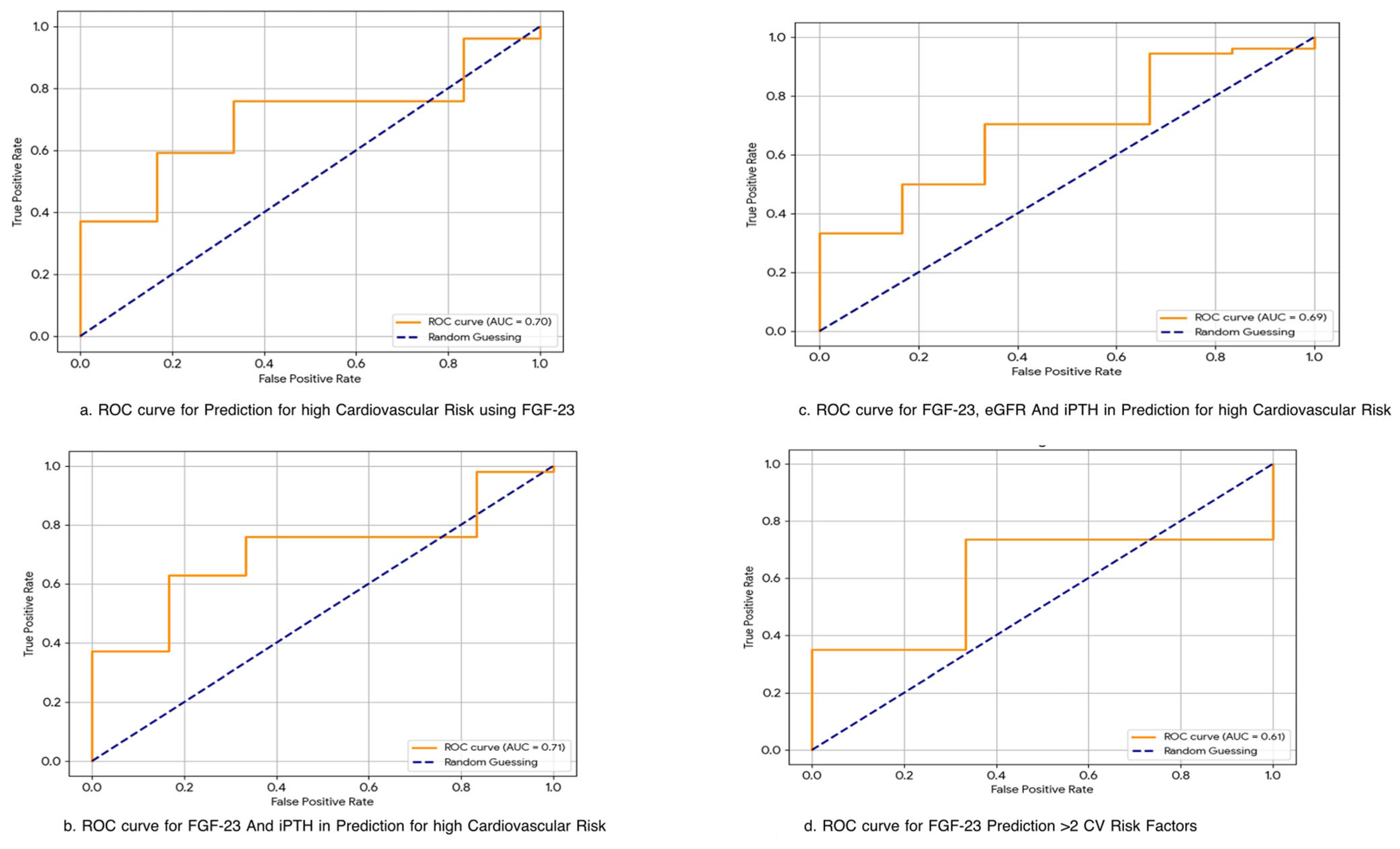
FGF-23 alone yielded an area under the curve (AUC) of 0.70 (95% CI: 0.56–0.84) for predicting high cardiovascular risk. A cutoff of 81.48 RU/mL provided a sensitivity of 76% and specificity of 67%. A multivariable model combining FGF-23, iPTH, and eGFR improved the AUC to 0.76 (95% CI: 0.64–0.88; p = 0.03). The FGF-23 + iPTH model yielded an AUC of 0.71. The results are summarized in Table 5 and Figure 2.
FGF-23 was significantly associated with key renal and mineral markers and demonstrated utility in identifying patients with subclinical cardiovascular changes. When used in combination with iPTH and eGFR, its predictive performance was enhanced, suggesting its potential role in multi-marker risk stratification in CKD.
These results support the clinical utility of FGF-23, especially in the early detection of cardiovascular risk among CKD patients.

4. Discussion

This study yields three key insights: First, fibroblast growth factor 23 (FGF-23) levels correlate robustly with renal dysfunction and mineral–bone disorder markers—specifically, serum phosphate and intact parathyroid hormone (iPTH). Second, elevated FGF-23 is significantly associated with early structural cardiovascular abnormalities, particularly left ventricular hypertrophy (LVH) and left atrial enlargement. Third, FGF-23 demonstrates moderate predictive capacity for cardiovascular remodeling in chronic kidney disease (CKD), with improved discriminative performance when combined with iPTH and estimated glomerular filtration rate (eGFR). These findings collectively position FGF-23 as not only a marker of mineral dysregulation but also a candidate biomarker for subclinical cardiovascular disease in CKD, adding evidence to the growing body of literature [15,16,17,18].
The observed association between FGF-23 and LA/LV structural changes reinforces preclinical data implicating FGF-23 as a direct cardiac modulator. Activation of FGFR4 by FGF-23 in cardiomyocytes has been shown to promote hypertrophic signaling through the calcineurin–NFAT pathway, independent of α-Klotho [9]. This mechanistic insight explains the preferential association of FGF-23 with concentric LV remodeling and LA dilation—early markers of cardiac strain—rather than with overt systolic dysfunction, such as reduced ejection fraction. Notably, this structural remodeling precedes clinical heart failure, aligning with findings from the CRIC study, which reported independent associations between FGF-23 and LVH as well as incident heart failure in CKD patients [19].
Our data further extend this relationship to vascular pathology. Carotid intima-media abnormalities in our cohort were more frequent among individuals with elevated FGF-23, supporting prior evidence linking FGF-23 to endothelial dysfunction and arterial stiffness via oxidative stress and suppressed Klotho expression [20,21]. Taken together, these findings suggest that FGF-23 contributes to both myocardial and vascular remodeling in early-stage CKD [11,18,22].
Unlike prior studies limited to dialysis populations, our cohort included patients across CKD stages, highlighting the relevance of FGF-23 even in earlier disease. In line with Ix et al.’s echocardiographic findings [13], we demonstrated that FGF-23 levels above 81.48 RU/mL were associated with abnormal cardiac structure. Importantly, our study is among the few to evaluate the predictive utility of FGF-23 using ROC analysis. The area under the curve (AUC) for FGF-23 alone was modest, but improved substantially (AUC = 0.76; sensitivity 75%, specificity 72%) when integrated with iPTH and eGFR in a multivariable model—suggesting additive prognostic value. This tri-marker approach is clinically attractive, particularly in resource-constrained settings, where access to advanced imaging is limited. FGF-23, alongside eGFR and iPTH, may serve as a cost-effective, non-invasive screen for incipient cardiovascular disease in CKD, informing early preventive strategies.
FGF-23 not only mirrors pathological processes but may represent a modifiable target. Clinical trials exploring dietary phosphate restriction, non-calcium phosphate binders, and FGFR inhibitors (e.g., burosumab) are underway to mitigate FGF-23-mediated cardiac and vascular effects [23]. Identification of high-risk individuals through biomarker profiling could allow targeted enrollment in such interventions. Additionally, the incorporation of FGF-23 into multimarker algorithms—including natriuretic peptides, troponins, and cardiac imaging—may enhance cardiovascular risk stratification in CKD, especially when coupled with machine-learning prediction tools [16,17,24].

4.1. Study Strengths and Limitations

Strengths of this study include its comprehensive phenotyping of both mineral metabolism and cardiovascular structure, and the inclusion of a non-dialysis-dependent CKD population. Nevertheless, certain limitations warrant acknowledgment. The cross-sectional design restricts causal inference, and the sample size limited subgroup analyses by CKD stage or etiology. Biomarker measurements were platform-specific and not standardized across assay types. While FGF-23 shows promise as a biomarker, the observed associations in our cohort are modest and warrant cautious interpretation. Longitudinal validation in multi-center cohorts will be essential to confirm the prognostic implications of our findings.

4.2. Future Directions

The limitations of our study could be mitigated in future research through the adoption of longitudinal cohort designs or prospective interventional studies, which would allow temporal sequencing and stronger causal inference. Larger multicenter studies would enhance statistical power and external validity, while advanced multivariable modeling—including adjustment for comorbidities, medication use, and inflammatory markers—could reduce residual confounding. Integration of mechanistic biomarkers and serial measurements of FGF-23, iPTH, and cardiovascular parameters would further strengthen the biological plausibility of associations. Together, these methodological refinements would help to minimize current limitations and provide more robust insights into the cardiorenal–mineral axis. Interventional trials evaluating whether a reduction in FGF-23 levels translates into cardiovascular benefit will also be pivotal. Finally, the development of composite risk prediction tools that integrate FGF-23 with structural, biochemical, and imaging markers could usher in a new paradigm of personalized cardiovascular risk management in CKD.

5. Conclusions

Fibroblast Growth Factor 23 is a robust biomarker of cardiovascular remodeling and mineral dysregulation in CKD. Elevated FGF-23 correlates with structural cardiac changes and demonstrates moderate predictive capacity for cardiovascular risk—particularly when combined with iPTH and eGFR. These findings advocate for its inclusion in integrative risk models and early intervention pathways. As our understanding of FGF-23 biology matures, its clinical utility in cardiovascular risk stratification and therapeutic targeting in CKD is likely to expand.
What Is Already Known:
  • FGF-23 is a phosphaturic hormone elevated early in chronic kidney disease (CKD) and has been implicated in left ventricular hypertrophy (LVH) and vascular dysfunction.
  • Mechanistic studies have shown that FGF-23 can induce myocardial remodeling via FGFR4-mediated pathways, independent of Klotho.
  • Large cohort studies (e.g., CRIC) have associated high FGF-23 with increased cardiovascular morbidity, especially in dialysis-dependent populations.
What This Study Adds:
  • Demonstrates strong correlations between FGF-23 and early structural cardiovascular changes, including LA/LV remodeling and diastolic dysfunction, even in non-dialysis CKD.
  • Proposes a novel multi-marker model (FGF-23 + iPTH + eGFR) with improved predictive power for cardiovascular risk (AUC = 0.76).
  • Introduces a stratified FGF-23-based cardiac risk phenotype, showing a dose–response relationship between FGF-23 levels and cardiac/vascular abnormalities.
  • Provides first-of-its-kind data from a South Asian CKD cohort, addressing a major gap in the global literature on cardiorenal risk profiling.

Supplementary Materials

The following supporting information can be downloaded at: https://www.mdpi.com/article/10.3390/jvd4040039/s1. Figure S1 Characteristics of subjects included in study, Figure S2 A–K: Scatter Plots of Correlations Between FGF-23 and Key Renal, Mineral, and Cardiometabolic Parameters.

Author Contributions

D.J.: Conceptualization, Data curation, Investigation, Methodology, Writing—original draft, Visualization, Supervision, Validation, Project administration, and Writing—review and editing. A.P.: Supervision, Validation, Methodology, and Writing—review and editing. N.W., H.S., A.G.: Writing—original draft and Writing—review and editing. Y.S.: Methodology, Visualization, Supervision, Validation, Writing—original draft, and Writing—review and editing. All authors have read and agreed to the published version of the manuscript.

Funding

This research received no external funding.

Institutional Review Board Statement

The study was conducted in accordance with the Declaration of Helsinki and approved by the Institutional Review Board of Subharti Medical College, Meerut (protocol code SMC/UECM/2023/750/327 and date of approval 30 October 2023) for studies involving humans.

Informed Consent Statement

Informed consent was obtained from all subjects involved in the study.

Data Availability Statement

Data are available from the corresponding author upon request.

Conflicts of Interest

The authors declare no conflicts of interest.

Abbreviations

The following abbreviations are used in this manuscript:
S. NoAbbreviationFull Form
1BUNBlood Urea Nitrogen
2CKDChronic Kidney Disease
3eGFREstimated Glomerular Filtration Rate
4FGF-23Fibroblast Growth Factor 23
5HDLHigh-Density Lipoprotein
6iPTHIntact Parathyroid Hormone
7LA/LVLeft Atrial-to-Left Ventricular Ratio
8LDLLow-Density Lipoprotein
9LVEFLeft Ventricular Ejection Fraction
10PAHPulmonary Artery Hypertension
11PTHParathyroid Hormone
12RWMARegional Wall Motion Abnormality

References

  1. Kovesdy, C.P. Epidemiology of chronic kidney disease: An update 2022. Kidney Int. Suppl. 2022, 12, 7–11. [Google Scholar] [CrossRef]
  2. Li, P.K.; Garcia-Garcia, G.; Lui, S.F.; Andreoli, S.; Fung, W.W.-S.; Hradsky, A.; Kumaraswami, L.; Liakopoulos, V.; Rakhimova, Z.; Saadi, G.; et al. Kidney health for everyone everywhere—From prevention to detection and equitable access to care. Braz. J. Med. Biol. Res. 2020, 53, e9614. [Google Scholar] [CrossRef]
  3. International Society of Nephrology. More Than 850 Million Worldwide Have Some Form of Kidney Disease: Help Raise Awareness. Available online: https://www.theisn.org/more-than-850-million-worldwide-have-some-form-of-kidney-disease-help-raise-awareness/ (accessed on 19 July 2025).
  4. Shah, A.; Hashmi, M.F.; Aeddula, N.R. Chronic Kidney Disease-Mineral Bone Disorder (CKD-MBD). In StatPearls [Internet]; StatPearls Publishing: Treasure Island, FL, USA, 2025. Available online: https://www.ncbi.nlm.nih.gov/books/NBK560742/ (accessed on 19 July 2025).
  5. Rodrigues, F.G.; Ormanji, M.S.; Heilberg, I.P.; Bakker, S.J.L.; de Borst, M.H. Interplay between gut microbiota, bone health and vascular calcification in chronic kidney disease. Eur. J. Clin. Investig. 2021, 51, e13588. [Google Scholar] [CrossRef]
  6. Stevens, P.E.; Levin, A.; Coresh, J.; Foster, B.; Francis, A.; Hall, R.K.; Herrington, W.G.; Hill, G.; Inker, L.A.; Kazancıoğlu, R.; et al. KDIGO 2024 Clinical Practice Guideline for the Evaluation and Management of Chronic Kidney Disease. Kidney Int. 2024, 105 (Suppl. S4), S117–S314. [Google Scholar] [CrossRef] [PubMed]
  7. Lee, C.; Yun, H.R.; Joo, Y.S.; Lee, S.; Kim, J.; Nam, K.H.; Jhee, J.H.; Park, J.T.; Yoo, T.-H.; Kang, S.-W.; et al. Framingham risk score and risk of incident chronic kidney disease: A community-based prospective cohort study. Kidney Res. Clin. Pract. 2019, 38, 49–59. [Google Scholar] [CrossRef] [PubMed]
  8. Faul, C.; Amaral, A.P.; Oskouei, B.; Hu, M.-C.; Sloan, A.; Isakova, T.; Gutierrez, O.M.; Aguillon-Prada, R.; Lincoln, J.; Hare, J.M.; et al. FGF-23 induces left ventricular hypertrophy. J. Clin. Investig. 2011, 121, 4393–4408. [Google Scholar] [CrossRef] [PubMed]
  9. Grabner, A.; Amaral, A.P.; Schramm, K.; Singh, S.; Sloan, A.; Yanucil, C.; Li, J.; Shehadeh, L.A.; Hare, J.M.; David, V.; et al. Activation of Cardiac Fibroblast Growth Factor Receptor 4 Causes Left Ventricular Hypertrophy. Cell Metab. 2015, 22, 1020–1032. [Google Scholar] [CrossRef]
  10. Gutierrez, O.M.; Januzzi, J.L.; Isakova, T.; Laliberte, K.; Smith, K.; Collerone, G.; Sarwar, A.; Hoffmann, U.; Coglianese, E.; Christenson, R.; et al. Fibroblast growth factor-23 and left ventricular hypertrophy in chronic kidney disease. Circulation 2009, 119, 2545–2552. [Google Scholar] [CrossRef]
  11. Scialla, J.J.; Xie, H.; Rahman, M.; Anderson, A.H.; Isakova, T.; Ojo, A.; Zhang, X.; Nessel, L.; Hamano, T.; Grunwald, J.E.; et al. Fibroblast growth factor-23 and cardiovascular events in CKD. J. Am. Soc. Nephrol. 2014, 25, 349–360. [Google Scholar] [CrossRef]
  12. Isakova, T.; Xie, H.; Yang, W.; Xie, D.; Anderson, A.H.; Scialla, J.; Wahl, P.; Gutiérrez, O.M.; Steigerwalt, S.; He, J.; et al. Fibroblast growth factor 23 and risks of mortality and end-stage renal disease in patients with chronic kidney disease. JAMA 2011, 305, 2432–2439. [Google Scholar] [CrossRef]
  13. Ix, J.H.; Katz, R.; Kestenbaum, B.; de Boer, J.H.; Chonchol, M.; Mukamal, K.J.; Rifkin, D.; Siscovick, D.S.; Sarnak, M.J.; Shlipak, M.G. Fibroblast growth factor-23 and death, heart failure, and cardiovascular events in community-living individuals (CHS). J. Am. Coll. Cardiol. 2012, 60, 200–207. [Google Scholar] [CrossRef]
  14. Kendrick, J.; Cheung, A.K.; Kaufman, J.S.; Greene, T.; Roberts, W.L.; Smits, G.; Chonchol, M. FGF-23 associates with death, cardiovascular events, and initiation of chronic dialysis. J. Am. Soc. Nephrol. 2011, 22, 1913–1922. [Google Scholar] [CrossRef]
  15. Vázquez-Sánchez, S.; Poveda, J.; Navarro-García, J.A.; González-Lafuente, L.; Rodríguez-Sánchez, E.; Ruilope, L.M.; Ruiz-Hurtado, G. An Overview of FGF-23 as a Novel Candidate Biomarker of Cardiovascular Risk. Front. Physiol. 2021, 12, 632260. [Google Scholar] [CrossRef]
  16. Heine, G.H.; Seiler, S.; Fliser, D. FGF-23: The rise of a novel cardiovascular risk marker in CKD. Nephrol. Dial. Transplant. 2012, 27, 3072–3081. [Google Scholar] [CrossRef] [PubMed]
  17. Kestenbaum, B.; Sachs, M.C.; Hoofnagle, A.N.; Siscovick, D.S.; Ix, J.H.; Robinson-Cohen, C.; Lima, J.A.C.; Polak, J.F.; Blondon, M.; Ruzinski, J.; et al. Fibroblast growth factor-23 and cardiovascular disease in the general population: The Multi-Ethnic Study of Atherosclerosis. Circ. Heart Fail. 2014, 7, 409–417. [Google Scholar] [CrossRef] [PubMed]
  18. Vogt, I.; Haffner, D.; Leifheit-Nestler, M. FGF23 and Phosphate–Cardiovascular Toxins in CKD. Toxins 2019, 11, 647. [Google Scholar] [CrossRef] [PubMed]
  19. Leidner, A.S.; Cai, X.; Zelnick, L.R.; Lee, J.; Bansal, N.; Pasch, A.; Nahsal, M.; Chen, J.; Anderson, A.H.; Sondheimer, J.H.; et al. Fibroblast Growth Factor 23 and Risk of Heart Failure Subtype: The CRIC (Chronic Renal Insufficiency Cohort) Study. Kidney Med. 2023, 5, 100723. [Google Scholar] [CrossRef]
  20. Lu, X.; Hu, M.C. Klotho/FGF23 Axis in Chronic Kidney Disease and Cardiovascular Disease. Kidney Dis. 2017, 3, 15–23. [Google Scholar] [CrossRef]
  21. Richter, B.; Haller, J.; Haffner, D.; Leifheit-Nestler, M. Klotho modulates FGF23-mediated NO synthesis and oxidative stress in human coronary artery endothelial cells. Pflug. Arch. 2016, 468, 1621–1635. [Google Scholar] [CrossRef] [PubMed]
  22. Vergara, N.; de Mier, M.V.P.; Rodelo-Haad, C.; Revilla-Gonzáles, G.; Membrives, C.; Díaz-Tocados, J.M.; Martínez-Moreno, J.M.; Torralbo, A.I.; Herencia, C.; Rodríguez-Ortiz, M.E.; et al. The direct effect of fibroblast growth factor 23 on vascular smooth muscle cell phenotype and function. Nephrol. Dial. Transplant. 2023, 38, 322–343. [Google Scholar] [CrossRef]
  23. Wolf, M. Update on fibroblast growth factor 23 in chronic kidney disease. Kidney Int. 2012, 82, 737–747. [Google Scholar] [CrossRef]
  24. Yildirim, M.; Salbach, C.; Mueller-Hennessen, M.; Frey, N.; Giannitsis, E. Diagnostic and Prognostic Evaluation of Novel Biomarkers Compared to ESC 0/1 h and 0/3 h Algorithms in Patients with Suspected Non-ST-Elevation Myocardial Infarction. J. Clin. Med. 2025, 14, 2957. [Google Scholar] [CrossRef]
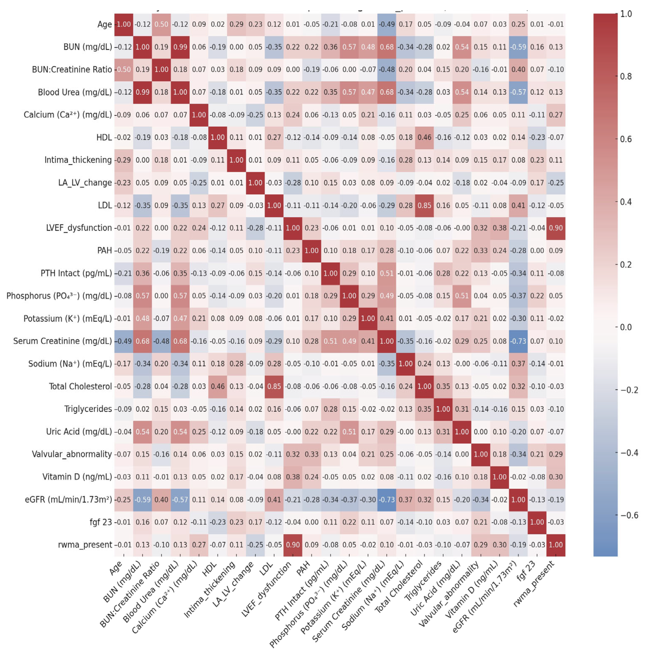
Figure 1. Heatmap of Correlation Matrix among Cardio–Renal–Mineral Markers. This heatmap depicts pairwise Pearson correlation coefficients (r) between continuous study variables. Positive correlations are shown in shades of red and negative correlations in shades of blue, with color intensity proportional to the correlation strength (scale bar at right: −0.6 to +1.0). Strong clustering of correlations can be observed among renal function markers (serum creatinine, blood urea, BUN, and eGFR), mineral metabolism parameters (FGF-23, iPTH, phosphorus, calcium), and lipid profile components (total cholesterol, LDL, HDL, triglycerides). Associations between cardiovascular measures (LVEF dysfunction, valvular abnormality, PAH, intima thickening, LA/LV change, RWMA presence) and renal/mineral parameters highlight the integrated nature of the cardiorenal–mineral axis.
Jvd 04 00039 g001
Figure 2. ROC Curves showing the Predictive Performance of FGF-23 for Cardiovascular Risk.
Jvd 04 00039 g002
Table 1. Baseline Characteristics of the Study Cohort (n = 60).
Variable/ParameterValueUnits
Demographics
Age56.9 ± 16.0years
Male sex31 (52%)
CKD Stage, n (%)
 Stage 1–212 (20%)
 Stage 318 (30%)
 Stage 410 (17%)
 Stage 520 (33%)
Renal Parameters
eGFR27.2 ± 25.5mL/min/1.73 m2
Serum Creatinine4.93 ± 3.51mg/dL
Blood Urea111.45 ± 76.51mg/dL
BUN51.92 ± 35.66mg/dL
BUN–Creatinine Ratio11.45 ± 5.56ratio (unitless)
Uric Acid6.81 ± 2.45mg/dL
Mineral Metabolism
FGF-23152.9 ± 105.7RU/mL
Intact PTH (iPTH)386.2 ± 370.8pg/mL
Vitamin D19.3 ± 11.2ng/mL
Calcium8.28 ± 0.94mg/dL
Phosphorus5.1 ± 2.5mg/dL
Electrolytes
Sodium139.75 ± 5.15mmol/L
Potassium4.71 ± 0.86mmol/L
Lipid Profile
Total Cholesterol141.5 ± 40.9mg/dL
LDL77.2 ± 31.9mg/dL
HDL35.8 ± 12.9mg/dL
Triglycerides148.6 ± 85.8mg/dL
Cardiac Parameters
LVEF56.4 ± 9.5%
Pulmonary Artery Pressure31.8 ± 10.4mmHg
Abbreviations: CKD, chronic kidney disease; eGFR, estimated glomerular filtration rate; BUN, blood urea nitrogen; FGF-23, fibroblast growth factor 23; iPTH, intact parathyroid hormone; LDL, low-density lipoprotein cholesterol; HDL, high-density lipoprotein cholesterol; LVEF, left ventricular ejection fraction.
Table 2. Significant Correlations with FGF-23.
ParameterCorrelation (r)p-Value
eGFR−0.2880.028
iPTH0.3610.006
Phosphorus0.3350.011
Creatinine0.2710.042
Table 3. Clinically Significant Correlations Among Mineral–Bone Metabolism, Renal Function, and Cardiovascular–Vascular Markers in Chronic Kidney Disease Patients as found in this study and their interpretation.
Thematic DomainVariable PairCorrelation Coefficient (r)p-ValueInterpretation
Mineral–Bone + Cardiac InterplayVitamin D vs. LVEF Dysfunction0.380.0032Suggests the protective role of vitamin D in preserving systolic function
Vitamin D vs. RWMA Present0.30.0202Higher vitamin D may be associated with reduced myocardial damage
Calcium vs. RWMA Present0.270.0406Possible role of calcium in myocardial excitability or ischemic susceptibility
Cardiorenal AxisValvular Abnormality vs. eGFR−0.340.007Declining GFR correlates with structural heart changes
PAH vs. Creatinine0.280.0292Worsening renal function linked with pulmonary hypertension
PAH vs. eGFR−0.280.0315Supports the presence of cardiorenal interaction
Vascular/Lipid/Age-Related ParametersAge vs. Intima Thickening0.290.0249Vascular aging evident through intimal changes
Sodium vs. Intima Thickening0.280.0324Electrolyte shifts may reflect vascular stiffness
HDL vs. Total Cholesterol0.460.0002Expected inverse pattern in lipid metabolism
Total Cholesterol vs. Triglycerides0.350.0067Co-association in dysmetabolism
Triglycerides vs. Uric Acid0.310.0145May indicate shared metabolic and oxidative stress pathways
FGF23/iPTH/eGFR vs. Cardiac–Vascular CorrelatesValvular Abnormality vs. eGFR−0.345Moderate inverse correlation; valvular disease may worsen with kidney function decline
PAH vs. eGFR−0.278Mild link of renal impairment with elevated pulmonary pressures
LA/LV Change vs. FGF230.168Weak positive association; possible early remodeling
LA/LV Change vs. iPTH0.149Weak trend
LA/LV Change vs. eGFR−0.086Minimal negative trend
LVEF Dysfunction vs. eGFR−0.213Mild inverse relationship, reinforcing cardiorenal link
Intima Thickening vs. FGF230.227Weak correlation; may reflect vascular remodeling in the CKD milieu
Abbreviations: CKD, chronic kidney disease; eGFR, estimated glomerular filtration rate; FGF-23, fibroblast growth factor 23; iPTH, intact parathyroid hormone; LVEF, left ventricular ejection fraction; RWMA, regional wall motion abnormality; LA/LV, left atrial-to-left ventricular ratio; PAH, pulmonary artery hypertension.
Table 4. Correlation of FGF-23 with Key Cardiovascular Structural and Vascular Markers in CKD Patients.
Cardiovascular/Vascular MarkerCorrelation with FGF-23 (r)Interpretation
Valvular Abnormality0.211Mild positive correlation; may reflect FGF-23’s role in valvular calcification in CKD
Pulmonary Arterial Hypertension (PAH)0.002No association; PAH likely driven by volume overload or diastolic dysfunction rather than FGF-23
LA/LV Structural Change0.168Weak trend; aligns with literature on FGF-23 promoting LV hypertrophy and remodeling
LVEF Dysfunction−0.036No significant correlation; FGF-23 appears unrelated to systolic dysfunction severity
Carotid Intima Thickening/Plaque0.227Mild positive correlation; suggests possible involvement in early vascular stiffness or subclinical atherosclerosis
Table 5. Predictive Utility of FGF-23 and Combined Biomarkers for Cardiovascular Risk Stratification in CKD.
Predictor(s)Target OutcomeAUC (Sensitivity, Specificity)Interpretation
FGF-23 aloneHigh Cardiovascular Risk *0.70 (76%, 67%)Moderate predictive value; may aid early screening
FGF-23 + iPTHHigh Cardiovascular Risk0.71Slightly improved discrimination compared to FGF-23 alone
FGF-23 + eGFR + iPTHHigh Cardiovascular Risk0.69No significant added value over FGF-23 alone
FGF-23 alone>2 Cardiovascular Risk Markers **0.61Low-to-moderate discriminatory power
FGF-23 + iPTH + eGFR>2 Cardiovascular Risk Markers0.76Best predictive model; supports multivariable biomarker approach
* High Cardiovascular Risk was defined as the presence of any one of the following: moderate-to-severe LVEF dysfunction, LA/LV hypertrophy or dilation, or carotid intima thickening/plaque. ** Presence of >2 cardiovascular risk markers out of: valvular abnormality, PAH, LA/LV change, LVEF dysfunction, carotid abnormalities, abnormal lipids (any one of HDL, LDL, TG, or total cholesterol).
Disclaimer/Publisher’s Note: The statements, opinions and data contained in all publications are solely those of the individual author(s) and contributor(s) and not of MDPI and/or the editor(s). MDPI and/or the editor(s) disclaim responsibility for any injury to people or property resulting from any ideas, methods, instructions or products referred to in the content.