Review

Beyond Endoleaks: A Holistic Management Approach to Late Abdominal Aortic Aneurysm Ruptures After Endovascular Repair

1
UCL Division of Surgery and Interventional Science, Royal Free Hospital, London NW3 2QG, UK
2
Department of Vascular Surgery, Royal Free Hospital, London NW3 2QG, UK
*
Author to whom correspondence should be addressed.
J. Vasc. Dis. 2025, 4(3), 24; https://doi.org/10.3390/jvd4030024
Submission received: 30 April 2025 / Revised: 27 May 2025 / Accepted: 18 June 2025 / Published: 22 June 2025
(This article belongs to the Section Peripheral Vascular Diseases)

Abstract

:
Late ruptures of abdominal aortic aneurysms post-endovascular aneurysm repair present a significant risk, occurring in about 0.9% of cases. The typical timeframe leading to rupture is roughly 37 months, with the primary factors often linked to endoleaks, especially types I and III, which sustain pressure within the aneurysm sac. The approaches to managing late ruptures consist of endovascular approaches, open surgical interventions, and conservative care, each customised to the patient’s specific characteristics. When feasible endovascular repair is favoured, additional stent grafts are deployed to seal endoleaks and offer lower perioperative mortality rates compared to those for open surgery. Open repair is considered when endovascular solutions fail or are not feasible. Conservative management with active monitoring and supportive treatment can be considered for haemodynamically stable non-surgical patients. Endovascular repair methods like fenestrated/branched EVAR (F/BEVAR) and parallel grafting (PGEVAR) are effective for complicated anatomies and show high technical success with reduced morbidity compared to that with open repairs. Chimney techniques and physician-modified endografts may help regain and broaden the sealing zone. Limb extensions with or without embolisation, interposition endografting, and whole-body relining are helpful options for type IB and type 3–5 endoleaks. Open surgical repair carries a higher perioperative mortality but may be essential in preventing death due to rupture following failed EVAR. The choice depends on the patient’s clinical stability and fitness for surgery in the absence of a viable endovascular alternative. This article discusses the available options for treating late rupture after EVAR, emphasising the importance of individualised treatment plans and the need for rigorous postoperative surveillance to prevent such complications.

1. Introduction

Late rupture of abdominal aortic aneurysm (rAAA) following EVAR is a significant concern in vascular and endovascular surgery, presenting a complex challenge. The incidence of late ruptures is relatively low, with studies reporting an incidence of approximately 0.9–3.1% [1,2]. However, the consequences of such ruptures are severe, often leading to high mortality rates of 20–60% [3]. Across three landmark randomised trials—IMPROVE, AJAX, and ECAR—the 1-year mortality rates for patients with rAAAs treated using an endovascular strategy were 38.6%, compared to 42.8% for those treated with open surgical repair. These results were consistent across the trials, with no statistically significant difference in survival between the two approaches [4,5,6]. A Greek multicentre study on the outcomes of surgery for aneurysm ruptures post-EVAR showed a 41% in-hospital mortality rate [3]. The mean time to rupture post-EVAR is reported to be around 37 months, although this can vary significantly [7]. The primary causes of late ruptures after EVAR are often related to endoleaks, particularly of types I and III [1]. Endoleaks result in continued pressurisation of the aneurysm sac, potentially leading to rupture if not adequately addressed. Type I endoleaks, which occur at the proximal or distal attachment sites, and type III endoleaks, which involve defects in the graft material or junctions, are particularly concerning [2]. Type II endoleaks, although less commonly associated with ruptures, can also contribute if they lead to significant sac expansion [7].
The management strategies for late rupture require a tailored approach based on the patient’s condition and the nature of the rupture. The three primary strategies include conservative management, endovascular approaches, and open surgical repair. Conservative management is generally reserved for patients who are not surgical candidates due to comorbidities and present with haemodynamically stable, contained ruptures. This approach involves close monitoring and supportive care, but it is not a definitive solution and carries a high risk of mortality if the rupture progresses [3]. Endovascular repair remains the preferred option for many cases of late ruptures after EVAR, particularly when the anatomy is suitable and appropriate device options are available. This approach can involve the placement of additional stent grafts to seal endoleaks or reinforce the existing repair. Studies have shown that endovascular treatment is associated with lower perioperative mortality compared to that under open surgical repair, making it an attractive option when feasible [8,9]. Open surgical repair, or conversion into open surgery, is often considered a last resort due to its invasiveness and higher associated morbidity and mortality. However, it remains a critical option for patients with an anatomy that is unsuitable for endovascular repair or when endovascular attempts have failed. Open repair involves direct surgical intervention to repair the aneurysm and address any complications such as graft migration or infection [3]. The choice of treatment strategy (Figure 1) is influenced by several factors, including the patient’s haemodynamic status, the presence and type of endoleak, and the availability of surgical expertise and resources. Haemodynamic stability at presentation is a crucial determinant of outcomes, with unstable patients experiencing significantly higher mortality rates.

2. The Treatment Options for an AAA Rupture Post-Endovascular Repair

When a rupture occurs after EVAR, the treatment options include conservative management, repeat endovascular intervention, or open surgical repair. The choice among these may be dynamic, especially early on in its presentation, and depends on several factors, including the patient’s haemodynamic stability, the anatomical suitability for further endovascular procedures, the nature and location of the endoleak or graft failure, and the resources and expertise available for a potentially complex repair (Table 1). The presence of an existing stent graft makes conservative management an option, especially in the early stages for haemodynamically stable patients. Continuation of conservative medical management only is, however, rarely an option in unstable patients at high risk of imminent demise due to ongoing extravasation. It may be considered only in patients who are not surgical candidates due to extreme frailty or comorbidities. Surgical intervention where feasible, either endovascular or open, is preferred and is associated with better outcomes [1].
Endovascular re-intervention is the preferred initial approach when feasible, especially in centres with the necessary expertise and resources. This strategy is supported by multiple studies and guidelines which indicate that endovascular repair is associated with lower perioperative mortality compared to that under open surgical repair. For example, pooled analyses and meta-analyses have shown the perioperative mortality rates for endovascular treatment of rAAAs to be around 20–25%, compared to 32–43% for open repair [10,11]. The endovascular options may include relining the existing graft, placing additional stent grafts to seal endoleaks, or using adjunctive techniques such as endovascular aneurysm sealing or chimney grafts to address challenging anatomies, such as short, migrated, or juxtarenal necks [12]. The feasibility of endovascular re-intervention depends on the patient’s anatomy, the type of endoleak, and the availability of the appropriate devices and expertise. In some series, up to 80% of patients with rAAAs were suitable for endovascular management, with the remainder requiring open repair due to anatomical or logistical constraints [13,14].
Open surgical repair remains an essential option, particularly when endovascular solutions are not feasible due to an unfavourable anatomy (e.g., short or wide necks, complex endoleaks), haemodynamic instability that precludes the time required for a complex endovascular setup, or the failure of endovascular re-intervention. Open repair is associated with higher perioperative mortality and morbidity, but it is sometimes the only viable option for swift and definitive haemorrhage control [15]. The decision to proceed with open repair is often made in the context of life-threatening instability or failed endovascular attempts or when the necessary endovascular devices or expertise are unavailable.
Table 1. Variables impacting the treatment triage for sac rupture post-EVAR.
ParameterConservativeEndovascularOpen SurgeryNotes
Haemodynamic StabilityOnly if stable (SBP > 90 mmHg), no ongoing shockPreferred if stable or moderately unstable (SBP 70–90 mmHg), responsive to fluidsPreferred if unstable and not unamenable to endovascular treatmentInstability and requirement for complex endovascular treatment often necessitates open repair [9]
Blood PressureSBP 90–120 mmHg, avoid hypertension and hypotensionSBP 70–90 mmHg, permissive hypotension to limit bleedingSBP 70–90 mmHg, rapid surgical aortic cross-clampingMaintain perfusion, avoid excessive hypertension [16]
Heart Rate<100 bpm preferred, no tachycardia<100 bpm preferred, tachycardia may indicate ongoing bleedingTachycardia (>100 bpm) may indicate ongoing bleedingTachycardia signals instability [17]
Time to InterventionConsidered, stable and non-surgical<90 min from arrival is ideal<90 min from arrival is idealDoor-to-intervention < 90 min (30–30–30 min) [16]
Patient FrailtySevere frailty (bedbound, poor baseline function)Moderate frailty (needs some assistance)Low/moderate frailty (independent)Frailty increases surgical risk
Heart FailureSevere (NYHA III-IV)Mild/moderate (NYHA I-II)No/mild heart failureHeart failure increases perioperative risk
Renal ImpairmentSevere (on dialysis, creatinine > 3 mg/dL)Mild/moderate (creatinine < 2.5 mg/dL)Mild/moderate (creatinine < 2.5 mg/dL)Renal impairment increases risk [9]
Anaesthesia SuitabilityNot suitable for any anaesthesiaLocal anaesthesia preferred, general if neededGeneral anaesthesia requiredLocal anaesthesia lowers mortality in EVAR [18]
AgeOld age (>90)Elderly (80–90)Younger (<80)Octogenarians more often treated with EVAR [19]
GenderEitherPreferable in femalesEither EVAR reduces mortality more in females [20]
Previous Abdominal SurgeryMultiple prior surgeries, hostile abdomenFeasible if access is possibleDifficult if adhesionsAdhesions complicate open repair [16]
Aneurysm AnatomyUnfavourable (short neck, severe angulation)Favourable (adequate neck, access)Unfavourable, open surgery if EVAR not possibleAdequate neck/iliac access essential for EVAR
Endoleak TypeType II, no expansionType I-VType I-V if endovascular not feasibleTypes I/III especially require intervention [1]
Graft InfectionPresent and unfit for surgeryHaemodynamic control with long-term antibioticsPresent and fit for surgeryInfection increases the risk of EVAR [21]
Institutional ExperienceLow experienceHigh experience: EVAR preferredHigh experience: open surgery possibleOutcomes better in high-volume centres [10]
Time Since Initial EVARRecent EVAR (<1 month), conservative if stableLate rupture (>1 year), EVAR or open, based on anatomyLate rupture, open surgery if EVAR not feasibleLate ruptures often endoleak-related [1]
Surveillance CompliancePoor compliance, unknown anatomyGood compliance: EVAR possibleGood compliance: endovascular not feasibleNon-compliance increases rupture risk
Blood LossNot applicableLower blood loss with EVARHigh blood loss with open surgeryEVAR reduces transfusion needs [9]
Life Expectancy<6 months>6 months>6 monthsConsider prognosis and quality of life
Access Vessel StatusPoor (occluded, small)Good (patent, adequate size)GoodAdequate access required for EVAR

2.1. Conservative Management

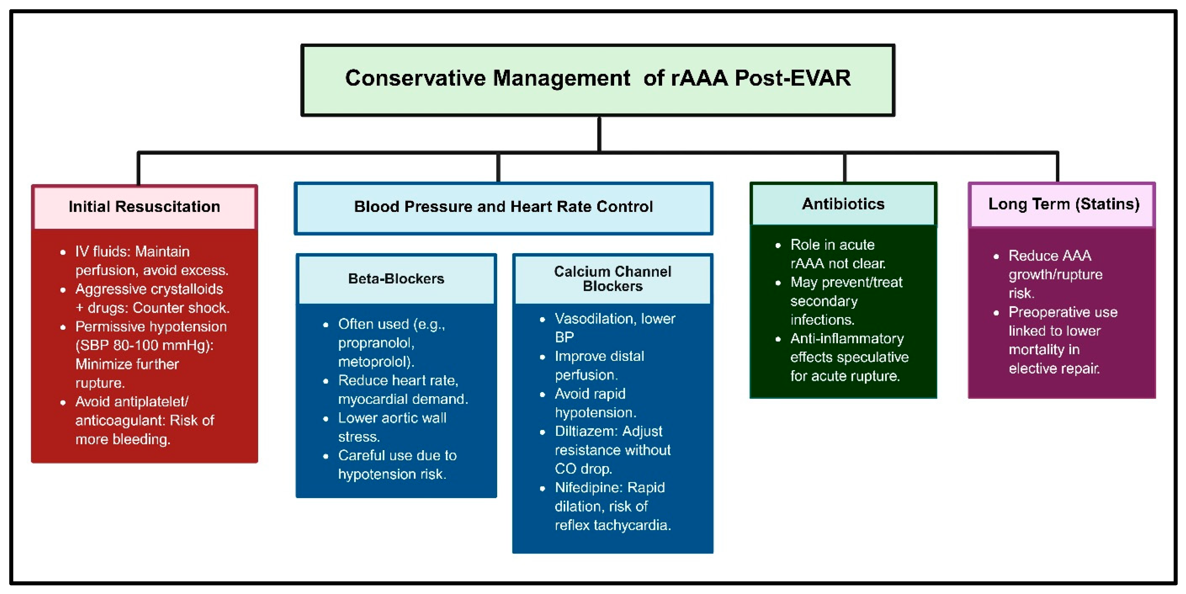
Patients with ruptured AAAs post-EVAR may present with symptoms similar to those of ruptured AAAs without prior EVAR, such as a sudden onset of severe abdominal or back pain, hypotension, and signs of shock. However, the presence of an endovascular graft can sometimes alter the clinical presentation such that the rupture may be contained for a longer period, potentially allowing more time for intervention [1]. The incidence and prevalence of mortality after rAAAs post-EVAR remain high, with studies indicating a perioperative mortality rate of approximately 24.5% for EVAR compared to 37.8% for open surgical repair [10]. Surgical intervention—endovascular or open—is the mainstay and the preferred treatment in suitable patients. Conservative management, as outlined in Figure 2, may be considered when a patient with an rAAA exhibits a stable haemodynamic status, showing no signs of severe shock or significant organ dysfunction. In such cases, immediate surgical/endovascular intervention may be delayed and in some circumstances withheld [22]. This is particularly so in patients who are not suitable candidates for surgical intervention due to high operative risk or the presence of comorbid conditions that outweigh the potential benefits of surgery [23]. Pharmacological management is essential, focusing on maintaining haemodynamic stability, reducing aortic wall stress, and preventing complications such as infections.
Intravenous (IV) fluids are essential in initial resuscitation. The aim is to maintain adequate perfusion to the vital organs while avoiding excessive administration of fluid, which could increase aortic wall stress and exacerbate bleeding. Initiating crystalloid infusions alongside pharmacologic interventions is vital to counteract hypovolemia [24]. Permissive hypotension is often employed, targeting a systolic blood pressure of around 80–100 mmHg and a diastolic pressure of 50–60 mmHg, to minimise the risk of worsening the rupture while ensuring sufficient organ perfusion [25,26]. Antiplatelet and anticoagulant therapies are generally avoided in the acute rupture setting due to the risk of exacerbating bleeding. While these agents may have a role in long-term management to prevent thrombotic complications, their use in the acute phase is contraindicated [27].
Beta-blockers, such as propranolol and metoprolol, are often employed due to their ability to reduce heart rate and myocardial oxygen demand. The rationale behind their use lies in the understanding that an increased cardiac output and heart rate can exacerbate aortic wall stress, thus elevating the risk of rupture. Lowering the heart rate through beta-blockade helps stabilise haemodynamics, particularly in tachycardic patients. Additionally, beta-blockers may indirectly stabilise the blood pressure by decreasing systemic vascular resistance and cardiac contractility, though their use should be carefully considered, as hypotension is common in ruptured cases [28].
Calcium channel blockers, like amlodipine, diltiazem, and nifedipine, may have a potential role in acute management. Their vasodilatory effects can be beneficial in mitigating vascular resistance and lowering blood pressure, particularly in hypertensive patients or those exhibiting signs of an increased cardiac output. They promote arterial relaxation and improve distal perfusion. However, it is essential to avoid rapid hypotension, especially in patients presenting with significant blood loss or shock. Diltiazem can be administered in a controlled manner to adjust systemic vascular resistance without inducing significant declines in cardiac output [29]. Nifedipine, a dihydropyridine calcium channel blocker, is utilised for its rapid vasodilatory effect; however, its use can lead to reflex tachycardia, which may not be ideal for patients with compromised haemodynamics [30].
Antibiotics, particularly those with anti-inflammatory properties like doxycycline, have been explored for their potential role in managing AAAs. While their primary use in the acute setting of rAAAs is not well-established, antibiotics may be administered to prevent or treat secondary infections, especially in cases where surgical intervention is delayed or complicated by infection. The anti-inflammatory effects of certain antibiotics could theoretically contribute to stabilising the aneurysm wall, although this application is more speculative and not supported by robust clinical evidence in the acute rupture scenario [31].
Statins have been shown to reduce AAA growth and rupture risk by suppressing inflammatory mediators, decreasing oxidative stress, and inhibiting extracellular matrix degradation. Preoperative statin use is associated with lower perioperative mortality following elective AAA repair, suggesting a potential benefit in the acute setting as well [32,33]. However, the immediate impact of statins in the acute rupture scenario is less clear, and their use would primarily be considered for long-term management post-repair.

2.2. Percutaneous Endovascular Repair for a Ruptured Abdominal Aortic Aneurysm (rPEVAR)

2.2.1. Fenestrated/Branched Endovascular Aneurysm Repair (F/BEVAR)

Fenestrated and branched endovascular aneurysm repair (F/BEVAR) is increasingly considered for the treatment of rAAA in patients who have previously undergone EVAR and are clinically stable enough and where suitable devices and expertise available. This approach is particularly relevant when the initial EVAR fails, often due to issues such as type Ia endoleaks, migration of the stent graft, or aneurysm progression, necessitating further intervention to prevent rupture. The use of F/BEVAR in these scenarios is supported by its ability to address complex aortic anatomies and extend the proximal seal zone, thereby mitigating the risk of rupture. The technical success of F/BEVAR in these settings is high, with studies reporting success rates of approximately 94.9% to 99% [34,35,36]. This high success rate is attributed to the ability of F/BEVAR to incorporate the renal and visceral arteries through custom fenestrations or branches, thus providing a robust solution for complex aneurysms.
The outcomes following F/BEVAR after failed EVAR are generally favourable, with the perioperative mortality rates ranging from 1.1% to 5% [37,38]. These figures are significantly lower than those associated with open repair, highlighting the safety profile of F/BEVAR. However, the procedure is not without risks. Complications such as spinal cord ischaemia, acute kidney injury, and endoleaks are reported, albeit at relatively low rates. For instance, spinal cord ischaemia occurs in approximately 2.4% to 4.7% of cases, while acute kidney injury is observed in about 6.8% of patients [36]. One study reported a persistent type Ia endoleak in 1 out of 85 patients (approximately 1.2%) after F/BEVAR following a failed EVAR [37]. These procedures can be tricky, challenging, and time-dependent in the rupture scenario. The NICE guidelines advise against complex repairs in ruptured AAAs, preferring open repair [39].
The situation is different with an existing failing EVAR endograft, in situ though failing, where feasible, endovascular options are less physiologically tasking, with better outcomes. The long-term outcomes also demonstrate the efficacy of F/BEVAR, with the one-year survival rates reported at 86% to 94% [35]. The freedom from re-intervention at one year is similarly high, ranging from 81% to 88% [38].

2.2.2. Parallel Graft Endovascular Aortic Repair (PGEVAR)

Parallel graft endovascular aortic repair (PGEVAR) is a complex procedure that can be used to treat rAAA. The decision to use PGEVAR is influenced by several factors, including the patient’s age, the anatomical complexity of the aneurysm, and the presence of comorbidities. The technical success of PGEVAR in treating rAAAs is generally high, with studies reporting success rates of around 90.9% for PGEVAR, compared to 100% for standard EVAR (sEVAR) in similar settings [40]. The 30-day mortality rate for PGEVAR is significantly lower than that for sEVAR, at 27.3% versus 50.7%, respectively [40]. This suggests that PGEVAR may offer a survival advantage in the acute phase following a rupture, particularly in anatomically complex cases where standard EVAR may not be feasible. The outcomes of PGEVAR are also influenced by the number and type of parallel grafts used. The use of multiple parallel grafts can increase the risk of type Ia endoleaks, which are a common complication requiring re-intervention. In one study, the presence of early type Ia endoleaks was associated with the implantation of two or more parallel grafts and insufficient oversizing of the main aortic stent graft [41]. Despite these challenges, the secondary success rate after re-intervention for endoleaks was reported to be 90% [41].
Age is a critical factor in the success of PGEVAR. Patients younger than 80 years tend to have better long-term survival rates, with the overall survival reported to be 75% after one year and 53% after three years [41]. The anatomical characteristics of the aneurysm also play a significant role. Juxtarenal aneurysms, for example, are associated with better outcomes compared to those of more complex aneurysms, such as suprarenal or thoracoabdominal types [41]. The regression of the aneurysm sac’s diameter post-PGEVAR is another positive prognostic indicator, correlating with improved survival rates.
The durability of parallel grafts is a crucial consideration in the long-term management of patients undergoing PGEVAR. Studies have shown high patency rates for parallel grafts, with secondary patency rates of 97.4% at one year and 94.1% at three years [41]. This durability is essential for maintaining the integrity of the repair and preventing future complications.
In terms of morbidity, PGEVAR is associated with a range of potential complications, including acute kidney injury, haematoma, and rebleeding at access sites [42]. These complications can be managed using the appropriate interventions, such as the placement of covered stents to address rebleeding. The presence of major adverse events is a significant factor affecting overall survival, highlighting the importance of careful patient selection and perioperative management.

2.2.3. Chimney EVAR (ChEVAR) and Periscope Techniques

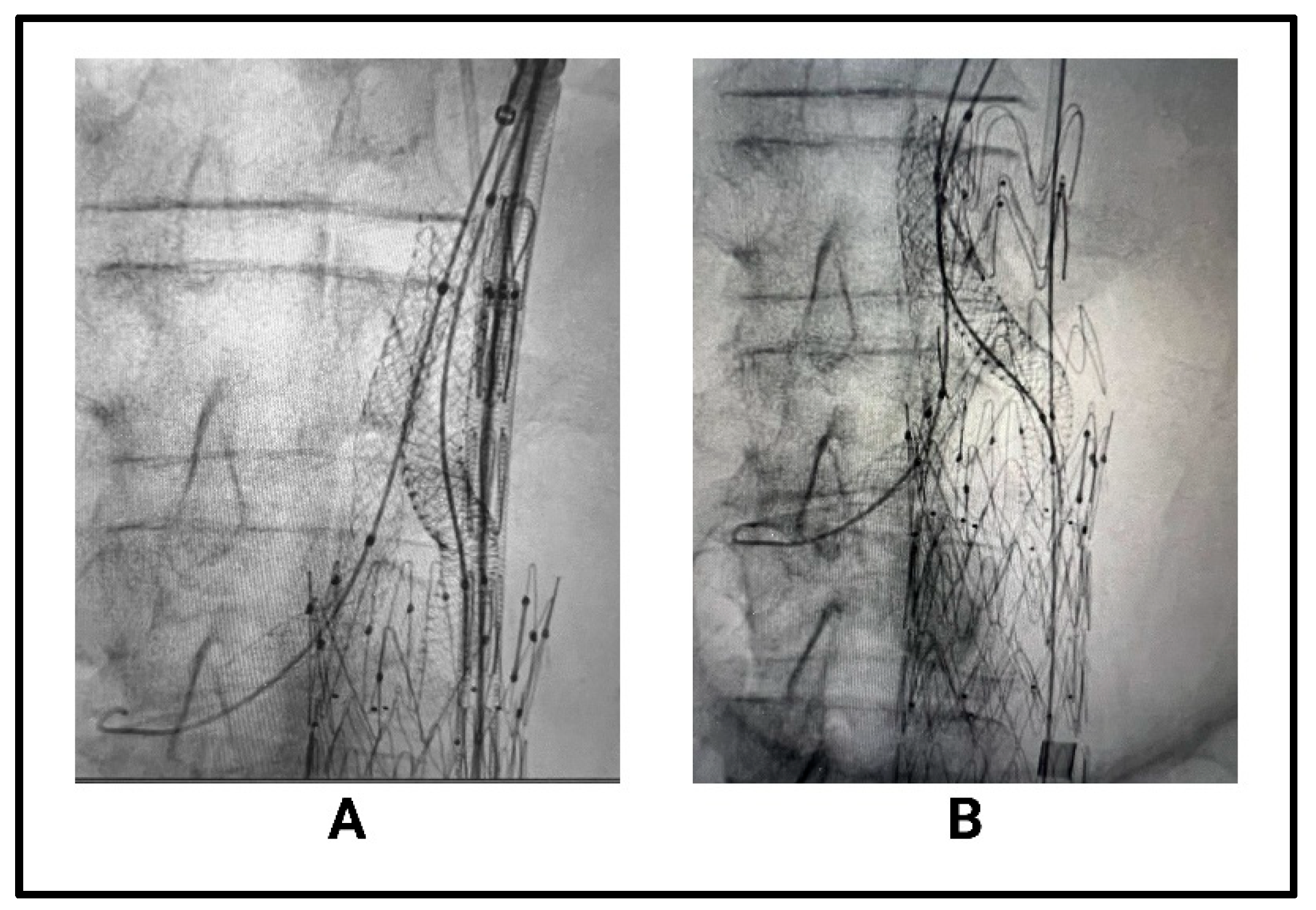
Chimney EVAR, ChEVAR, is often employed when there is a need to extend the proximal landing zone of the stent graft to ensure adequate sealing and prevent endoleaks, which are a common complication following EVAR. This technique involves placing additional stents, or “chimneys,” parallel to the main aortic stent graft to maintain perfusion to vital branch vessels such as the renal or mesenteric arteries (Figure 3). This approach is particularly useful in cases of juxtarenal or pararenal aneurysms where the aneurysm neck is short or angulated, making standard EVAR challenging [43].
The outcomes of ChEVAR are generally favourable, with studies reporting acceptable rates of morbidity and mortality. A study involving 77 patients undergoing ChEVAR for symptomatic and ruptured AAAs reported a 30-day mortality rate of 11.4% for ruptured cases, which is comparable to the outcomes of standard EVAR in similar settings [44]. Another study highlighted that the use of ChEVAR in French university hospitals resulted in a 30-day mortality rate of 11.7%, with a primary patency rate of 97.4% at six months [45]. These figures suggest that ChEVAR can be a viable option for managing complex aneurysms, particularly when anatomical challenges preclude the use of standard EVAR techniques.
Age and comorbidities play a significant role in the success of ChEVAR. The median age of patients undergoing ChEVAR is typically in the early 70s, with a significant proportion of patients classified as American Society of Anaesthesiologists (ASA) class III or higher, indicating a high level of surgical risk [44]. Despite these challenges, the technique has been shown to provide durable results, with the long-term follow-up (17 months) data indicating high patency rates (97%) and low rates of re-intervention (2% for type I endoleaks) [46].
Anatomical factors such as the diameter and morphology of the aneurysm neck are critical determinants of ChEVAR’s success. Aneurysms with short or angulated necks are more likely to benefit from the chimney technique, as it allows for the extension of the sealing zone and reduces the risk of type I endoleaks [47,48]. The incidence of type I endoleaks in ChEVAR procedures is reported to be around 11.9%, with most cases being managed conservatively or through secondary interventions [45,46].
The presence of comorbidities such as hypertension, diabetes, and smoking history can also impact the outcomes of ChEVAR. Patients with a history of tobacco use, for example, have been shown to have a higher prevalence of a hostile neck anatomy, which can complicate the procedure and increase the risk of complications [48].

2.2.4. Physician-Modified Endografts (PMEGs)

Physician-modified endografts (PMEGs) play a crucial role in the urgent management of ruptures or symptomatic aneurysms after failed EVAR, especially when custom-made devices are not available due to time constraints. In these life-threatening situations, the rapid assembly and deployment of PMEGs, often within an hour, allow for immediate intervention [49]. PMEGs are particularly indicated for patients with a complex aortic anatomy or those who are at prohibitively high risk for open surgical repair. This includes cases where the aneurysm involves the branch vessels or when the patient’s comorbidities or previous surgeries make an open repair unfeasible [50]. In the emergency setting, such as a contained or free rupture, PMEGs can be rapidly constructed by modifying off-the-shelf endografts with fenestrations or branches to maintain perfusion to the vital arteries while excluding the aneurysm sac [51]. Case reports and series have demonstrated the successful use of PMEGs in ruptured thoracoabdominal and aortic arch aneurysms, even in patients who were not candidates for open surgery due to active bleeding or recent major operations [52].
The technical success rates for PMEGs in urgent cases are high, with systematic reviews reporting rates above 97% [53]. However, the risk of major adverse events is higher in the emergency setting compared to that in elective cases, with 30-day mortality rates of approximately 24.6% in urgent cases versus 11.6% in elective cases, reflecting the severity of the underlying pathology and the critical condition of these patients [54]. Despite these risks, PMEGs offer a feasible and often life-saving alternative when no other endovascular or surgical options are available in the required timeframe [50].

2.2.5. Interposition Iliac Limb Extensions and Embolisation for Type 1B and Type 3 Endoleaks

After AAA rupture following failed EVAR due to a type 1B or type 3 endoleak, immediate and effective re-exclusion of the aneurysm sac is essential to prevent ongoing haemorrhage and improve survival. The incidence of type 1B and type 3 endoleaks after EVAR is relatively low, but when they occur, they are associated with a significant risk of rupture and require prompt intervention. The risk factors for late distal type 1 endoleaks include the short length of the CIA, the large diameter of the CIA, and inadequate oversizing of the iliac limb [55]. In this acute setting, endovascular techniques such as iliac limb extensions, internal iliac artery (IIA) embolisation, and, in select cases, interposition grafting are the primary strategies for salvage. Iliac limb extension is particularly indicated when the distal seal of the original EVAR graft has failed, often due to aneurysmal degeneration or anatomical changes in the common iliac artery (CIA), resulting in a type 1B endoleak, or when a type 3 endoleak occurs near the iliac limb due to modular disconnection or a fabric tear. In the context of ruptures, rapid extension into the external iliac artery (EIA) is often necessary to achieve a new, secure seal and halt bleeding. This approach is supported by evidence showing that external iliac extension (EIE) is more likely to result in significant aneurysm sac shrinkage compared to that under the bell-bottom technique (BBT) or iliac branch endoprosthesis (IBE), which is crucial for durable exclusion after rupture, although EIE may be associated with higher rates of limb-related complications and reoperations. However, in the setting of rupture, the need for immediate and effective aneurysm exclusion outweighs these risks, making EIE the preferred option when anatomically feasible [56,57,58].
IIA embolisation is often performed in conjunction with iliac limb extension when the extension would otherwise occlude the IIA’s origin, risking retrograde perfusion of the aneurysm sac and persistent endoleaks. Both coil and plug embolisation techniques are feasible and safe, with technical success rates exceeding 95% and no significant increase in the endoleak rates compared to those under EVAR without embolisation. The main indication for IIA embolisation is the need to extend the stent graft into the EIA to achieve an adequate seal, particularly when the CIA is aneurysmal or too short. Meanwhile, embolisation can increase the risk of buttock claudication and, less commonly, pelvic ischaemia. Claudication is generally transient and resolves over time. The choice between concomitant and staged embolisation does not significantly affect operative time, contrast use, or renal function, but concomitant embolisation may be associated with a higher incidence of transient buttock claudication. In the acute rupture setting, concomitant embolisation is often favoured due to its expediency, for haemostasis, and to avoid the need for multiple procedures [59,60].
Interposition endografting is used to realign and seal the disconnected endograft. This is useful in type 3A endoleaks in which the limb junctions can leak significantly or even disconnect entirely with full arterial pressurisation of the sac. A suitable-size limb with sufficient overlap with the adjoining limbs is deployed to re-establish haemostasis. This procedure is relatively quick and can be undertaken under local anaesthesia in many cases [58].

2.3. Open Surgical Repair After Failed Endovascular Repair

The decision to proceed with open surgical repair is often influenced by the failure of EVAR, which can occur due to complications such as endoleaks, graft migration, or aneurysm rupture. The success of open surgical repair is contingent upon several anatomical, physiological, and pathological factors, as well as the timing and indications for the procedure.
The perioperative mortality rates for open surgical repair of ruptured AAAs are significantly higher compared to those for endovascular approaches. A meta-analysis reported a pooled perioperative mortality of 37.8% for open surgical repair, compared to 24.5% for EVAR [10]. Despite its higher mortality, open surgical repair remains a definitive treatment option, particularly when EVAR is not feasible or has failed. The choice between endovascular and open repair is often dictated by the patient’s anatomical suitability for endovascular intervention, the availability of suitable devices and expertise in the context, and the urgency of the rupture, as well as overall physiological status.
Anatomical factors play a crucial role in determining the suitability of open surgical repair. Patients with a complex aneurysm morphology, such as a short or angulated proximal neck, neck thrombi, and calcification with a failed EVAR in situ, may not be ideal candidates for a straightforward endovascular solution and may require open repair [61]. Additionally, the presence of a hostile anatomy, such as conical aortic necks, can lead to EVAR failure, necessitating conversion into open surgery. Physiological factors, including the patient’s haemodynamic stability and comorbid conditions, also influence the decision to proceed with open surgical repair. Patients who are haemodynamically unstable or have significant comorbidities may not tolerate the physiological stress of open surgery, making EVAR a more suitable option if anatomically feasible [62]. However, in cases where EVAR is not possible, open repair may be the only viable option despite the increased risk. Pathological factors, such as the presence of inflammatory changes or infections, can complicate the management of ruptured AAAs. Inflammatory AAAs present unique challenges due to the thickening of the aortic wall and dense adhesions, which can increase the risk of iatrogenic injury during open repair [63].
The success of open surgical repair is also influenced by institutional factors, such as the experience and volume of the surgical centre. Higher institutional caseloads have been associated with improved outcomes for open repair, highlighting the importance of specialised centres for managing complex AAA cases [10].

3. Imaging Surveillance After EVAR

Imaging surveillance after EVAR plays a crucial role in managing patients with AAAs to prevent complications such as ruptures. The primary imaging modalities used for surveillance include computed tomography angiography (CTA), magnetic resonance imaging (MRI), and duplex ultrasound (DU). Each modality has its own sensitivity and specific role in the surveillance protocol (Table 2).
CTA is often considered the gold standard for post-EVAR surveillance due to its high sensitivity, of around 94% [64], in detecting endoleaks and other complications. However, it involves exposure to radiation and the risk of contrast-induced nephropathy, which limits its frequent use [65]. MRI, while avoiding radiation, is less commonly used due to its higher cost, but it provides excellent soft tissue contrast and is useful in patients with contraindications to iodinated contrast, with 85% sensitivity [65]. Colour duplex ultrasound (CDU) is a non-invasive and cost-effective alternative with no radiation exposure, but it has lower sensitivity (77%) [66] compared to that for CTA in detecting certain types of endoleaks [67]. Type I endoleaks are often challenging for ultrasound to detect due to their location at the proximal or distal attachment sites of the stent graft, which may not be well visualised using ultrasound techniques [66]. Type III endoleaks, which occur due to defects in the graft material or junctions between components, may be challenging to detect, especially if they are small or located in complex anatomical regions [68]. Contrast-enhanced ultrasound (CEU) has been shown to have a sensitivity comparable to that of CTA, at 97% [69], making it a promising alternative in certain cases.
Surveillance typically begins one month post-EVAR, with subsequent imaging at regular intervals. The frequency of surveillance can vary based on the initial findings and patient risk factors. The European Society for Vascular Surgery suggests that patients with no detectable endoleak at 30 days can have their follow-up extended to five years, although this approach may miss significant complications [70]. Annual imaging is therefore recommended, with more frequent surveillance in the first year due to the higher incidence of complications during this period [16]. The incidence of ruptures in patients without follow-up imaging is a significant concern. Non-compliance with the surveillance protocols can lead to missed detection of complications such as endoleaks, which can result in aneurysm expansion and rupture. Studies have shown that non-compliant patients have a higher risk of adverse outcomes, including increased mortality rates [71]. The incidence of ruptures in non-compliant patients can be as high as 6% over five years, compared to less than 1% in compliant patients [72]. The rate of aneurysm expansion that warrants intervention post-EVAR is typically an increase in diameter of more than 5 mm, which is considered significant and may necessitate re-intervention [73]. This threshold is used to guide the decisions on further treatment to prevent ruptures.
Table 2. Imaging modalities for surveillance after failed EVAR and ruptured abdominal aneurysm.
Imaging ModalityInitial/Follow-Up Use Per GuidelinesImpact of Patient Frailty and
Haemodynamics
Treatment Method’s InfluenceKey Evidence/
Guideline Notes
CT Angiography (CTA)Gold standard for initial and follow-up imaging; typically at 1, 6, and 12 months and then annually [65,74]Haemodynamically unstable or frail patients may not tolerate contrast/radiation; risk of nephropathy [67]Open or repeated endovascular repair often requires CTA for planning and post-procedure assessments [74]High sensitivity/specificity; guidelines recommend CTA for initial assessment and when complications are suspected
Dual-Energy CT (DECT) and Dual-Energy CT Angiography (DECTA)Used as an advanced alternative to CTA, especially for complex cases [75]Similar limitations to CTA; may be less suitable for unstable/frail patientsUseful for detailed vascular assessments post-interventionOffers improved vascular imaging; not yet standard in all guidelines
Duplex Ultrasound (DUS)Increasingly used for routine follow-up, especially after first year if stable [73,76]Well-tolerated in frail/unstable patients; no contrast/radiationConservative management or stable post-repair patients often monitored with DUSLower sensitivity than that of CTA but safe and cost-effective for long-term surveillance
Contrast-Enhanced Ultrasound (CEUS)Can replace CTA for follow-up; high sensitivity for endoleak detection [67,77]Safe for frail patients; no nephrotoxic contrastUseful for all treatment types, especially when CTA is contraindicatedSensitivity/specificity comparable to that of CTA; guidelines support use when CTA is risky or inconclusive
Abdominal X-rayUsed adjunctively for stent position; not for endoleak detection [78]Minimal impact from frailty and haemodynamicsUsed in all management types for device integrityNot a standalone modality; complements DUS in some protocols
MRISometimes used if CTA contraindicated (e.g., renal impairment) [79]Preferred in patients with contrast allergies or renal dysfunctionUsed for complex cases or when other imaging is inconclusiveHigher endoleak detection than that of CTA in some studies; less common in routine protocols

4. Expert Opinion

Ruptured abdominal aortic aneurysms after failed endovascular repair, particularly when the rupture is still contained by the endograft, present a complex clinical scenario that demands a highly individualised, anatomy-driven, and haemodynamics-sensitive approach. The management strategy must be tailored to the patient’s physiological status, the specific failure mode of the EVAR, and the anatomical features of the aorta and the endograft.
The first and most critical determinant is the patient’s haemodynamic status. In haemodynamically stable patients with a contained rupture, there is a narrow window for detailed imaging and multidisciplinary planning. Conservative management is rarely appropriate but may be considered in exceptional cases where the patient is unfit for any intervention due to extreme comorbidities. However, the natural history of contained ruptures is unpredictable, and most patients will eventually require intervention due to the high risk of progression to free rupture and death [80]. Conservative management is generally reserved for those with minimal symptoms, no evidence of ongoing haemorrhage, and prohibitive surgical risk and should only be considered after a thorough multidisciplinary discussion. In unstable patients, rapid control of bleeding is paramount. Endovascular options, such as aortic occlusion balloons, can be used as a temporising measure to stabilise the patient and allow for definitive repair. If endovascular re-intervention is not feasible due to anatomical constraints or device availability or if the patient deteriorates despite initial stabilisation, open surgical conversion may be indicated.
For the majority of patients, especially those who are haemodynamically stable, endovascular re-intervention is the preferred approach due to its lower perioperative mortality compared to that under open repair [81]. The most common causes of post-EVAR ruptures are type I and III endoleaks, often associated with device migration, component separation, or inadequate initial sealing. These are frequently amenable to endovascular solutions, provided the anatomy is suitable [82]. The choice of endovascular technique is dictated by the location and nature of the endoleak; the quality and length of the available landing zones; and the involvement of the visceral or iliac branches.
For proximal (type IA) or distal (type IB) endoleaks, advanced endovascular techniques are required when the standard cuffs or extensions cannot achieve an adequate seal. Fenestrated or branched EVAR (F/BEVAR) is indicated when the proximal or distal sealing zones are inadequate due to short, angulated, or diseased necks or when the visceral or iliac branches are involved. F/BEVAR allows for extension of the sealing zone while preserving the flow to the vital branches, making it particularly suitable for anatomically complex cases. The use of F/BEVAR is especially advantageous in patients with short or angulated necks or when the aneurysm extends to or involves the visceral segment. In urgent settings where custom devices are not available, physician-modified endografts (PMEGs) can be rapidly adapted to the patient-specific anatomy, providing a life-saving option in anatomically challenging cases [1,10].
Parallel graft techniques, such as chimney EVAR (ChEVAR), are also employed to extend the proximal seal zone, particularly when immediate off-the-shelf solutions are required and the anatomy is suitable for parallel stenting of the visceral branches. ChEVAR is most appropriate when the proximal neck is short or diseased and when rapid intervention is necessary. This technique involves placing covered stents in the visceral arteries alongside the main endograft, allowing for extension of the sealing zone without compromising branch perfusion [83]. ChEVAR is especially useful in emergent situations where time does not permit the fabrication of custom devices. The anatomical factors influencing the choice of technique include the length and quality of the proximal and distal landing zones, the involvement of the visceral or iliac branches, the presence of thrombi or calcification, and the configuration of the failed endograft. F/BEVAR and ChEVAR are particularly useful in cases with short or angulated necks or when the visceral branches are at risk. PMEGs provide flexibility in urgent cases with a complex anatomy.
When a type 1B endoleak is identified, particularly in the setting of common iliac artery (CIA) aneurysmal degeneration or an inadequate distal seal, extension of the endograft limb into the external iliac artery (EIE) is a well-established technique. This often necessitates embolisation of the internal iliac artery (IIA) to prevent retrograde perfusion and endoleaks, which can be performed either concomitantly or as a staged procedure. Studies have shown that EIE is associated with greater aneurysm sac regression compared to that under the bell-bottom technique (BBT) and iliac branch endoprosthesis (IBE), although it carries a higher rate of limb-related complications and a slightly increased incidence of type 1B endoleaks [56]. Coil embolisation of the IIA is generally safe, with buttock claudication being the most common complication, but serious ischaemic events are rare, making this a viable option in patients unfit for open repair [84]. Selective IIA coverage without embolisation may also be considered in certain anatomical scenarios, with similar midterm outcomes and a low incidence of re-intervention [85].
For type III endoleaks, which result from component separation or fabric disruption, relining with additional endograft components is often effective. The choice of relining device depends on the location and extent of the defect, as well as the compatibility with the existing endograft. Persistent type II endoleaks rarely cause ruptures but may contribute to sac expansion and, in the context of rupture, should be addressed if identified as the culprit. Embolisation techniques or conversion into open repair may be required if the endovascular options fail.
When endovascular options are exhausted or not feasible due to anatomical constraints, open surgical repair remains the definitive treatment. Open conversion involves explantation of the failed endograft and reconstruction of the aortoiliac segment, which may include an aorto-bi-iliac or an aorto-bifemoral bypass, with or without reimplantation of the visceral or hypogastric arteries depending on the extent of disease and prior interventions. In cases where the iliac arteries are unsuitable for direct anastomosis, extra-anatomic bypasses such as axillo-femoral or femoro-femoral crossover grafts may be required. Open repair is associated with higher morbidity and mortality, particularly in the emergent setting and in patients with significant comorbidities, but it remains the gold standard when infection is present or when the endovascular techniques fail to achieve durable exclusion of the aneurysm sac [58,86]. Infection is a special consideration; if the rupture is associated with graft infection, open repair with graft explantation and extensive debridement is generally required, as endovascular solutions are associated with higher rates of recurrent infection and related complications [21]. The decision to proceed with endovascular versus open repair in the setting of infection must be made on a case-by-case basis, weighing the risks of recurrent infection against the patient’s ability to tolerate open surgery.

Author Contributions

Conceptualization, R.R. and O.A.; methodology, R.R. and O.A.; writing—original draft preparation, R.R.; writing—review and editing, R.R. and O.A. All authors have read and agreed to the published version of the manuscript.

Funding

This research received no external funding.

Conflicts of Interest

The authors declare no conflict of interest.

References

  1. Antoniou, G.; Georgiadis, G.; Antoniou, S.; Neequaye, S.; Brennan, J.; Torella, F.; Vallabhaneni, S. Late Rupture of Abdominal Aortic Aneurysm After Previous Endovascular Repair. J. Endovasc. Ther. 2015, 22, 734–744. [Google Scholar] [CrossRef]
  2. Saratzis, A.; Bown, M.; Sayers, R. Commentary: Late Rupture After Endovascular Aneurysm Repair. J. Endovasc. Ther. 2015, 22, 745–747. [Google Scholar] [CrossRef]
  3. Moulakakis, K.; Lazaris, A.; Georgiadis, G.; Kakkos, S.; Papavasileiou, V.G.; Antonopoulos, C.; Papapetrou, A.; Katsikas, V.; Klonaris, C.; Geroulakos, G. A Greek Multicentre Study Assessing the Outcome of Late Rupture After Endovascular Abdominal Aortic Aneurysm Repair. J. Vasc. Surg. 2024, 80, 288. [Google Scholar] [CrossRef]
  4. Sweeting, M.; Ulug, P.; Powell, J.; Desgranges, P.; Balm, R. Ruptured Aneurysm Trials: The Importance of Longer-term Outcomes and Meta-analysis for 1-year Mortality. Eur. J. Vasc. Endovasc. Surg. 2015, 50, 297–302. [Google Scholar] [CrossRef] [PubMed]
  5. Alnefaie, S.; Alzahrani, Y.; Alzahrani, B. A Comparison of Endovascular Aneurysm Repair and Open Repair for Ruptured Aortic Abdominal Aneurysms. Cureus 2022, 14, e25672. [Google Scholar] [CrossRef] [PubMed]
  6. Desgranges, P.; Kobeiter, H.; Katsahian, S.; Bouffi, M.; Gouny, P.; Favre, J.; Alsac, J.; Sobocinski, J.; Julia, P.; Alimi, Y.; et al. Editor’s Choice—ECAR (Endovasculaire ou Chirurgie dans les Anévrysmes aorto-iliaques Rompus): A French Randomized Controlled Trial of Endovascular Versus Open Surgical Repair of Ruptured Aorto-iliac Aneurysms. Eur. J. Vasc. Endovasc. Surg. 2015, 50, 303–310. [Google Scholar] [CrossRef]
  7. Greiner, A.; Schleimer, K.; Jalaie, H.; Gombert, A.; Jacobs, M.; Kalder, J. Late rupture after EVAR: A new trend? J. Cardiovasc. Surg. 2014, 55 (Suppl. 1), 169–174. [Google Scholar]
  8. Yilmaz, N.; Peppelenbosch, N.; Cuypers, P.; Tielbeek, A.; Duijm, L.; Buth, J. Emergency Treatment of Symptomatic or Ruptured Abdominal Aortic Aneurysms: The Role of Endovascular Repair. J. Endovasc. Ther. 2002, 9, 449–457. [Google Scholar] [CrossRef]
  9. Antoniou, G.; Georgiadis, G.; Antoniou, S.; Pavlidis, P.; Maras, D.; Sfyroeras, G.; Georgakarakos, E.; Lazarides, M. Endovascular repair for ruptured abdominal aortic aneurysm confers an early survival benefit over open repair. J. Vasc. Surg. 2013, 58, 1091–1105. [Google Scholar] [CrossRef]
  10. Kontopodis, N.; Galanakis, N.; Antoniou, S.; Tsetis, D.; Ioannou, C.; Veith, F.; Powell, J.; Antoniou, G. Meta-Analysis and Meta-Regression Analysis of Outcomes of Endovascular and Open Repair for Ruptured Abdominal Aortic Aneurysm. Eur. J. Vasc. Endovasc. Surg. 2020, 59, 399–410. [Google Scholar] [CrossRef]
  11. Zhang, S.; Feng, J.; Li, H.-Y.; Zhang, Y.; Lu, Q.; Jing, Z. Open surgery (OS) versus endovascular aneurysm repair (EVAR) for hemodynamically stable and unstable ruptured abdominal aortic aneurysm (rAAA). Heart Vessel. 2016, 31, 1291–1302. [Google Scholar] [CrossRef] [PubMed]
  12. Böckler, D.; Holden, A.; Krievins, D.; De Vries, J.; Peters, A.; Geisbüsch, P.; Reijnen, M. Extended use of endovascular aneurysm sealing for ruptured abdominal aortic aneurysms. Semin. Vasc. Surg. 2016, 29, 106–113. [Google Scholar] [CrossRef] [PubMed]
  13. Ogino, H.; Isogai, N.; Kume, N.; Shibutani, S.; Yashiro, H.; Takahara, M.; Fujimura, N. Evaluating the Effectiveness and Clinical Outcomes of Endovascular Aneurysm Repair-First Approach for Ruptured Abdominal Aortic Aneurysm in Japan. J. Endovasc. Ther. 2024, 15266028241248337. [Google Scholar] [CrossRef]
  14. McGreevy, D.; Pirouzram, A.; Gidlund, K.; Nilsson, K.; Hörer, T. A 12-year experience of endovascular repair for ruptured abdominal aortic aneurysms in all patients. J. Vasc. Surg. 2022, 77, 741–749. [Google Scholar] [CrossRef]
  15. Veith, F.; Veith, F.; Lachat, M.; Mayer, D.; Malina, M.; Holst, J.; Mehta, M.; Verhoeven, E.; Larzon, T.; Gennai, S.; et al. Collected World and Single Center Experience With Endovascular Treatment of Ruptured Abdominal Aortic Aneurysms. Ann. Surg. 2009, 250, 818–824. [Google Scholar] [CrossRef]
  16. Chaikof, E.; Dalman, R.; Eskandari, M.; Jackson, B.; Lee, A.; Mansour, A.M.H.; Mastracci, T.; Mell, M.; Murad, H.; Nguyen, L.; et al. The Society for Vascular Surgery practice guidelines on the care of patients with an abdominal aortic aneurysm. J. Vasc. Surg. 2018, 67, 2–77.e2. [Google Scholar] [CrossRef]
  17. Peppelenbosch, N.; Yilmaz, N.; Marrewijk, C.; Buth, J.; Cuypers, P.; Duijm, L.; Tielbeek, A. Emergency treatment of acute symptomatic or ruptured abdominal aortic aneurysm. Outcome of a prospective intent-to-treat by EVAR protocol. Eur. J. Vasc. Endovasc. Surg. 2003, 26, 303–310. [Google Scholar] [CrossRef] [PubMed]
  18. Cilingiroglu, M.; Inanc, H. Local or general anesthesia for EVAR in patients with ruptured AAA? Catheter. Cardiovasc. Interv. 2022, 100, 938. [Google Scholar] [CrossRef]
  19. Beck, A.; Sedrakyan, A.; Mao, J.; Venermo, M.; Faizer, R.; Debus, S.; Behrendt, C.; Scali, S.; Altreuther, M.; Schermerhorn, M.; et al. Variations in Abdominal Aortic Aneurysm Care: A Report From the International Consortium of Vascular Registries. Circulation 2016, 134, 1948–1958. [Google Scholar] [CrossRef]
  20. Png, C.; Pendleton, A.; Altreuther, M.; Budtz-Lilly, J.; Gunnarsson, K.; Kan, C.-D.; Khashram, M.; Laine, M.; Mani, K.; Pederson, C.; et al. Effect of EVAR on International Ruptured AAA Mortality—Sex and Geographic Disparities. J. Clin. Med. 2024, 13, 2464. [Google Scholar] [CrossRef]
  21. Shirasu, T.; Kuno, T.; Yasuhara, J.; Yokoyama, Y.; Takagi, H.; Cullen, M.; Kent, K.; Clouse, W. Recurrent infection is more common after endovascular versus open repair of infected abdominal aortic aneurysm: Systematic review and meta-analysis. J. Vasc. Surg. 2021, 75, 348–355.e10. [Google Scholar] [CrossRef]
  22. Grootes, I.; Barrett, J.; Ulug, P.; Rohlffs, F.; Laukontaus, S.; Tulamo, R.; Venermo, M.; Greenhalgh, R.M.; Sweeting, M. Predicting Risk of Rupture and Rupture-Preventing Reinterventions Following Endovascular Abdominal Aortic Aneurysm Repair. Br. J. Surg. 2018, 105, 1294–1304. [Google Scholar] [CrossRef]
  23. Troisi, N.; Bertagna, G.; Torri, L.; Canovaro, F.; D’Oria, M.; Adami, D.; Berchiolli, R. The Management of Ruptured Abdominal Aortic Aneurysms: An Ongoing Challenge. J. Clin. Med. 2023, 12, 5530. [Google Scholar] [CrossRef] [PubMed]
  24. Teng, B.; Xie, C.; Zhao, Y.; Wang, Z. Studies Related to Ruptured Abdominal Aortic Aneurysms in the Past 10 Years (2011-2020): A Bibliometric Analysis. Med. Sci. Monit. 2021, 28, e935006. [Google Scholar] [CrossRef]
  25. Wang, Y.-D.; Liu, Z.; Ren, J.; Xiang, M.-X. Pharmacological Therapy of Abdominal Aortic Aneurysm: An Update. Curr. Vasc. Pharmacol. 2018, 16, 114–124. [Google Scholar] [CrossRef] [PubMed]
  26. Li, R.; Liu, Y.; Jiang, J. Research advances in drug therapy for abdominal aortic aneurysms over the past five years: An updated narrative review. Int. J. Cardiol. 2022, 372, 93–100. [Google Scholar] [CrossRef] [PubMed]
  27. Cameron, S.; Russell, H.; Owens, A. Antithrombotic therapy in abdominal aortic aneurysm: Beneficial or detrimental? Blood 2018, 132, 2619–2628. [Google Scholar] [CrossRef]
  28. Siordia, J.A. Beta-Blockers and Abdominal Aortic Aneurysm Growth: A Systematic Review and Meta-Analysis. Curr. Cardiol. Rev. 2021, 17, e230421187502. [Google Scholar] [CrossRef]
  29. Mieth, A.; Revermann, M.; Babelova, A.; Weigert, A.; Schermuly, R.T.; Brandes, R.P. L-type calcium channel inhibitor diltiazem prevents aneurysm formation by blood pressure-independent anti-inflammatory effects. Hypertension 2013, 62, 1098–1104. [Google Scholar] [CrossRef]
  30. Puertas-Umbert, L.; Almendra-Pegueros, R.; Jimenez-Altayo, F.; Sirvent, M.; Galán, M.; Martínez-González, J.; Rodríguez, C. Novel pharmacological approaches in abdominal aortic aneurysm. Clin. Sci. 2023, 137, 1167–1194. [Google Scholar] [CrossRef]
  31. Chen, J.; Hu, L.; Liu, Z. Medical treatments for abdominal aortic aneurysm: An overview of clinical trials. Expert Opin. Investig. Drugs 2024, 33, 979–992. [Google Scholar] [CrossRef] [PubMed]
  32. Hosseini, A.; Sahranavard, T.; Reiner, Ž.; Jamialahmadi, T.; Dhaheri, Y.; Eid, A.; Sahebkar, A. Effect of statins on abdominal aortic aneurysm. Eur. J. Pharm. Sci. 2022, 178, 106284. [Google Scholar] [CrossRef]
  33. Salata, K.; Syed, M.; Hussain, M.A.; de Mestral, C.; Greco, E.; Mamdani, M.; Tu, J.V.; Forbes, T.L.; Bhatt, D.L.; Verma, S.; et al. Statins Reduce Abdominal Aortic Aneurysm Growth, Rupture, and Perioperative Mortality: A Systematic Review and Meta-Analysis. J. Am. Heart Assoc. 2018, 7, e008657. [Google Scholar] [CrossRef] [PubMed]
  34. Asirwatham, M.; Konanki, V.; Lucas, S.; Grundy, S.; Zwiebel, B.; Shames, M.; Arnaoutakis, D. Comparative Outcomes of Physician-Modified Fenestrated/Branched Endovascular Aortic Aneurysm Repair in the Setting of Prior Failed EVAR. J. Vasc. Surg. 2023, 78, 1153–1161. [Google Scholar] [CrossRef]
  35. Schanzer, A.; Beck, A.; Eagleton, M.; Farber, M.; Oderich, G.; Schneider, D.; Sweet, M.; Crawford, A.; Timaran, C. Results of fenestrated and branched endovascular aortic aneurysm repair after failed infrarenal endovascular aortic aneurysm repair. J. Vasc. Surg. 2019, 72, 849–858. [Google Scholar] [CrossRef]
  36. Nana, P.; Kölbel, T.; Behrendt, C.; Kouvelos, G.; Giannoukas, A.; Haulon, S.; Spanos, K. Systematic review on re-intervention with fenestrated or branched devices after failed previous endovascular aortic aneurysm repair. J. Vasc. Surg. 2022, 77, 1806–1814.e2. [Google Scholar] [CrossRef] [PubMed]
  37. Hostalrich, A.; Mesnard, T.; Soler, R.; Girardet, P.; Kaladji, A.; Baptiste, E.J.; Malikov, S.; Reix, T.; Ricco, J.; Chaufour, X. Prospective Multicentre Cohort Study of Fenestrated and Branched Endografts After Failed Endovascular Infrarenal Aortic Aneurysm Repair with Type Ia Endoleak. Eur. J. Vasc. Endovasc. Surg. 2021, 62, 540–548. [Google Scholar] [CrossRef]
  38. Juszczak, M.; Vezzosi, M.; Nasr, H.; Claridge, M.; Adam, D. Fenestrated-Branch Endovascular Repair After Prior Abdominal Aortic Aneurysm Repair. Eur. J. Vasc. Endovasc. Surg. 2021, 62, 728–737. [Google Scholar] [CrossRef]
  39. Powell, J.; Wanhainen, A. Analysis of the Differences Between the European Society for Vascular Surgery 2019 and National Institute for Health and Care Excellence 2020 Guidelines for Abdominal Aortic Aneurysm. Eur. J. Vasc. Endovasc. Surg. 2020, 60, 7–15. [Google Scholar] [CrossRef]
  40. Nana, P.; Volakakis, G.; Spanos, K.; Kouvelos, G.; Bareka, M.; Arnaoutoglou, E.; Giannoukas, A.; Matsagkas, M. Endovascular Repair of Ruptured Abdominal Aortic Aneurysms Using the Endurant™ Endograft. J. Clin. Med. 2024, 13, 5282. [Google Scholar] [CrossRef]
  41. Kopp, R.; Stachowski, L.; Puippe, G.; Zimmermann, A.; Menges, A. Long-Term Outcomes of Endovascular Aortic Repair with Parallel Chimney or Periscope Stent Grafts for Ruptured Complex Abdominal Aortic Aneurysms. J. Clin. Med. 2025, 14, 234. [Google Scholar] [CrossRef] [PubMed]
  42. Mosher, E.; Reitz, K.; Andraska, E.; Sridharan, N.; Alie-Cusson, F.; Makaroun, M.; Liang, N. Parallel grafting in ruptured aortic aneurysms with previous open infrarenal surgical repair. Ann. Vasc. Surg.—Brief Rep. Innov. 2021, 2, 100029. [Google Scholar] [CrossRef]
  43. Jennings, J.; Sheahan, L.; Gloss, C.; Vogel, T.; Bath, J. Multiple Chimney Endografts (ChEVAR) for Ruptured Pararenal Aortic Aneurysm. Ann. Vasc. Surg. 2021, 75, 531.e1–531.e6. [Google Scholar] [CrossRef]
  44. Jernigan, E.G.; Nguyen Tran, N.; Qato, K.; Giangola, G.; Carroccio, A.; Conway, A.M. Outcomes of chimney/snorkel endovascular repair for symptomatic and ruptured abdominal aortic aneurysms. J. Vasc. Surg. 2021, 74, 1117–1124. [Google Scholar] [CrossRef]
  45. Touma, J.; Caradu, C.; Sylvestre, R.; Settembre, N.; Schneider, F.; Moia, A.; Ahmed, B.; Lebas, B.; Gaudric, J.; Alsac, J.; et al. Multicentre Experience with the Chimney Technique for Abdominal Aortic Aneurysms in French University Hospitals. Eur. J. Vasc. Endovasc. Surg. 2020, 59, 776–784. [Google Scholar] [CrossRef]
  46. Lindblad, B.; Jabr, A.; Holst, J.; Malina, M. Chimney Grafts in Aortic Stent Grafting: Hazardous or Useful Technique? Systematic Review of Current Data. Eur. J. Vasc. Endovasc. Surg. 2015, 50, 722–731. [Google Scholar] [CrossRef]
  47. Sierra-Juárez, M.; Valderrama-Treviño, A.; González-Martínez, I.; Mendoza-Barrera, G.; Ramos-Peralta, M. Initial experience of endovascular aneurysm repair at the Dr. Eduardo Liceaga General Hospital of Mexico. Int. Surg. J. 2024, 11, 697–702. [Google Scholar] [CrossRef]
  48. Rabellino, M.; Chiabrando, J.; Garagoli, F.; Foscolo, M.A.; Fleitas, M.; Chas, J.; Caro, V.; Bluro, I.; Shinzato, S. Incidence of endoleak type IA in patients undergoing chimney endovascular aortic repair (ChEVAR) vs. standard endovascular repair. Arch. Peru. Cardiol. Y Cirugía Cardiovasc. 2024, 5, 22–28. [Google Scholar] [CrossRef]
  49. Kimball, A.; Mydłowska, A.; Beck, A. Physician-modified endografts for urgent and emergent aortic pathology. Semin. Vasc. Surg. 2021, 34, 215–224. [Google Scholar] [CrossRef]
  50. Pyun, A.; Han, S. Contemporary indications, techniques, and outcomes of physician-modified endografts for the treatment of complex abdominal and thoracoabdominal aortic aneurysms. Semin. Vasc. Surg. 2022, 35, 364–373. [Google Scholar] [CrossRef]
  51. Solano, M.A.; Keller, M.M.; Colon, M.J.P.; Patel, M.R.; Timaran, M.C.; Kirkwood, M.M.; Baig, M.S. Physician Modified Endograft for Ruptured Dissecting Aortic Arch Aneurysm. Vasc. Endovasc. Surg. 2024, 58, 876–883. [Google Scholar] [CrossRef] [PubMed]
  52. Torrealba, J.; Panuccio, G.; Kölbel, T.; Gandet, T.; Heidemann, F.; Rohlffs, F. Physician-Modified Endograft With Inner Branches for the Treatment of Complex Aortic Urgencies. J. Endovasc. Ther. 2021, 29, 697–704. [Google Scholar] [CrossRef]
  53. Melo, G.; Prendes, C.; Caldeira, D.; Stana, J.; Rantner, B.; Wanhainen, A.; Oderich, G.; Tsilimparis, N. Systematic Review and Meta-analysis of Physician Modified Endografts for Treatment of Thoraco-Abdominal and Complex Abdominal Aortic Aneurysms. Eur. J. Vasc. Endovasc. Surg. 2022, 64, 188–199. [Google Scholar] [CrossRef]
  54. Oderich, G.; Ribeiro, M. Techniques of Physician-Modified Endovascular Grafts (PMEGs) for Incorporation of Renal Mesenteric Arteries. In Endovascular Aortic Repair: Current Techniques with Fenestrated, Branched and Parallel Stent-Grafts; Springer International Publishing: Berlin/Heidelberg, Germany, 2017; pp. 671–688. [Google Scholar] [CrossRef]
  55. Gray, D.; Samaan, C.; Oikonomou, K.; Gruber-Rouh, T.; Schmitz-Rixen, T.; Derwich, W. Age and Oversizing Influence Iliac Dilatation after EVAR. J. Clin. Med. 2022, 11, 7113. [Google Scholar] [CrossRef] [PubMed]
  56. Bahroloomi, D.; Qato, K.; Nguyen, N.; Schreiber-Gregory, D.; Conway, A.; Giangola, G.; Carroccio, A. External Iliac Extension Causes Greater Aneurysm Sac Regression than Bell-bottom Technique or Iliac Branch Endoprosthesis for Repair of Concomitant Infra-renal Aortic and Iliac Artery Aneurysm. J. Vasc. Surg. 2022, 76, 132–140. [Google Scholar] [CrossRef]
  57. Naughton, P.; Park, M.; Kheirelseid, E.; O’Neill, S.; Rodriguez, H.; Morasch, M.; Madhavan, P.; Eskandari, M. A comparative study of the bell-bottom technique vs hypogastric exclusion for the treatment of aneurysmal extension to the iliac bifurcation. J. Vasc. Surg. 2012, 55, 956–962. [Google Scholar] [CrossRef] [PubMed]
  58. Mascoli, C.; Faggioli, G.; Gallitto, E.; Longhi, M.; Abualhin, M.; Pini, R.; Massoni, C.; Freyrie, A.; Stella, A.; Gargiulo, M. Planning and Endograft Related Variables Predisposing to Late Distal Type I Endoleaks. Eur. J. Vasc. Endovasc. Surg. 2019, 58, 334–342. [Google Scholar] [CrossRef]
  59. Lee, C.; Dougherty, M.; Calligaro, K. Concomitant unilateral internal iliac artery embolization and endovascular infrarenal aortic aneurysm repair. J. Vasc. Surg. 2006, 43, 903–907. [Google Scholar] [CrossRef]
  60. Fontana, F.; Coppola, A.; Ferrario, L.; De Marchi, G.; Macchi, E.; Zorzetto, G.; Franchin, M.; Piffaretti, G.; Tozzi, M.; Carcano, G.; et al. Internal Iliac Artery Embolization within EVAR Procedure: Safety, Feasibility, and Outcome. J. Clin. Med. 2022, 11, 7399. [Google Scholar] [CrossRef]
  61. Armbruster, C. Outcomes of Elective Abdominal Aortic Aneurysm Management: Open Surgical Versus Endovascular Repair. 2021. Available online: https://idun.augsburg.edu/cgi/viewcontent.cgi?article=2103&context=etd (accessed on 15 April 2025).
  62. Visser, J.; Bosch, J.; Hunink, M.; Van Dijk, L.; Hendriks, J.; Poldermans, D.; Van Sambeek, M. Endovascular repair versus open surgery in patients with ruptured abdominal aortic aneurysms: Clinical outcomes with 1-year follow-up. J. Vasc. Surg. 2006, 44, 1148–1155. [Google Scholar] [CrossRef]
  63. Capoccia, L.; Riambau, V. Endovascular repair versus open repair for inflammatory abdominal aortic aneurysms. Cochrane Database Syst. Rev. 2015, 2015, CD010313. [Google Scholar] [CrossRef] [PubMed]
  64. Sommer, W.; Becker, C.; Haack, M.; Rubin, G.; Weidenhagen, R.; Schwarz, F.; Nikolaou, K.; Reiser, M.; Johnson, T.; Clevert, D. Time-resolved CT angiography for the detection and classification of endoleaks. Radiology 2012, 263, 917–926. [Google Scholar] [CrossRef] [PubMed]
  65. Zaiem, F.; Almasri, J.; Tello, M.; Prokop, L.; Chaikof, E.; Murad, M. A systematic review of surveillance after endovascular aortic repair. J. Vasc. Surg. 2018, 67, 320. [Google Scholar] [CrossRef] [PubMed]
  66. Mirza, T.; Karthikesalingam, A.; Jackson, D.; Walsh, S.; Holt, P.; Hayes, P.; Boyle, J. Duplex ultrasound and contrast-enhanced ultrasound versus computed tomography for the detection of endoleak after EVAR: Systematic review and bivariate meta-analysis. Eur. J. Vasc. Endovasc. Surg. 2010, 39, 418–428. [Google Scholar] [CrossRef]
  67. Brazzelli, M.; Hernández, R.; Sharma, P.; Robertson, C.; Shimonovich, M.; Maclennan, G.; Fraser, C.; Jamieson, R.; Vallabhaneni, S. Contrast-enhanced ultrasound and/or colour duplex ultrasound for surveillance after endovascular abdominal aortic aneurysm repair: A systematic review and economic evaluation. Health Technol. Assess. 2018, 22, 1–220. [Google Scholar] [CrossRef]
  68. Kapetanios, D.; Kontopodis, N.; Mavridis, D.; McWilliams, R.G.; Giannoukas, A.D.; Antoniou, G.A. Meta-analysis of the accuracy of contrast-enhanced ultrasound for the detection of endoleak after endovascular aneurysm repair. J. Vasc. Surg. 2019, 69, 280–294.e286. [Google Scholar] [CrossRef]
  69. Gürtler, V.; Sommer, W.; Meimarakis, G.; Kopp, R.; Weidenhagen, R.; Reiser, M.; Clevert, D. A comparison between contrast-enhanced ultrasound imaging and multislice computed tomography in detecting and classifying endoleaks in the follow-up after endovascular aneurysm repair. J. Vasc. Surg. 2013, 58, 340–345. [Google Scholar] [CrossRef]
  70. Väärämäki, S.; Uurto, I.; Hahl, T.; Suominen, V. Reliability and safety of individualized follow-up based on the 30-day CTA after endovascular aneurysm repair (EVAR). Ann. Vasc. Surg. 2022, 86, 305–312. [Google Scholar] [CrossRef]
  71. Grima, M.; Karthikesalingam, A.; Holt, P. Multicentre Post-EVAR Surveillance Evaluation Study (EVAR-SCREEN). Eur. J. Vasc. Endovasc. Surg. 2019, 57, 521–526. [Google Scholar] [CrossRef]
  72. Geraedts, A.; De Mik, S.; Ubbink, D.; Koelemay, M.; Balm, R. Postoperative surveillance and long-term outcome after endovascular aortic aneurysm repair in the Netherlands: Study protocol for the retrospective ODYSSEUS study. BMJ Open 2020, 10, e033584. [Google Scholar] [CrossRef]
  73. Blecha, M.; Scali, S.; Stone, D.; Mao, J.; Goodney, P.; Lemmon, G. Duplex Ultrasound Only Surveillance After Endovascular Abdominal Aortic Aneurysm Repair is Associated with Favorable Long-Term Outcomes. Ann. Vasc. Surg. 2024, 108, 112–126. [Google Scholar] [CrossRef] [PubMed]
  74. Kazimierczak, W.; Kazimierczak, N.; Serafin, Z. Review of Clinical Applications of Dual-Energy CT in Patients after Endovascular Aortic Repair. J. Clin. Med. 2023, 12, 7766. [Google Scholar] [CrossRef] [PubMed]
  75. Charalambous, S.; Perisinakis, K.; Kontopodis, N.; Papadakis, A.E.; Maris, T.G.; Ioannou, C.V.; Karantanas, A.; Tsetis, D. Dual-energy CT angiography in imaging surveillance of endovascular aneurysm repair–preliminary study results. Eur. J. Radiol. 2022, 148, 110165. [Google Scholar] [CrossRef]
  76. Rakemaa, L.; Laukontaus, S.; Aho, P.; Tulamo, R.; Laine, M.; Venermo, M. Ultrasound Surveillance is Feasible After Endovascular Aneurysm Repair. Eur. J. Vasc. Endovasc. Surg. 2019, 58, e651–e652. [Google Scholar] [CrossRef]
  77. Bredahl, K.; Taudorf, M.; Lönn, L.; Vogt, K.; Sillesen, H.; Eiberg, J. Contrast Enhanced Ultrasound can Replace Computed Tomography Angiography for Surveillance After Endovascular Aortic Aneurysm Repair. Eur. J. Vasc. Endovasc. Surg. 2016, 52, 729–734. [Google Scholar] [CrossRef]
  78. Harrison, G.; Oshin, O.; Vallabhaneni, S.; Brennan, J.; Fisher, R.; McWilliams, R. Surveillance after EVAR based on duplex ultrasound and abdominal radiography. Eur. J. Vasc. Endovasc. Surg. 2011, 42, 187–192. [Google Scholar] [CrossRef]
  79. Secchi, F.; Capra, D.; Monti, C.B.; Mobini, N.; Ortiz, M.D.M.G.; Trimarchi, S.; Mazzaccaro, D.; Righini, P.; Nano, G.; Sardanelli, F. Safe follow-up after endovascular aortic repair with unenhanced MRI: The SAFEVAR study. Diagnostics 2022, 13, 20. [Google Scholar] [CrossRef]
  80. Andersson, M.; Talvitie, M.; Benson, L.; Roy, J.; Roos, H.; Hultgren, R. A Population Based Study of Post-EVAR Rupture during 15 years. J. Vasc. Surg. 2021, 74, 701–710.e3. [Google Scholar] [CrossRef]
  81. Hinchliffe, R.; Bruijstens, L.; MacSweeney, S.; Braithwaite, B. A randomised trial of endovascular and open surgery for ruptured abdominal aortic aneurysm—Results of a pilot study and lessons learned for future studies. Eur. J. Vasc. Endovasc. Surg. 2006, 32, 506–513. [Google Scholar] [CrossRef]
  82. Schlösser, F.; Gusberg, R.; Dardik, A.; Lin, P.; Verhagen, H.; Moll, F.; Muhs, B. Aneurysm rupture after EVAR: Can the ultimate failure be predicted? Eur. J. Vasc. Endovasc. Surg. 2009, 37, 15–22. [Google Scholar] [CrossRef]
  83. Mehta, M.; Paty, P.; Roddy, S.; Taggert, J.; Sternbach, Y.; Kreienberg, P.; Chang, B.; Darling, R. Treatment options for delayed AAA rupture following endovascular repair. J. Vasc. Surg. 2011, 53, 14–20. [Google Scholar] [CrossRef] [PubMed]
  84. Criado, F.; Wilson, E.; Velazquez, O.; Carpenter, J.; Barker, C.; Wellons, E.; Abul-Khoudoud, O.; Fairman, R. Safety of coil embolization of the internal iliac artery in endovascular grafting of abdominal aortic aneurysms. J. Vasc. Surg. 2000, 32, 684–688. [Google Scholar] [CrossRef] [PubMed]
  85. Papazoglou, K.; Sfyroeras, G.; Zambas, N.; Konstantinidis, K.; Kakkos, S.; Mitka, M. Outcomes of endovascular aneurysm repair with selective internal iliac artery coverage without coil embolization. J. Vasc. Surg. 2012, 56, 298–303. [Google Scholar] [CrossRef] [PubMed]
  86. Shehata, R.; Aslam, J.; Beng, N. 276 Favorable Outcomes for Endovascular Aneurysm Repair with Aorto-Uni-Iliac (AUI) Stent Grafts for Treatment of Abdominal Aortic Aneurysms (AAA) by a Single Team in 8 Years at South Mersey Arterial (SMArt): A Tertiary Care Vascular Surgery Centre. Br. J. Surg. 2023, 110, znad258.755. [Google Scholar] [CrossRef]
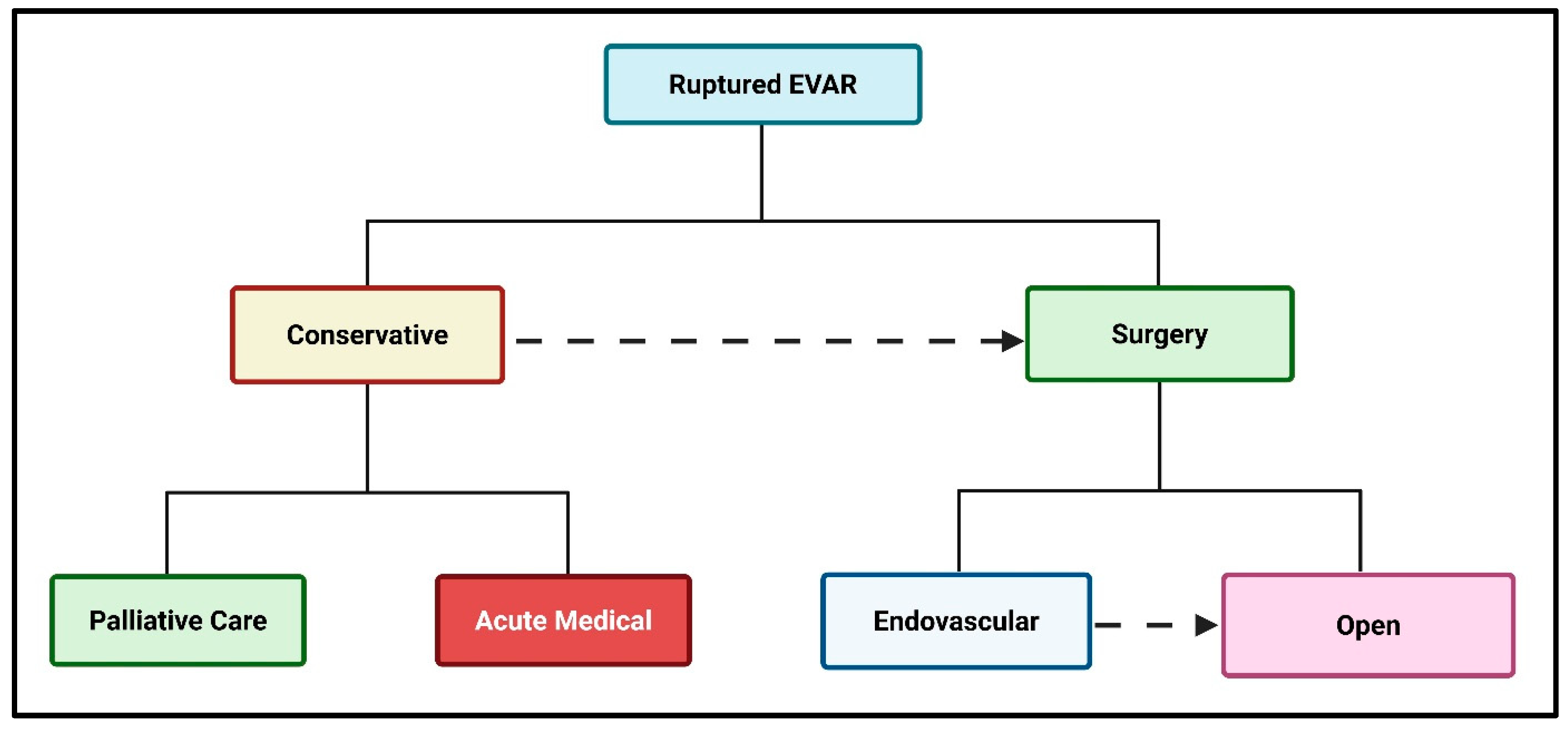
Figure 1. Management of late AAA rupture post-endovascular repair.
Jvd 04 00024 g001
Figure 2. Conservative management of rAAA post-EVAR.
Jvd 04 00024 g002
Figure 3. Chimney extension of previous EVAR for type 1A (A); an aortic extension graft deployed with superior mesenteric and renal arterial chimneys in place (B).
Jvd 04 00024 g003
Disclaimer/Publisher’s Note: The statements, opinions and data contained in all publications are solely those of the individual author(s) and contributor(s) and not of MDPI and/or the editor(s). MDPI and/or the editor(s) disclaim responsibility for any injury to people or property resulting from any ideas, methods, instructions or products referred to in the content.