
Non-Invasive Diagnosis of Angioma Serpiginosum Plantaris: High-Resolution Dermoscopy, High-Frequency Ultrasound, and Line-Field Confocal Optical Coherence Tomography


Abstract
1. Introduction
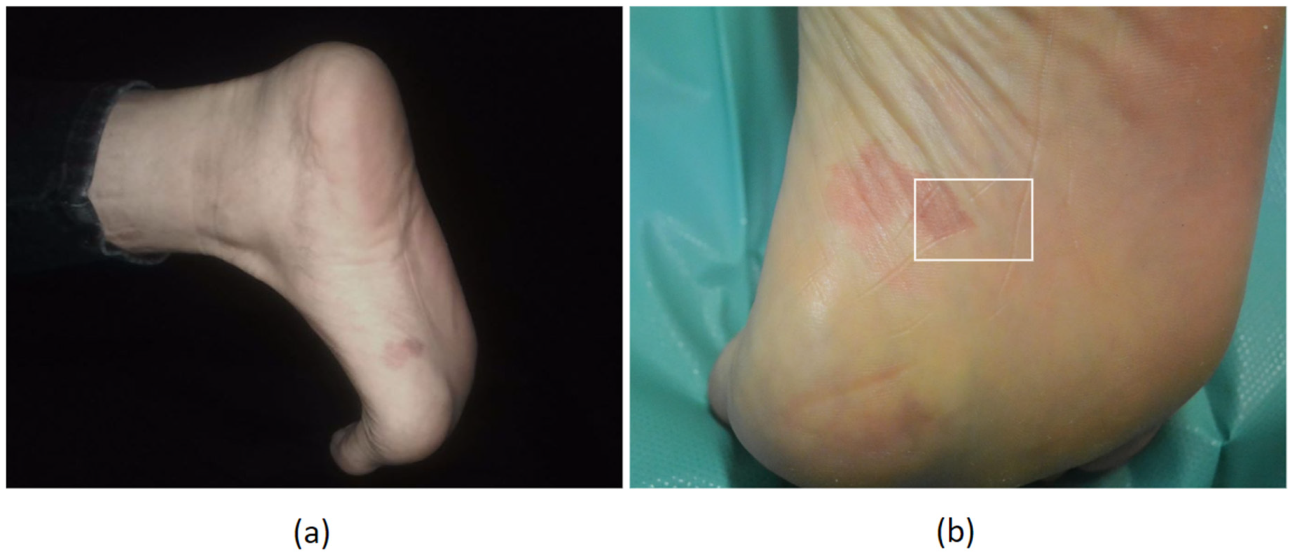
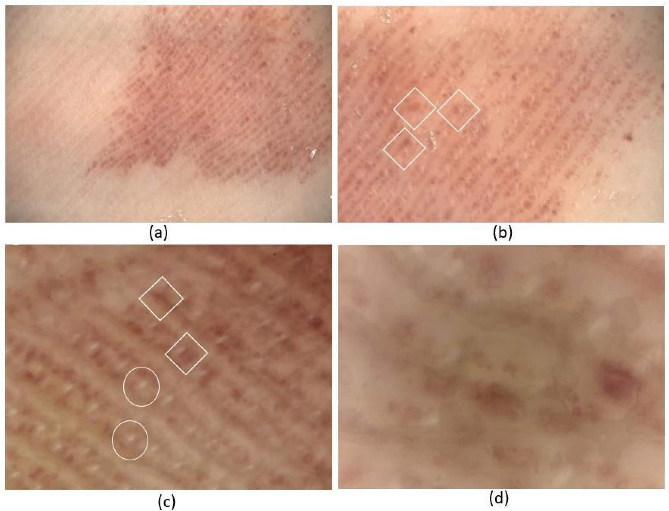
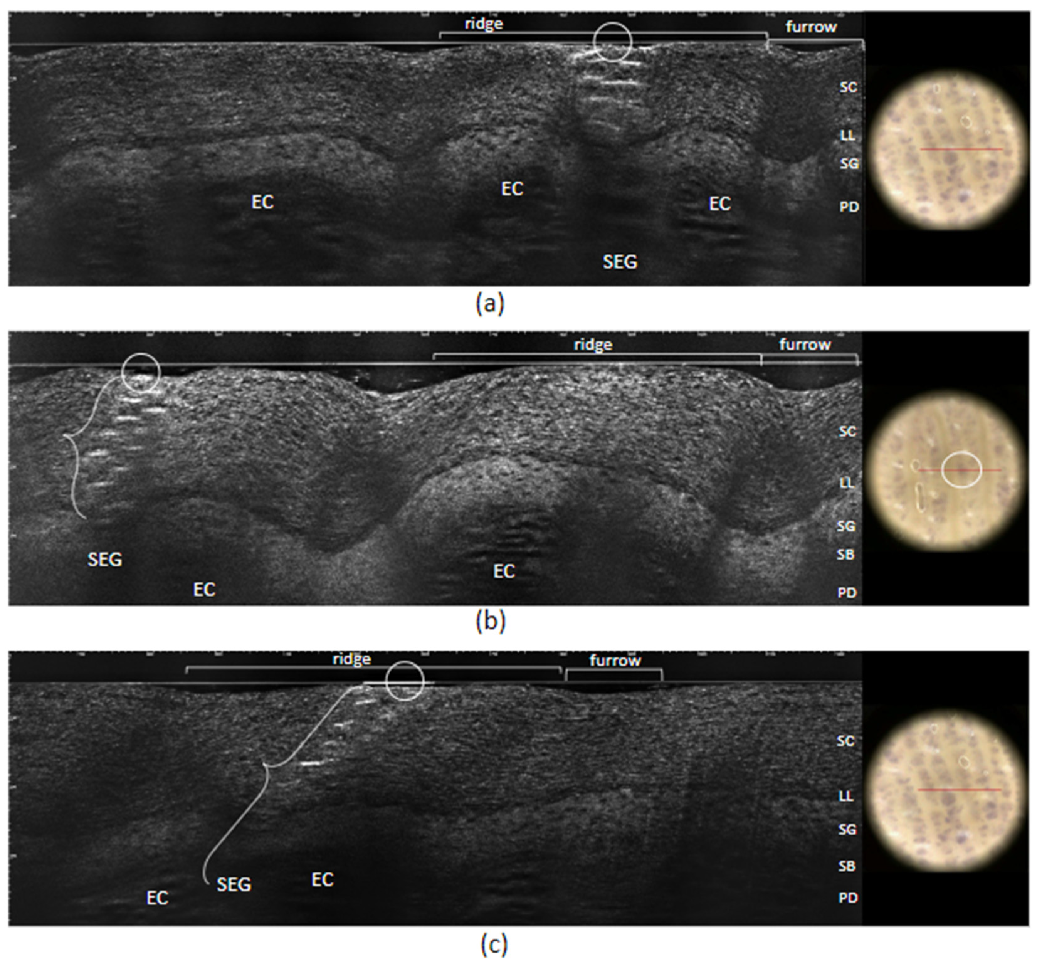
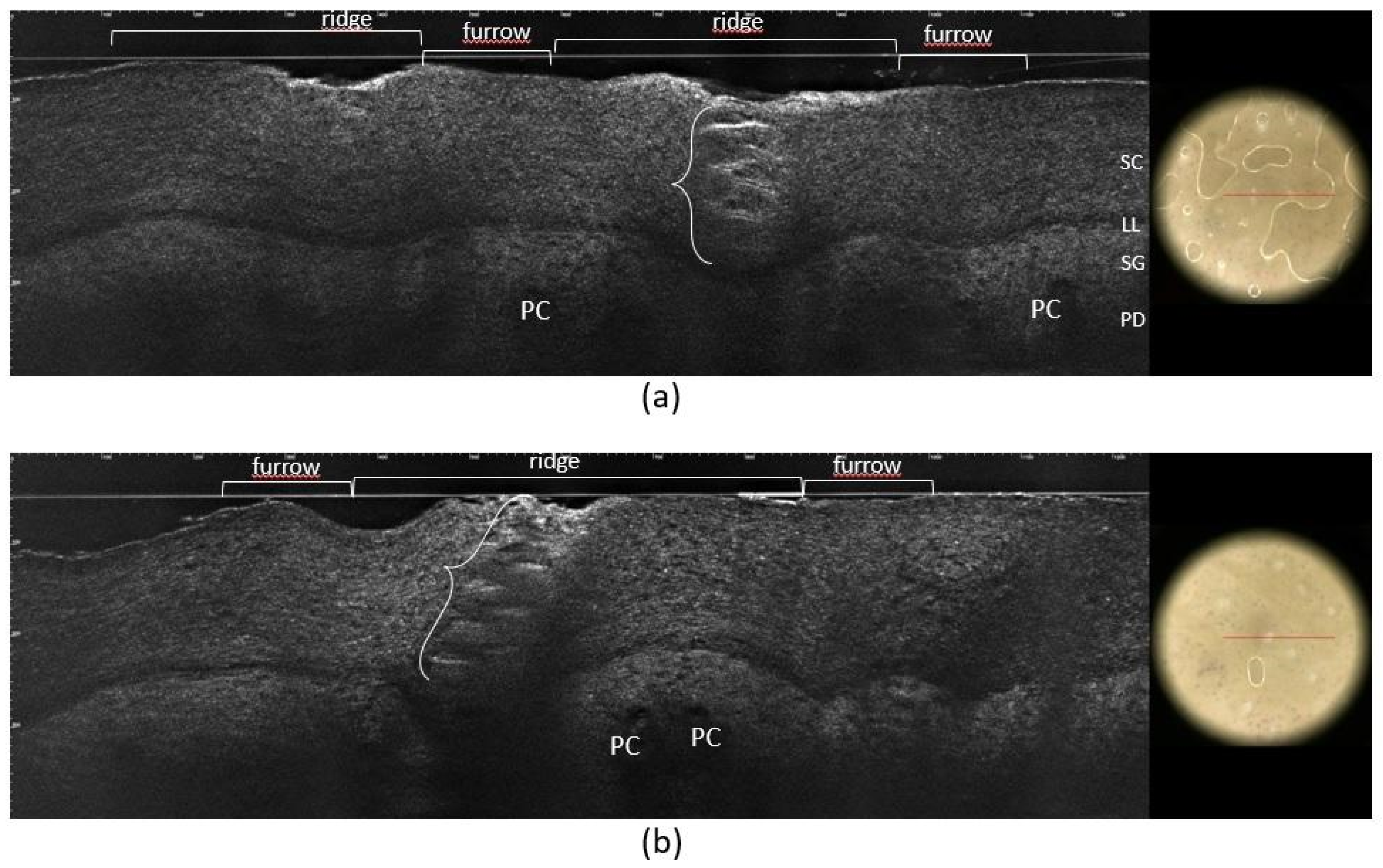
2. Case Report
3. Discussion
4. Conclusions
Author Contributions
Funding
Institutional Review Board Statement
Informed Consent Statement
Data Availability Statement
Conflicts of Interest
References
- Hutchinson, J. A peculiar form of serpiginous and infective naevoid disease. Arch. Surg. 1889, 1, 275. [Google Scholar]
- Radcliffe Crocker, H. Diseases of the Skin; Blakiston’s Press: Philadelphia, PA, USA, 1905; p. 646. [Google Scholar]
- Sammarco, E.; Ametrano, O. Angioma serpiginosum: Two cases in children and review of literature. Dermatol. Rep. 2022, 14, 9260. [Google Scholar] [CrossRef]
- Namazi, M.R.; Handjani, F. Angioma serpiginosum. Dermatol Online J. 2003, 9, 19. [Google Scholar] [CrossRef] [PubMed]
- Ghanadan, A.; Kamyab-Hesari, K. Dermoscopy of angioma serpiginosum: A case report. Int. J. Dermatol. 2014, 53, 1505–1507. [Google Scholar]
- Sandhu, K.; Gupta, S. Angioma serpiginosum: Report of two unusual cases. J. Eur. Acad. Dermatol. Venereol. 2005, 19, 127–128. [Google Scholar] [CrossRef] [PubMed]
- Ohnishi, T.; Nagayama, T.; Morita, T.; Miyazaki, T.; Okada, H.; Ohara, K.; Watanabe, S. Angioma serpiginosum: A report of 2 cases identified using epiluminescence microscopy. Arch. Dermatol. 1999, 135, 1366–1368. [Google Scholar] [CrossRef]
- Erkek, E.; Bozdogan, O.; Akarsu, C.; Atasoy, P.; Koçak, M. Absence of Estrogen and Progesterone Receptors Around the Affected Vessels of Angioma Serpiginosum. Am. J. Clin. Dermatol. 2006, 7, 383–386. [Google Scholar] [CrossRef] [PubMed]
- Neumann, E. Some new observations on the genesis of angioma serpiginosum. Acta Derm. Venereol. 1971, 51, 194–198. [Google Scholar] [PubMed]
- Katta, R.; Wagner, A. Angioma serpiginosum with extensive cutaneous involvement. J. Am. Acad. Dermatol. 2000, 42, 384–385. [Google Scholar] [CrossRef]
- Vather, D.; Harvey, G.; Agnew, K.; Purvis, D. Two cases of unusually early onset extensive angioma serpiginosum. Australas. J. Dermatol. 2022, 64, e91–e93. [Google Scholar] [CrossRef]
- Gupta, I.; Dayal, S.; Sahu, P.; Sen, R. Angioma Serpiginosum with Soft Tissue Hypertrophy and Palmar Involvement: A Rare Presentation. Indian J. Dermatol. 2023, 68, 354. [Google Scholar] [PubMed]
- Das, A.; Savant, S.S.; Kumar, P.; Hassan, S. Late-onset Segmental Angioma Serpiginosum. Indian J. Dermatol. 2016, 61, 226–227. [Google Scholar] [CrossRef] [PubMed]
- Tognetti, L.; Pianigiani, E.; Ierardi, F.; Cartocci, A.; Fiorani, D.; Quattro, M.; Caini, M.; Oranges, T.; Cinotti, E.; Cevenini, G.; et al. A new clinical and dermoscopic monitoring of infantile hemangiomas treated with oral propranolol. Dermatol. Ther. 2020, 33, e14283. [Google Scholar] [CrossRef] [PubMed]
- Tognetti, L.; Nunziata, L.; Cinotti, E.; Rubegni, P. Acroangiodermatitis with atypical presentation: Diagnostic imaging with high-frequency ultrasound, high-resolution dermoscopy and line-field optical coherence tomography. J. Eur. Acad. Dermatol. Venereol. 2023. [Google Scholar] [CrossRef] [PubMed]
- Tognetti, L.; Fimiani, M.; Rubegni, P. Benign dermoscopic parallel ridge pattern in plantar hyperpigmentation due to capecitabine. Dermatol. Pract. Concept. 2015, 5, 79–81. [Google Scholar] [CrossRef] [PubMed]
- Tognetti, L.; Carraro, A.; Lamberti, A.; Cinotti, E.; Suppa, M.; Perrot, J.L.; Rubegni, P. Kaposi sarcoma of the glans: New findings by line field confocal optical coherence tomography examination. Ski. Res. Technol. 2021, 27, 285–287. [Google Scholar] [CrossRef] [PubMed]
- Tognetti, L.; Cinotti, E.; Coriolani, G.; Suppa, M.; Perrot, J.L.; Vascotto, M.; Grosso, S.; Rubegni, P. Cutaneous lesions of Anderson-Fabry disease examined with a novel technique: Line-field confocal optical coherence tomography. J. Eur. Acad. Dermatol. Venereol. 2022, 36, E371–E373. [Google Scholar] [CrossRef] [PubMed]
- Tognetti, L.; Fiorani, D.; Suppa, M.; Cinotti, E.; Fontaine, M.; Marmol, V.; Rubegni, P.; Perrot, J. Examination of circumscribed palmar hypokeratosis with line-field confocal optical coherence tomography: Dermoscopic, ultrasonographic and histopathologic correlates. Indian J. Dermatol. Venereol. Leprol. 2020, 86, 206–208. [Google Scholar] [CrossRef] [PubMed]
- Freites-Martinez, A.; Moreno-Torres, A.; Núñez, A.H.; Martinez-Sanchez, D.; Huerta-Brogeras, M.; Borbujo, J. Angioma serpiginosum: Report of an unusual acral case and review of the literature. An. Bras. Dermatol. 2015, 90, 26–28. [Google Scholar] [CrossRef]
- Lo, Y.; Chen, Y. Acral angioma serpiginosum: Clinicopathologic and dermoscopic presentation. Australas. J. Dermatol. 2019, 60, E211–E213. [Google Scholar] [CrossRef]
- Bayramgurler, D.; Filinte, D.; Kiran, R. Angioma serpiginosum with sole involvement. Eur. J. Dermatol. 2008, 18, 708–709. [Google Scholar] [PubMed]
- Chen, J.-H.; Wang, K.-H.; Hu, C.-H.; Chiu, J.-S. Atypical Angioma Serpiginosum. Yonsei Med. J. 2008, 49, 509–513. [Google Scholar] [CrossRef] [PubMed]
- Diociaiuti, A.; Cutrone, M.; Rotunno, R.; De Vito, R.; Neri, I.; Pisaneschi, E.; El Hachem, M. Angioma serpiginosum: A case report and review of the literature. Ital. J. Pediatr. 2019, 45, 53. [Google Scholar] [CrossRef] [PubMed]
- Zaldivar Fujigaki, J.L.; Anjum, F. Schamberg Disease. In StatPearls [Internet]; StatPearls Publishing: Treasure Island, FL, USA, 2024. [Google Scholar]
- Sukanya, G.; Sane, R.R.; Ravi, V.N.; Nehete, S.S. Unilateral Nevoid Telangiectasia: An Overlooked Entity. Indian J. Dermatol. 2022, 67, 423–424. [Google Scholar] [PubMed]
- Villela-Segura, U. Dermoscopy as an Important Tool for Differentiating Unilateral Nevoid Telangiectasia and Angioma Serpiginosum. Dermatol. Pract. Concept. 2019, 9, 306–307. [Google Scholar] [CrossRef] [PubMed]
- Aranguren-López, I.; Vildósola-Esturo, S.; Arias-Camisón, I.; López-Pestaña, A. Circumscribed Plantar Hypokeratosis. Actas Dermo-Sifiliográficas 2019, 110, 619–621. [Google Scholar] [CrossRef] [PubMed]
- Nazzaro, G.; Ponziani, A.; Brena, M.; Cavicchini, S. Dermoscopy confirms diagnosis of circumscribed plantar hypokeratosis. J. Am. Acad. Dermatol. 2017, 76, S43–S45. [Google Scholar] [CrossRef]
- Chauvel-Picard, J.; Bérot, V.; Tognetti, L.; Cano, C.O.; Fontaine, M.; Lenoir, C.; Pérez-Anker, J.; Puig, S.; Dubois, A.; Forestier, S.; et al. Line-field confocal optical coherence tomography as a tool for three-dimensional in vivo quantification of healthy epidermis: A pilot study. J. Biophotonics 2021, 15, e202100236. [Google Scholar] [CrossRef]


| Year | Author ref | Gender | Age (Years) | Age of Onset | Acral Skin Involved |
|---|---|---|---|---|---|
| 2008 | Bayramgurler [22] | F | 16 | Early childhood | Whole left lower limb involving left sole |
| 2008 | Chen [23] | F | 48 | 43 | Left sole to toes |
| 2015 | Freites-Martinez [20] | F | 35 | 30 | Right toe and sole |
| 2019 | Diociaiuti [24] | M | 14 | From birth | Both the lower limbs including the soles |
| 2019 | Lo [21] | F | 51 | 40 | Left sole |
| 2024 | Our Case | F | 29 | 19 | Left sole |
Disclaimer/Publisher’s Note: The statements, opinions and data contained in all publications are solely those of the individual author(s) and contributor(s) and not of MDPI and/or the editor(s). MDPI and/or the editor(s) disclaim responsibility for any injury to people or property resulting from any ideas, methods, instructions or products referred to in the content. |
© 2024 by the authors. Licensee MDPI, Basel, Switzerland. This article is an open access article distributed under the terms and conditions of the Creative Commons Attribution (CC BY) license (https://creativecommons.org/licenses/by/4.0/).
Share and Cite
Tognetti, L.; La Marca, F.; Cinotti, E.; Rubegni, P. Non-Invasive Diagnosis of Angioma Serpiginosum Plantaris: High-Resolution Dermoscopy, High-Frequency Ultrasound, and Line-Field Confocal Optical Coherence Tomography. J. Vasc. Dis. 2024, 3, 152-160. https://doi.org/10.3390/jvd3020013
Tognetti L, La Marca F, Cinotti E, Rubegni P. Non-Invasive Diagnosis of Angioma Serpiginosum Plantaris: High-Resolution Dermoscopy, High-Frequency Ultrasound, and Line-Field Confocal Optical Coherence Tomography. Journal of Vascular Diseases. 2024; 3(2):152-160. https://doi.org/10.3390/jvd3020013
Chicago/Turabian StyleTognetti, Linda, Francesca La Marca, Elisa Cinotti, and Pietro Rubegni. 2024. "Non-Invasive Diagnosis of Angioma Serpiginosum Plantaris: High-Resolution Dermoscopy, High-Frequency Ultrasound, and Line-Field Confocal Optical Coherence Tomography" Journal of Vascular Diseases 3, no. 2: 152-160. https://doi.org/10.3390/jvd3020013
APA StyleTognetti, L., La Marca, F., Cinotti, E., & Rubegni, P. (2024). Non-Invasive Diagnosis of Angioma Serpiginosum Plantaris: High-Resolution Dermoscopy, High-Frequency Ultrasound, and Line-Field Confocal Optical Coherence Tomography. Journal of Vascular Diseases, 3(2), 152-160. https://doi.org/10.3390/jvd3020013