Article

Small Paracentral Acute Middle Maculopathy Lesions as Biomarker of Vascular Morbidity: Natural Course

by
Dmitrii S. Maltsev
*,
Alexei N. Kulikov
,
Maria A. Burnasheva
,
Yana A. Kalinicheva
and
Alexander S. Vasiliev
Department of Ophthalmology, Military Medical Academy, 21, Botkinskaya Str., St. Petersburg 194044, Russia
*
Author to whom correspondence should be addressed.
J. Vasc. Dis. 2024, 3(1), 67-76; https://doi.org/10.3390/jvd3010006
Submission received: 30 May 2023 / Revised: 22 July 2023 / Accepted: 4 January 2024 / Published: 1 February 2024
(This article belongs to the Section Peripheral Vascular Diseases)

Abstract

:
Background: To evaluate the incidence rate of small paracentral acute middle maculopathy (PAMM) lesions in healthy fellow eyes of patients with retinal vein occlusions (RVO). Methods: Patients with unilateral branch RVO or central RVO who were followed up for at least one year were included. Fellow healthy eyes were examined with 6 mm optical coherence tomography angiography scans at baseline and at the end of the follow-up. Small resolved PAMM lesions were displayed using structural en face projections of the slab between two segmentation lines of the outer plexiform layer with 0 µm and −9 µm offset. New lesions were identified by overlaying baseline and final structural en face projections. Results: A total of 41 eyes of 41 patients (12 females and 29 males, mean age 63.5 ± 10.1 years) were included. The mean follow-up was 23.6 ± 11.4 months. Among all RVO patients, small resolved PAMM lesions in healthy fellow eyes were found in 39 patients (95.1%). In the whole cohort, the median number of PAMM lesions was 5.5 (95% CI 3.0 to 8.0) (mean 6.5 ± 5.7) at baseline, which increased statistically significantly to 5.5 (95% CI 3.8 to 8.2) (mean 6.7 ± 5.7) at the end of the follow-up period (p = 0.03). Eight (19.5%) patients demonstrated new lesions, with a median number of new lesions of 1 (ranged 1 to 2). Conclusions: Small PAMM lesions seem to be a slowly progressing retinal vascular biomarker.

1. Introduction

The retina is an ideal subject for in vivo assessment of the status of microcirculation. A number of imaging modalities allow us to obtain high-definition images of retinal vessels from which we can extrapolate the status of the vascular system as a whole. This includes color photography of the eye fundus and fluorescein angiography, both of which have been used to display the retinal vascular network for decades. However, color fundus photography has limited resolution, and fluorescein angiography is an invasive procedure. Recently, optical coherence tomography angiography (OCTA) has been introduced in clinical practice, which has allowed for reproducible, noninvasive in vivo visualization of the retinal microcirculation at the level of individual capillaries [1,2]. As a result, more than a dozen OCTA indices and vascular biomarkers have been suggested as descriptive characteristics of retinal microcirculation, and have been shown to be related to ocular and systemic vascular pathology.
Paracentral acute middle maculopathy (PAMM) is a retinal vascular phenomenon discovered with OCTA, which is characterized by regional hypoperfusion of the deep capillary plexus followed by secondary postischemic loss of the inner nuclear layer [3,4]. Symptomatic PAMM has been associated with various pharmacological agents and ocular and systemic disorders. As has been recently identified, small lesions share similar morphological characteristics with typical PAMM lesions, but occur asymptomatically and are diagnosed almost exclusively in resolved form [5]. These small resolved PAMM lesions have been found close to the large retinal arteries and seem to be caused by hypoperfusion in individual small arterial branches [6]. Small PAMM lesions have been shown to be highly prevalent among patients with retinal vein occlusions (RVO) [5], arterial hypertension [7], diabetes, and different stages of diabetic retinopathy [8], as well as among patients with cardiovascular disorders and high risk of vascular events [9]. Despite extensive study, the natural course of these lesions is not known. Particularly, we do not know the rate of their occurrence, which is information that would be helpful in estimating the progression of comorbidities of the cardiovascular system. The aim of this study, therefore, was to evaluate the incidence rate of small PAMM lesions. Since, in a previous study, we observed a high prevalence of small resolved PAMM lesions in healthy fellow eyes of patients with RVO, these patients were chosen for the current study as a high-risk cohort for the incidence of new lesions.

2. Material and Methods

This study followed the ethical standards stated in the Declaration of Helsinki and was approved (Approval ID #207) by the Local Ethics Committee of the Military Medical Academy (St. Petersburg). All individuals signed written informed consent before participating in the study. The study included patients with unilateral branch retinal vein occlusion (BRVO) or central retinal vein occlusion (CRVO) who were followed up in our clinic for at least one year between February 2018 and February 2023.
Medical history, including RVO duration, presence of hypertension, and smoking status, was collected from each participant. Exclusion criteria were a history of any other known diseases of the posterior eye segment, glaucoma, diabetes mellitus, optical media opacities, quality of OCTA image Q7 or lower, and the presence of retinal vein occlusion in both eyes.
All patients received a comprehensive ophthalmic examination and OCTA with RTVue-XR Avanti (Optovue, Fremont, CA, USA). Before examination, all eyes were dilated with mydriatic eye drops. For the fellow healthy eye of each participant, a 6 mm OCTA scan was obtained. This scan was assembled from 2 orthogonal three-dimensional scans, each of 400 repeated B-scans consisting of 400 A-scans. All of the resulting scans were reviewed using AngioVue software (version 2017.1.0.150) by two experienced specialists (D.S.M. and Ya.A.K.). Structural en face projections were generated using two segmentation lines of the outer plexiform layer with 0 µm and −9 µm offsets (Figure 1).
As has been observed in previous studies, this slab displays all small resolved PAMM lesions as relatively sharply delineated black areas [5,10]. To confirm the presence of lesions, all cross-sectional scans were checked for the presence of inner nuclear layer thinning with outer plexiform layer elevation, which corresponds to the lesion viewed on en face projection.
En face projections were generated from the two visits with greatest time between them. These two projections were then uploaded to external software and overlayed using the large retinal vessels for alignment. Subsequently, the images were switched back and forth multiple times to register the appearance of any new lesions.
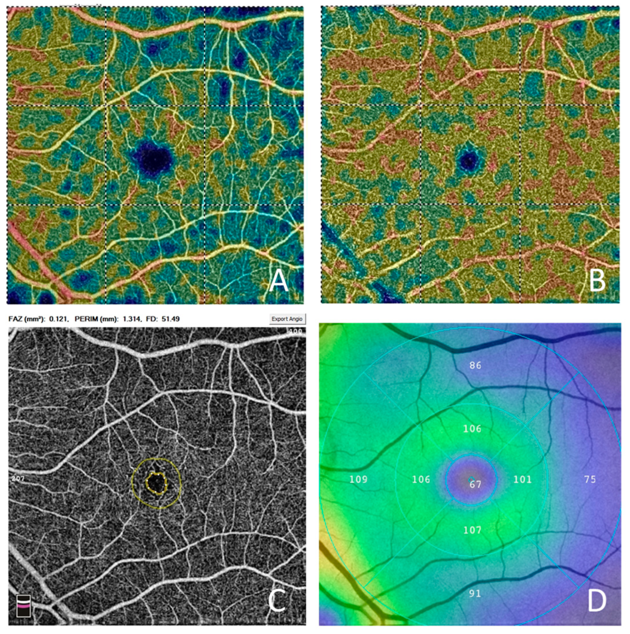
To establish whether the changes in PAMM lesions were associated with longitudinal remodeling of retinal microcirculation, we also inspected other OCTA parameters, including the vessel density of the superficial capillary plexus (SCP), the vessel density of the deep capillary plexus (DCP), and the foveal avascular zone (FAZ) area. All parameters were taken from the same 6 mm OCTA scan used to evaluate PAMM lesions. All values were automatically generated by Angiovue software. The vessel density value was defined as the vessel density of the whole OCTA image in each plexus. The FAZ area was defined as an area delineated by the terminal capillaries around the center of the fovea displayed in the full retina slab (Figure 2).
Additionally, we evaluated the link between PAMM lesions and the changes in ganglion cell complex (GCC) thickness. The GCC thickness was defined as the distance between the inner retinal surface and the border between the inner plexiform and inner nuclear layers. It was calculated automatically using Angiovue software as the mean inner retinal thickness of the 6 mm circle, excluding the foveal zone.
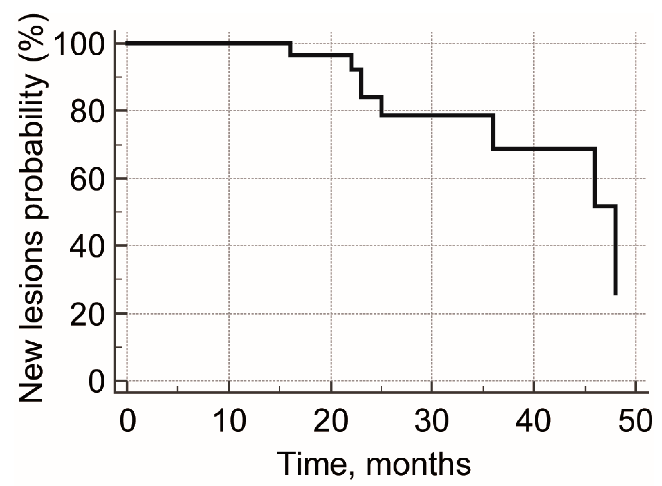
Statistical analysis was performed using MedCalc 18.4.1 (MedCalc Software, Ostend, Belgium). All data are presented as the mean ± standard deviation or as the median and 95% confidence interval (CI). The Wilcoxon test was used to compare changes in the number of PAMM lesions at the beginning and end of the follow-up. Repeated measure analysis of variance (ANOVA) was used to compare the vessel density of SCP and DCP, the FAZ area, and the inner retinal thickness at the beginning and end of the follow-up. A Kaplan–Meier curve was constructed to assess the rate of appearance of new small resolved PAMM lesions. A p-value of 0.05 or less was considered to be statistically significant.

3. Results

A total of 41 eyes of 41 patients (12 females and 29 males with a mean age at baseline of 63.5 ± 10.1 years) were included. All except four patients had arterial hypertension (90.2%). The mean follow-up was 23.6 ± 11.4 months (ranged from 12 to 48 months). The mean age at the end of the follow-up was 65.1 ± 10.2 years. Twenty-five patients had BRVO (8 females and 17 males, mean age 62.4 ± 10.9), and 16 patients had CRVO (4 females and 12 males, mean age 65.1 ± 8.8 years).
Among all RVO patients, small resolved PAMM lesions in healthy fellow eyes were found in 39 patients (95.1%), while no acute small PAMM lesions were observed. In BRVO and CRVO patients, small resolved PAMM lesions in healthy fellow eyes were found in 24 (96.0%) and 15 (93.8%) patients, respectively (p > 0.05) (Table 1).
Over the whole cohort, the median number of small resolved PAMM lesions in healthy fellow eyes was 5.5 (95% CI 3.0 to 8.0) (mean 6.5 ± 5.7) at baseline, which increased statistically significantly to a median value of 5.5 (95% CI 3.8 to 8.2) (mean 6.7 ± 5.7) at the end of the follow-up (p = 0.03). Among patients who had new lesions (eight patients, six males, and two females (19.5%)), five patients had BRVO (62.5%) and three patients had CRVO (37.5%). The median age in this group was 67.5 years (ranged from 48 to 78 years), and the median follow-up was 24 months (ranged from 16 to 48 years). The median number of small resolved PAMM lesions at baseline was 3.5 (ranged from 0 to 22), and the median number of new lesions was one (ranged from one to two) (Figure 3). The estimated incidence rate of small resolved PAMM lesions was calculated to be as high as one lesion per ten patient-years (Figure 4).
In the subgroup of patients without changes in the number of PAMM lesions, the vessel densities of SCP at the beginning and end of the follow-up were 48.1 ± 5.0% and 47.5 ± 5.3% (p = 0.22); the vessel densities of DCP at the beginning and end of the follow-up were 50.1 ± 6.9% and 49.8 ± 7.4% (p = 0.76); the FAZ areas at the beginning and end of the follow-up were 0.25 ± 0.09 mm2 and 0.26 ± 0.09 mm2 (p = 0.23); and the GCC thicknesses at the beginning and end of the follow-up were 96.9 ± 9.8 µm and 95.8 ± 11.4 µm (p = 0.05).
Among patients who demonstrated new PAMM lesions, the vessel densities of SCP at the beginning and end of the follow-up were 47.1 ± 3.9% and 47.0 ± 3.2% (p = 0.94); the vessel densities of DCP at the beginning and end of the follow-up were 47.8 ± 7.0% and 50.6 ± 7.0% (p = 0.39); the FAZ areas at the beginning and end of the follow-up were 0.23 ± 0.08 mm2 and 0.24 ± 0.08 mm2 (p = 0.26); and the GCC thicknesses at the beginning and end of the follow-up were 98.5 ± 3.4 µm and 97.5 ± 4.1 µm (p = 0.15).
No statistically significant differences were found in the changes in OCTA parameters between subgroups of patients with and without increases in the number of PAMM lesions (p > 0.05).

4. Discussion

PAMM was initially described by Sarraf in 2013 as a subtype of acute macular neuroretinopathy with a specific level of retinal ischemia focused on the inner nuclear layer. These lesions are found in a diverse cohort of patients and were defined as hyperreflective bands involving the inner nuclear layer on structural OCT. As a form of acute macular neuroretinopathy, these lesions cause relative paracentral scotoma and either no or minimal changes on fluorescein angiography [11]. Later, the introduction of OCTA shed light on the pathophysiology of PAMM, implying the involvement of deep capillary plexus hypoperfusion in the acute phase of PAMM [3]. This explained the selective damage to the inner nuclear layer (INL) in the acute phase of PAMM and the thinning of this layer in the chronic phase. Associated conditions potentially leading to retinal vascular injury and paracentral acute middle maculopathy include eye compression injury causing global ocular ischemia, sickle cell disease, Purtscher’s retinopathy, inflammatory occlusive retinal vasculitis, post-H1N1 vaccine, hypertensive retinopathy, migraine, and upper respiratory infection [12]. Today, PAMM is considered, therefore, not as a subtype of acute macular neuroretinopathy, but as a distinct form of retinal ischemia [13].
The pathophysiology of PAMM includes increased extraction of oxygen from retinal arterial vessels due to the decrease in blood flow velocity and low oxygen saturation at the venular pole of retinal circulation [14]. The lesion, in such cases, belongs to a region supplied by a distinct retinal vessel and varies in size from a quarter of the optic disc to lesions covering more than a half of the macula. A significant proportion of symptomatic PAMM lesions are associated with the cilioretinal artery [15], which agrees with deteriorated choriocapillaris perfusion in both eyes of patients with unilateral PAMM [16]. Although the nature of PAMM as an arterial or venous lesion was previously unclear, we now have enough data indicating the arterial nature of symptomatic PAMM. Initial reports describing PAMM with OCTA indicated a decrease in the vessel density of the deep capillary plexus [3]. A subsequent evaluation of OCTA data not only showed damage to the deep capillary plexus, but also indicated a decrease in vessel density in the superficial capillary plexus [4]. This cannot be caused by PAMM itself, since SCP does not depend on deep retinal circulation. Rather, SCP represents simultaneous changes in the retinal microvasculature due to a common underlying reason such as hypertension. Today, PAMM may, therefore, be considered not as a distinct disease, but as a specific biomarker for vascular disorders.
Symptomatic PAMM lesions vary in terms of severity of ischemia and their localization to distinct retinal vessels, including arteriolar, fern-like, and globular patterns [17]. The fern-like pattern follows the small arterial vessels of a macular branch, or even the arterial vessels of a whole macular area. Arteriolar lesions seem to have deeper hypoperfusion and may be considered as the next step in the evolution of PAMM, following on from the fern-like pattern [18,19]. Globular pattern seems to be the most severe, since it reflects global hypoperfusion of the retinal arteriolar microcirculation such as that seen in microembolic events. All of this suggests that PAMM patterns result from different underlying mechanisms. Specifically, the arteriolar pattern results from transient occlusion of a large retinal arteriolar branch with complete restoration of blood flow, while the fern-like pattern represents elevated intraluminal pressure in the central retinal vein, which transmits to the venular and far arteriolar pole of retinal microcirculation [17].
Over years of accurate reporting of PAMM cases, the spectrum of potential causative factors has expanded to several dozen systemic disorders, external factors, and drugs. This includes sickle cell disease [20,21]; giant-cell arteritis [22,23]; embolic events, including those after filler injections [24,25]; and different ocular procedures, such as vitrectomy [26]. Other sporadic factors include migraine [27], viral infections [28], and COVID-19 [29]. However, in young patients, symptomatic PAMM often has no established etiology. The diversity of causative factors indicates that PAMM is a universal retinal vascular condition rather than a distinct retinal disease, and can be used as a biomarker for vascular morbidity or as a biomarker in retinal vascular disorders [30]. This suggestion received further confirmation after small resolved PAMM lesions were described.
The first mention of these lesions was made in relation to Behcet’s disease, and was described as a local INL thinning and outer plexiform layer (OPL) elevation, but without explanation of their nature [31]. Today, taking into account arterial occlusions in Behcet’s disease, the appearance of PAMM lesions in this condition is hardly surprising.
Due to their size and asymptomatic character, the detection of small PAMM lesions is more complex than typical PAMM lesions and requires a thorough review of all cross-sectional scans of the macular volume pattern by an experienced specialist. Moreover, the detection of these lesions could be inaccurate without an analysis of en face projections. In a number of papers, a thin slab between two OPL lines with 0 µm and −9 µm offsets was shown to be effective in displaying small resolved PAMM lesions as dark perivascular spots [5]. Moreover, the latter is the only comprehensive approach for the quantitative analysis of these small lesions, and it was used in the current study.
An analysis of the fellow eyes of unilateral RVO patients revealed a high prevalence of those lesions and their perivascular distribution [5]. Neither vessel density nor severity of venous occlusion were found to be associated with the presence of PAMM lesions among RVO eyes. No difference in the prevalence of the lesions was found between CRVO and BRVO. These data were obtained retrospectively and, although the odds ratio for the presence of small resolved PAMM lesions in unilateral RVO was high, the risk of RVO in eyes with small resolved PAMM lesions is not known. These lesions were also found to be strongly associated with hypertension and high risk of cardiovascular events, such as stroke [9,30]. Despite their high prevalence among patients with cardiovascular morbidity, cross-sectional studies have reported small PAMM lesions in healthy persons, a result which seems to be dependent on the age of the study cohort and may be as high as 19.3% [5]. Although this may suggest that some of the healthy participants had undiagnosed cardiovascular disorders, at the same time, it agrees with the idiopathic characteristic of some typical PAMM lesions.
Taking into account the perivascular distribution of the lesions, the name “retinal ischemic perivascular lesions” (RIPL) has been suggested for this phenomenon [9]. However, this name does not reflect the nature of the lesions and does not account for those lesions which seemingly have no relation to large retinal vessels. In 2019, Spaide also described this type of lesion, in an otherwise healthy person, as micro-(diminutive) PAMM. That patient demonstrated a small positive scotoma with no deterioration of visual acuity. No substantial changes were observed with ophthalmoscopy or fluorescein angiography. In the acute stage, this diminutive lesion showed a typical hyperreflective band at the level of INL, but on a limited area of 175 µm. After 4 weeks, symptoms reduced and completely disappeared [32].
The detailed microanatomy of small resolved PAMM lesions, explored with OCTA, revealed their association with small branches of relatively large retinal arteries. These vessels branch from the main trunk at a right angle and supply a small paravascular region of the retina. It has been suggested that the Venturi effect is involved in the hypoperfusion of these small vessels due to the high difference in vessel diameter, high blood flow velocity in the main trunk, and the branching at 90°. All of these factors could theoretically lead to a slowing of blood flow in small arterial branches, and this effect would only increase with higher arterial pressure and blood flow velocity. Another potential mechanism may include constriction of the main trunk, which could reduce blood flow in these small arterial branches [6]. Although embolic nature is quite typical for symptomatic PAMM, it does not stand up to criticism when small resolved PAMM lesions are observed in a high proportion of healthy young persons or patients with mild hypertension without other comorbidities.
Despite intense study, there is almost no information on the formation of these small lesions. Only two papers have reported these small lesions in their acute phase [5,32]. It is, therefore, not known whether these lesions occur evenly over time or are associated with some systemic acute events such as hypertensive crisis or stroke. The rate of occurrence of these lesions is also unclear.
In this paper, we have evaluated a group of patients with retinal vein occlusions and cardiovascular morbidity where the prevalence of small resolved PAMM lesions was high. We found that, over a mean period of two years, no more than 20% of patients exhibited new small PAMM lesions. And even in those eyes, the number of new lesions was very low, around one new lesion, varying from one to two. All of this indicates that the process of the formation of new small PAMM lesions is quite slow, possibly evenly distributed over time. This may, therefore, represent the effects of systemic risk factors like hypertension instead of direct association with some single-time events. In this study, we therefore obtained further validation of this phenomenon for its use as a biomarker of cardiovascular morbidity and possibly for the assessment of its long-term compensation.
Interestingly, when we were evaluating OCTA parameters, including the vessel density of SCP and DCP and the FAZ area, which are well-known biomarkers of vascular morbidity, we found no changes in these values in either subgroup with or without new PAMM lesions. This fact supports the suggestion that small resolved PAMM lesions represent a sensitive and independent biomarker for vascular morbidity. This also highlights the difficulties of the conventional OCTA metric which, although highly reproducible, can hardly be used for the evaluation of subtle changes in an individual patient [33,34]. In this study, we also evaluated changes in GCC complex in patients with and without progression of PAMM lesions. Since open-angle glaucoma is a risk factor for RVO, and since these two conditions share common systemic risk factors, changes in the inner retina could be expected in studied eyes even after exclusion of glaucoma patients [35]. In our study, we found no association between GCC changes and the appearance of new PAMM lesions.
This study has some limitations. Firstly, the study was retrospective and included a relatively small number of patients. Also, the results of the study cannot be extrapolated to other cohorts, such as diabetic patients or patients with severe cardiovascular disorders, who may have different occurrence rates of small PAMM lesions. However, this is, to the best of our knowledge, the first study assessing the natural course of these lesions.

5. Conclusions

In conclusion, in this study, we have evaluated the natural course of small PAMM lesions, a biomarker of vascular morbidity, in the healthy fellow eyes of RVO patients. Over a mean two-year period, these lesions slowly grew in number by at least one new lesion in 20% of patients. The estimated incidence rate of these lesions was one new lesion per 10 patient years. The absence of notable corresponding changes in the main quantitative OCTA parameters, including the vessel density of SCP and DCP and the FAZ area, indicates that small resolved PAMM lesions may better reflect the natural course of retinal microvasculature alteration under the action of systemic risk factors.

Author Contributions

Conceptualization, D.S.M. and M.A.B.; methodology, M.A.B. and Y.A.K.; software, Y.A.K.; validation, A.N.K., D.S.M. and M.A.B.; formal analysis, Y.A.K.; investigation, M.A.B. and A.S.V.; resources, A.N.K.; data curation, D.S.M.; writing—original draft preparation, D.S.M.; writing—review and editing, D.S.M. and A.N.K.; visualization, A.S.V. and Y.A.K.; supervision, A.N.K.; project administration, D.S.M. All authors have read and agreed to the published version of the manuscript.

Funding

This research received no external funding.

Institutional Review Board Statement

The study was conducted in accordance with the Declaration of Helsinki and approved by the Local Ethics Committee of the Military Medical Academy (Approval ID: #207 from 18 February 2020).

Informed Consent Statement

Informed consent was obtained from all subjects involved in the study.

Data Availability Statement

The primary data are available from Dmitrii S. Maltsev via email [email protected].

Conflicts of Interest

The authors declare no conflicts of interest.

Abbreviations

BRVO, branch retinal vein occlusion; CRVO, central retinal vein occlusion; confidence interval; DCP, deep capillary plexus; FAZ, foveal avascular zone; INL, inner nuclear layer; OCTA, optical coherence tomography; OPL, outer plexiform layer; PAMM, paracentral acute middle maculopathy; RVO, retinal vein occlusion; SCP, superficial capillary plexus.

References

  1. Jia, Y.; Wei, E.; Wang, X.; Zhang, X.; Morrison, J.C.; Parikh, M.; Lombardi, L.H.; Gattey, D.M.; Armour, R.L.; Edmunds, B.; et al. Optical Coherence Tomography Angiography of Optic Disc Perfusion in Glaucoma. Ophthalmology 2014, 121, 1322–1332. [Google Scholar] [CrossRef] [PubMed]
  2. Jia, Y.; Tan, O.; Tokayer, J.; Potsaid, B.; Wang, Y.; Liu, J.J.; Kraus, M.F.; Subhash, H.; Fujimoto, J.G.; Hornegger, J.; et al. Split-spectrum amplitude-decorrelation angiography with optical coherence tomography. Opt. Express 2012, 20, 4710–4725. [Google Scholar] [CrossRef] [PubMed]
  3. Nemiroff, J.; Kuehlewein, L.; Rahimy, E.; Tsui, I.; Doshi, R.; Gaudric, A.; Gorin, M.B.; Sadda, S.; Sarraf, D. Assessing Deep Retinal Capillary Ischemia in Paracentral Acute Middle Maculopathy by Optical Coherence Tomography Angiography. Am. J. Ophthalmol. 2016, 162, 121–132.e1. [Google Scholar] [CrossRef]
  4. Kulikov, A.N.; Maltsev, D.S.; Leongardt, T.A. Retinal Microvasculature Alteration in Paracentral Acute Middle Maculopathy and Acute Macular Neuroretinopathy. Retin. Cases Brief Rep. 2018; ahead of print. [Google Scholar] [CrossRef]
  5. Maltsev, D.S.; Kulikov, A.N.; Burnasheva, M.A.; Chhablani, J. Prevalence of resolved paracentral acute middle maculopathy lesions in fellow eyes of patients with unilateral retinal vein occlusion. Acta Ophthalmol. 2019, 98, e22–e28. [Google Scholar] [CrossRef] [PubMed]
  6. Maltsev, D.S.; Kulikov, A.N.; Burnasheva, M.A.; Freund, K.B. Vascular Microanatomy of Small Resolved Paracentral Acute Middle Maculopathy Lesions. Ophthalmol. Retin. 2020, 5, 928–934. [Google Scholar] [CrossRef] [PubMed]
  7. Burnasheva, M.A.; Maltsev, D.S.; Kulikov, A.N.; Sherbakova, K.A.; Barsukov, A.V. Association of Chronic Paracentral Acute Middle Maculopathy Lesions with Hypertension. Ophthalmol. Retin. 2020, 4, 504–509. [Google Scholar] [CrossRef]
  8. Maltsev, D.S.; Kulikov, A.N.; Burnasheva, M. Association of Resolved Paracentral Acute Middle Maculopathy Lesions with Diabetic Retinopathy. J. Curr. Ophthalmol. 2022, 34, 318–322. [Google Scholar] [CrossRef]
  9. Long, C.P.; Chan, A.X.; Bakhoum, C.Y.; Toomey, C.B.; Madala, S.; Garg, A.K.; Freeman, W.R.; Goldbaum, M.H.; DeMaria, A.N.; Bakhoum, M.F. Prevalence of subclinical retinal ischemia in patients with cardiovascular disease—A hypothesis driven study. EClinicalMedicine 2021, 33, 100775. [Google Scholar] [CrossRef]
  10. Maltsev, D.S.; Kulikov, A.N.; Kazak, A.A.; Burnasheva, M.A. Status of Choriocapillaris in Fellow Eyes of Patients with Unilateral Retinal Vein Occlusions. Ophthalmic Surg. Lasers Imaging Retin. 2021, 52, 23–28. [Google Scholar] [CrossRef]
  11. Sarraf, D.; Rahimy, E.; Fawzi, A.A.; Sohn, E.; Barbazetto, I.; Zacks, D.N.; Mittra, R.A.; Klancnik, J.M.; Mrejen, S.; Goldberg, N.R.; et al. Paracentral Acute Middle Maculopathy: A New Variant of Acute Macular Neuroretinopathy Associated with Retinal Capillary Ischemia. JAMA Ophthalmol. 2013, 131, 1275–1287. [Google Scholar] [CrossRef]
  12. Chen, X.; Rahimy, E.; Sergott, R.C.; Nunes, R.P.; Souza, E.C.; Choudhry, N.; Cutler, N.E.; Houston, S.K.; Munk, M.R.; Fawzi, A.A.; et al. Spectrum of Retinal Vascular Diseases Associated with Paracentral Acute Middle Maculopathy. Am. J. Ophthalmol. 2015, 160, 26–34.e1. [Google Scholar] [CrossRef] [PubMed]
  13. Dansingani, K.K.; Freund, K.B. Paracentral Acute Middle Maculopathy and Acute Macular Neuroretinopathy: Related and Distinct Entities. Am. J. Ophthalmol. 2015, 160, 1–3.e2. [Google Scholar] [CrossRef] [PubMed]
  14. Bakhoum, M.F.; Freund, K.B.; Dolz-Marco, R.; Leong, B.C.; Baumal, C.R.; Duker, J.S.; Sarraf, D. Paracentral Acute Middle Maculopathy and the Ischemic Cascade Associated with Retinal Vascular Occlusion. Arch. Ophthalmol. 2018, 195, 143–153. [Google Scholar] [CrossRef] [PubMed]
  15. Pichi, F.; Fragiotta, S.; Freund, K.B.; Au, A.; Lembo, A.; Nucci, P.; Sebastiani, S.; Hernandez, J.C.G.; Interlandi, E.; Pellegrini, F.; et al. Cilioretinal artery hypoperfusion and its association with paracentral acute middle maculopathy. Br. J. Ophthalmol. 2019, 103, 1137–1145. [Google Scholar] [CrossRef] [PubMed]
  16. Maltsev, D.S.; Kulikov, A.N.; Burnasheva, M.A. Choriocapillaris alteration in patients with paracentral acute middle maculopathy. Eur. J. Ophthalmol. 2022, 32, 3622–3628. [Google Scholar] [CrossRef]
  17. Sridhar, J.; Shahlaee, A.; Rahimy, E.; Hong, B.K.; Khan, M.A.; Maguire, J.I.; Dunn, J.P.; Mehta, S.; Ho, A.C. Optical Coherence Tomography Angiography and En Face Optical Coherence Tomography Features of Paracentral Acute Middle Maculopathy. Arch. Ophthalmol. 2015, 160, 1259–1268.e2. [Google Scholar] [CrossRef] [PubMed]
  18. Falavarjani, K.G.; Phasukkijwatana, N.; Freund, K.B.; Cunningham, E.T.; Kalevar, A.; McDonald, H.R.; Dolz-Marco, R.; Roberts, P.K.; Tsui, I.; Rosen, R.; et al. En Face Optical Coherence Tomography Analysis to Assess the Spectrum of Perivenular Ischemia and Paracentral Acute Middle Maculopathy in Retinal Vein Occlusion. Am. J. Ophthalmol. 2017, 177, 131–138. [Google Scholar] [CrossRef]
  19. Zhang, Z.; Jiang, Y.; Huang, X.; Wu, Z.; Ke, B. Clinical Characteristics of Paracentral Acute Middle Maculopathy in Eyes with Retinal Vascular Occlusion Diseases in Chinese Patients. J. Ophthalmol. 2021, 2021, 8867570. [Google Scholar] [CrossRef]
  20. Hussnain, S.A.; Coady, P.A.; Stoessel, K.M. Paracentral acute middle maculopathy: Precursor to macular thinning in sickle cell retinopathy. BMJ Case Rep. 2017, 2017, bcr2016216124. [Google Scholar] [CrossRef]
  21. Ong, S.S.; Ahmed, I.; Scott, A.W. Association of Acute Macular Neuroretinopathy or Paracentral Acute Middle Maculopathy with Sickle Cell Disease. Ophthalmol. Retin. 2021, 5, 1146–1155. [Google Scholar] [CrossRef]
  22. Ahuja, A.S.; El-Dairi, M.A.; Hadziahmetovic, M.; Gospe, S.M. Paracentral Acute Middle Maculopathy as a Manifestation of Giant Cell Arteritis. J. Neuro-Ophthalmol. 2021, 41, e153–e156. [Google Scholar] [CrossRef] [PubMed]
  23. Broyles, H.; Chacko, J.; Chancellor, J.; LoRusso, F.; Phillips, P.H.; Mashayekhi, A.; Uwaydat, S. Paracentral Acute Middle Maculopathy as the Initial Presentation of Giant Cell Arteritis. J. Neuro-Ophthalmol. 2021, 41, e157–e159. [Google Scholar] [CrossRef] [PubMed]
  24. Echegaray, J.J.; Echegaray, P.M.; Townsend-Pico, W. Penile Filler Injection Leading to Silicone Embolism Syndrome and Paracentral Acute Middle Maculopathy. Ophthalmic Surg. Lasers Imaging Retin. 2022, 53, 635–638. [Google Scholar] [CrossRef] [PubMed]
  25. de Oliveira, B.M.R.; de Souza, L.M.; Andrade, G.C.; Lobos, C.Z.M.; de Souza, E.C.; de Moraes, N.S.B. Purtscher-like retinopathy and paracentral acute middle maculopathy caused by industrial silicone embolism. Retin. Cases Brief Rep. 2022, 16, 372–374. [Google Scholar] [CrossRef] [PubMed]
  26. Nakashima, H.; Iwama, Y.; Tanioka, K.; Emi, K. Paracentral Acute Middle Maculopathy following Vitrectomy for Proliferative Diabetic Retinopathy. Ophthalmology 2018, 125, 1929–1936. [Google Scholar] [CrossRef] [PubMed]
  27. Dasari, V.R.; Selliyan, A.; Gratton, S.M. Paracentral acute middle maculopathy in a patient with frequent migraine with aura: A case report. Retin. Cases Brief Rep. 2022, 16, 550–552. [Google Scholar] [CrossRef] [PubMed]
  28. Shahlaee, A.; Sridhar, J.; Rahimy, E.; Shieh, W.S.; Ho, A.C. Paracentral acute middle maculopathy associated with postviral purtscher-like retinopathy. Retin. Cases Brief Rep. 2019, 13, 50–53. [Google Scholar] [CrossRef]
  29. Çebi, A.Y.; Kılıçarslan, O.; Uçar, D. Coincident Acute Macular Neuroretinopathy and Paracentral Acute Middle Maculopathy in COVID-19. Turk. J. Ophthalmol. 2023, 53, 120–123. [Google Scholar] [CrossRef]
  30. Madala, S.; Adabifirouzjaei, F.; Lando, L.; Yarmohammadi, A.; Long, C.P.; Bakhoum, C.Y.; Goldbaum, M.H.; Sarraf, D.; DeMaria, A.N.; Bakhoum, M.F. Retinal Ischemic Perivascular Lesions, a Biomarker of Cardiovascular Disease. Ophthalmol. Retin. 2022, 6, 865–867. [Google Scholar] [CrossRef]
  31. Kido, A.; Uji, A.; Morooka, S.; Kuroda, Y.; Arichika, S.; Akagi, T.; Tsujikawa, A. Outer Plexiform Layer Elevations as a Marker for Prior Ocular Attacks in Patients with Behcet’s Disease. Investig. Opthalmol. Vis. Sci. 2018, 59, 2828–2832. [Google Scholar] [CrossRef]
  32. Spaide, R.F. Diminutive paracentral acute middle maculopathy lesion. Retin. Cases Brief Rep. 2021, 15, 643–646. [Google Scholar] [CrossRef] [PubMed]
  33. Mukkamala, L.; Nguyen, M.; Chang, M.; Park, S.S. Repeatability of Vascular Density Measurement of the Three Retinal Plexus Layers Using OCT Angiography in Pathologic Eyes (OCTA Vascular Density Repeatability of Three Plexus Layers). Clin. Ophthalmol. 2021, 15, 93–103. [Google Scholar] [CrossRef] [PubMed]
  34. You, Q.; Freeman, W.R.; Weinreb, R.N.; Zangwill, L.; Manalastas, P.I.C.; Saunders, L.J.; Nudleman, E. Reproducibility of vessel density measurement with optical coherence tomography angiography in eyes with and without retinopathy. Retina 2017, 37, 1475–1482. [Google Scholar] [CrossRef] [PubMed]
  35. Yin, X.; Li, J.; Zhang, B.; Lu, P. Association of glaucoma with risk of retinal vein occlusion: A meta-analysis. Acta Ophthalmol. 2019, 97, 652–659. [Google Scholar] [CrossRef]
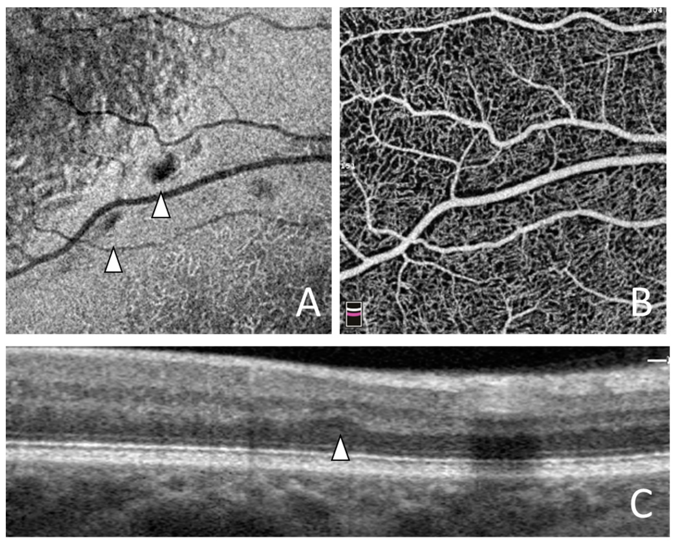
Figure 1. Optical coherence tomography angiography imaging of small resolved paracentral acute middle maculopathy lesions with 3 mm scan pattern. (A) Structural en face projection shows small resolved paracentral acute middle maculopathy lesions (arrowheads). (B) Corresponding optical coherence tomography angiography projection of superficial capillary plexus shows no changes in retinal microvasculature. (C) Cross-sectional scan shows small resolved paracentral acute middle maculopathy lesion (arrowhead).
Jvd 03 00006 g001
Figure 2. Optical coherence tomography angiography parameters assessed in the study. (A) Heat-map displaying vessel density of superficial capillary plexus (50.8%). (B) Heat-map displaying vessel density of deep capillary plexus (59.2%). (C) Superficial capillary plexus slab displaying foveal avascular zone area (0.121 mm2). (D) Heat-map displaying inner retinal thickness (97.6 µm).
Jvd 03 00006 g002
Figure 3. Natural course of small resolved paracentral acute middle maculopathy lesions observed with a 6 mm optical coherence tomography angiography scan pattern. (A) Structural en face projection shows small resolved paracentral acute middle maculopathy lesions (arrows) at baseline. (B) Structural en face projection shows previously existing and new (arrowheads) small resolved paracentral acute middle maculopathy lesions after 26 months of follow-up. (C) Structural en face projection of outer retina slab shows position of cross-sectional scan (green line) in (E). (D) Structural en face projection of outer retina slab shows position of cross-sectional scan (green line) in (F). (E) Cross-sectional scan shows no small resolved paracentral acute middle maculopathy lesions at baseline. (F) Cross-sectional scan shows new small resolved paracentral acute middle maculopathy lesions (arrowhead) after 26 months of follow-up.
Jvd 03 00006 g003
Figure 4. Kaplan–Meier curve showing cumulative probability of occurrence of new small resolved paracentral acute middle maculopathy lesions in healthy fellow eyes of patients with unilateral retinal vein occlusions.
Jvd 03 00006 g004
Table 1. Characteristics of the study cohort.
RVO (n = 41)BRVO (n = 25)CRVO (n = 16)
Age (y)63.46 ± 10.1362.44 ± 10.9565.06 ± 8.77
Males/Females29/1217/812/4
PAMM lesions at baseline6.5 ± 5.75.4 ± 4.78.3 ± 6.8
Disclaimer/Publisher’s Note: The statements, opinions and data contained in all publications are solely those of the individual author(s) and contributor(s) and not of MDPI and/or the editor(s). MDPI and/or the editor(s) disclaim responsibility for any injury to people or property resulting from any ideas, methods, instructions or products referred to in the content.