Article

The Effects of Psilocybin on Lipopolysaccharide-Induced Inflammation in THP-1 Human Macrophages

Department of Biological Sciences, University of Lethbridge, Lethbridge, AB T1K 3M4, Canada
*
Authors to whom correspondence should be addressed.
Psychoactives 2024, 3(1), 48-64; https://doi.org/10.3390/psychoactives3010004
Submission received: 12 December 2023 / Revised: 22 January 2024 / Accepted: 26 January 2024 / Published: 28 January 2024

Abstract

:
Psilocybin, an innate compound produced by mushrooms belonging to the Psilocybe genus, is primarily known for its agonistic effects on the serotonin 5-HT2A receptor. This receptor’s functioning is involved in many neurological processes. In the context of this research, our primary aim was to comprehensively investigate the influence of psilocybin as a serotonin receptor agonist on the intricate cascade of events involved in THP-1 macrophages stimulated by lipopolysaccharide (LPS). THP-1 monocyte cells were subjected to differentiation into macrophages through a controlled incubation with phorbol 12-myristate 13-acetate (PMA). The next step involved the induction of an inflammatory response by exposing THP-1 macrophages to 500 ng/mL LPS for 4 h. Subsequently, we triggered the activation of the second phase of the NLRP3 inflammasome by introducing adenosine triphosphate (ATP) immediately following LPS stimulation. Our findings have revealed a dose-dependent inverse correlation between psilocybin exposure and the production of LPS-induced proinflammatory cytokines and proteins. Our work indicates that psilocybin likely mediates these responses by influencing key signaling pathways, including NF-κB, IL-6/TYK2/STAT3, and TYK2/STAT1.

1. Introduction

Inflammation represents a physiological reaction orchestrated by the immune system and can be activated by a diverse array of stimuli, involving pathogens, compromised cells, and harmful substances. These triggers have the capacity to elicit acute or chronic inflammatory reactions within vital organs, potentially culminating in tissue injury or the onset of various diseases. Both infectious and non-infectious agents, as well as cellular damage, have the ability to activate inflammatory cells and initiate intricate signaling pathways associated with inflammation, with the most frequently involved pathways being nuclear factor-κB (NF-κB), inflammasomes, and JAK-STAT pathways [1,2,3,4,5].
LPS has the capacity to function as both Pathogen-Associated Molecular Patterns (PAMPs) and Damage-Associated Molecular Patterns (DAMPs). It can engage Toll-like receptors, specifically TLR4, initiating a cascade of signaling pathways, including NF-κB, interferon regulatory factor (IRF), and MAPK pathways. These signaling events subsequently trigger the synthesis and release of proinflammatory cytokines and chemokines, facilitating an immune response characterized by inflammation [6,7,8].
Likewise, transcription factors STAT3 and STAT1 play a substantial role in the expression of multiple genes encoding proinflammatory cytokines. The IL-6/JAK/STAT3 pathway has gained significant attention as a potential target for innovative therapeutic strategies in the treatment of diverse inflammatory conditions [9,10,11].
Additionally, immune cells possess NOD-like receptors (NLRs) that detect pathogenic patterns in the cell’s cytoplasm, leading to the activation of inflammasomes. The NLRP3 inflammasome is extensively studied due to its association with inflammatory disorders, obesity, diabetes, and neurodegenerative diseases [12,13]. The activation of the NLRP3 inflammasome occurs in two steps. Initially, immune cells recognize insults, activating NF-κB and increasing the expression of IL-1β, IL-18, and NLRP3. In the second step, various processes like potassium ion efflux, reactive oxygen species generation, and decreased cAMP levels collectively activate the NLRP3 inflammasome. This activation induces the cleavage of Pro-Caspase-1 and thereby the conversion of Pro-IL-1β and Pro-IL-18 into their active forms, IL-1β and IL-18, respectively [12,14,15,16]. The activity of certain membrane channels, such as Pannexin-1 (PANX-1)/P2X7, can influence the assembly of the NLRP3 inflammasome. PANX-1 activation is linked to ATP efflux, subsequently triggering the activation of P2X7. This, in turn, causes alterations in the intracellular ion content and thereby the activation of the NLRP3 inflammasome [17,18].
Dysregulated NLRP3 inflammasome activation and subsequent increased IL-1β production by macrophages resident in the pancreas have been linked to β-cell apoptosis, loss of β-cells, and the progression of T2DM [19].
Serotonin, also referred to as 5-hydroxytryptamine (5-HT), plays a vital role as a peripheral hormone in various organs. Its activity is mediated through various components such as different classes of mammalian serotonin receptor subtypes, the serotonin transporter (SERT), and its binding to effector proteins. Serotonin signaling is present in almost all immune cells, and recent research has shed light on its immunoregulatory functions. For instance, in monocytes/macrophages, serotonin has been observed to influence the release of proinflammatory cytokines by activating serotonin receptors [20]. In this line, it has been found that the selective activation of the 5-HT2A receptor can suppress inflammatory responses in in primary aortic smooth muscle cells induced by TNFα [21]. In vivo studies have substantiated the efficacy of activation of the 5-HT2A receptor in mitigating proinflammatory responses. In this context, it has been observed that the systemic and selective activation of the 5-HT2A receptor is associated with the downregulation of proinflammatory genes, contributing to the reduction of systemic inflammation [22].
Psychedelic compounds interact with serotonin receptors and their different subtypes located throughout the brain. These receptors play a role in regulating various processes such as emotions, moods (including anxiety and aggression), cognition, sexual behavior, learning, memory, and appetite [23,24]. It is important to remember that these receptors are present throughout the entire nervous system in the body [25].
Psilocybin, among all psychedelic drugs, is known for its notably favorable safety profile [26]. Psilocybin, a natural compound which is produced by the Psilocybe genus of mushrooms, has agonistic effects on many serotonin receptors, in particular on the 5-HT2A receptor. In fact, it has been demonstrated that psilocybin is able to modulate 5-HT2A receptor activation and modify downstream gene transcription and translation, and thereby it is considered as a safe serotonin receptor agonist [27]. Previously, we demonstrated that psilocybin could regulate proinflammatory responses in the brains of LPS-induced mice. Building on this, we observed that psilocybin reduces inflammation in the brains of animals with LPS-induced neuroinflammation [28]. The findings from this study prompted further investigation into the anti-inflammatory effects of psilocybin.
In the present study, our objective was to elucidate the effects of psilocybin on lipopolysaccharide (LPS)-induced inflammation in THP-1 macrophages. Our findings indicate that psilocybin has the potential to modify the inflammatory responses in LPS-induced THP-1 macrophages, and this effect is, at least in part, attributed to alterations in NF-κB, STAT3, and STAT1 phosphorylation.

2. Materials and Methods

2.1. Cell Cultures and Experimental Conditions

In this study, THP-1 monocytes (catalog number: ATCC TIB-202, sourced from the American Type Culture Collection, Rockville, MD, USA) were employed. The THP-1 cell line, a human leukemia monocytic cell line, is widely utilized for exploring responses and mechanisms related to inflammation. Cell cultures were established in cell culture plates utilizing RPMI 1640 medium (catalog number: 350-000-CL, procured from Wisent Inc., Saint-Jean-Baptiste, QC, Canada). Further, 10% fetal bovine serum (FBS) was added to the medium (catalog number: 10082147, obtained from Fisher Scientific Company, Ottawa, ON, Canada). The incubation of cultures was conducted at +37 °C with a CO2 concentration of 5%. Subculture was executed every 48 h, and cells at passages 8 to 13 were selected for all experimental procedures.
For the differentiation of monocytes into macrophages, non-adherent monocytes underwent exposure to 50 ng/mL phorbol-12-myristate-13-acetate (PMA) (product number: BML-PE160-0005, procured from Enzo Farmingdale, NY, USA) for 48 h. To nullify the effects of PMA on macrophage responses to LPS/ATP and psilocybin (product number: 520-52-50, Applied Pharmaceutical Innovation, Edmonton, AB, Canada), the medium was discarded, cells were rinsed twice with PBS 1X (without calcium and magnesium, catalog number: 311-010-CL, Wisent Inc., Saint-Jean-Baptiste, QC, Canada), followed by a 24 h recovery period in a PMA-free medium. Note that the psilocybin employed in this study was synthesized and purified by HPLC, and its purity, as per HPLC analysis, was found to be 99.12%. After purchase, psilocybin was stored at 20 °C, in a dark place.
Subsequently, cells were pre-treated with psilocybin for 1 h, followed by incubation with a medium containing 500 ng/mL lipopolysaccharide (LPS) from Escherichia coli O111:B4 (catalog number: L4391, sourced from EMD Millipore Corporation, Temecula, CA, USA) for 4 h to induce proinflammatory responses. The LPS stock solution was prepared by dissolving 1 mg of sterile powder in 1 mL of medium, and the resulting stock was stored at −20 °C.
Cells treated with LPS underwent incubation with 5 mM adenosine 5′-triphosphate (ATP) disodium salt hydrate (product ID: A6419, sourced from EMD Millipore Corporation, Temecula, CA, USA) for 30 min to initiate the assembly of the NLRP3 inflammasome. It is essential to mention that, for the preparation of the ATP stock, the powder was dissolved in the medium, and the pH was adjusted to 7 using NaOH, followed by filtration of the stock, and storage at −20 °C. In this study, we examined the effects of three different doses of psilocybin on both LPS-stimulated and unstimulated THP-1 macrophages. The objective was to understand the impact of psilocybin on general inflammatory responses and also on LPS-mediated inflammatory responses in THP-1 macrophages. To ensure that psilocybin’s activity does not interfere with LPS induction, we pre-treated the cells with psilocybin before the induction with LPS. It is important to highlight that we conducted optimization studies to determine the optimal dose of LPS and the duration of LPS and ATP incubation. Moreover, as extracellular ATP levels primarily contribute to the formation of the NLRP3 inflammasome, we studied the response of NLRP3, Pro-IL-1β, and IL-1β to psilocybin in LPS + ATP (LA)-induced THP-1 macrophages, whereas for other proteins and gene expression studies, we used LPS-induced THP-1 macrophages.

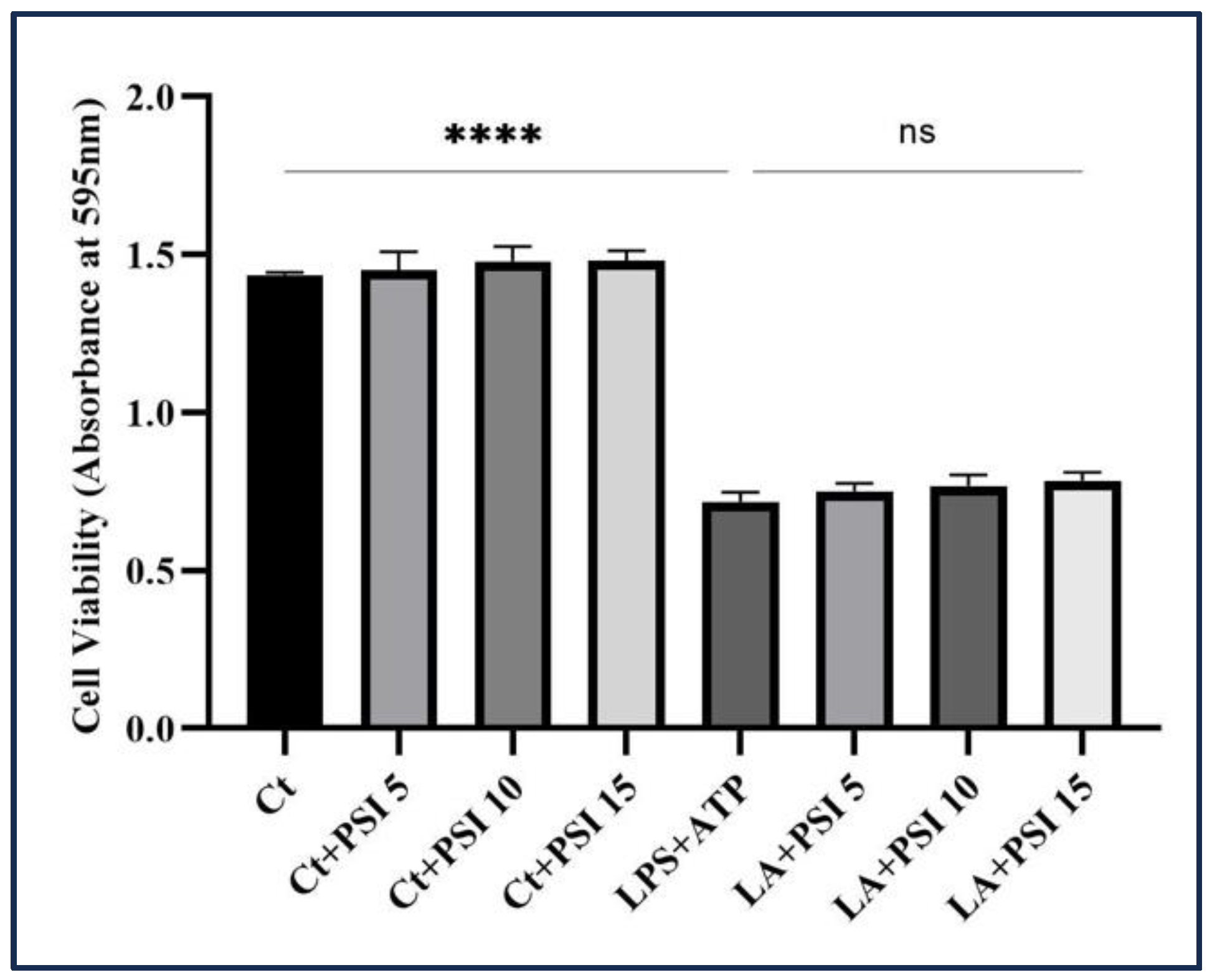
2.2. Cytotocity Assay

To evaluate the effect of psilocybin on THP-1 macrophages’ survival induced by LA, we employed an MTT assay. Initially, THP-1 monocyte cells were treated with PMA and subsequently dispensed into separate wells with 1 × 10⁴ cells/mL in a 96-well plate. It is worth mentioning that we seeded a high number of THP-1 monocyte cells in each well. This decision is based on the fact that THP-1 monocytes, upon differentiation into macrophages, acquire macrophage-like properties, including the loss of their ability to undergo cell division [29]. After one-hour pretreatment with various concentrations of psilocybin, the cells were then incubated with LPS (500 ng/mL) and ATP (5 mM), followed by incubation at +37 °C with a 5% carbon dioxide concentration for the following 24 h.
Following the incubation period, a metabolic activity assay was performed. The content of each well was incubated with a solution of 3-(4,5-Dimethylthiazol-2-yl)-2,5-diphenyltetrazolium bromide (MTT) labeling reagent (product ID: 11465007001, Millipore Sigma Canada Ltd., Oakville, ON, Canada). Subsequently, the microplate was placed in a suitable environment for 4 h. Subsequently, a solubilizing solution was added, and the plate was left to incubate overnight. The solubilizing solution consists of SDS 10% in diluted hydrochloric acid (HCl). The absorbance of the resultant solution was assessed at a particular wavelength using a plate reader (FLUOstar Omega, BMG LABTECH, Offenburg, Germany).

2.3. Immunoblotting

Post cellular treatments, cell lysis was carried out with RIPA lysis buffer (comprising 150 mM NaCl, 50 mM Tris pH 8, 0.5% Deoxycholate, 0.1% SDS, 1% Triton X-100, 1 mM Na3Vo4, 1 mM phenylmethylsulfonyl fluoride (PMSF), and 1 mM EDTA, sourced from Abcam). Whole cell lysates were subsequently stored under suitable conditions. After quantifying total protein, a specific quantity of proteins was loaded for gel electrophoresis. Depending on the abundance of the studied proteins, varying amounts of total proteins (50 µg for low abundance and 100 µg for high abundance proteins) were utilized for gel electrophoresis. The resolved proteins underwent electrotransfer onto polyvinylidene difluoride (PVDF) membranes (Amersham Hybond® P membranes, product number: RPN2020F, obtained from GE Healthcare, Oakville, ON, Canada); membranes were then blocked with 5% nonfat milk in PBS + 0.1% Tween® 20 Detergent (PBST) before being incubated with primary antibodies. This was succeeded by an overnight incubation with primary antibodies at 4 °C. Information regarding the primary antibodies is provided in Table S1. The membranes were subjected to three washes with PBST, followed by incubation with relevant secondary antibodies for 2 h at room temperature, and then a second round of wash with PBST. Immunoreactivity was detected using peroxidase-conjugated antibodies, and the reaction was visualized using the ECL Plus Western Blotting Detection System (GE Healthcare, Oakville, ON, Canada). Densitometric analyses were conducted using ImageJ 1.53K. Band intensities were quantified and normalized against the intensity of housekeeping proteins. Two housekeeping proteins, GAPDH and β-actin, were employed to normalize western blot results. The use of two housekeeping proteins as controls aimed to ensure that the quantification of bands was not affected by incomplete blotting due to air bubbles. To ensure consistent results, the responses of all examined proteins on the same mem-brane were normalized against the same controls; thus the same control image is pre-sented on all figures. For membrane stripping, a mild stripping buffer was employed, consisting of 10 mL Tween 20, 1 g SDS, 15 g glycine, per 1 L of distilled water, with a pH of 2.2 as per the instructions provided by Abcam.

2.4. Gene Expression Profiling by Real-Time PCR

In the examination of gene transcription, RNA extraction from the cells was carried out employing a commercially available TRIzol® Reagent (CAT#15596018, purchased from Invitrogen, Life Technologies Inc., Burlington, ON, Canada), adhering to the guidelines provided by the manufacturer. Following the measurement of RNA quantity using Nanodrop 2000c (ThermoFisher Scientific, Waltham, MA, USA), the complementary DNA was synthesized with the iScript™ Reverse Transcription Supermix (CAT: 1708841, obtained from BioRad Laboratories, Saint-Laurent, QC, Canada); total RNA in the amount of 1 μg was used. To set up qRT-PCR reactions, 1 μL of cDNA was combined with SsAdvancedTM Universal Inhibitor-Tolerant SYBR Green Supermix (product ID: 1725017, Bio-Rad Laboratories, Saint-Laurent, QC, Canada), along with 2.5 mM of each primer according to the manufacturer’s instructions. The qRT-PCR primers were designed using the PrimerQuest™ Tool, an online software, and procured from Eurofins (Ottawa, ON, Canada) (refer to Table S2).

2.5. Enzyme-Linked Immunosorbent Assay (ELISA)

The quantification of IL-β release and its response to the treatments in LPS + ATP groups were assessed using the Human IL-1β/IL-1F2 Quantikine ELISA Kit (Cat number DLB50 from R&D Systems, Inc., Minneapolis, MN, USA) according to the manufacturer’s instructions. After the addition of the Stop Solution, optical density measurements were recorded at 450 nm and 570 nm employing SpectraMax (SpectraMax i3x from Molecular Devices, San Jose, CA, USA) microplate reader. To compensate for potential optical distortions in the plate, the values at 570 nm were subtracted from those at 450 nm. Due to the notably high quantity of secreted IL-1β, the supernatants underwent a 50-fold dilution.

2.6. Statistical Analysis

The data analysis was conducted utilizing GraphPad Prism 8.0 software. All results are expressed as mean values ± standard deviation (SD). Statistical significance was evaluated employing one-way analysis of variance (ANOVA), followed by the application of Dunnett’s test for comparing mean values between the LPS and LA groups with other treatment groups.

3. Results

3.1. Psilocybin Does Not Affect LA-Stimulated THP-1Macrophage Cell Survival

According to the findings from the cell viability assay, the combination of LPS and ATP noticeably decreased macrophage viability after 24 h. Conversely, none of the psilocybin concentrations had an adverse effect on the viability of LA-induced macrophages (Figure 1).

3.2. Psilocybin Modulates the Protein Levels of IL-6 and Cox-2 in THP-1 Macrophages Stimulated by LPS

In order to evaluate the anti-inflammatory characteristics of psilocybin, this investigation analyzed how psilocybin impacts the protein levels of IL-6, Pro-TNF-α, and Cox-2.
Our findings revealed distinct responses to varying psilocybin concentrations. Specifically, 10 μM and 15 μM psilocybin effectively reduced Cox-2 protein levels, whereas 5 μM psilocybin increased the levels of this proinflammatory protein in THP-1 macrophages challenged by LPS (Figure 2). Furthermore, only the 10 μM concentration of psilocybin demonstrated a moderating effect on IL-6 levels, while 15 μM psilocybin enhanced the production of this cytokine. Notably, none of the three psilocybin concentrations reduced Pro-TNF-α level: instead, both 5 μM and 15 μM psilocybin led to an increase in the levels of this proinflammatory cytokine (Figure 2).

3.3. The Introduction of Psilocybin Inhibits NLRP3 Activation

In our western blot analysis, we observed that 15 μM psilocybin effectively reduced the concentrations of both premature and active IL-1β in THP-1 macrophages stimulated by LPS, while 5 μM psilocybin demonstrated an increasing impact on the levels of both forms of IL-1β (Figure 3a). The IL-1β/Pro-IL-1β ratio, a suitable indicator for assessing the second phase, displayed a decrease in response to all three concentrations of psilocybin (Figure 3a). Furthermore, 5 μM and 10 μM psilocybin were found to decrease the level of NLRP3 protein, while 15 μM psilocybin increased the level of this protein in LPS-induced THP-1 macrophages (Figure 3b).

3.4. Psilocybin Downregulates the Levels of P-NF-κB, P-STAT3, P-TYK2, and P-STAT1 Transcription Factors

To see the signaling pathways involved, we investigated the response of NF-κB activation, a protein complex responsible for regulating the transcription of cytokine genes such as IL-1β, IL-6, TNF-α, and COX-2, to three various doses of psilocybin [30,31]. Our western blot analysis showed that the administration of 10 μM and 15 μM psilocybin led to the downregulation of NF-κB phosphorylation and the inhibition of subsequent activation, while 5 μM psilocybin increased the activation of this transcription factor (Figure 4a). Interestingly, psilocybin did not demonstrate any mitigating influence on total NF-κB (Figure 4a). Psilocybin’s inhibitory effects on NF-κB activation prevented the translocation of this transcription factor into the nucleus, as indicated by the NF-κB/P-NF-κB ratio.
The IL-6/JAK/STAT3 signaling cascade is implicated in a multitude of biological processes. Within this pathway, TYK2, belonging to the Janus kinase (JAK) family, exerts phosphorylation and activation on STAT3, thereby instigating its translocation into the nucleus and subsequent engagement in transcriptional activities [32]. Our findings indicate that psilocybin has inhibitory effects on both P-TYK2 and P-STAT3 (Figure 4b,c), suggesting its mitigating impact on the TYK2/STAT3 axis. Moreover, psilocybin demonstrated a reduction in the phosphorylated form of STAT1 (Figure 4e).

3.5. Psilocybin Decreases PANX-1 Levels

PANX-1 is a widely distributed protein recognized for creating expansive channels with elevated conductivity. Its participation in diverse inflammatory and injury responses underscores its importance. These channels play a role in augmenting extracellular ATP levels, activation of P2X7 receptors, assembly of NLRP3 inflammasomes, and release of proinflammatory cytokines, specifically IL-1β [33].
In our study, we observed that psilocybin does not show an inhibitory effect on the cleavage of PANX-1 in macrophages challenged by LPS (Figure 5). However, it exhibited a negative impact on the overall level of total PANX-1 protein in LPS-induced THP-1 macrophages (Figure 5).

3.6. The Administration of Psilocybin Changed the Levels of SR-2A and SR-2B Receptors in LPS-Induced THP-1 Macrophages

Psilocybin exhibits a strong binding affinity for serotonin receptors 2A (SR-2A) and 2B (SR-2B). These receptors, which belong to the larger family of serotonin receptors, play significant roles in a wide range of physiological and psychological processes [24]. Our findings demonstrate that the exposure to LPS reduces the levels of both SR-2A and SR-2B in LPS-induced THP-1 macrophages. Addition of psilocybin changed the levels of both receptors (Figure 6).

3.7. Psilocybin Likely Exerts Its Inhibitory Effect on the Levels of Pro-Il-1β and Pro-TNFα by Mitigating Their Transcription

We assessed the levels of some proinflammatory transcripts in THP-1 macrophages stimulated by LPS following psilocybin treatment. Our aim was to identify any potential correlation between the mitigating effects of psilocybin on proinflammatory cytokines/proteins and the suppression of NF-κB, STAT3, and STAT1 activation. Our findings demonstrated that psilocybin led to a downregulation of IL-1β and TNFα transcription in THP-1 macrophages challenged by LPS (Figure 7). However, none of the administered doses decreased IL-6 and COX-2 mRNA levels. Similarly, psilocybin did not display an inhibitory impact on the expression of NLRP3, PANX-1, and P2X7. Interestingly, the response patterns of some examined transcripts were comparable to the response of the activated form of NF-κB to various concentrations of psilocybin (Figure 7).

3.8. Only the Highest Dose of Psilocybin Exhibited a Suppressive Effect on the Release of IL-1β into the Extracellular Environment

An ELISA assay was performed using the supernatant of the treated cells to investigate any potential correlation between the response of the mature form of IL-1β production and its secretion. Treatment with psilocybin at a concentration of 15 μM suppressed the release of the mature form of IL-1β in the medium in LA-induced THP-1 macrophages (Figure 8). Importantly, the secretion pattern of IL-1β in response to different doses of psilocybin was comparable to that of active IL-1β and corelated with the findings obtained from western blot analysis for Pro-IL-1β.

4. Discussion

Psilocybin (4-phosphoryloxy-N, N-dimethyltryptamine), classified as a substituted indole alkylamine, is categorized as a hallucinogenic tryptamine. Psilocybin is the primary mind-altering substance found in so-called magic mushrooms [34]. Alongside its recognized recreational, spiritual, and religious applications, there is substantial medical potential supported by both personal accounts and scientific research [35].
In this study, our objective was to examine the impact of psilocybin on proinflammatory responses using an in vitro model.
There is limited research on the impact of psilocybin on inflammation. One such study by Nkadimeng et al. (2020) explored its potential anti-inflammatory effects. They focused on a specific species of magic mushrooms called Psilocybe natalensis. The study demonstrated that the analgesic, antioxidant, and anti-inflammatory properties of these mushrooms varied depending on the dosage administered. To evaluate these effects, the researchers treated LPS-induced macrophages with three different extracts derived from Psilocybe natalensis mushrooms, namely ethanol and cold and hot water extracts. The results of the study confirmed the antioxidant capabilities of Psilocybe natalensis mushrooms, as all three extracts effectively inhibited the nitric oxide induced by LPS. Additionally, the study showed that these mushrooms possessed anti-inflammatory characteristics through the inhibition of prostaglandin E2 and IL-1β cytokine synthesis [36].
The present study initially aimed to examine the ability of psilocybin to decrease COX-2, Pro-TNFα, IL-1β, IL-6 and Pro-IL-1β levels in LA/LPS-induced THP-1 macrophages. We found that the administration of certain doses of psilocybin significantly reduced IL-6 and COX-2 protein levels in THP-1 macrophages challenged by LPS (see Figure 2). These results align with the study by Nkadimeng (2020) et al., which demonstrated that extract treatment leads to a decrease in prostaglandin E2 production mediated by COX-2 and IL-6. Psilocybin also decreased Pro-IL-1β, mature IL-1β (see Figure 3a), and secreted IL-1β in a dose-response manner (see Figure 8); Nkadimeng et al. (2020) also found psilocybin to decrease the level of secreted IL-1β.
To understand the potential mechanisms of psilocybin’s effects on IL-6, COX-2, and IL-1β mRNA levels, we examined its impact on key inflammation-related transcription factors: NF-κB, STAT1, and STAT3. Our results indicate that psilocybin at doses of 10 and 15 μM downregulated the elevated levels of phosphorylated NF-κB in LPS-induced macrophages (Figure 4a), thereby suppressing its activity. Psilocybin also attenuated the activation of TYK2, STAT3, and STAT1 transcription factors, further supporting its inhibitory influence on the transcription of proinflammatory proteins.
Additionally, the qRT-PCR results revealed that certain doses of psilocybin downregulated IL-1β and TNFα at the transcriptional level. This effect may be linked to psilocybin’s inhibitory impact on NF-kB, STAT3, and STAT1 activation in LPS-induced macrophages. The inhibitory effect of psilocybin on the activation of NF-kB and subsequent transcription of proinflammatory genes is likely mediated through the downregulation of IL-6/TYK2 proteins, observed in our experiments. This may be further supported by the fact that only the 10 μM and 15 μM doses of psilocybin mitigated the phosphorylation of both NF-kB and TYK2, while 5 μM psilocybin did not show any significant impact on the phosphorylation of TYK2, and it even increased the phosphorylation of NF-kB. Accordingly, Yang et al. (2005) established that interferon α induces the activation of NF-κB in cells lacking JAK1 by utilizing a pathway that relies on TYK2. This observation implies that TYK2 phosphorylation is involved in the phosphorylation of NF-κB and the consequent proinflammatory responses [37]. STAT3 functions as a transcription factor that, once activated, promotes the expression of various proinflammatory cytokines, including IL-17, IL-23, and IL-8 [11]. Notably, IL-6 plays a crucial role in activating STAT3 during inflammation, and the IL-6/STAT3 axis has emerged as a promising target for the development of novel treatments for inflammatory disorders. Excessive activation of NF-κB results in elevated levels of IL-6, which in turn can initiate the phosphorylation of JAKs such as TYK2. This phosphorylation event subsequently activates STAT3 and even NF-kB itself, facilitating their movement into the cell nucleus and ultimately leading to an increased transcription of cytokine genes [38]. The activation of STAT3 leads to a cooperative interplay with NF-κB, resulting in an increase in the phosphorylation and subsequent translocation of NF-κB into the nucleus [39,40], which ultimately results in cytokine storm. According to our findings, 10 μM and 15 μM of psilocybin significantly inhibit the TYK2/STAT3 pathway (Figure 4), suggesting a potential mechanism through which psilocybin may modulate the proinflammatory responses leading to a cytokine storm.
Apart from the suppressive effects of psilocybin on the TYK2/STAT3 axis, it appears to have a mitigating effect on the TYK2/STAT-1 axis as well (Figure 4). This suggests an additional mechanism by which psilocybin can downregulate proinflammatory responses in macrophages stimulated with LPS. Accordingly, it has been established that the activation of STAT1 induced by LPS is responsible for the production of IL-6, involving both NF-κB-dependent and independent pathways [9]. It is worth mentioning that while 5 μM does not show any significant impact on the mitigation of TYK2 phosphorylation, this dose mitigates the phosphorylation of both STAT3 and STAT1, suggesting the possible involvement of other JAKs in mediating the inhibitory impact of 5 μM psilocybin on the phosphorylation of these transcription factors.
It is worth mentioning that, despite psilocybin’s inhibitory effects on IL-6 and COX-2 protein levels, no effect on the mRNA levels of IL-6 and COX-2 was observed (Figure 7). This suggests that specific dose(s) of psilocybin may exert their inhibitory effects on the post-transcriptional regulation of these proteins. The pattern of expression of COX-2 and IL-6 and COX-2 mRNA to psilocybin in macrophages treated with LPS was consistent with that of the TNFα transcript and was associated with changes in P-NF-κB upon psilocybin administration. This may suggest a potential role of NF-κB as a target through which psilocybin exerts its effects on the transcription of these proinflammatory cytokines.
LPS induction of macrophages results in an increase in the transcription of Pro-IL-1β and NLRP3 [41], which in turn results in the formation of NLRP3 inflammasome and subsequent production of active IL-1β. Exposure to 5 and 10 μM psilocybin lowered the concentration of NLRP3 in THP-1 cells challenged by LA. This reduction is likely due to post-translational processes, as no inhibitory effect was observed in the mRNA level (Figure 7). In addition, the alleviating effect of 10 and 15 μM of psilocybin on the increased levels of Pro-IL-1β, which is likely mediated through the downregulation of its transcription (Figure 3a and Figure 7), provides substantial evidence of psilocybin’s inhibitory influence on the initial stage of activation of the NLRP3 inflammasome. Furthermore, the administration of psilocybin resulted in the downregulation of the IL-1β/Pro-IL-1β ratio in LA-induced THP-1 macrophages (see Figure 3a). This suggests that all three doses of psilocybin may also inhibit the second phase of NLRP3 inflammasome activation in addition to suppressing the initial phase.
During apoptosis, PANX-1, a transmembrane protein found in various cells including macrophages, undergoes cleavage by Caspase-3 and 7 at its C-terminus. This cleavage leads to the opening of PANX-1 channels and an increase in membrane permeability [42,43]. As a result, extracellular ATP levels rise, triggering the activation of the P2X7 receptor and leading to an influx of potassium ions into the cell. Intracellular potassium levels are known to be associated with the initiation of the second phase of NLRP3 inflammasome activation [17]. Exposure to psilocybin diminished the levels of total PANX-1 (see Figure 5). This reduction is likely due to its influence on post-transcriptional processes, as indicated by the qRT-PCR results (see Figure 7). This suggests that psilocybin may target an additional pathway through which it exerts inhibitory effects on the second phase of NLRP3 inflammasome activation. Furthermore, we observed that 5 μM psilocybin increased the cleavage and subsequent opening of PANX-1 channel, while neither 10 μM nor 15 μM had a significant impact on this process (see Figure 5). This provides evidence of a potential link between apoptosis, PANX-1 cleavage, and the stimulatory effect of 5 μM psilocybin on the response of P-NF-κB, Pro-IL-1β, IL-6, and COX-2.
In general, our results show that psilocybin impacts the production of inflammation-related cytokines in LPS/LA-induced macrophages in a complex manner. The findings suggest that the effect of psilocybin on the production of proinflammatory cytokines is dose dependent and sometimes bi-phasic or has a bell-curve response. For instance, the 10 and 15 μM concentrations of psilocybin decreased the protein levels of IL-1β and COX-2, while 5 μM increased the levels of these proteins. Additionally, 10 μM psilocybin reduced the level of IL-6, but 15 μM increased the level of this protein. Moreover, while 10 μM psilocybin did not impact the level of Pro-TNFα, the 5 and 15 μM concentrations increased its level. The dose-dependent effects of psilocybin on the production of proinflammatory cytokines could be attributed to its complex interactions with the immune system. Additional research is required for a comprehensive understanding of the underlying mechanisms involved.
Monocytes/macrophages possess various serotonin receptors such as 5-HT1E, 5-HT2B, 5-HT1A, 5-HT2A, 5-HT3, 5-HT4, and 5-HT7 [20]. Psilocybin exhibits agonistic effects on several serotonin receptors, particularly 5-HT2A and 5-HT2B receptors. Previous studies have demonstrated that psilocybin can modulate the activation of these receptors, leading to alterations in downstream gene transcription and translation [27]. The relationship between the activation of 5-HT2 receptor(s) and anti-inflammatory properties has been extensively established [20,44,45]. In our current report, we demonstrated that psilocybin was capable of restoring the diminished levels of serotonin receptors 5-HT2A and 5-HT2B in THP-1 macrophages induced by LPS (Figure 6). This discovery may introduce a novel pathway through which psilocybin exerts its inhibitory effects on the dysregulated proinflammatory cytokines and proteins.
In summary, the key findings of this study are as follows (Figure 9):
1.
Psilocybin demonstrates a dose-dependent mitigation of elevated levels of Pro-IL-1β, IL-1β, IL-6, and COX-2 proteins in LA/LPS-induced THP-1 macrophages.
2.
Specific doses of psilocybin mitigate the activities of NF-κB, STAT1, and STAT3 transcription factors, potentially serving as a target through which psilocybin downregulates TNFα and IL-1β expression.
3.
Psilocybin, at certain doses, appears to exert mitigatory effects on STAT3 activation by influencing TYK-2 activation.
4.
The reduction in PANX-1 levels likely contributes to the mitigatory impact of specific psilocybin doses on the NLRP3 assembly in LPS-induced THP-1 macrophages.
5.
Psilocybin demonstrates a stimulatory effect on the levels of SR-2A and SR-2B in LPS-induced THP-1 macrophages (Figure 9).

5. Conclusions

Our finding suggests that specific doses of psilocybin have the potential to reduce the elevated levels of proinflammatory proteins and cytokines such as IL-1β, IL-6, and COX-2 within macrophages treated with LPS/LA. This reduction can be mediated through various pathways and axes, including NF-κB, TYK2/STAT1, TYK2/STAT3, and the NLRP3 inflammasome assembly. We also observed that 5 μM psilocybin had a stimulatory effect on P-NF-κB, IL-1β, IL-6, and COX-2, while 10 μM psilocybin mitigated the levels of these proinflammatory-related proteins and cytokines. This suggests that the impact of psilocybin on inflammatory responses is dose-dependent, and the response of each proinflammatory protein/cytokine to psilocybin may vary.

6. Future Direction

In this work, we have provided evidence of the inhibitory impact of specific doses of psilocybin on the production of proinflammatory proteins and cytokines in LPS/LA-induced THP-1 macrophages. To further validate our results, it is recommended to conduct experiments with other cell lines and in vivo systems.
Additionally, considering that psilocybin inhibits the IL-1β/Pro-IL-1β ratio, future research could explore the impact of psilocybin on the second phase of NLRP3 inflammasome activation, focusing on post-translational modifications of NLRP3 in response to psilocybin.
We have also observed the inhibitory impact of psilocybin on STAT3 and STAT1 activation. Further investigations could explore the response of proinflammatory cytokines controlled by these transcription factors, including IL-8, IL-17, and IL-23.
While our study found that psilocybin administration increased the levels of two serotonin receptors in LPS-induced macrophages, further research is needed to establish connections between these findings and the responses of the proinflammatory proteins and cytokines studied here.

Supplementary Materials

The following supporting information can be downloaded at: https://www.mdpi.com/article/10.3390/psychoactives3010004/s1, Table S1: Information on the primary antibodies; Table S2: The sequences of primers used for q-RT-PCR; The whole western blot images.

Author Contributions

Conceptualization, I.K. and E.G.G.; methodology, E.G.G., B.W. and D.L.; validation, I.K., D.L., B.W. and O.K.; formal analysis, E.G.G., B.W., D.L. and I.K.; resources, I.K. and O.K.; data curation, E.G.G. and I.K.; writing—original draft preparation, E.G.G.; writing—review and editing, E.G.G., I.K. and O.K.; visualization, E.G.G.; supervision, I.K. and O.K.; project administration, I.K. and O.K.; funding acquisition, I.K. All authors have read and agreed to the published version of the manuscript.

Funding

This research was funded by MITACS, grant number IT28033.

Institutional Review Board Statement

Not applicable.

Informed Consent Statement

Not applicable.

Data Availability Statement

Raw western blot images were submitted to Psychoactives.

Acknowledgments

We acknowledge the technical support given by Rommy Rodriguez-Juarez and Rocio Rodriguez-Juarez.

Conflicts of Interest

The authors declare no conflicts of interest. The funders had no role in the design of the study; in the collection, analyses, or interpretation of data; in the writing of the manuscript; or in the decision to publish the results.

References

  1. Kotas, M.E.; Medzhitov, R. Homeostasis, inflammation, and disease susceptibility. Cell 2015, 160, 816–827. [Google Scholar] [CrossRef]
  2. Rock, K.L.; Latz, E.; Ontiveros, F.; Kono, H. The sterile inflammatory response. Annu. Rev. Immunol. 2009, 28, 321–342. [Google Scholar] [CrossRef]
  3. Schett, G.; Neurath, M.F. Resolution of chronic inflammatory disease: Universal and tissue-specific concepts. Nat. Commun. 2018, 9, 3261. [Google Scholar] [CrossRef]
  4. Furman, D.; Campisi, J.; Verdin, E.; Carrera-Bastos, P.; Targ, S.; Franceschi, C.; Ferrucci, L.; Gilroy, D.W.; Fasano, A.; Miller, G.W. Chronic inflammation in the etiology of disease across the life span. Nat. Med. 2019, 25, 1822–1832. [Google Scholar] [CrossRef] [PubMed]
  5. Chen, L.; Deng, H.; Cui, H.; Fang, J.; Zuo, Z.; Deng, J.; Li, Y.; Wang, X.; Zhao, L. Inflammatory responses and inflammation-associated diseases in organs. Oncotarget 2018, 9, 7204. [Google Scholar] [CrossRef] [PubMed]
  6. Tang, D.; Kang, R.; Coyne, C.B.; Zeh, H.J.; Lotze, M.T. PAMP s and DAMP s: Signal 0s that spur autophagy and immunity. Immunol. Rev. 2012, 249, 158–175. [Google Scholar] [CrossRef] [PubMed]
  7. Nunes-Alves, C. New LPS receptors discovered. Nat. Rev. Immunol. 2014, 14, 583. [Google Scholar] [CrossRef] [PubMed]
  8. Cen, X.; Liu, S.; Cheng, K. The role of toll-like receptor in inflammation and tumor immunity. Front. Pharmacol. 2018, 9, 878. [Google Scholar] [CrossRef] [PubMed]
  9. Metwally, H.; Tanaka, T.; Li, S.; Parajuli, G.; Kang, S.; Hanieh, H.; Hashimoto, S.; Chalise, J.P.; Gemechu, Y.; Standley, D.M. Noncanonical STAT1 phosphorylation expands its transcriptional activity into promoting LPS-induced IL-6 and IL-12p40 production. Sci. Signal. 2020, 13, eaay0574. [Google Scholar] [CrossRef] [PubMed]
  10. Wen, Z.; Zhong, Z.; Darnell, J.E., Jr. Maximal activation of transcription by Statl and Stat3 requires both tyrosine and serine phosphorylation. Cell 1995, 82, 241–250. [Google Scholar] [CrossRef]
  11. Yu, H.; Pardoll, D.; Jove, R. STATs in cancer inflammation and immunity: A leading role for STAT3. Nat. Rev. Cancer 2009, 9, 798–809. [Google Scholar] [CrossRef]
  12. Mangan, M.S.; Olhava, E.J.; Roush, W.R.; Seidel, H.M.; Glick, G.D.; Latz, E. Targeting the NLRP3 inflammasome in inflammatory diseases. Nat. Rev. Drug Discov. 2018, 17, 588–606. [Google Scholar] [CrossRef]
  13. Donath, M.Y.; Shoelson, S.E. Type 2 diabetes as an inflammatory disease. Nat. Rev. Immunol. 2011, 11, 98–107. [Google Scholar] [CrossRef] [PubMed]
  14. Shao, B.-Z.; Xu, Z.-Q.; Han, B.-Z.; Su, D.-F.; Liu, C. NLRP3 inflammasome and its inhibitors: A review. Front. Pharmacol. 2015, 6, 262. [Google Scholar] [CrossRef]
  15. He, Y.; Hara, H.; Núñez, G. Mechanism and regulation of NLRP3 inflammasome activation. Trends Biochem. Sci. 2016, 41, 1012–1021. [Google Scholar] [CrossRef]
  16. Tschopp, J.; Schroder, K. NLRP3 inflammasome activation: The convergence of multiple signalling pathways on ROS production? Nat. Rev. Immunol. 2010, 10, 210–215. [Google Scholar] [CrossRef]
  17. Chen, K.W.; Demarco, B.; Broz, P. Pannexin-1 promotes NLRP3 activation during apoptosis but is dispensable for canonical or noncanonical inflammasome activation. Eur. J. Immunol. 2020, 50, 170–177. [Google Scholar] [CrossRef]
  18. Schneider, S.L.; Ross, A.L.; Grichnik, J.M. Do inflammatory pathways drive melanomagenesis? Exp. Dermatol. 2015, 24, 86–90. [Google Scholar] [CrossRef] [PubMed]
  19. Ghasemi-Gojani, E.; Kovalchuk, I.; Kovalchuk, O. Cannabinoids and terpenes for diabetes mellitus and its complications: From mechanisms to new therapies. Trends Endocrinol. Metab. 2022, 33, 828–849. [Google Scholar] [CrossRef]
  20. Herr, N.; Bode, C.; Duerschmied, D. The effects of serotonin in immune cells. Front. Cardiovasc. Med. 2017, 4, 48. [Google Scholar] [CrossRef]
  21. Yu, B.; Becnel, J.; Zerfaoui, M.; Rohatgi, R.; Boulares, A.H.; Nichols, C.D. Serotonin 5-hydroxytryptamine2A receptor activation suppresses tumor necrosis factor-α-induced inflammation with extraordinary potency. J. Pharmacol. Exp. Ther. 2008, 327, 316–323. [Google Scholar] [CrossRef]
  22. Nau, F., Jr.; Yu, B.; Martin, D.; Nichols, C.D. Serotonin 5-HT2A receptor activation blocks TNF-α mediated inflammation in vivo. PLoS ONE 2013, 8, e75426. [Google Scholar] [CrossRef]
  23. Halberstadt, A.L.; Geyer, M.A. Multiple receptors contribute to the behavioral effects of indoleamine hallucinogens. Neuropharmacology 2011, 61, 364–381. [Google Scholar] [CrossRef] [PubMed]
  24. Nichols, D.E.; Nichols, C.D. Serotonin receptors. Chem. Rev. 2008, 108, 1614–1641. [Google Scholar] [CrossRef]
  25. Mohammad-Zadeh, L.; Moses, L.; Gwaltney-Brant, S. Serotonin: A review. J. Vet. Pharmacol. Ther. 2008, 31, 187–199. [Google Scholar] [CrossRef] [PubMed]
  26. Hendricks, P.S.; Johnson, M.W.; Griffiths, R.R. Psilocybin, psychological distress, and suicidality. J. Psychopharmacol. 2015, 29, 1041–1043. [Google Scholar] [CrossRef]
  27. Ling, S.; Ceban, F.; Lui, L.M.W.; Lee, Y.; Teopiz, K.M.; Rodrigues, N.B.; Lipsitz, O.; Gill, H.; Subramaniapillai, M.; Mansur, R.B.; et al. Molecular Mechanisms of Psilocybin and Implications for the Treatment of Depression. CNS Drugs 2022, 36, 17–30. [Google Scholar] [CrossRef]
  28. Zanikov, T.; Gerasymchuk, M.; Ghasemi Gojani, E.; Robinson, G.I.; Asghari, S.; Groves, A.; Haselhorst, L.; Nandakumar, S.; Stahl, C.; Cameron, M. The Effect of Combined Treatment of Psilocybin and Eugenol on Lipopolysaccharide-Induced Brain Inflammation in Mice. Molecules 2023, 28, 2624. [Google Scholar] [CrossRef]
  29. Liu, T.; Huang, T.; Li, J.; Li, A.; Li, C.; Huang, X.; Li, D.; Wang, S.; Liang, M. Optimization of differentiation and transcriptomic profile of THP-1 cells into macrophage by PMA. PLoS ONE 2023, 18, e0286056. [Google Scholar] [CrossRef]
  30. Liu, T.; Zhang, L.; Joo, D.; Sun, S.-C. NF-κB signaling in inflammation. Signal Transduct. Target. Ther. 2017, 2, 17023. [Google Scholar] [CrossRef]
  31. Lim, J.W.; Kim, H.; Kim, K.H. Nuclear factor-κB regulates cyclooxygenase-2 expression and cell proliferation in human gastric cancer cells. Lab. Investig. 2001, 81, 349–360. [Google Scholar] [CrossRef] [PubMed]
  32. Johnson, D.E.; O’Keefe, R.A.; Grandis, J.R. Targeting the IL-6/JAK/STAT3 signalling axis in cancer. Nat. Rev. Clin. Oncol. 2018, 15, 234–248. [Google Scholar] [CrossRef] [PubMed]
  33. Yanguas, S.C.; Willebrords, J.; Johnstone, S.R.; Maes, M.; Decrock, E.; De Bock, M.; Leybaert, L.; Cogliati, B.; Vinken, M. Pannexin1 as mediator of inflammation and cell death. Biochim. Biophys. Acta (BBA)-Mol. Cell Res. 2017, 1864, 51–61. [Google Scholar] [CrossRef] [PubMed]
  34. Passie, T.; Seifert, J.; Schneider, U.; Emrich, H.M. The pharmacology of psilocybin. Addict. Biol. 2002, 7, 357–364. [Google Scholar] [CrossRef]
  35. Lowe, H.; Toyang, N.; Steele, B.; Valentine, H.; Grant, J.; Ali, A.; Ngwa, W.; Gordon, L. The therapeutic potential of psilocybin. Molecules 2021, 26, 2948. [Google Scholar] [CrossRef]
  36. Nkadimeng, S.M.; Nabatanzi, A.; Steinmann, C.M.; Eloff, J.N. Phytochemical, cytotoxicity, antioxidant and anti-inflammatory effects of Psilocybe natalensis magic mushroom. Plants 2020, 9, 1127. [Google Scholar] [CrossRef]
  37. Yang, C.H.; Murti, A.; Valentine, W.J.; Du, Z.; Pfeffer, L.M. Interferonα Activates NF-κB in JAK1-deficient Cells through aTYK2-dependentPathway. J. Biol. Chem. 2005, 280, 25849–25853. [Google Scholar] [CrossRef]
  38. Gojani, E.G.; Wang, B.; Li, D.-P.; Kovalchuk, O.; Kovalchuk, I. Anti-Inflammatory Effects of Minor Cannabinoids CBC, THCV, and CBN in Human Macrophages. Molecules 2023, 28, 6487. [Google Scholar] [CrossRef]
  39. Suryavanshi, S.V.; Zaiachuk, M.; Pryimak, N.; Kovalchuk, I.; Kovalchuk, O. Cannabinoids Alleviate the LPS-Induced Cytokine Storm via Attenuating NLRP3 Inflammasome Signaling and TYK2-Mediated STAT3 Signaling Pathways In Vitro. Cells 2022, 11, 1391. [Google Scholar] [CrossRef]
  40. Hirano, T. IL-6 in inflammation, autoimmunity and cancer. Int. Immunol. 2021, 33, 127–148. [Google Scholar] [CrossRef] [PubMed]
  41. Kelley, N.; Jeltema, D.; Duan, Y.; He, Y. The NLRP3 Inflammasome: An Overview of Mechanisms of Activation and Regulation. Int. J. Mol. Sci. 2019, 20, 3328. [Google Scholar] [CrossRef] [PubMed]
  42. Chekeni, F.B.; Elliott, M.R.; Sandilos, J.K.; Walk, S.F.; Kinchen, J.M.; Lazarowski, E.R.; Armstrong, A.J.; Penuela, S.; Laird, D.W.; Salvesen, G.S. Pannexin 1 channels mediate ‘find-me’signal release and membrane permeability during apoptosis. Nature 2010, 467, 863–867. [Google Scholar] [CrossRef] [PubMed]
  43. Sandilos, J.K.; Chiu, Y.-H.; Chekeni, F.B.; Armstrong, A.J.; Walk, S.F.; Ravichandran, K.S.; Bayliss, D.A. Pannexin 1, an ATP Release Channel, Is Activated by Caspase Cleavage of Its Pore-associated C-terminal Autoinhibitory Region. J. Biol. Chem. 2012, 287, 11303–11311. [Google Scholar] [CrossRef]
  44. Cloëz-Tayarani, I.; Petit-Bertron, A.F.; Venters, H.D.; Cavaillon, J.M. Differential effect of serotonin on cytokine production in lipopolysaccharide-stimulated human peripheral blood mononuclear cells: Involvement of 5-hydroxytryptamine2A receptors. Int. Immunol. 2003, 15, 233–240. [Google Scholar] [CrossRef] [PubMed]
  45. Kubera, M.; Maes, M.; Kenis, G.; Kim, Y.-K.; Lasoń, W. Effects of serotonin and serotonergic agonists and antagonists on the production of tumor necrosis factor α and interleukin-6. Psychiatry Res. 2005, 134, 251–258. [Google Scholar] [CrossRef]
Figure 1. The effect of psilocybin on the survival of THP-1 macrophage cells treated with LA. The MTT assay was performed to measure the impact of psilocybin on LA-induced THP-1 cell viability. Administering various concentrations of psilocybin to macrophages induced by LPS and ATP did not negatively impact the viability of the cells. The presented data represent the mean values (with standard deviation, SD), n = 3. Figure abbreviations include Ct (control), PSI (psilocybin), and LA (LPS + ATP). Asterisks denote significant differences, where four—p < 0.0001; ns—non-significant.
Psychoactives 03 00004 g001
Figure 2. Western blot analysis of Cox-2, IL-6, and Pro-TNFα in response to psilocybin. Whole cellular lysates prepared from LPS-induced and uninduced THP-1 macrophages were subjected to western blot analysis using antibodies to IL-6, Pro-TNFα, and Cox2; GAPDH served as a loading control. Relative densitometry was measured using ImageJ. All data are presented as mean values ± SD, n = 3 measurements. The abbreviations are: Ct (control), PSI (psilocybin). Asterisks indicate significant differences, where 4 asterisks denote a p-value < 0.0001, while “ns” denotes non-significance. The whole western blots were showed in the supplementary materials.
Psychoactives 03 00004 g002
Figure 3. The effects of psilocybin on Pro-IL-1β, mature IL-1β, and NLRP3 in THP-1 macrophages. (a) Immunoblotting of Pro-IL-1β and active IL-1β in both LA-induced and uninduced THP-1 macrophages in response to psilocybin treatment. β-actin served as a loading control. (b) Western blot analysis of the level of NLRP3 protein in both LA-stimulated and unstimulated THP-1 macrophages in response to psilocybin. β–actin served as a loading control. Relative densitometry was measured using ImageJ 1.53k. All data are presented as mean values ± SD, n = 3 measurements. Abbreviations: Ct (control), PSI (psilocybin), and LA (LPS + ATP) The asterisks indicate significant distinctions, where three means p < 0.001 and four means p < 0.0001. supplementary materials.
Psychoactives 03 00004 g003
Figure 4. The impact of psilocybin on P-NF-kB P65 (Ser536), total-NF-kB P65, P-STAT3, total-STAT3, P-TYK2, total-TYK2, and P-STAT1 in THP-1 macrophages at protein level. (a) The impact of three doses of psilocybin on the levels of P-NF-κB P65 (Ser536), total-NF-kB P65, and P-NF-κB P65 (Ser536)/NF-κB P65 in both LPS-induced and uninduced THP-1 macrophages. (b) Psilocybin’s impact on the expression of P-STAT3 and total-STAT3 at protein level in THP-1 macrophages stimulated by LPS. (c) Psilocybin’s effect on the responses of P-TYK2 and TYK2 proteins in LPS-challenged THP-1 macrophages. (d) Levels of P-NF-kB P65, total-NF-kB P65, P-STAT3, total-STAT3, P-TYK2, total-TYK2, and actin proteins in the western blot images. (e) The impact of three doses of psilocybin on the response of P-STAT1 in LPS-stimulated THP-1 macrophages. As a loading control, β-actin was employed, and ImageJ was utilized to measure relative densitometry. All data are expressed as mean values with the standard deviation (±SD) based on three measurements (n = 3). Abbreviations: Ct (control), PSI (psilocybin). Asterisks denote the significance levels: one indicates p < 0.05; two mean p < 0.01; three shows p < 0.001; four indicate p < 0.0001; ns means non-significant. supplementary materials.
Psychoactives 03 00004 g004
Figure 5. Western blot analysis of PANX-1 (55 kDa) and cleaved-PANX-1 in THP-1 macrophages in response to psilocybin. Whole cellular lysates prepared from both LPS-induced and uninduced THP-1 macrophages were subjected to western blot analysis using antibodies to PANX-1 and cleaved PANX-1, As a loading control, β-actin was employed, and ImageJ was utilized to measure relative densitometry. All data are expressed as mean values with the standard deviation (±SD) based on three measurements (n = 3). Abbreviations: Ct (control) and PSI (psilocybin). Asterisks denote the significant differences: p < 0.0001; ns—non-significant. supplementary materials.
Psychoactives 03 00004 g005
Figure 6. The response of SR-2A and SR-2B receptors to psilocybin in THP-1 macrophages. Whole cellular lysates prepared from LPS-induced and uninduced THP-1 macrophages were subjected to western blot analysis using antibodies to SR-2A and SR-2B; GAPDH and β-actin served as loading controls. Densitometry was conducted using ImageJ. The data are expressed as mean values with the standard deviation (±SD) based on three measurements (N = 3). Abbreviations: Ct (control), PSI (psilocybin), SR-2A (serotonin receptor 2A), and SR-2B (serotonin receptor 2B). The asterisks indicate the level of significance: one—p < 0.05; two—p < 0.01; three—p < 0.001; four—p < 0.0001; ns—no significance. supplementary materials.
Psychoactives 03 00004 g006
Figure 7. The impact of psilocybin on the expression profiles of Pro-IL-1β, IL-6, TNFα, Cox-2, Nlrp3, PANX-1, and P2X7 genes in THP-1 cells. THP-1 macrophages were treated with psilocybin for 1 h, followed by the stimulation of the cells with LPS for the next 4 h. Subsequently, total RNA was extracted from the cells and subjected to qPCR analysis. The data are represented as mean values accompanied by the standard deviation (±SD), calculated from a sample size of N = 3, normalized against GAPDH mRNA. Abbreviations: Ct (control), PSI (psilocybin). The asterisks show significant difference, one—p < 0.05; two—p < 0.01; three—p < 0.001; four—p < 0.0001; ns—non-significant.
Psychoactives 03 00004 g007
Figure 8. ELISA analysis of secreted IL-1β in LA-challenged THP-1 macrophages in response to psilocybin. ELISA was conducted to determine the impact of psilocybin on IL-1β release. The data are expressed as mean values with the standard deviation (±SD) based on three measurements (N = 3). Abbreviations: Ct (control), PSI (psilocybin), and LA (LPS + ATP). The asterisks denote significant difference, where one indicates p < 0.05; three show p < 0.001; four mean p < 0.0001; ns indicates non-significant.
Psychoactives 03 00004 g008
Figure 9. The effects of 10 μM psilocybin on inflammatory responses in LPS/LA-induced THP-1 macrophages. The dose of 10 μM psilocybin decreases the concentrations of proinflammatory proteins, including Pro-IL-1β, COX-2, IL-6, NLRP3, and PANX-1. Additionally, the administration of 10 μM psilocybin inhibits the activities of NF-κB, TYK2, STAT3, and STAT1, downregulating Pro-IL-1β as well as Pro-TNFα. The reduction of IL-6/P-TYK2 could play a crucial role in the inhibitory effect of psilocybin on the activation of NF-κB, STAT1, and STAT3. This finding strongly supports the potential of psilocybin to inhibit cytokine storms. Notably, the downregulation of PANX-1 protein by 10 μM psilocybin in LPS-induced macrophages may play a crucial role in suppressing the assembly of NLRP3 inflammasome, Pro-Caspase-1 cleavage into active Caspase-1, and the subsequent synthesis of active IL-1β.
Psychoactives 03 00004 g009
Disclaimer/Publisher’s Note: The statements, opinions and data contained in all publications are solely those of the individual author(s) and contributor(s) and not of MDPI and/or the editor(s). MDPI and/or the editor(s) disclaim responsibility for any injury to people or property resulting from any ideas, methods, instructions or products referred to in the content.

