Review

Racial and Gender Disparities in Clinical Trial Representation for Age-Related Macular Degeneration Treatments: A Scoping Review

1
Anne Burnett Marion School of Medicine, Texas Christian University, Fort Worth, TX 76129, USA
2
Prevention of Blindness from Glaucoma and Age Related Macular Degeneration, Floral Park, NY 11001, USA
3
David & Ilene Flaum Eye Institute, University of Rochester Medical Center, Rochester, NY 14642, USA
*
Author to whom correspondence should be addressed.
J. Clin. Transl. Ophthalmol. 2025, 3(3), 16; https://doi.org/10.3390/jcto3030016
Submission received: 12 May 2025 / Revised: 19 June 2025 / Accepted: 1 August 2025 / Published: 13 August 2025
(This article belongs to the Special Issue Retinal Diseases: Recent Advances in Diagnosis and Treatment)

Abstract

Background/Objective: Age-related macular degeneration (AMD) is a leading cause of irreversible vision loss. Intravitreal anti-vascular endothelial growth factor (VEGF) therapy is the primary treatment for neovascular AMD. This study aimed to assess racial, ethnic, and gender representation in U.S.-based randomized controlled trials (RCTs) of anti-VEGF therapies. Methods: A systematic PubMed search identified 19 eligible RCTs. Titles and abstracts were screened, and demographic data were independently extracted and cross-verified. Chi-squared analysis was used to evaluate disparities in participant representation. Risk of bias was assessed using the ROBIS checklist. Results: Among 8003 participants across 19 trials, 92.3% were Caucasian. Asian, African American, Hispanic/Latino, and American Indian participants collectively comprised just over 5%. This underrepresentation of non-Caucasian groups was statistically significant (p < 0.01, df = 4) and not associated with study sponsorship. Gender analysis showed 59% female and 41% male participation, which was not statistically significant (p = 0.83, df = 1). Conclusions: Non-Caucasian populations remain significantly underrepresented in anti-VEGF RCTs for AMD. This raises concerns about the generalizability of trial findings to diverse populations. Future clinical trials must prioritize inclusive recruitment to ensure equitable, evidence-based care for all patients.

1. Introduction

1.1. History of Age-Related Macular Degeneration

AMD causes progressive central vision loss. Its pathogenesis stems from genetic, metabolic, environmental, and lifestyle factors that damage the retinal pigment epithelium (RPE) [1]. As RPE cells deteriorate, lipid- and protein-rich drusen accumulate. These deposits are readily visible on fundus exam and optical coherence tomography [1]. “Dry” or non-exudative AMD is characterized by the presence of drusen and thickening of Bruch’s membrane (a layer separating the choroid from the RPE layer within the eye), but lacks the proliferation and leakage of the abnormal vasculature underneath Bruch’s membrane. The hallmark of “wet” or exudative disease is the neovascularization and aberrant vascular proliferation, which untreated, can result in retinal detachment and permanent visual loss [2].
Early mentions of AMD, starting in 1852, began with non-descriptive reports of thickening within Bruch’s membrane, which slowly began occurring in patients around the age of 70 [3]. Drusen was next officially classified and described by Wedl; however, its role in AMD still remained unclear for several years to follow [3,4]. The theory of drusen’s etiology continued to fluctuate over the years following, and was thought to be possibly due to leukocyte activity within the retina, choroid, and membrane [4]. Despite its suggested etiology, years later, the exact underlying etiology remains incompletely understood [2]. From the 1850s to 1965, classifications and descriptions of AMD varied widely within the field. Following 1965, the terminology utilized to classify the diagnosis and stages of AMD became more uniform and specific. In 1995, the early and late stages of disease were finally classified by Alan Bird in “Age-Related Macular Disease: Etiology and Clinical Management.” Most notably, Bird helped to subclassify “late disease” by reporting if there was a presence or absence of neovascularization, geographic atrophy, or if fluid leakage was present [5].

1.2. Epidemiology of Age-Related Macular Degeneration

AMD is the leading cause of central vision loss and legal blindness in U.S. adults over age 50. Legal blindness is defined as best-corrected visual acuity worse than 20/200 or a visual field under 20° in the better-seeing eye [1]. Specifically, of the patients diagnosed with AMD, it is estimated that ~8.7–9.0% will become completely blind [6]. Several risk factors have been identified to be associated with the development of AMD. Unmodifiable risk factors include age > 50 years old, family history of neovascular age-related macular degeneration (NVAMD), race/ethnicity, gender, and eye color. Modifiable risk factors include tobacco use, obesity, diet, cardiovascular disease, cataract surgery, ocular sun exposure, and medications (Table 1) [7,8].
In reference to risk stratification across age groups, AMD prevalence has shown to increase from 3.5% in populations of 55–59 years old, to over 17% in those above 85 years old [1]. In addition to age, another unmodifiable risk factor identified exists between different races and ethnicities. In one study of 6176 patients, prevalence of AMD was 2.4% in African American populations, 4.2% in Hispanic, 4.6% in Asians, and 5.4% in Caucasian [8]. In another study differentiating between early and late AMD as well, there was a respective prevalence of 5.3% and 4.1% of early and late in Caucasian populations, 4.5% and 2.2% in Asian, 3.3% and 0.8% in Hispanics, and 1.6% and 0.4% in African American populations [9]. In addition to the well-studied risk factors above, other less conclusive risk factors exist, including female gender, uncontrolled hypertension, and prolonged use of specific medications (including vasodilators, beta blockers, prolonged NSAID use, dihydropyridine and non-dihydropyridine calcium channel blockers, and thiazide diuretics (Table 1) [7,9,10].
Notwithstanding the risk and incidence varying across race and ethnic groups, there continues to be a lack of equal representation in the clinical trials evaluating new therapeutics. Between 2000 and 2010, AMD, diabetic retinopathy (DR), and glaucoma clinical trials globally had a significant predominance of Caucasian participants, resulting in a stark underrepresentation of Asian, Hispanic, and African American populations [11], emphasizing that the issue is not only within AMD research. Yet, despite the higher prevalence of NVAMD in Caucasian populations, the significant underrepresentation of other ethnic/racial groups in AMD trials raises concern about whether the current data driving AMD treatment recommendations can be broadly applied across all populations. In clinical trials conducted between 2000 and 2010, 91.51% of participants were Caucasian, 5.19% Asian, 0.16% African American, and 5.69% Hispanic or Latina. Conversely, between 2010 and 2020, 85.10% of participants were Caucasian, 11.16% Asian, 0.26% African American, and 9.51% Hispanic/Latinx [12]. Though there was a slight improvement in representation in AMD clinical trials from 2010 to 2020, the forecasted overrepresentation of Caucasian participants is projected to continue and worsen between 2030 and 2050, rather than improve. Several theories exist to explain the continued underrepresentation of all racial groups, but likely are secondary to a combination of multiple factors [13]. Structural, socioeconomic, and demographic barriers to enrollment to research exist, and likely strongly contribute to present issues. Additionally, lack of trust within the medical system likely plays a role as well. Despite the multiple proposed etiologies for the stark underrepresentation, no single consensus can be agreed upon [13]. Given the complexity underlying the continued underrepresentation within AMD clinical trials, more attention is needed in future studies to uncover the root of the issue. This mismatch threatens the validity and equity of trial findings and underscores the need for more targeted patient recruitment [14].

1.3. History of Anti-VEGF Therapy and Its Role in AMD

Anti-VEGF intravitreal injections gained traction in the treatment of AMD in the early 2000s after Genentech of San Francisco, CA, in 1997, started its phase 1 trial analyzing bevacizumab for colon cancer via VEGFR-1/2 antagonism [15,16,17]. Its efficacy sparked a new research era within the ophthalmology community into anti-VEGF injections for the management of AMD, DME (diabetic macular edema), RVO (retinal vein occlusion), and ROP (retinopathy of prematurity) [18]. Despite never gaining FDA approval, bevacizumab has been used off-label for ophthalmological use following small studies indicating efficacy in improving visual acuity, vascularization, and OCT findings [19]. Its efficacy is suspected to be due to its mechanism of antagonizing new blood vessel growth, a major characteristic of AMD’s pathogenesis [20,21].
In 2004, Pegaptanib (Macugen) was FDA-approved as an anti-VEGF agent in the use of AMD following the publication of VISION (VEGF Inhibition Study in Ocular Neovascularization) [22]. VISION demonstrated Pegaptanib bound specifically to the 165 isoform of VEGF to inhibit the intracellular cascade learning to aberrant vascularization and traction to the retina (Table 2) [16,17]. Ranibizumab (Lucentis) was the next agent granted FDA approval in 2006 following the MARINA (Minimally Classic Occult Trial of the Anti-VEGF Antibody Ranibizumab in the Treatment of Neovascular AMD) and ANCHOR (Anti-VEGF Antibody for the Treatment of Predominantly Classic CNV (choroidal neovascularization) in AMD) [19,23]. The goal of the development of Ranibizumab was to target VEGF in a slightly different manner, focusing on the antigen-binding fragment targeting site A of the VEGF molecule [24]. In addition to targeting a different aspect, Ranibizumab was thought to have improved retinal penetration given its smaller molecular weight, and a decreased risk of clogging the trabecular meshwork, resulting in secondary ocular hypertensive episodes [25]. New research in 2011 continued to further the field with the publication of VIEW (VEGF Trap-Eye: Investigation of Efficacy and Safety in Wet WMD), supporting the FDA approval of aflibercept (Eylea) [26]. Aflibercept is a chimeric fusion protein which functions as a decoy antigen, binding to VEGF blocking its activation (Table 2) [2,27]. Most recently, in 2019, Brolucizumab (Beovu) gained FDA approval as an anti-VEGF agent in the treatment of AMD after the release of the HAWK (Efficacy and Safety of RTH258 versus Aflibercept, Study 1) and HARRIER (Efficacy and Safety of RTH 258 versus Aflibercept-Study 2) (2020) [28,29].
As continued research into anti-VEGF agents has highlighted, these agents have proven to be an effective treatment in the management of AMD, DME, and RVO [30].
Numerous studies have evaluated the most commonly used anti-VEGF agents (Bevacizumab, Ranibizumab, and Aflibercept) for their comparable efficacy, and comparable improvement of visual acuity and central foveal thickness (CFT) [31]. Given that the different medications have proven to be equivalent in functionality and morphology, ocular improvements provided the largest difference in their respective half-lives and injection schedules based on medication pharmacokinetics and pharmacodynamics [32,33]. The estimated vitreous and systemic half-life of 0.5 mg Ranibizumab is 4.75 days and 5.8 days, respectively [32]. For Bevacizumab in patients who received a single dose (1.25 mg), the vitreous half-life was calculated to be 6.7 days and systemic 18.7 days. For Aflibercept injections of 2.0 mg, the vitreous half-life was 9.0 days, and systemic was 11.4 days [32,34].
Given the varying mechanisms of action and access, treatment costs have continued to vary considerably between the agents [35]. Bevacizumab costs between USD 50 and 100 per dose, while Ranibizumab and Aflibercept often exceed USD 1800 per dose [4]. The Center for Value-Based Medicine published a cost–benefit analysis reviewing 168,400 patients over the course of an eleven-year time period receiving routine injections to manage their NVAMD. The cost was further divided into the direct cost of using predominantly Bevacizumab, Ranibizumab, or Aflibercept over the time period. When Bevacizumab (Avastin) monotherapy was predominantly utilized over the 11-year-period, the associated treatment cost was calculated to be USD 14,722 [36]. The cost associated with Ranibizumab (Lucentis) and Aflibercept (Eylea) over the same timeframe was calculated to be USD 106,582 and USD 61,811, respectively [25,37].
In general, the overall efficacy of the different anti-VEGF agents is relatively similar, as patient response to each different formulation is highly variable [38]. However, the variations in pharmacokinetics and injection schedule are likely to contribute to the associated cost [36]. This variable-associated cost to patients can begin to guide medication choice, as well as patient-specific insurance coverage [39]. This variability suggests that access to treatment is likely additionally influenced by economic and insurance factors, which could introduce biases in treatment outcomes across different demographic groups [30].
Table 3, from Dickson and James “Medicare Part B Spending on Macular Degeneration Treatments,” highlights the most common anti-VEGF agents (Avastin, Eylea, and Lucentis), and the cost and add-on payments who have Medicare Part B [15].

1.4. Study Rationale

The current literature supports the prevalence of AMD to be 2.4% (African American), 4.2% (Hispanic), 4.6% (Chinese), and 5.4% (Caucasian); all with the highest prevalence within the 75–84 age range [7]. Despite AMD’s lower incidence in non-White populations, ensuring that AMD treatments are validated across diverse racial groups will encompass better patient care across larger patient populations. This study conducts a demographic analysis to investigate racial and gender disparities in clinical trial representation for AMD therapies, promoting equitable treatment outcomes [40,41].

2. Methods

A systematic literature search was conducted in PubMed using the following Boolean query: (“age-related macular degeneration” OR “AMD” OR “ARMD”) AND (“anti-VEGF” OR “vascular endothelial growth factor”) AND (“randomized controlled trial” OR “RCT”) AND (“race” OR “ethnicity” OR “gender” OR “demographics”). The last search was conducted in December of 2023. The screening and selection process followed PRISMA format as detailed in Figure 1. Three independent reviewers screened titles and abstracts that were identified by Rayyan software (an AI-powered systematic Review Management Platform (Rayyan v4.0)), which identified RCTs evaluating intravitreal anti-VEGF therapy for AMD. The three authors subsequently reviewed and edited the output and take full responsibility for the content of the publication. Eligible studies were required to (1) be conducted in the United States, (2) be RCTs involving anti-VEGF treatment for AMD, and (3) report participant-level data on race, ethnicity, and gender. We defined race categories as Caucasian, African American, Asian, Hispanic/Latino, American Indian/Alaska Native, Multiple, and Other; ethnicity as Hispanic vs. non-Hispanic; and gender as male vs. female. All available time points and subgroup measures reported within each trial were extracted. Additional variables collected included sample size, study duration, year of publication, intervention agent, dosing regimen, and funding source. When demographic or study details were missing or unclear, we searched ClinicalTrials.gov and sponsor websites; if unresolved, those variables were noted as “Not Reported”. The restriction to U.S.-based studies was implemented to ensure consistency in demographic reporting, as U.S. trials are more likely to adhere to federal standards for collecting and reporting race and gender data. Of the 31 studies initially identified, 19 met the full inclusion criteria after full-text review. Five independent reviewers extracted demographic data, and discrepancies were resolved by consensus. Demographic data were cross-referenced with entries in clinicaltrials.gov. Priority was given to ensure that comprehensive data was obtained. When necessary, trial sponsor websites, company websites of major anti-VEGF manufacturers (Genentech and Regeneron) were consulted to confirm accuracy, and Supplementary Materials were also reviewed to ensure consistency and accuracy was maintained. In cases when discrepancies could not be resolved, those data points were excused from aggregate analyses to maintain accuracy and transparency. Risk of bias was assessed independently by two reviewers using a ROBIS (Risk of Bias in Systematic Reviews) checklist. Discrepancies were resolved by consensus. Risk of bias findings were incorporated into the interpretation of results and are discussed in detail in the Section 4. For synthesis, trials were grouped by race categories and separately by gender, creating contingency tables of observed versus expected counts for each demographic group. Reported percentages were converted to absolute counts using each study’s sample size; missing demographic counts were excluded from the synthesis if not recoverable. Chi-squared tests were utilized to assess the difference between observed and expected demographic distributions based on the U.S Census data. For each chi-squared analysis, the p-value and degrees of freedom (df) were reported. A significance threshold of p < 0.01 was utilized. All results were visualized to illustrate disparities in representation. This scoping review was conducted according to the Preferred Reporting Items for Systematic Reviews and Meta-Analyses Extension for Scoping Reviews (PRISMA-ScR) guidelines [42].
No formal heterogeneity analyses (e.g., subgroup or meta-regression) were performed, as this scoping review focused on descriptive disparities rather than effect-size estimation. No sensitivity analyses were conducted. Assessment of reporting bias was not performed, as our outcomes were descriptive demographic counts rather than effect estimates. No formal certainty or confidence assessment (e.g., GRADE) was undertaken, given the scoping nature of the review and absence of pooled effect measures.

3. Results

Within the 19 RCTs that met the inclusion criteria, the average study length was 11.89 months. The average number of participants included in each study was 421.21, with Heier in 2012 containing the largest patient population of 2419 and Khanani in 2020 with the smallest patient population of 76 patients; 14/19 studies analyzed the intravitreal use of Ranibizumab (Lucentis), 4/19 reported on Aflibercept (Eylea), 3/19 analyzed Bevacizumab (Avastin), 2/19 reported on Faricimab (Vabysmo), 1/19 reported results of intravitreal use of RTH258 (Brolucizumab), and 1/19 analyzed Acrizanib (Table 4).
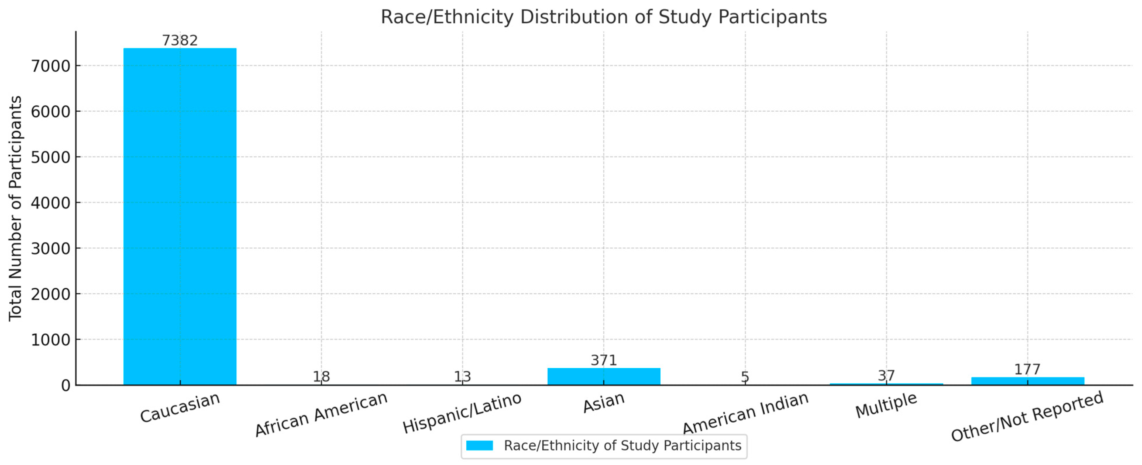
The racial and ethnic representation of participants within each study gave the following results: 92.3% identified as Caucasian, 0.2% as African American, 0.2% as Hispanic/Latino, 4.6% as Asian, 0.06% as American Indian, 0.4% as Multiple, accounting for over 5.06%, and 2.2% were not identified specifically, or identified as “other”. A chi-squared analysis conducted of the racial and ethnic representation within the studies showed a statistically significant difference in the representation (p < 0.0001) (df = 4). This pronounced disparity in representation skewed heavily toward the representation of Caucasian participants, as represented in Table 5 and Figure 2.
Gender analysis of the 8003 study participants was also conducted. The statistical analysis did not reveal a statistically significant difference (p < 0.83) between the representation of females vs. male participants, as the average across all studies revealed 4660 (58%) female participants, and 3343 (42%) male participants (Figure 3) [44].
Lastly, the funding and sponsorship behind each study was analyzed. The goal was to observe if there was a difference in representation within study participants with different funding and sponsorship; and if more equal representation of ethnic and racial groups existed at public vs. private vs. pharmaceutical institutions. Two of the nineteen studies were funded by private research institutions, including a total of 836 participants. Four of the nineteen studies were funded by public research institutions, including a total of 1273 patients. Thirteen of the nineteen studies were funded by various pharmaceutical companies, which analyzed a majority of the patients included, cumulating at 5894 of the total 8003 patients within this review (Table 6). Our analysis did show a statistically significant difference between the ethnic, racial, or gender representation across the various studies (p < 0.001) (Table 4). However, there was no correlation between the study sponsorship (public vs. private vs. pharmaceutical) and the distribution of ethnic, gender, or racial representation.

4. Discussion

The results revealed significant racial disparities in RCT representation for anti-VEGF therapies in AMD, highlighting a gap in current research. These findings highlight the need for improved accuracy in epidemiologic representation in clinical trials to ensure the efficacy and safety of treatments across a larger patient population. As shown in Table 4, Caucasians comprised 92.3% of participants, with all other racial and ethnic groups together accounting for just over 5%. This pronounced underrepresentation underscores the need for future trials to adopt targeted recruitment strategies that mirror real-world disease demographics. Studies have shown that the prevalence of AMD differs significantly among racial groups, with Caucasian populations having the highest prevalence at 5.4%, followed by Chinese (4.6%), Hispanic (4.2%), and African American populations (2.4%) [6]. Despite these differences, the representation in clinical trials does not reflect the actual distribution of AMD in the general population.
Our analysis also uncovered a slight predominance of female participants (58%) compared to males (42%) in AMD trials. While this difference was not statistically significant, women’s higher representation could reflect greater healthcare utilization rates among women or gender-specific recruitment strategies in clinical trials [44]. However, this gender imbalance could implicate potential gaps in trial inclusivity or may be secondary to increased life expectancy amongst women [45,46]. Additionally, unlike other medical specialties, ophthalmology clinical trials may not face the same gender-specific barriers to participation, such as concerns related to pregnancy, reproductive health, or gendered perceptions of disease. Recruitment strategies for AMD trials may therefore be more inclusive by default.
Trial underrepresentation is not unique to AMD trials. Similar patterns have been observed in other clinical trials, where minority groups are also significantly underrepresented [7,46]. Underrepresentation is a historical issue and continues to be a challenge in medical research [47]. The lack of diversity in clinical trials raises concerns about the generalizability of results across different racial and ethnic groups. Genetic and environmental factors that vary among populations can influence disease progression and treatment response [47]. For instance, the risk factors for AMD, including age, family history, smoking, obesity, and cardiovascular disease, may have different impacts across racial groups [48]. The significant underrepresentation of non-Caucasian individuals may contribute to potential disparities in diagnosis, management, and clinical outcomes across diverse populations. This can be seen in a study comparing the same treatment regimen for NVAMD using ranibizumab monotherapy in Caucasian and non-Caucasian populations, showing increased visual acuity improvement in the Caucasian population with fewer overall injections when compared to non-Caucasian populations [49,50,51]. RCT underrepresentation may also mask side effects among understudied populations, leading to clinicians facing uncertainty when prescribing treatments validated primarily in one demographic, potentially leading to hesitancy or inconsistent care for minority patients. As a result, there could be policy implications where regulatory agencies limit drug indications to populations studied in clinical trials, potentially restricting access for underrepresented groups. This can delay or prevent the use of effective therapies in these populations. Although this has not occurred with anti-VEGF medications in AMD, improving trial recruitment will prevent it from ever happening. In December of 2023, the FDA put a mandate in place for improving diversity. Recent policy efforts, such as Diverse and Equitable Participation in Clinical Trials (DEPICT) Act, aim to increase accountability and promote the enrollment of diverse populations in clinical research. These initiatives require sponsors to report participant demographics and implement strategies to improve representation [52].
It is important to note that this stark underrepresentation of non-Caucasian individuals is not unique to AMD research alone. The underrepresentation of non-Caucasian individuals exists in trials analyzing therapeutics for primary open-angle glaucoma and diabetic retinopathy as well [53,54,55,56]. By drawing attention to this lack of representation in current research across the field as a whole, the hope is to spur on equal representation of participants in future studies to make results more generalizable across all patient populations.
Economic considerations further complicate accessibility, as the substantial cost of anti-VEGF therapy necessitates comprehensive cost-effectiveness analyses to manage resource allocation. While specific comparisons of expenditure between AMD, DR, and glaucoma treatments are not readily available in the current literature, the economic burden of these conditions is substantial. A 2022 report estimated the total economic impact of late-stage AMD in the USA at USD 49.1 billion, with USD 27.5 billion in direct healthcare costs and USD 21.6 billion in indirect costs [57]. In comparison, glaucoma-related U.S. healthcare costs are estimated at USD 2.5 billion annually, including USD 1.9 billion in direct costs and USD 0.6 billion in indirect costs [58]. For diabetic eye disease, diabetes-related blindness alone costs approximately USD 500 million per year in the United States, with diabetic retinopathy patients incurring noticeably higher medical costs than those with other diabetes-related conditions [3,4,59].
This is merely a glimpse into the economic burden of ophthalmic conditions, with AMD appearing to have the highest overall economic impact among the three. These costs underscore the importance of early detection, effective management, and the development of cost-effective treatment strategies to mitigate their economic burden.

Outcome Statements: Limitations and Assessment of Bias

This study has several limitations that should be acknowledged. First, the analysis was restricted to clinical trials conducted in the United States, which may limit the generalizability of the findings to global trials. Trials conducted exclusively in the United States were only included to ensure consistency and reliability in the reporting of race and gender demographics, given the standardization requirements for demographic data collection that are mandated by federal regulatory and funding agencies. Second, the inclusion criteria required trials to report both racial and gender demographics, thus excluding studies that did not disclose this information. Third, the categorization of race and ethnicity in clinical trials were inconsistent, with some studies grouping participants into broad categories such as “other”, which may obscure disparities within specific subgroups. Additionally, reliance on published trials may introduce publication bias, as studies with significant findings or industry sponsorship are more likely to be published, potentially skewing the demographic data. We also acknowledge that our reliance on a limited number of databases may have excluded relevant trials, particularly those published in non-indexed journals or conducted outside major funding networks. Finally, a large number of the RCTs included were incidentally funded by pharmaceutical companies. Given trial funding has the potential to influence study design and reporting practices, the difference in funding across the trials included may have also affected the demographic representation as well.
The ROBIS (Risk of Bias in Systematic Reviews) checklist was utilized to analyze the risk of bias within the review [60]. Eligibility criteria were prospectively defined to align with the study’s objective of evaluating racial, ethnic, and gender disparities in AMD trials. Inclusion was restricted to U.S.-based RCTs reporting race, ethnicity, and gender demographics. While these criteria ensured relevance to the U.S. population, they potentially introduced selection bias. To mitigate this, the search strategy incorporated PubMed and cross-referencing with ClinicalTrials.gov to capture a comprehensive sample of eligible trials. Nonetheless, we recognize that this selection bias may prevent the global generalizability of our study data, but the focus of this study was comparing the epidemiology of U.S. RCTs to the U.S. AMD patient population. A systematic PubMed search was conducted using predefined terms to minimize retrieval bias. Three independent reviewers performed title/abstract screening using Rayyan software, followed by full-text reviews, reducing subjectivity. However, the exclusion of non-English studies and reliance on a single database (PubMed) may have introduced language and database biases. To address this, trial registries and pharmaceutical company websites were cross-referenced to identify additional studies. Data extraction was performed independently by five researchers using standardized forms, enhancing reproducibility. Demographic variables were explicitly prioritized, and discrepancies were resolved through consensus. While funding sources were documented, the predominance of industry-sponsored trials (13/19 studies) raises concerns about sponsorship bias, as pharmaceutical-funded trials may selectively report outcomes; however, since this study’s focus is on demographic data, selective outcome reports may not impact this study’s findings as significantly. This is still acknowledged as a potential confounder in interpreting generalizability. Furthermore, the reviewers did not emphasize the results on the basis of their statistical significance, reducing interpretation bias. On the basis of the ROBIS review, there is a low risk of bias within this review [60].

5. Recommendations

In order to address the limitations and disparities identified in this study, we recommend several strategies to improve both transparency and epidemiologic accuracy of clinical research. Regulatory agencies should mandate standardized reporting of race, ethnicity, and gender in clinical trials, thereby enabling more robust meta-analyses of demographic characteristics which allow better tracking participation progress and the strategies’ effectiveness. Researchers are encouraged to adopt targeted recruitment strategies like community engagement, culturally sensitive outreach, and partnerships with healthcare institutions to improve enrollment as needed. This can be achieved via long-term partnerships with patients, advocacy groups, and community leaders from underrepresented populations [61]. Additionally, future clinical trials should incorporate subgroup analyses to analyze the efficacy and safety of anti-VEGF therapies across different racial and ethnic groups, which may help identify variations in treatment response. It is critical to address barriers to clinical trial participation, such as transportation costs, language barriers, and medical mistrust, particularly in the underrepresented population. An actionable strategy to approach these issues includes implementing ongoing cultural competency training for clinical trial staff to enhance understanding and communication with various populations. Additionally, offering multilingual staff and materials can further contribute to building community trust. Additionally, offering financial incentives to participants or free transportation could also address these barriers. These can be funded via funding agencies incentivizing research that accurately reflects epidemiological diversity by prioritizing grants and awards for studies committed to inclusive recruitment, in alignment with the NIH Revitalization Act [62]. Promoting longitudinal studies of demographic trends over time will help evaluate the impact of these initiatives on improving diversity in clinical trials. Lastly, in light of recent federal policy changes, we recommend that future clinical trials adhere to the requirements of the FDA’s DEPICT Act, which directly mandates the creation of Diversity Action Plans, demographic-specific enrollment targets, and transparent reporting of participant diversity [52]. Although most trials in our analysis predate this legislation, aligning future research with these standards will help address persistent disparities and enhance the relevance and equity of clinical evidence in AMD care.

6. Conclusions

This study highlights significant racial disparities in the representation of participants in clinical trials for anti-VEGF therapies targeting AMD. While Caucasian individuals constituted the overwhelming majority of trial participants (92.3%), Asian, African American, Hispanic/Latino, and American Indian groups were collectively underrepresented, making up just over 5%. Although gender representation was more balanced, with females comprising 59% of participants, the lack of racial diversity raises concerns about the generalizability of trial results and the equitable application of these therapies across diverse populations. The findings underscore the urgent need for more inclusive clinical trial practices to ensure that anti-VEGF treatments are safe and effective for all patients, regardless of race or ethnicity. Genetic, environmental, and socioeconomic factors that vary across populations can influence disease progression and treatment response, making diverse representation essential for developing evidence-based therapies. Addressing disparities requires improving recruitment strategies, standardizing demographic reporting, and conducting targeted studies in underrepresented populations.
In the meantime, to support and educate patients on modifiable risk factors of AMD should continue to be emphasized within the field [31]. Smoking is associated with a 2–4× increased risk of AMD diagnosis, and more rapid disease progression to geographic atrophy after diagnosis [9,10,27,56,57]. Smoking cessation is imperative to encourage; however, it is important to note, even after cessation, prior smokers may still have a modestly increased risk of disease progression due to the pathological oxidative effects of cigarette smoke within the RPE cells and choroidal vessels [28,51]. Diets high in antioxidant carotenoids, such as lutein and zeaxanthin, in green leafy vegetables, are encouraged as they may provide a protective benefit [4]. Thus, many clinicians continue to recommend adhering to a diet similar to the Mediterranean diet rather than specifying adherence to a low margarine consumption. The Mediterranean diet is rich in antioxidants, and it naturally is high in fruits, vegetables, grains, nuts, lean meat and fish, dairy, limited red meat, and preference of olive oil over butter [27,28]. Other highly associated risk factors include cardiovascular disease, hypertension, atherosclerosis, obesity, and sunlight exposure given each of their associations with increasing retinal oxidative stress and suspected disease progression (see Table 1) [28].
In conclusion, as the global burden of AMD continues to rise, fostering diversity in research will be critical to developing treatments that are effective, safe, and applicable to the full spectrum of patients affected by this debilitating condition. Future research should incorporate intentional strategies to enhance racial and ethnic diversity in clinical trial enrollment, mandate standardized demographic reporting, enable adequality powered subgroup analyses, and investigate population specific genetic, environmental, and social determinants to improve the external validity and equality of AMD treatment outcomes. We urge researchers to prioritize diverse recruitment, clinicians to advocate for representative studies, and policymakers to support and enforce inclusive clinical trial standards.

Author Contributions

Conceptualization: K.A., A.S. and D.P.; Methodology: K.A., A.S. and T.J.; Software: A.S., T.J. and I.Z.; Formal Analysis: T.J., A.S. and I.Z.; Investigation: T.J., A.S. and I.Z.; Data Curation: T.J., A.S., I.Z. and K.A.; Writing—original Draft Preparation: T.J., A.S., I.Z., D.P. and K.A.; Writing—review and editing: T.J., A.S., I.Z. and K.A.; Visualization: A.S., D.P. and K.A.; Supervision: K.A.; Project Administration: K.A.; Funding Acquisition: K.A. All authors have read and agreed to the published version of the manuscript.

Funding

This research received no external funding.

Acknowledgments

During the preparation of this manuscript, the authors utilized Rayyan software, an AI-powered systematic Review Management Platform (Rayyan v4.0) for the purposes of identifying RCTs evaluating intravitreal anti-VEGF therapy for AMD. The authors have reviewed and edited the output and take full responsibility for the content of this publication.

Conflicts of Interest

The authors declare no conflict of interest.

References

  1. Wong, W.L.; Su, X.; Li, X.; Cheung, C.M.; Klein, R.; Cheng, C.Y.; Wong, T.Y. Global prevalence of age-related macular degeneration and disease burden projection for 2020 and 2040: A systematic review and meta-analysis. Lancet Glob. Health 2014, 2, 106. [Google Scholar] [CrossRef]
  2. Fleckenstein, M.; Schmitz-Valckenberg, S.; Chakravarthy, U. Age-Related Macular Degeneration: A Review. JAMA 2024, 331, 147–157. [Google Scholar] [CrossRef]
  3. de Jong, P.T.V.M. Elusive drusen and changing terminology of AMD. R. Coll. Ophthalmol. 2018, 32, 904–914. [Google Scholar] [CrossRef]
  4. de Jong, P.T.V.M. A historical analysis of the quest for the origins of aging macula disorder, the tissues involved, and its terminology. Ophthalmol. Eye Dis. 2016, 8 (Suppl. S1), 5–14. [Google Scholar] [CrossRef]
  5. Bird, A.C. Age-related macular disease: Aetiology and clinical management. Community Eye Health 1999, 12, 8–9. [Google Scholar] [PubMed]
  6. Mitchell, P.; Liew, G.; Gopinath, B.; Wong, T.Y. Age-related macular degeneration. Lancet 2018, 392, 1147–1159. [Google Scholar] [CrossRef]
  7. Klein, R.; Klein, B.E.K.; Knudtson, M.D.; Wong, T.Y.; Cotch, M.F.; Liu, K.; Burke, G.; Saad, M.F.; Jacobs, D.R., Jr. Prevalence of age-related macular degeneration in 4 racial/ethnic groups in the multi-ethnic study of atherosclerosis. Ophthalmology 2006, 113, 373–380. [Google Scholar] [CrossRef] [PubMed]
  8. Kodjikian, L.; Rezkallah, A.; Decullier, E.; Aulagner, G.; Huot, L.; Mathis, T.; GEFAL Study Group. Early predictive factors of visual loss at 1 year in neovascular age-related macular degeneration under anti-vascular endothelial growth factor. Ophthalmol. Retin. 2022, 6, 109–115. [Google Scholar] [CrossRef]
  9. Tomany, S.C.; Wang, J.J.; Van Leeuwen, R.; Klein, R.; Mitchell, P.; Vingerling, J.R.; Klein, B.E.; Smith, W.; De Jong, P.T. Risk factors for incident age-related macular degeneration: Pooled findings from 3 continents. Ophthalmology 2004, 111, 1280–1287. [Google Scholar] [CrossRef] [PubMed]
  10. Tomany, S.C.; Cruickshanks, K.J.; Klein, R.; Klein, B.E.K.; Knudtson, M.D. Sunlight and the 10-year incidence of age-related maculopathy: The beaver dam eye study. Arch. Ophthalmol. 2004, 122, 750–757. [Google Scholar] [CrossRef]
  11. Whitescarver, T.D.; Hobbs, S.D.; Wade, C.I.; Winegar, J.W.; Colyer, M.H.; Reddy, A.K.; Drayna, P.M.; Justin, G.A. A history of anti-VEGF inhibitors in the ophthalmic literature: A bibliographic review. J. Vitreoretin. Dis. 2020, 5, 304–312. [Google Scholar] [CrossRef] [PubMed]
  12. Fisher, D.E.; Klein, B.E.K.; Wong, T.Y.; Rotter, J.I.; Li, X.; Shrager, S.; Burke, G.L.; Klein, R.; Cotch, M.F. Incidence of age-related macular degeneration in a multi-ethnic united states population: The multi-ethnic study of atherosclerosis. Ophthalmology 2016, 123, 1297–1308. [Google Scholar] [CrossRef]
  13. Berkowitz, S.T.; Groth, S.L.; Gangaputra, S.; Patel, S. Racial/ethnic disparities in ophthalmology clinical trials resulting in US food and drug administration drug approvals from 2000 to 2020. JAMA Ophthalmol. 2021, 139, 629–637. [Google Scholar] [CrossRef] [PubMed]
  14. Qu, C.; Zheng, Y.; Zhou, M.; Zhang, Y.; Shen, F.; Cao, J.; Xu, L.M. Value of bevacizumab in treatment of colorectal cancer: A meta-analysis. World J. Gastroenterol. 2015, 21, 5072–5080. [Google Scholar] [CrossRef]
  15. Dickson, S.R.; James, K.E. Medicare Part B Spending on Macular Degeneration Treatments Associated with Manufacturer Payments to Ophthalmologists. JAMA Health Forum. 2023, 4, e232951. [Google Scholar] [CrossRef] [PubMed]
  16. Rosenfeld, P.J.; Brown, D.M.; Heier, J.S.; Boyer, D.S.; Kaiser, P.K.; Chung, C.Y.; Kim, R.Y.; MARINA Study Group. Ranibizumab for neovascular age-related macular degeneration. N. Engl. J. Med. 2006, 355, 1419–1431. [Google Scholar] [CrossRef]
  17. Rosenfeld, P.J.; Moshfeghi, A.A.; Puliafito, C.A. Optical coherence tomography findings after an intravitreal injection of bevacizumab (avastin) for neovascular age-related macular degeneration. Ophthalmic Surg. Lasers Imaging 2005, 36, 331–335. [Google Scholar] [CrossRef]
  18. Holz, F.G.; Dugel, P.U.; Weissgerber, G.; Hamilton, R.; Silva, R.; Bandello, F.; Larsen, M.; Weichselberger, A.; Wenzel, A.; Schmidt, A.; et al. Single-chain antibody fragment VEGF inhibitor RTH258 for neovascular age-related macular degeneration: A randomized controlled study. Ophthalmology 2016, 123, 1080–1089. [Google Scholar] [CrossRef]
  19. Brown, G.C.; Brown, M.M.; Rapuano, S.B.; Boyer, D. A cost-benefit analysis of VEGF-inhibitor therapy for neovascular age-related macular degeneration in the united states. Am. J. Ophthalmol. 2021, 223, 405–429. [Google Scholar] [CrossRef]
  20. Lushchyk, T.; Amarakoon, S.; Martinez-Ciriano, J.P.; van den Born, L.I.; Baarsma, G.S.; Missotten, T. Bevacizumab in age-related macular degeneration: A randomized controlled trial on the effect of injections every 4 weeks, 6 weeks and 8 weeks. Acta Ophthalmol. 2013, 91, 456. [Google Scholar] [CrossRef]
  21. Schauwvlieghe, A.M.E.; Dijkman, G.; Hooymans, J.M.; Verbraak, F.D.; Hoyng, C.B.; Dijkgraaf, M.G.; Peto, T.; Vingerling, J.R.; Schlingemann, R.O. Comparing the effectiveness of bevacizumab to ranibizumab in patients with exudative age-related macular degeneration. the BRAMD study. PLoS ONE 2016, 11, e0153052. [Google Scholar] [CrossRef]
  22. Diabetic Retinopathy Clinical Research Network; Wells, J.A.; Glassman, A.R.; Jampol, L.M.; Aiello, L.P.; Antoszyk, A.N.; Arnold-Bush, B.; Baker, C.W.; Bressler, N.M.; Browning, D.J.; et al. Aflibercept, bevacizumab, or ranibizumab for diabetic macular edema. N. Engl. J. Med. 2015, 372, 1193–1203. [Google Scholar] [CrossRef] [PubMed]
  23. Brown, D.M.; Kaiser, P.K.; Michels, M.; Soubrane, G.; Heier, J.S.; Kim, R.Y.; Sy, J.P.; Schneider, S.; ANCHOR Study Group. Ranibizumab versus verteporfin for neovascular age-related macular degeneration. N. Engl. J. Med. 2006, 355, 1432–1444. [Google Scholar] [CrossRef]
  24. Heier, J.S.; Khanani, A.M.; Quezada Ruiz, C.; Basu, K.; Ferrone, P.J.; Brittain, C.; Figueroa, M.S.; Lin, H.; Holz, F.G.; Patel, V.; et al. Efficacy, durability, and safety of intravitreal faricimab up to every 16 weeks for neovascular age-related macular degeneration (TENAYA and LUCERNE): Two randomised, double-masked, phase 3, non-inferiority trials. Lancet 2022, 399, 729–740. [Google Scholar] [CrossRef] [PubMed]
  25. Meyer, C.H.; Holz, F.G. Preclinical aspects of anti-VEGF agents for the treatment of wet AMD: Ranibizumab and bevacizumab. Eye 2011, 25, 661–672. [Google Scholar] [CrossRef]
  26. Heier, J.S.; Wykoff, C.C.; Waheed, N.K.; Kitchens, J.W.; Patel, S.S.; Vitti, R.; Perlee, L.; Chu, K.W.; Leal, S.; Asmus, F.; et al. Intravitreal combined aflibercept + anti-platelet-derived growth factor receptor β for neovascular age-related macular degeneration: Results of the phase 2 CAPELLA trial. Ophthalmology 2020, 127, 211–220. [Google Scholar] [CrossRef]
  27. Merle, B.M.J.; Silver, R.E.; Rosner, B.; Seddon, J.M. Adherence to a mediterranean diet, genetic susceptibility, and progression to advanced macular degeneration: A prospective cohort study. Am. J. Clin. Nutr. 2015, 102, 1196–1206. [Google Scholar] [CrossRef]
  28. Merle, B.M.J.; Colijn, J.M.; Cougnard-Grégoire, A.; de Koning-Backus, A.P.M.; Delyfer, M.N.; Kiefte-de Jong, J.C.; Meester-Smoor, M.; Féart, C.; Verzijden, T.; Samieri, C.; et al. Mediterranean diet and incidence of advanced age-related macular degeneration: The EYE-RISK consortium. Ophthalmology 2019, 126, 381–390. [Google Scholar] [CrossRef] [PubMed]
  29. Jaffe, G.J.; Ciulla, T.A.; Ciardella, A.P.; Devin, F.; Dugel, P.U.; Eandi, C.M.; Masonson, H.; Monés, J.; Pearlman, J.A.; Quaranta-El Maftouhi, M.; et al. Dual antagonism of PDGF and VEGF in neovascular age-related macular degeneration: A phase IIb, multicenter, randomized controlled trial. Ophthalmology 2017, 124, 224–234. [Google Scholar] [CrossRef]
  30. Böhni, S.C.; Bittner, M.; Howell, J.P.; Bachmann, L.M.; Faes, L.; Schmid, M.K. Comparison of eylea® with lucentis® as first-line therapy in patients with treatment-naïve neovascular age-related macular degeneration in real-life clinical practice: Retrospective case-series analysis. BMC Ophthalmol. 2015, 15, 109. [Google Scholar] [CrossRef]
  31. García-Quintanilla, L.; Luaces-Rodríguez, A.; Gil-Martínez, M.; Mondelo-García, C.; Maroñas, O.; Mangas-Sanjuan, V.; González-Barcia, M.; Zarra-Ferro, I.; Aguiar, P.; Otero-Espinar, F.J.; et al. Pharmacokinetics of intravitreal anti-VEGF drugs in age-related macular degeneration. Pharmaceutics 2019, 11, 365. [Google Scholar] [CrossRef] [PubMed]
  32. Meer, E.A.; Oh, D.H.; Brodie, F.L. Time and distance cost of longer acting anti-VEGF therapies for macular degeneration: Contributions to drug cost comparisons. Clin. Ophthalmol. 2022, 16, 4273–4279. [Google Scholar] [CrossRef] [PubMed]
  33. Eldem, B.M.; Muftuoglu, G.; Topbaş, S.; Çakir, M.; Kadayifcilar, S.; Özmert, E.; Bahçecioğlu, H.; Sahin, F.; Sevgi, S.; SALUTE Study Group. A randomized trial to compare the safety and efficacy of two ranibizumab dosing regimens in a turkish cohort of patients with choroidal neovascularization secondary to AMD. Acta Ophthalmol. 2015, 93, 458. [Google Scholar] [CrossRef]
  34. Lally, D.R.; Hill, L.; Amador-Patarroyo, M.J. Subretinal fluid resolution and visual acuity in patients with neovascular age-related macular degeneration: A HARBOR post hoc analysis. Ophthalmol. Retin. 2022, 6, 1054–1060. [Google Scholar] [CrossRef]
  35. Khanani, A.M.; Patel, S.S.; Ferrone, P.J.; Osborne, A.; Sahni, J.; Grzeschik, S.; Basu, K.; Ehrlich, J.S.; Haskova, Z.; Dugel, P.U. Efficacy of every four monthly and quarterly dosing of faricimab vs ranibizumab in neovascular age-related macular degeneration: The STAIRWAY phase 2 randomized clinical trial. JAMA Ophthalmol. 2020, 138, 964–972. [Google Scholar] [CrossRef]
  36. Ross, E.L.; Hutton, D.W.; Stein, J.D.; Bressler, N.M.; Jampol, L.M.; Glassman, A.R.; Diabetic Retinopathy Clinical Research Network. Cost-effectiveness of aflibercept, bevacizumab, and ranibizumab for diabetic macular edema treatment: Analysis from the diabetic retinopathy clinical research network comparative effectiveness trial. JAMA Ophthalmol. 2016, 134, 888–896. [Google Scholar] [CrossRef]
  37. Jackson, T.L.; Slakter, J.; Buyse, M.; Wang, K.; Dugel, P.U.; Wykoff, C.C.; Boyer, D.S.; Gerometta, M.; Baldwin, M.E.; Price, C.F. A randomized controlled trial of OPT-302, a VEGF-C/D inhibitor for neovascular age-related macular degeneration. Ophthalmology 2023, 130, 588–597. [Google Scholar] [CrossRef]
  38. Kertes, P.J.; Galic, I.J.; Greve, M.; Williams, G.; Baker, J.; Lahaie, M.; Sheidow, T. Efficacy of a treat-and-extend regimen with ranibizumab in patients with neovascular age-related macular disease: A randomized clinical trial. JAMA Ophthalmol. 2020, 138, 244–250. [Google Scholar] [CrossRef]
  39. Poor, S.H.; Weissgerber, G.; Adams, C.M.; Bhatt, H.; Browning, D.J.; Chastain, J.; Ciulla, T.A.; Ferriere, M.; Gedif, K.; Glazer, L.C.; et al. A randomized, double-masked, multicenter trial of topical acrizanib (LHA510), a tyrosine kinase VEGF-receptor inhibitor, in treatment-experienced subjects with neovascular age-related macular degeneration. Am. J. Ophthalmol. 2022, 239, 180–189. [Google Scholar] [CrossRef]
  40. Dugel, P.U.; Koh, A.; Ogura, Y.; Dugel, P.U.; Koh, A.; Ogura, Y.; Jaffe, G.J.; Schmidt-Erfurth, U.; Brown, D.M.; Gomes, A.V.; et al. HAWK and HARRIER: Phase 3, multicenter, randomized, double-masked trials of brolucizumab for neovascular age-related macular degeneration. Ophthalmology 2020, 127, 72–84. [Google Scholar] [CrossRef] [PubMed]
  41. CATT Research Group; Martin, D.F.; Maguire, M.G.; Maguire, M.G.; Ying, G.S.; Grunwald, J.E.; Fine, S.L.; Jaffe, G.J. Ranibizumab and bevacizumab for neovascular age-related macular degeneration. N. Engl. J. Med. 2011, 364, 1897–1908. [Google Scholar] [CrossRef]
  42. Tricco, A.C.; Lillie, E.; Zarin, W.; O’Brien, K.K.; Colquhoun, H.; Levac, D.; Moher, D.; Peters, M.D.J.; Horsley, T.; Weeks, L.; et al. PRISMA Extension for Scoping Reviews (PRISMA-ScR): Checklist and Explanation. Ann. Intern. Med. 2018, 169, 467–473. [Google Scholar] [CrossRef]
  43. Mehta, A.; Steel, D.H.; Muldrew, A.; Peto, T.; Reeves, B.C.; Evans, R.; Chakravarthy, U.; IVAN Study Investigators. Associations and Outcomes of Patients with Submacular Hemorrhage Secondary to Age-related Macular Degeneration in the IVAN Trial. Am. J. Ophthalmol. 2022, 236, 89–98. [Google Scholar] [CrossRef] [PubMed]
  44. Colijn, J.M.; Buitendijk, G.H.S.; Prokofyeva, E.; Alves, D.; Cachulo, M.L.; Khawaja, A.P.; Cougnard-Gregoire, A.; Merle, B.M.J.; Korb, C.; Erke, M.G.; et al. Prevalence of age-related macular degeneration in europe: The past and the future. Ophthalmology 2017, 124, 1753–1763. [Google Scholar] [CrossRef] [PubMed]
  45. Galdas, P.M.; Cheater, F.; Marshall, P. Men and health help-seeking behaviour: Literature review. J. Adv. Nurs. 2005, 49, 616–623. [Google Scholar] [CrossRef] [PubMed]
  46. Ibrahim, F.N.; Sivaprasad, S.; Cheung, C.M.G. Gender and ethnic diversity in randomised clinical trials in age-related macular degeneration and diabetic macular oedema. Eye 2025, 39, 1249–1253. [Google Scholar] [CrossRef]
  47. Oh, S.S.; Galanter, J.; Thakur, N.; Pino-Yanes, M.; Barcelo, N.E.; White, M.J.; de Bruin, D.M.; Greenblatt, R.M.; Bibbins-Domingo, K.; Wu, A.H.; et al. Diversity in clinical and biomedical research: A promise yet to be fulfilled. PLoS Med. 2015, 12, e1001918. [Google Scholar] [CrossRef]
  48. Schaneman, J.; Kagey, A.; Soltesz, S.; Stone, J. The role of comprehensive eye exams in the early detection of diabetes and other chronic diseases in an employed population. Popul. Health Manag. 2010, 13, 195–199. [Google Scholar] [CrossRef]
  49. Mohamed, R.; Gadhvi, K.; Mensah, E. What Effect Does Ethnicity Have on the Response to Ranibizumab Therapy in Neovascular Age-Related Macular Degeneration? Ophthalmologica 2018, 240, 157–165. [Google Scholar] [CrossRef] [PubMed]
  50. Ogurtsova, K.; da Rocha Fernandes, J.D.; Huang, Y.; Linnenkamp, U.; Guariguata, L.; Cho, N.H.; Cavan, D.; Shaw, J.E.; Makaroff, L.E. IDF diabetes atlas: Global estimates for the prevalence of diabetes for 2015 and 2040. Diabetes Res. Clin. Pract. 2017, 128, 40–50. [Google Scholar] [CrossRef]
  51. Yau, J.W.Y.; Rogers, S.L.; Kawasaki, R.; Lamoureux, E.L.; Kowalski, J.W.; Bek, T.; Chen, S.J.; Dekker, J.M.; Fletcher, A.; Grauslund, J.; et al. Global prevalence and major risk factors of diabetic retinopathy. Diabetes Care 2012, 35, 556–564. [Google Scholar] [CrossRef]
  52. Guymer, R.H.; Chong, E.W. Modifiable risk factors for age-related macular degeneration. Med. J. Aust. 2006, 184, 455–458. [Google Scholar] [CrossRef]
  53. Di Muro, F.M.; Dangas, K.; Ortega, R.; Vogel, B.; Batchelor, W.B.; Douglas, P.S.; Mehran, R. Preserving and Promoting Clinical Trial Representativeness: A Review of Existing Strategies and the Path Forward. JAMA Cardiol. 2025. [Google Scholar] [CrossRef]
  54. Allison, K.; Patel, D.G.; Greene, L. Racial and Ethnic Disparities in Primary Open-Angle Glaucoma Clinical Trials: A Systematic Review and Meta-analysis. JAMA Netw. Open 2021, 4, e218348. [Google Scholar] [CrossRef]
  55. Greenlee, T.E.; Malhotra, N.A.; Iyer, A.I.; Conti, T.F.; Chen, A.X.; Singh, R.P. Association of Socioeconomic Health Care Disparities with Use of Anti-Vascular Endothelial Growth Factor and Visual Acuity Outcomes in Patients with Diabetic Macular Edema. Ophthalmic Surg. Lasers Imaging Retin. 2022, 53, 380–391. [Google Scholar] [CrossRef] [PubMed]
  56. Guttman Krader, C. Identifying Gaps in Anti-VEGF Treatment Among Minorities with Diabetic Macular Edema. Modern Retina. 1 December 2020. Available online: https://www.modernretina.com/view/identifying-gaps-in-anti-vegf-treatment-among-minorities-with-dme (accessed on 7 May 2025).
  57. Feldman, R.M.; Cioffi, G.A.; Liebmann, J.M.; Weinreb, R.N. Current knowledge and attitudes concerning cost-effectiveness in glau-coma pharmacotherapy: A glaucoma specialists focus group study. Clin. Ophthalmol. 2020, 14, 729–739. [Google Scholar] [CrossRef]
  58. Lee, P.P.; Walt, J.G.; Doyle, J.J.; Kotak, S.V.; Evans, S.J.; Budenz, D.L.; Chen, P.P.; Coleman, A.L.; Feldman, R.M.; Jampel, H.D.; et al. A Multicenter, Retrospective Pilot Study of Resource Use and Costs Associated with Severity of Disease in Glaucoma. Arch. Ophthalmol. 2006, 124, 12–19. [Google Scholar] [CrossRef] [PubMed]
  59. Zhang, X.; Low, S.; Kumari, N.; Wang, J.; Ang, K.; Yeo, D.; Yip, C.C.; Tavintharan, S.; Sum, C.F.; Lim, S.C. Direct medical cost associated with diabetic retinopathy severity in type 2 diabetes in sin-gapore. PLoS ONE 2017, 12, e0180949. [Google Scholar] [CrossRef]
  60. ROBIS (Risk of Bias in Systematic Reviews). Available online: https://www.bristol.ac.uk/population-health-sciences/projects/robis/ (accessed on 7 May 2025).
  61. Oyer, R.A.; Hurley, P.; Boehmer, L.; Steeby Bruinooge, S.; Levit, K.; Barrett, N.; Benson, A.B.; Bernick, L.A.; Byatt, L.; Charlot, M.; et al. Increasing Racial and Ethnic Diversity in Cancer Clinical Trials: An American Society of Clinical Oncology and Association of Community Cancer Centers Joint Research Statement. J. Clin. Oncol. 2022, 40, 2163–2171. [Google Scholar] [CrossRef]
  62. Cruz, J.G.; Patel, D.; Allison, K. Diabetic Retinopathy and Vision Loss: A Public Health Concern. J. Biomed. Res. Environ. Sci. 2024, 5, 691–698. [Google Scholar] [CrossRef]
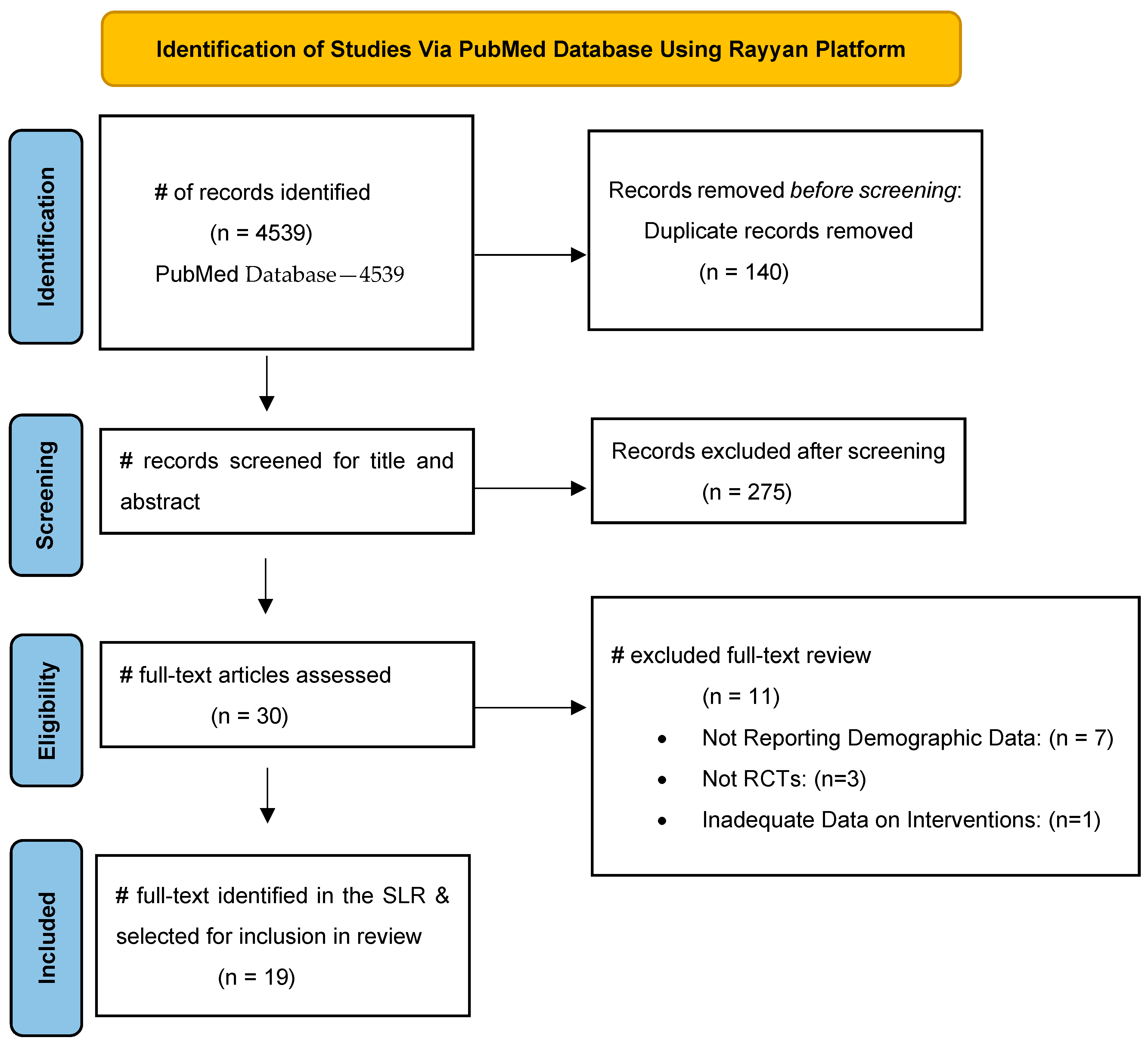
Figure 1. PRISMA flowchart. Identification of studies via PubMed database using Rayyan.
Jcto 03 00016 g001
Figure 2. Race and ethnic distribution of study participants.
Jcto 03 00016 g002
Figure 3. Gender distribution of study participants.
Jcto 03 00016 g003
Table 1. Description of associated risk factors of AMD.
Risk FactorDescription
AgeAge is the most significant risk factor for the development of ARMD. The incidence of ARMD increases with age, particularly after the age of 50. Diagnosis is most common in individuals over the age of 65 specifically
Genetic FactorsFamily history of exudative ARMD significantly increases the risk. Specific genetic variations have also been linked to higher susceptibility (CFH, ARMS2)
Female GenderWomen are more likely to develop ARMD than men, likely in part due to longer life expectancy and hormonal factors (increased estrogen)
SmokingSmoking is one of the proven modifiable risk factors in the development of ARMD. There is a two- to three-fold increased risk of ARMD among smokers
Race and EthnicityCaucasians are at a higher risk compared to other racial groups. Hispanics and African Americans have a lower risk of a development.
Hypertension (High Blood Pressure)Uncontrolled sustained hypertension can result in microvascular retinal complications, increasing the risk of ARMD secondary to pre-existing retinal damage.
ObesityObesity results in microvascular and macrovascular complications systemically, as well as in the retina. Associated presence of metabolic syndrome and high cholesterol also slightly increases the risk
Dietary FactorsLow intake of antioxidants, including, vitamins C, E, zinc, and omega-3 fatty acids increases the risk. A poor diet lacking green leafy vegetables and high in saturated fats has been linked with a higher prevalence
Prolonged Sunlight ExposureUltraviolet light from the sun (UV-A and UV-B) increases the risk of ARMD, due to the cumulative retina damage over time.
Light-Colored EyesSimilar to prolonged sunlight exposure, light-colored eyes (blue, green, etc.) have an increased risk due to a decreased amount of UV protection
Cardiovascular DiseasePresence of diagnosed cardiovascular diseases including, heart disease, atherosclerosis, hyper cholesterol, etc., can impact retinal blood flow, contributing to ARMD development
Prolonged Use of Specific MedicationsVasodilators: Prolonged use of vasodilating agents (ex: nitrates, beta blockers, calcium channel blockers) can impact retinal circulation long term
Beta Blockers: Similar to vasodilators as above, decreased pressure and circulating volume can result in decreased retinal circulation, increasing risk of ARMD
Calcium Channel Blockers: Both dihydropyridine and non-dihydropyridine subtypes may increase vasculature leak, contributing to the pathogenesis of ARMD
Thiazide Diuretics: Long term use of thiazide diuretics can impact electrolyte balance directly, resulting in aberrant retinal circulation
Low Serum Levels of Lutein and ZeaxanthinCarotenoids found in green leafy vegetables can help to protect the retina from oxidative damage long-term. Low serum levels of carotenoids for an extended period of time can result in increased risk of ARMD development
Cataract SurgeryCataract surgery can change the mechanism of retinal exposure to UV exposure from sunlight. Specifically, patients who underwent cataract surgery at a young age have a slightly increased risk of ARMD development
Chronic Systemic and/or Ocular InflammationProlonged systemic and/or retinal inflammation have both been linked to a slightly increased risk over ARMD development. The low-grade background inflammation is suspected to contribute to eventual retinal damage
Table 2. Properties of anti-vascular endothelial growth factor (VEGF) antibodies for age-related macular degeneration (AMD).
PropertiesRanibizumab (Lucentis)Bevacizumab (Avastin)Aflibercept (Eylea)Faricimab (Vabysmo)RTH258 (Brolucizumab)Acrizanib
Year of FDA Approval2006N/A201120222019N/A
ClassAntibody FragmentMonoclonal AntibodyFusion ProteinAntibody FragmentAntibody FragmentProtein Tyrosine Kinase
MW (KDa)48149115149260.4454
Net ChargeNegativeNegativeSlightly PositiveNegativeNegativeNegative
Binding TargetVEGF-AVEGF-AVEGF-A, VEGF-B, PlGFVEGF-A, Ang-2VEGF-AVEGFR-1, EGFR
Table 3. Medicare Part B cost and add-on payments for age-related macular degeneration therapies.
TherapyMean Spending per Unit, USDMean Units per ClaimMean Spending per Claim, USDMean No. of Claims per Beneficiary per YearMean Annual Spending per Beneficiary, USDMean Annual Add-On Payment per Beneficiary, USD
Bevacizumab (Avastin)76.32141068.483.94167.07171.80
Aflibercept (Eylea)923.5621847.125.09235.60380.76
Ranibizumab (Lucentis)333.5551667.755.18505.52350.66
Under the Medicare Part B program, ophthalmologists are reimbursed at the average sales price (ASP) for a drug plus an add-on payment (6% of the ASP by statute but was limited to 4.3% from 2013–2024 due to a federal budget sequester). The mean annual add-on payment is calculated as the difference between mean annual spending and mean annual spending divided by 1.043, as the mean annual spending already includes the add-on amount. b Mean spending per unit values were from the 2020 Medicare Part B dashboard. c Mean units per claim and mean claims per beneficiary per year were calculated from Medicare Physician and Other Practitioners’ utilization data from 2020 for ophthalmologists.
Table 4. Overview of study characteristics included within results and analysis; part 1 [2,16,18,19,20,21,22,23,24,25,26,29,30,35,36,37,38,39,43].
First Author, Year PublishedMean Age (Years)Male (%)Female (%)Intravitreal Drug UsedFinal Data Stratified by Ethnicity or Race
Rosenfeld, 20067735.264.8RanibizumabNo
Heier, 20127642.957.1Ranibizumab/AfliberceptNo
Heier, 2022>5037.362.7FaricimabNo
Lushchyk, 2013>654060BevacizumabNo
Holz, 20167646.153.9RTH258No
Schauwvlieghe, 2016784555Bevacizumab/RanibizumabNo
Khanani, 2020794258Ranibizumab/FaricimabNo
Eldem, 2015705248RanibizumabNo
Kertes, 20207939.760.3RanibizumabNo
Lally, 20227740.659.4RanibizumabNo
Jaffee, 2017783862RanibizumabNo
Poor, 20227754.445.6Topical AcrizanibNo
Jackson, 20237639.760.3RanibizumabNo
Khanani, 20227645.254.8Brolucizumab/AfliberceptNo
Nguyen, 20127534.565.5RanibizumabNo
Mohamed, 2018863367RanibizumabYes
Heier, 2020794060Ranibizumab/AfliberceptNo
Gillies, 2020774951Ranibizumab/AfliberceptNo
Mehta, 2022793961Bevacizumab/RanibizumabNo
Table 5. Overview of study characteristics included within results and analysis; part 2 [2,16,18,19,20,21,22,23,24,25,26,29,30,35,36,37,38,39,43].
First Author, Year PublishedStudy DesignTrial Duration (Months)Number of Patients AnalyzedWhite (%)Black or African American (%)Hispanic or Latino (%)Asian (%)American Indian or Alaska Native (%)Multiple (%)Not Reported or Other (%)
Rosenfeld, 2006RCT2471696.6000003.4
Heier, 2012RCT12241984.70.311.20003.8
Heier, 2022RCT2813393.621.403.20.20.40.4
Lushchyk, 2013RCT12120100000000
Holz, 2016RCT619498.60001.400
Schauwvlieghe, 2016RCT1232798000020
Khanani, 2020RCT127697.61.401.1000
Eldem, 2015RCT1277100000000
Kertes, 2020RCT2446694.300005.70
Lally, 2022RCT1834997.40.23.62.10.20.30
Jaffee, 2017RCT644997.6000002.4
Poor, 2022RCT39087.8008.9000
Jackson, 2023RCT1136698.6000001.4
Khanani, 2022RCT2453597.80.600.9000.6
Nguyen, 2012RCT1015175.90024.1000
Mohamed, 2018RCT5217840000016
Heier, 2020RCT250593.60.604.70.20.40
Gillies, 2020RCT327893.30.405.3001
Mehta, 2022RCT253599000000
Table 6. Distribution of study sponsorship and patient enrollment.
Sponsorship SourceNumber of StudiesTotal PatientsGender Distribution (Number of Patients)Racial Distribution (# of Patients)
MaleFemaleCaucasianAfrican AmericanHispanicAsianAI *M **NR ***
Private28363005368120000024
Public4127351775612230003047
Pharmaceutical1358942483341153471813371237106
Total1980033300470373821813371537177
* = American Indian/Alaska Native. ** = Multiple. *** = Not Reported or Other.
Disclaimer/Publisher’s Note: The statements, opinions and data contained in all publications are solely those of the individual author(s) and contributor(s) and not of MDPI and/or the editor(s). MDPI and/or the editor(s) disclaim responsibility for any injury to people or property resulting from any ideas, methods, instructions or products referred to in the content.