Case Report

Visual Rehabilitation in Post Mild Traumatic Brain Injury. Case-Based Review

by
Carmen López-de-la-Fuente
*,
Hermes Barriga-Longás
and
Elvira Orduna-Hospital
*
Department of Applied Physics, University of Zaragoza, 50009 Zaragoza, Spain
*
Authors to whom correspondence should be addressed.
J. Clin. Transl. Ophthalmol. 2023, 1(1), 25-34; https://doi.org/10.3390/jcto1010005
Submission received: 13 December 2022 / Revised: 5 January 2023 / Accepted: 21 February 2023 / Published: 24 February 2023

Abstract

:
Background: Traumatic brain injury (TBI) can cause visual dysfunction affecting binocularity, spatial orientation, posture, and balance. Currently, there are several options for treating manifested visual disturbances; vision therapy is one of the possible treatment options. Methods: A 14-year-old female fainted and sustained trauma to the upper-temporal part of her right eyebrow. The patient presented with eye pain, a decreased visual field, and blurred vision and she exhibited great difficulties when reading. Both neurological tests and exploration of the anterior and posterior ocular segments showed results within normal limits. The patient was diagnosed with fusional vergence dysfunction, associated with accommodative infacility and oculomotor dysfunction. To eliminate her symptoms, a visual rehabilitation program was implemented; it consisted of accommodative, anti-suppressive, vergential, motility, hand–eye coordination, and peripheral vision exercises. Results: The symptoms manifested by the patient gradually dissipated throughout the course of therapy. However, not all optometric parameters reached normal values after visual therapy, which could indicate a traumatic injury that limited the achievement of normal optometric ranges. Conclusions: After a mild traumatic brain injury (mTBI), it is necessary to carry out a complete examination of the patient’s visual function. This visual examination must include an analysis of vergences, ocular motility, and the accommodative system to obtain an accurate diagnosis. The outcomes of the present clinical case imply that visual therapy should be considered as a treatment in cases of mTBIs.

1. Introduction

“Concussion or Traumatic brain injury (TBI) is defined as a head injury with a Glasgow Coma Scale (GCS) between 13 and 15, and neuroimaging, if performed, does not demonstrate an acute abnormality” [1]. A mild traumatic brain injury (mTBI) has a better prognosis if diagnosed and treated early [2] and can cause visual dysfunction affecting binocularity, spatial orientation, posture, and balance [3]. It is a common injury after road accidents and falls, accounting for between 70% and 90% of mild brain injuries [4]. An mTBI, or concussion, occurs after a severe impact to the head, directly or indirectly, that causes a change in brain function [5].
Generally, patients who suffer an mTBI do not require hospitalisation, although they may experience a multitude of motor, sensory, perceptual, and psychological difficulties, as well as different visual problems, at some point. Many neurological signs and symptoms caused by an mTBI can be very subtle and transitory. Some of these (such as balance, cognitive issues, or sensory disturbances) may persist over the usual recovery phase, which may predispose patients to future injuries [5]. Symptoms generally disappear within three months to a year after the accident. Subjective symptoms include pain, depression, post-traumatic stress, anxiety and other associated factors, as well as self-reported symptoms like headache, fatigue, and self-perceived cognitive deficits [4].
Patients who have suffered an mTBI often present with difficulties in their motor coordination and visuospatial perception. However, the consequences of traumatic damage on visual and fine motor coordination are unknown [6]. Approximately 90% of people who have suffered an mTBI have an oculomotor disability. Between 10% and 40% experience an accommodative ocular alteration. In addition, half have reported photosensitivity symptoms [7]. Therefore, visual symptoms include photosensitivity, visual field loss and visual motion sensitivity, as well as blurred and double vision, eyestrain, sensitivity to light, and difficulty sustaining attention during a visual task [8,9,10,11,12,13]. It is common for patients after an mTBI to experience visual symptoms even though they have good VA. For this reason, binocular vision assessment, accommodation, and ocular motility are necessary to detect possible sequelae after trauma. According to some authors, an mTBI should be treated in a multidisciplinary manner if the patient suffers from persistent symptoms that last for more than a month after the traumatic event [9]. The visual symptoms associated with this type of injury in the paediatric population can have a great impact on the near-vision tasks that children perform at school, leading to reading difficulties [12].
Binasal occlusion, prisms, filters, and visual therapy are some of the alternative treatments used with patients who have persistent visual symptoms that emerge after a TBI. The effectiveness of these various forms of treatments has not yet been proven due to the small number of studies carried out, so there are currently no standardized measurements. Vision therapy is effective in highlighting symptoms in patients and even improving their visual function. In any case, one of the main problems in these cases is making a good enough prognosis to predict the temporary duration of vision therapy required by each individual [14].
The aim of this case report is to show the efficacy of a visual therapy program with the purpose of improving the functional skills involved in visual efficacy to reduce the symptoms caused by an mTBI in a 14-year-old patient.

2. Case History

A 14-year-old female fainted and sustained trauma to the upper-temporal part of her right eyebrow two months ago. She went to the emergency department of the Miguel Servet University Hospital in Zaragoza (Spain), where she underwent various tests and was discharged that same day. However, her symptoms, including a decreased visual field (Figure 1A), blurred far and near vision, and ocular pain progressively increased and mainly affected the right eye (RE). She also complained of difficulty completing her homework when she had to look at books or notebooks placed on the table to the right or left. None of these visual symptoms were present prior to the trauma, as reported by the patient.
At the time, she was not taking any medication or wearing any kind of visual correction. She was admitted to the Miguel Servet Children’s Hospital for a week for tests, but nothing significant was found. Both neurological tests, including magnetic resonance imaging (MRI), anterior and posterior ocular segments evaluation, and intraocular pressure measurement revealed normal results. She was treated with analgesics to relieve eye pain. It was decided to treat the case jointly between the ophthalmology department of the Miguel Servet University Hospital and the optometry area of the University of Zaragoza, where a visual efficiency evaluation and subsequent treatment were carried out. Informed consent was obtained from the parents of the patient to publish the details of this case.

2.1. Pre-Treatment Clinical Findings

The pre-treatment study measurements assessed are shown in Table 1. Refraction under cycloplegia shows hyperopia and astigmatism, but the patient did not tolerate it. Decreased uncorrected visual acuity (VA) in the RE was also observed. The lens prescription was postponed due to the accommodation dysfunction. The patient was not able to relax the accommodation, especially with the right eye. She had blurriness in both far vision and near vision with the cycloplegic prescription.
Although anisocoria with greater miosis of the RE (the right pupil diameter was 3 mm and the left pupil diameter was 5 mm when illuminated) was observed in the pupillary examination, the pupillary reflexes did not present any alteration. Deviation was assessed with the cover test in the primary gaze position both at distance and near vision, obtaining esophoria and hyperphoria of the RE in both cases. Because the patient showed difficulties in changing the fixation from the right to the left gaze, the red filter test was performed. The results are displayed in Table 2. The deviation was incomitant. Although in levoversion the deviation was compensated with base-down (BD) in the left eye (LE) (hypodeviation of the RE), in the rest of the gaze positions, it was compensated with base-up (BU) in the LE (hyperdeviation of the RE). The patient did not perceive diplopia in the three central gaze positions, but she saw a double image when looking to the right or to the left.
The near point of convergence (NPC) was broken at 11 cm and recovered at 15 cm; both positive and negative fusional vergences were diminished in near vision. During the accommodation assessment, unequal accommodation was observed between both eyes, obtaining values below the norm in the RE and normal in the LE, with problems in the RE to lighten both positive and negative lenses, as well as a fluctuating value in the monocular estimation method (MEM) retinoscopy in the RE.
The patient had poor fixation in both eyes. Both the follow-ups and saccades were evaluated using the Northern State University College of Optometry (NSUCO) test. During the follow-ups, the patient presented large jumps and refixations. In addition, in the evaluation of saccadic movements, hypo- and hyper-refixations and greater imprecision movements with RE were observed.
The visual field of the RE (Figure 1A) showed a cloverleaf pattern, indicating a nonorganic visual field loss. In this case, the fixation and accommodation problems that the patient presented were likely the reason why the performance of this test was incorrect.
The patient had an unconjugated visual system, with greater difficulty in fusion of both eyes’ images in certain gaze positions due to the vertical component. She also exhibited reduced vergences, the problem being greater with convergence in near vision. Furthermore, unequal accommodation was noted between both eyes, with accommodative infacility and great difficulties in clearing both negative and positive lenses with the RE. Finally, fixation, pursuit, and saccadic movements were outside the normal range for her age.
Therefore, the patient presented fusional vergence dysfunction, accommodative infacility, and ocular motility dysfunction. The diagnosis of fusional vergence dysfunction is based on the reduced ability to diverge and converge, and accommodative infacility is based on the difficulty in stimulating and relaxing accommodation.
It was recommended that the patient start a visual therapy program, which is a set of vision therapy sessions designed to improve the functional skills involved in visual efficacy and to reduce symptoms. The use of a book stand was also suggested both at home and at school to allow the patient to place reading material in the visual field where she did not manifest as much difficulty in fusion. For this reason, it was recommended that she stand in a central area in the classroom so that the blackboard or screen was in the place where she had the best vision. The refraction prescription was delayed due to the patient’s accommodation problems. The vertical prism prescription was dismissed in this initial moment due to the inconcomitancy of deviation.

2.2. Treatment Timeline and Therapeutic Interventions

An 18-session vision therapy program was carried out at the university clinic for 1 h per week, complemented by exercises performed by the patient at home for approximately 15 min per day.
During the therapy program, the sequence was as follows: monocular, biocular and binocular, and the integration of visual skills. During the different phases, accommodative, vergences, motility, anti-suppression, eye–hand coordination, and peripheral vision exercises were performed. The detailed protocol can be found in the Supplementary Material.

3. Follow-Up and Outcomes

Throughout the course of therapy, the main symptoms shown were diplopia and blurriness, more predominantly on the right side. The patient also had serious problems when performing saccadic jumps from left to right and vice versa. During the vision therapy sessions, the patient had great difficulty correctly locating objects in space with her RE but not with her LE; therefore, activities to improve this ability had to be included during rehabilitation.
When accommodation improvement was achieved in both eyes, the patient was prescribed RE: +1.50 D − 0.50 D × 5° with VA of 20/16 and in the LE: +1.50 D with a VA of 20/16. Following this, she observed that her symptoms diminished.
After the vision therapy program, the patient’s symptoms disappeared; she did not report headaches, eyestrain, or pain and did not notice a decrease in the visual field (Figure 1C,D). The problem of fixation that the patient presented, as well as the blurriness that she reported, especially in the RE, would be related to the abnormal data that appeared in the first campimetry (Figure 1A). She was very comfortable with the current prescription, which she found to be better than being without glasses. A new assessment was performed at that time (Table 1), in which the cover test showed orthophoria in the primary position of far and near gaze. However, with Maddox, esophoria and hyperphoria of the RE were still observed. In the Worth test, the patient reported fusion at far vision and near vision. In the Random Dot 2-S Stereopsis test, 30 s of arc were obtained. The visual skills involved in visual efficacy were at a correct level, and the test values were within normal limits for her age. Ocular motility was normalised in fixation, saccadic, and tracking, as well as the values of the NPC and vergential ranges. However, there was still difficulty in the divergence with the vergence facility test. For accommodation, there was slight difficulty with negative lenses both in binocular and monocular vision and in positive relative accommodation (PRA). The amplitude of accommodation (AA) continued to be unequal between both eyes, with lower values than those she presented before the therapy started. When assessing the accommodative response, an accommodative delay was found, but on this occasion, the fluctuation that it exhibited in the first assessment was not observed.

4. Discussion

Although visual symptoms are very common in mTBIs, optometric examination is usually not included in the visual assessment protocol for this type of patient. Sheman et al. [15] carried out a study with adolescents to evaluate the sensitivity and specificity of physician-administered screening for detecting convergence and accommodative disorders post-concussion. They found that when only the NPC, the AA, and a questionnaire of symptoms were completed, a high number of false positives were obtained. This could be avoided if a complete optometric examination was carried out, which should include tests to assess vergence, accommodation, and ocular motility.
In that same study, a high percentage of patients with post-concussion symptoms (70%) had at least one oculomotor diagnosis, such as vergence dysfunction (60%), accommodation disorders (56%), accommodative insufficiency (35%), and convergence insufficiency (34%); 47% of participants had more than one oculomotor diagnosis after concussion [15]. In a population of people younger than 21 years old, Raghuram found that 89% of patients with post-concussion symptoms had a remote NPC, 95% of them had an oculomotor problem, 84% presented with accommodation problems, and 54% had vergence dysfunction [16]. Others have found high binocular vision rates and accommodation and ocular motility disorders in both adults and children with persistent symptoms after an mTBI [15]. Furthermore, NPC measured within the first 14 days after a concussion appears to be related to increased symptoms (headaches, dizziness, or blurred vision) and prolonged recovery beyond 28 days post-concussion [17].
Studies have compared vergences and accommodation dynamics between patients with an mTBI and a control group. Several authors have found that patients with an mTBI had a slower maximum velocity for convergence than controls [18,19]. In the study by Green et al. [20], all mTBI subjects who reported objectively-assessed monocular accommodative responses with the WAM-5500 open-field infrared autorefractor (Grand Seiko; Hiroshima, Japan) had responses which were slower and more variable over time compared to those in the control group. According to Ciuffreda [10], vergence and accommodation dynamics, in terms of latency and response time, are abnormal in individuals with an mTBI. Szymanowicz et al. [18] found that in the mTBI group, the convergence and divergence responses were more variable and slower, and the vergence latency was greater, compared to the control group.
Fusional vergence dysfunction is typically characterized by having phorias that fall within the normal range, as well as reduced positive and negative fusional vergence [14]. However, in this patient, the esophoria was significant in both near and far vision, in addition to obtaining a distant NPC. Numerous studies indicate that this finding is common in patients with a concussion [15,16,21,22]. However, in this case, as some authors indicate, obtaining a remote NPC is not synonymous with convergence insufficiency [16,23]. According to our clinical experience, fusional vergence dysfunction is usually milder than that found in this case. It is important to note the incomitant vertical deviation that was observed with the red filter. Regarding accommodation, although the diagnosis was accommodative infacility, the patient exhibited unequal accommodation between both eyes, with an AA, monocular accommodative facility, and MEM altered in the RE, with problems in clearing both positive and negative lenses.
Alvarez et al. [24] compared various parameters (symptoms, clinical examination findings, and convergence and divergence objective measurements) between a group of children with typical convergence insufficiency, another group with convergence insufficiency and persistent post-concussion symptoms, and a control group, suggesting that post-concussion convergence insufficiency could be a different entity from typical convergence insufficiency and not a more severe form of convergence insufficiency. The authors suggested that diagnostic protocols for typical convergence insufficiency might not be suitable for patients who have experienced an mTBI. Different authors suggest that certain parameters, such as pupillary diameter, vergence, and accommodation dynamics, or maximum eye movement speed, can serve as potentially non-invasive physiological biomarkers for an mTBI/concussion [10,24,25,26].
Wiecek [23], who also found possible differences between typical binocular vision disorders and accommodation and those seen after an mTBI, obtained paradoxical outcomes when analysing data on the most frequent accommodative dysfunctions after concussion. Of the patients who failed the vergence flexibility test (50%), the majority had more difficulty clearing positive lenses (relaxing accommodation), but in turn, the most frequent finding was a reduced AA (54.3%), which would indicate difficulty in accommodation. Our patient had the same experience, with the exception that it was only in the RE. These values would not fit with those usually found for convergence insufficiency or accommodative infacility.
In our case, the results derived in the initial evaluation in terms of vergences and accommodation are not the usual ones that could be expected in a case of typical binocular or accommodative dysfunction. Thus, for other binocular and accommodative dysfunctions, the diagnostic and treatment protocols may not be the same in patients with brain damage. In addition, our patient failed to normalise certain binocular and accommodative parameters, which may be related to the appearance of the binocular and accommodative vision dysfunctions associated with an acute head injury, which were not neurodevelopmental in nature. It would hence be necessary to know what the specific treatment would be for this type of condition, both in children and adults. Although studies are emerging on mTBI vision therapy treatment [18,27,28], there is little scientific evidence for persistent visual symptom treatment following an mTBI with other techniques, such as binasal occlusion, yoked prisms, vertical prisms and filters, and vestibular training [12].
There are studies suggesting that the same vergence and accommodation therapy could be used for both typical and post-mTBI accommodative insufficiency, but there is no research confirming the efficacy of this or any other therapeutic intervention to improve binocular function in patients with post-mTBI convergence insufficiency [13,29].
Some authors, based on the differences in the vergence and accommodation dynamics between patients with binocular and accommodative dysfunctions who have or have not suffered from an mTBI, consider it necessary to use smooth and step vergence procedures during treatment to rehabilitate both the fast component and the slow component of these visual skills [10,14,30].
An object is not usually fixed in a stable way. Motor responses in the natural environment include saccadic, tracking and vergence eye movements, in addition to accommodation involvement. Whether due to the observer movement, the environment, or because the subject tries to focus on an object at a different distance, a vergence (which is usually not symmetrical for both eyes) and accommodation combination must occur during the pursuit of motor responses and saccades that allow us to quickly fixate and focus on targets in our natural 3D visual environment [31]. Therefore, to integrate the different visual skills, exercises that combine vergence procedures with versions and saccades should be performed in the final phases of therapy [14].

5. Conclusions

After an mTBI, it is necessary to carry out a complete examination of the patient’s visual function. This visual examination must include an analysis of vergences, ocular motility, and the accommodative system to obtain an accurate diagnosis. Optometric vision therapy is an effective treatment option to eliminate visual symptoms and improve performance in daily living tasks. A vision therapy program consists of a series of personalised exercises whose purpose is to improve the different visual abilities of the patient to obtain single, neat, comfortable, and efficient vision.

Supplementary Materials

The following supporting information can be downloaded at: https://www.mdpi.com/article/10.3390/jcto1010005/s1. References [32,33,34,35,36] are cited in the supplementary materials.

Author Contributions

Conceptualization, C.L.-d.-l.-F., H.B.-L. and E.O.-H.; methodology, C.L.-d.-l.-F., H.B.-L. and E.O.-H.; validation, C.L.-d.-l.-F., H.B.-L. and E.O.-H.; formal analysis, C.L.-d.-l.-F., H.B.-L. and E.O.-H.; investigation, C.L.-d.-l.-F., H.B.-L. and E.O.-H.; resources, C.L.-d.-l.-F. and E.O.-H.; data curation, H.B.-L.; writing—original draft preparation, C.L.-d.-l.-F., H.B.-L. and E.O.-H.; writing—review and editing, C.L.-d.-l.-F., H.B.-L. and E.O.-H.; visualization, C.L.-d.-l.-F. and E.O.-H.; supervision, C.L.-d.-l.-F. All authors have read and agreed to the published version of the manuscript.

Funding

This research received no external funding.

Institutional Review Board Statement

The study was conducted in accordance with the Declaration of Helsinki, and approved by the Comité de Ética de la Investigación de la Comunidad de Aragón (CEICA) with reference PI21-074.

Informed Consent Statement

Informed consent was obtained from the subject involved in the study. Written informed consent has been obtained from the patient to publish this paper.

Data Availability Statement

Not applicable.

Conflicts of Interest

The authors declare no conflict of interest.

References

  1. Martin, J.; Olavarria, G.; Stanton, B. Pediatric Neurosurgery in Primary Care, an Issue of Pediatric Clinics of North America, Ebook ed.; Elsevier Health Sciences: Philadelphia, PA, USA, 2021; Volume 68, No. 4. [Google Scholar]
  2. Crampton, A.; Teel, E.; Chevignard, M.; Gagnon, I. Vestibular-ocular Reflex Dysfunction Following Mild Traumatic Brain Injury: A Narrative Review. Neurochirurgie 2021, 67, 231–237. [Google Scholar] [CrossRef] [PubMed]
  3. Padula, W.V.; Capo-Aponte, J.E.; Padula, W.V.; Singman, E.L.; Jenness, J. The Consequence of Spatial Visual Processing Dysfunction Caused by Traumatic Brain Injury (TBI). Brain Inj. 2017, 31, 589–600. [Google Scholar] [CrossRef] [PubMed]
  4. Cassidy, J.D.; Cancelliere, C.; Carroll, L.J.; Côté, P.; Hincapié, C.A.; Holm, L.W.; Hartvigsen, J.; Donovan, J.; Nygren-de Boussard, C.; Kristman, V.L.; et al. Systematic Review of Self-Reported Prognosis in Adults After Mild Traumatic Brain Injury: Results of the International Collaboration on Mild Traumatic Brain Injury Prognosis. Arch. Phys. Med. Rehabil. 2014, 95, S132–S151. [Google Scholar] [CrossRef] [PubMed]
  5. Stuart, S.; Parrington, L.; Martini, D.; Peterka, R.; Chesnutt, J.; King, L. The Measurement of Eye Movements in Mild Traumatic Brain Injury: A Structured Review of an Emerging Area. Front. Sport. Act. Living 2020, 2, 5. [Google Scholar] [CrossRef] [PubMed]
  6. Benassi, M.; Frattini, D.; Garofalo, S.; Bolzani, R.; Pansell, T. Visuo-motor Integration, Vision Perception and attention in mTBI Patients. Preliminary Findings. PLoS ONE 2021, 16, e0250598. [Google Scholar] [CrossRef]
  7. Fimreite, V.; Willeford, K.T.; Ciuffreda, K.J. Effect of Chromatic Filters on Visual Performance in Individuals with Mild Traumatic Brain Injury (mTBI): A pilot study. J. Optom. 2016, 9, 231–239. [Google Scholar] [CrossRef] [Green Version]
  8. Barrett, K.; Ward, A.B.; Boughey, A.; Jones, M.; Mychalkiw, W. Sequelae of Minor Head Injury: The Natural History of Post-Concussive Symptoms and their Relationship to Loss of Consciousness and Follow-up. Emerg. Med. J. 1994, 11, 79–84. [Google Scholar] [CrossRef]
  9. Yengo-Kahn, A.M.; Reynolds, R.A.; Bonfield, C.M. Mild Traumatic Brain Injury in Children. Pediatr. Clin. N. Am. 2021, 68, 857–874. [Google Scholar] [CrossRef]
  10. Ciuffreda, K.J.; Thiagarajan, P. Objectively-based Vergence and Accommodative Dynamics in Mild Traumatic Brain Injury (mTBI): A mini review. Vision Res. 2022, 191, 107967. [Google Scholar] [CrossRef]
  11. Pearson, A.L.; de Haerne, C.M.; Malvankar-Mehta, M.S.; Bursztyn, L.L.C.D. Visual Abnormalities in Chronic Post-Concussion Syndrome. Adv. Ophthalmol. Optom. 2020, 5, 171–185. [Google Scholar] [CrossRef]
  12. Barton, J.; Ranalli, P. Vision tHerapy: Occlusion, Prisms, Filters, and vEstibular Exercises for Mild Traumatic Brain Injury. Surv. Ophthalmol. 2020, 66, 346–353. [Google Scholar] [CrossRef] [PubMed]
  13. Gallaway, M.; Scheiman, M.; Mitchell, G.L. Vision Therapy for Post-Concussion Vision Disorders. Optom. Vis. Sci. Off. Publ. Am. Acad. Optom. 2017, 94, 68–73. [Google Scholar] [CrossRef] [PubMed] [Green Version]
  14. Scheiman, M.; Wick, B. Clinical Management of Binocular Vision: Heterophoric, Accommodative, and Eye Movement Disorders; Lippincott Williams & Wilkins: Philadelphia, PA, USA, 2008. [Google Scholar]
  15. Scheiman, M.; Grady, M.F.; Jenewein, E.; Shoge, R.; Podolak, O.E.; Howell, D.H.; Master, C.L. Frequency of Oculomotor Disorders in Adolescents 11 to 17 yEars of aGE with Concussion, 4 to 12 Weeks Post Injury. Vision Res. 2021, 183, 73–80. [Google Scholar] [CrossRef] [PubMed]
  16. Raghuram, A.; Cotter, S.A.; Gowrisankaran, S.; Kanji, J.; Howell, D.R.; Meehan, W.P., 3rd; Shah, A.S. Postconcussion: Receded Near Point of Convergence is not Diagnostic of Convergence Insufficiency. Am. J. Ophthalmol. 2019, 206, 235–244. [Google Scholar] [CrossRef]
  17. Walker, G.A.; Wilson, J.C.; Seehusen, C.N.; Provance, A.J.; Howell, D.R. Is Near Point of Convergence Associated with Symptom Profiles or Recovery in Adolescents After Concussion? Vision Res. 2021, 184, 52–57. [Google Scholar] [CrossRef]
  18. Szymanowicz, D.; Ciuffreda, K.J.; Thiagarajan, P.; Ludlam, D.P.; Green, W.; Kapoor, N. Vergence in Mild Traumatic Brain Injury: A Pilot Study. J. Rehabil. Res. Dev. 2012, 49, 1083–1100. [Google Scholar] [CrossRef]
  19. Thiagarajan, P.; Ciuffreda, K.J.; Ludlam, D.P. Vergence Dysfunction in Mild Traumatic Brain Injury (mTBI): A Review. Ophthalmic Physiol. Opt. J. Br. Coll. Ophthalmic Opt. 2011, 31, 456–468. [Google Scholar] [CrossRef]
  20. Green, W.; Ciuffreda, K.J.; Thiagarajan, P.; Szymanowicz, D.; Ludlam, D.P.; Kapoor, N. Accommodation in Mild Traumatic Brain Injury. J. Rehabil. Res. Dev. 2010, 47, 183–199. [Google Scholar] [CrossRef]
  21. Master, C.L.; Scheiman, M.; Gallaway, M.; Goodman, A.; Robinson, R.L.; Master, S.R.; Grady, M.F. Vision Diagnoses Are Common After Concussion in Adolescents. Clin. Pediatr. 2016, 55, 260–267. [Google Scholar] [CrossRef]
  22. Kawata, K.; Rubin, L.H.; Lee, J.H.; Sim, T.; Takahagi, M.; Szwanki, V.; Bellamy, A.; Darvish, K.; Assari, S.; Henderer, J.D.; et al. Association of Football Subconcussive Head Impacts with Ocular Near Point of Convergence. JAMA Ophthalmol. 2016, 134, 763–769. [Google Scholar] [CrossRef] [Green Version]
  23. Wiecek, E.K.; Roberts, T.L.; Shah, A.S.; Raghuram, A. Vergence, Accommodation, and Visual Tracking in Children and Adolescents Evaluated in a Multidisciplinary Concussion Clinic. Vision Res. 2021, 184, 30–36. [Google Scholar] [CrossRef] [PubMed]
  24. Alvarez, T.L.; Yaramothu, C.; Scheiman, M.; Goodman, A.; Cotter, S.A.; Huang, K.; Chen, A.M.; Grady, M.; Mozel, A.E.; Podolak, O.E.; et al. Disparity Vergence Differences Between Typically Occurring and Concussion-related Convergence Insufficiency Pediatric Patients. Vision Res. 2021, 185, 58–67. [Google Scholar] [CrossRef] [PubMed]
  25. Ciuffreda, K.J.; Ludlam, D.P.; Yadav, N.K.; Optom, B.S. Convergence Peak Velocity: An Objective, Non-Invasive, Oculomotor-Based Biomarker for Mild Traumatic Brain Injury (mTBI)/Concussion. Vis. Dev. Rehabil. 2018, 4, 6–11. [Google Scholar]
  26. Ciuffreda, K.J.; Joshi, N.R.; Truong, J.Q. Understanding the Effects of Mild Traumatic Brain Injury on the Pupillary Light Reflex. Concussion 2017, 2, CNC36. [Google Scholar] [CrossRef] [Green Version]
  27. Kapoor, N.; Ciuffreda, K.J.; Han, Y. Oculomotor Rehabilitation in Acquired Brain Injury: A Case Series. Arch. Phys. Med. Rehabil. 2004, 85, 1667–1678. [Google Scholar] [CrossRef]
  28. Thiagarajan, P.; Ciuffreda, K.J. Article 4 Accommodative and Vergence Dysfunctions in mTBI: Treatment Effects and Systems Correlations; State University of New York: New York, NY, USA, 2014. [Google Scholar]
  29. Scheiman, M.M.; Talasan, H.; Mitchell, G.L.; Alvarez, T.L. Objective Assessment of Vergence after Treatment of Concussion-Related CI: A Pilot Study. Optom. Vis. Sci. Off. Publ. Am. Acad. Optom. 2017, 94, 74–88. [Google Scholar] [CrossRef] [PubMed] [Green Version]
  30. Barton, J.J.S.; Ranalli, P.J. Vision Therapy: Ocular Motor Training in Mild Traumatic Brain Injury. Ann. Neurol. 2020, 88, 453–461. [Google Scholar] [CrossRef]
  31. Candy, T.R.; Cormack, L.K. Recent Understanding of Binocular Vision in the Natural Environment with Clinical Implications. Prog. Retin. Eye Res. 2021, 88, 101014. [Google Scholar] [CrossRef]
  32. Clinical Guidelines|AOA [Internet]. Available online: https://www.aoa.org/practice/clinical-guidelines?sso=y (accessed on 4 January 2023).
  33. Scheiman, M.; Wick, B. Clinical Management of Binocular Vision, 4th ed.; Lippincott Williams and Wilkins: Philadelphia, PA, USA, 2014. [Google Scholar]
  34. Griffin, J.R.; Grisham, J.D. Binocular Anomalies. Diagnosis and Vision Therapy, 4th ed.; Butterworth-Heinemann: Boston, MA, USA, 2002. [Google Scholar]
  35. Portela-Camino, J.A.; Martín-González, S.; Ruiz-Alcocer, J.; Illarramendi-Mendicute, I.; Garrido-Mercado, R. An Evaluation of the Agreement between a Computerized Stereoscopic Game Test and the TNO Stereoacuity Test. Clin. Optom. 2021, 13, 181–190. [Google Scholar] [CrossRef]
  36. Portela-Camino, J.A.; Martín-González, S.; Ruiz-Alcocer, J.; Illarramendi-Mendicute, I.; Garrido-Mercado, R. A Random Dot Computer Video Game Improves Stereopsis. Optom. Vis. Sci. 2018, 95, 523–535. [Google Scholar] [CrossRef]
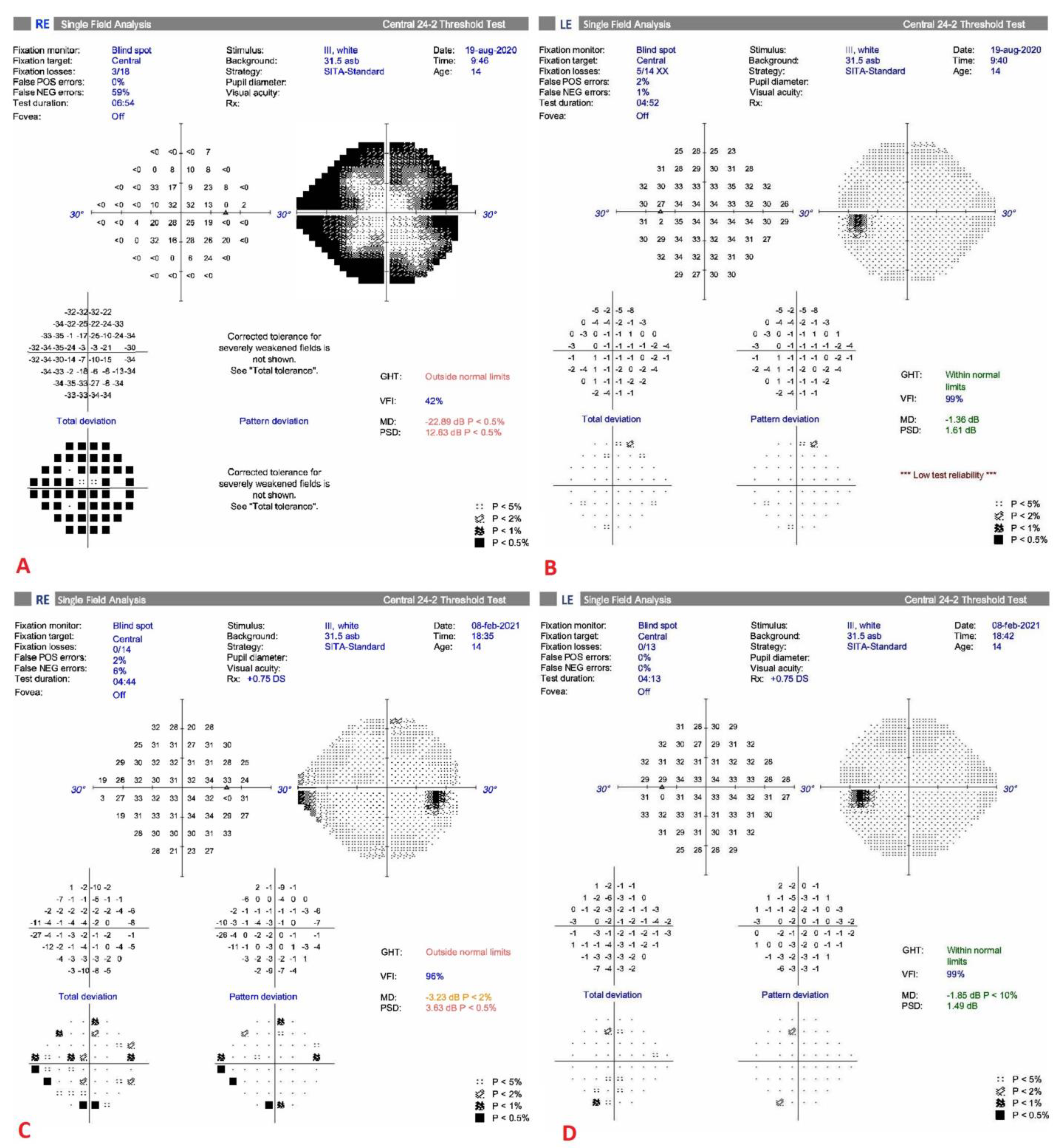
Figure 1. (A) Humphrey visual field (HVF, threshold 24-2) of the right eye (RE) after a mild traumatic brain injury (mTBI) and before starting visual therapy showing generalized loss of sensitivity. (B) HVF 24-2 of the left eye (LE) after an mTBI and before starting visual therapy. (C) HVF 24-2 of the RE after visual therapy showing generalized improvement in sensitivity and (D) HVF 24-2 of the LE after visual therapy. Abbreviations: RE: right eye; LE: left eye; POS: positive; NEG: negative; GHT: glaucoma hemifield test; VFI: visual field index; MD: mean deviation; PSD: pattern standard deviation.
Jcto 01 00005 g001
Table 1. Pre-treatment and post-treatment subjective clinical measures. The normal range of each test for the age patient are in the shaded column.
TestPre-Treatment Vision Exam ResultsPost-Treatment Vision Exam ResultsNormal Range
Uncorrected VA (Snellen) (RE/LE)20/3220/1620/1620/1620/20
Cycloplegic refraction (RE/LE)+1.75 D − 0.50 D × 180°+1.25 D − 0.50 D × 180°+1.50 D −0.50 D × 5°+1.50 DN/A
Non-cycloplegic refraction (RE/LE)+0.00 D+0.25 D+1.25 D − 0.50 D × 5°+1.25 DN/A
Stereopsis (Random Dot 2-S Test)200”30”20”
Worth testFusionFusionFusion
Cover test (distance)8 Δ esophoria, 2 Δ hyperphoria REOrtophoria0–2 ∆ exophoria
Cover test (near)8 Δ esophoria, 2 Δ hyperphoria REOrtophoria0–6 ∆ exophoria
Maddox (distance)8 ∆ esophoria, 6 ∆ hyperphoria RE6 ∆ esophoria, 2 ∆ hyperphoria RE0–2 ∆ exophoria
Maddox (near)8 ∆ esophoria, 5 ∆ hyperphoria RE8 ∆ esophoria, 2 ∆ hyperphoria RE0–6 ∆ exophoria
NPC (break/recovery)11 cm/15 cm7/8 cm1–3” break/3–5” recovery
NFV prism bar (near)−/10/6 ∆8/14/8 ∆13/10 ∆
PFV prism bar (near)6/14/14 ∆18/25/20 ∆19/14 ∆
NFV prism bar (distance)−/6/4 ∆6/8/4 ∆7/4 ∆
PFV prism bar (distance)8/20/18 ∆6/12/8 ∆11/7 ∆
Vergence facility 3/12 ∆4 cpm9 cpm15 cpm
Monocular AA (RE/LE)8.00 D13.00 D10.5 D13 D13 D
Monocular accommodative facility (RE/LE)8 cpm difficulty clearing negative and positive lenses13 cpm9 cpm10 cpm11 cpm
NRA/PRA+1.75/−0.50 D+2.25/−1.50 D+2.00/−2.37 D
MEM (RE/LE)Fluctuating value+1.50 D+2.00 D+2.00 D+0.50 D
Pursuits (NSUCO)5/1/4/45/3/5/55/4/4/5
Saccades (NSUCO)5/1/4/45/5/5/55/3/4/5
Abbreviations: AA: amplitude of accommodation; LE: left eye; MEM: monocular estimation method; N/A: not applicable; NFV: negative fusional vergence; NPC: near point of convergence; NRA/PRA: negative relative accommodation/positive relative accommodation; NSUCO: Northeastern State University College of Optometry; PFV: positive fusional vergence; RE: right eye; VA: visual acuity.
Table 2. Pre-treatment results of the red filter test.
DextroversionCentral AreaLevoversion
4 Δ BO5 Δ BU LE-8 Δ BO5 Δ BU LE
10 Δ BO8 Δ BU LE-12 Δ BO5 Δ BD LE
16 Δ BO8 Δ BU LE--
Abbreviations: BD: base-down; BO: base-out; BU: base-up; LE: left eye.
Disclaimer/Publisher’s Note: The statements, opinions and data contained in all publications are solely those of the individual author(s) and contributor(s) and not of MDPI and/or the editor(s). MDPI and/or the editor(s) disclaim responsibility for any injury to people or property resulting from any ideas, methods, instructions or products referred to in the content.