
Integrating Machine Learning with Medical Imaging for Human Disease Diagnosis: A Survey †
Abstract
1. Introduction
2. Machine Learning Applications in Healthcare
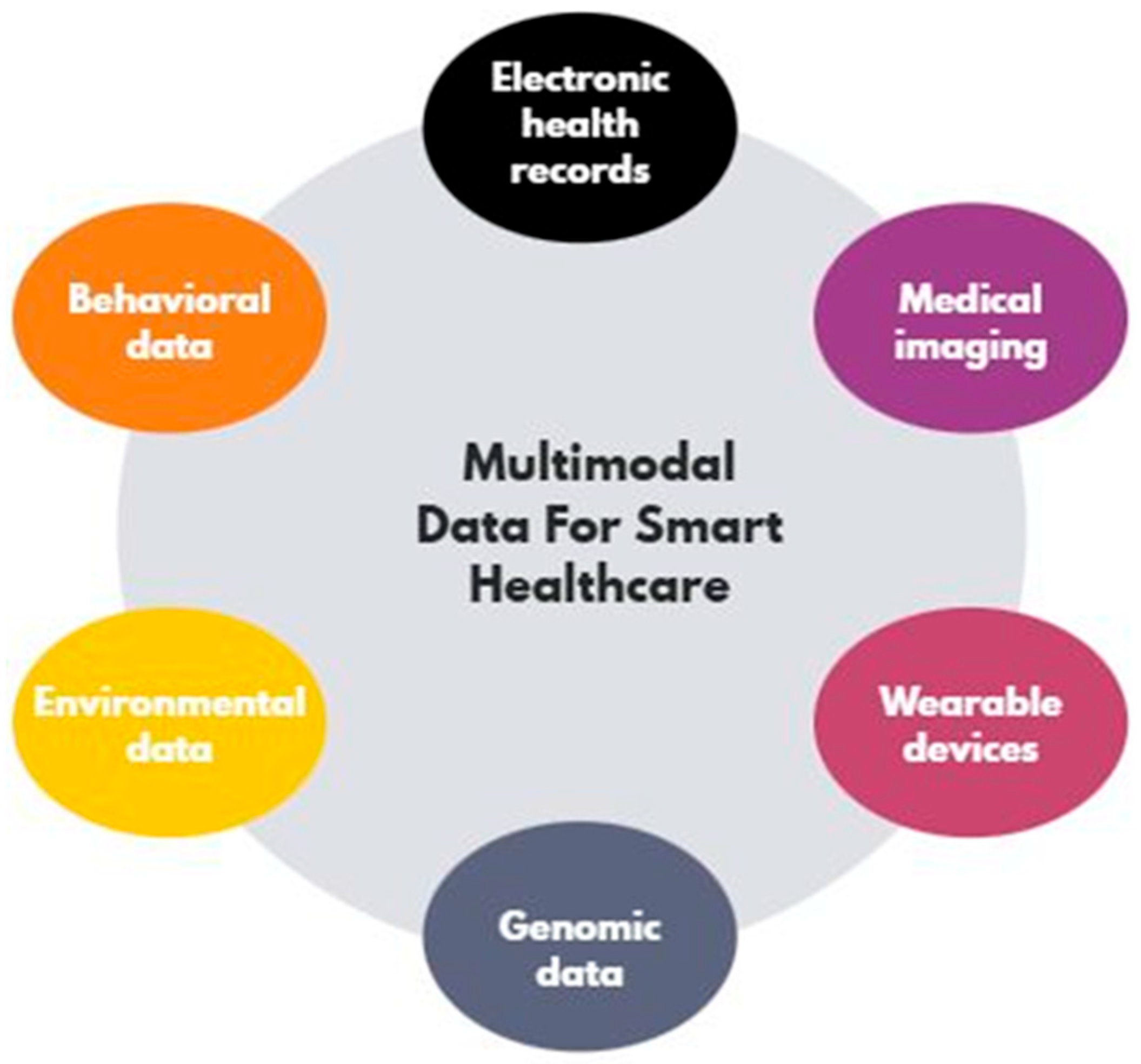
3. Data Modalities in Healthcare
4. Medical Imaging Types
- X-ray: Discovered in 1895, X-rays produce 2D images based on the variable absorption of radiation. They are used to detect fractures, lung diseases, cancers, osteoporosis, and dental cavities. Fast and cost-effective, they require precautions due to radiation exposure [10].
- CT (computed tomography): Developed in 1971, CT enhances X-rays by producing 3D images with better resolution and contrast. It is widely used in cardiology, oncology, and vascular radiology to detect tumors, lesions, and evaluate cancer spread, particularly breast cancer. Unlike X-rays, CT differentiates [10].
- MRI (magnetic resonance imaging): MRI, invented in the 1980s, visualizes abnormalities undetectable by other techniques like CT scans or X-rays. Unlike these methods, which map tissue density, MRI maps proton energy propagation. It uses a powerful magnetic field to align protons and a radiofrequency current to disrupt their balance, capturing images of soft tissues rich in water and fat [10].
- Ultrasound: Medical ultrasound uses a probe emitting ultrasonic signals to visualize internal body tissues such as muscles, tendons, and ovaries in real time. It allows for the observation of tissue structure, blood flow, and anomalies like cysts without harmful radiation. Its advantages include safety, portability, and real-time imaging, but it requires a qualified professional and provides a limited field of view [10].
5. Applications of Machine Learning in Medical Imaging
- Segmentation: In medical image analysis, segmentation is essential for enhancing diagnostic accuracy. It isolates relevant regions from surrounding tissues, allowing clinicians to focus on key areas for better assessment. This is particularly important for detecting and monitoring diseases like prostate and lung cancer, as it highlights anatomical and pathological structures with clarity [2].
- Classification: Classification is vital for accurate diagnosis, as it categorizes segmented regions such as distinguishing between benign and malignant tumors. Using machine learning, it analyzes features from images to assess disease severity and support treatment decisions. Alongside segmentation, classification ensures reliable and precise medical diagnosis [2].
6. ML Methods and Models: Previous Work
6.1. Lung Cancer
6.2. Prostate Cancer
6.3. Liver Disease
6.4. Heart Disease
6.5. Brain Disease
- Efficient Feature Selection Technique: A computationally efficient feature selection method is needed to eliminate data cleaning steps while enhancing the accuracy of disease prediction [11].
- Small Sample Sizes: The main challenge faced in most studies lies in insufficient data to train the model. To train advanced models such as ANN or random forest, a sufficiently large sample is required to avoid overfitting and improve generalization [12].
7. Conclusions and Perspectives
Author Contributions
Funding
Institutional Review Board Statement
Informed Consent Statement
Data Availability Statement
Conflicts of Interest
References
- Kumar, Y.; Koul, A.; Singla, R.; Ijaz, M.F. Artifcial intelligence in disease diagnosis: A systematic literature review, synthesizing framework and future research agenda. J. Ambient. Intell. Humaniz. Comput. 2023, 14, 8459–8486. [Google Scholar] [CrossRef] [PubMed]
- Yeasmin, M.N.; Al Amin, M.; Joti, T.J.; Aung, Z.; Azim, M.A. Advances of AI in image-based computer-aided diagnosis: A review. Array 2024, 23, 100357. [Google Scholar] [CrossRef]
- Dubey, S.; Tiwari, G.; Singh, S.; Goldberg, S.; Pinsky, E. Using machine learning for healthcare treatment planning. Front. Artif. Intell. 2023, 6, 1124182. [Google Scholar] [CrossRef] [PubMed]
- Ozdemir, B.; Pacal, I. A robust deep learning framework for multiclass skin cancer classification. Sci. Rep. 2025, 15, 4938. [Google Scholar] [CrossRef] [PubMed]
- Gupta, R.; Srivastava, D.; Sahu, M.; Tiwari, S.; Ambasta, R.K.; Kumar, P. Artificial intelligence to deep learning: Machine intelligence approach for drug discovery. Mol. Divers. 2021, 25, 1315–1360. [Google Scholar] [CrossRef] [PubMed]
- Kaur, E.; Bans, A.; Eze, U.O.; Singh, J. Artificial Intelligence in Healthcare: A Prospective Approach. Geeta Univ. J. 2023, 1, 1–12. [Google Scholar]
- Ramesh, J.; Aburukba, R.; Sagahyroon, A. A remote healthcare monitoring framework for diabetes prediction. Healthc. Technol. Lett. Using Mach. Learn. 2021, 8, 45–57. [Google Scholar] [CrossRef] [PubMed]
- Zhao, B.; Waterman, R.S.; Urman, R.D.; Gabriel, R.A. A Machine Learning Approach to Predicting Case Duration for Robot-Assisted Surgery. J. Med. Syst. 2019, 43, 32. [Google Scholar] [CrossRef] [PubMed]
- Shaik, T.; Tao, X.; Li, L.; Xie, H.; Velásquez, J.D. A survey of multimodal information fusion for smart healthcare: Mapping the journey from data to wisdom. Inf. Fusion 2024, 102, 102040. [Google Scholar] [CrossRef]
- Goyal, B.; Dogra, A.; Agrawal, S.; Sohi, B.S. Noise Issues Prevailing in Various Types of Medical Images. Biomed. Pharmacol. J. 2018, 11, 1227–1237. [Google Scholar] [CrossRef]
- Dhara, A.K.; Mukhopadhyay, S.; Dutta, A.; Garg, M.; Khandelwal, N. A Combination of Shape and Texture Features for Classification of Pulmonary Nodules in Lung CT Images. J. Digit. Imaging 2016, 29, 466–475. [Google Scholar] [CrossRef] [PubMed]
- Nair, S.S.; Devi, V.N.M.; Bhasi, S. Enhanced lung cancer detection: Integrating improved random walker segmentation with artificial neural network and random forest classifier. Heliyon 2024, 10, e29032. [Google Scholar] [CrossRef] [PubMed]
- Aldoj, N.; Lukas, S.; Dewey, M.; Penzkofer, T. Semi-automatic classification of prostate cancer on multi-parametric MR imaging using a multi-channel 3D convolutional neural network. Eur. Radiol. 2019, 30, 1243–1253. [Google Scholar] [CrossRef]
- Şerbănescu, M.S.; Manea, N.C.; Streba, L.; Belciug, S.; Pleşea, I.E.; Pirici, I.; Bungărdean, R.M.; Pleşea, R.M. Automated Gleason grading of prostate cancer using transfer learning from general-purpose deep-learning networks. Rom. J. Morphol. Embryol. 2020, 61, 149–155. [Google Scholar] [CrossRef]
- Chen, Q.; Hu, S.; Long, P.; Lu, F.; Shi, Y.; Li, Y. A Transfer Learning Approach for Malignant Prostate Lesion Detection on Multiparametric MRI. Sage J. 2019, 18, 1–9. [Google Scholar] [CrossRef] [PubMed]
- Prakash, N.N.; Rajesh, V.; Namakhwa, D.L.; Pande, S.D.; Ahammad, S.H. A DenseNet CNN-based liver lesion prediction and classification for future medical diagnosis. Sci. Afr. 2023, 20, e01629. [Google Scholar] [CrossRef]
- Prakash, K.; Saradha, S. Efficient prediction and classification for cirrhosis disease using LBP, GLCM and SVM from MRI images. Mater. Today Proc. 2023, 81, 383–388. [Google Scholar] [CrossRef]
- Alsekait, D.M.; Shdefat, A.Y.; Nabil, A.; Nawaz, A.; Rana, M.R.R.; Ahmed, Z.; Fathi, H.; AbdElminaam, D.S. Heart-Net: A Multi-Modal Deep Learning Approach for Diagnosing Cardiovascular Diseases. Comput. Mater. Contin. 2024, 80, 3967–3990. [Google Scholar] [CrossRef]
- Lu, S.; Wang, S.H.; Zhang, Y.D. Detection of abnormal brain in MRI via improved AlexNet and ELM optimized by chaotic bat algorithm. Neural Comput. Appl. 2020, 33, 10799–10811. [Google Scholar] [CrossRef]

| Type of Disease | Publisher | Publication Year | Dataset | Methodology | Performance Metrics |
|---|---|---|---|---|---|
| Lung Cancer [11] | Journal of Digital Imaging | 2016 | LIDC-IDRI (CT Scan) | Shape-based, margin-based, and texture-based feature extraction techniques; feature selection using A-z and p-values; SVM classifier. | AUC (Az) performance: Configuration 1: 95% Configuration 2: 88% Configuration 3: 84% |
| Lung Cancer [12] | Heliyon | 2024 | LIDC-IDRI (CT Scan) | Improved random walker with artificial neural network and random forest classifier. | Accuracy: Random Forest classifier: 99.6% ANN: 94.8% |
| Prostate Cancer [13] | European Radiology | 2019 | ProstateX challenge (MRI) | Multi-channel 3D convolutional neural network, cross-validation | AUC: 89% Sensitivity: 88.6% Specificity: 90.5% |
| Prostate Cancer [14] | Romanian Journal of Morphology and Embryology | 2020 | Included 439 images from 83 prostate cancer patients | GoogleNet AlexNet | Accuracy: GoogleNet: 60.9% AlexNet: 61.17% |
| Prostate Cancer [15] | Sage Journals | 2019 | ProstateX challenge | VGG-16 InceptionV3 | AUC: VGG-16: 83% InceptionV3: 81% |
| Liver Disease [16] | Scientific African | 2023 | Kaggle Liver CT Scan Dataset and real-time images collected from Government General Hospital Vijayawada | Region-growing, Dens-Net CNN | accuracy: 98.34% sensitivity: 99.72% recall: 97.84% |
| Liver Disease [17] | Materials Today: Proceedings | 2023 | MVISS 3.0 T and gaggle liver patient dataset | LBP, GLCM, SVM | - |
| Heart Disease [18] | Computers, Materials & Continua | 2024 | Sunnybrook Cardiac Data (HNET-DSI) Kaggle datasets of cardiac disease patients (HNET-DSII) Github-datasets of cardiac disease patients (HNET-DSIII) | Heart-Net | Accuracy of: HNET-DSI: 92.56% HNET-DSII: 93.45% HNET-DSIII: 91.89% |
| Brain Disease [19] | Neural Computing and Applications | 2020 | Brain MRIs | BN-AlexNet-ELM-CBA | Sensitivity: 97.14% Specificity: 95.71% Accuracy: 96.43% |
Disclaimer/Publisher’s Note: The statements, opinions and data contained in all publications are solely those of the individual author(s) and contributor(s) and not of MDPI and/or the editor(s). MDPI and/or the editor(s) disclaim responsibility for any injury to people or property resulting from any ideas, methods, instructions or products referred to in the content. |
© 2025 by the authors. Licensee MDPI, Basel, Switzerland. This article is an open access article distributed under the terms and conditions of the Creative Commons Attribution (CC BY) license (https://creativecommons.org/licenses/by/4.0/).
Share and Cite
Roman, A.; Taib, C.; Dhaiouir, I.; El Khatir, H. Integrating Machine Learning with Medical Imaging for Human Disease Diagnosis: A Survey. Comput. Sci. Math. Forum 2025, 10, 12. https://doi.org/10.3390/cmsf2025010012
Roman A, Taib C, Dhaiouir I, El Khatir H. Integrating Machine Learning with Medical Imaging for Human Disease Diagnosis: A Survey. Computer Sciences & Mathematics Forum. 2025; 10(1):12. https://doi.org/10.3390/cmsf2025010012
Chicago/Turabian StyleRoman, Anass, Chaymae Taib, Ilham Dhaiouir, and Haimoudi El Khatir. 2025. "Integrating Machine Learning with Medical Imaging for Human Disease Diagnosis: A Survey" Computer Sciences & Mathematics Forum 10, no. 1: 12. https://doi.org/10.3390/cmsf2025010012
APA StyleRoman, A., Taib, C., Dhaiouir, I., & El Khatir, H. (2025). Integrating Machine Learning with Medical Imaging for Human Disease Diagnosis: A Survey. Computer Sciences & Mathematics Forum, 10(1), 12. https://doi.org/10.3390/cmsf2025010012