Next Article in Journal
Case History: Improved Maxillary Growth and Development Following Digit Sucking Elimination and Orofacial Myofunctional Therapy
Previous Article in Journal
Muscle Tension Dysphonia in Singers and Professional Speakers with Ankyloglossia: Impact of Treatment with Lingual Frenuloplasty and Orofacial Myofunctional Therapy
 
 
International Journal of Orofacial Myology and Myofunctional Therapy is published by MDPI from Volume 51 Issue 1 (2025). Previous articles were published by another publisher in Open Access under a CC-BY (or CC-BY-NC-ND) licence, and they are hosted by MDPI on mdpi.com as a courtesy and upon agreement with the previous journal publisher.
Article

Videofluoroscopy of the Oral Phase of Swallowing in Eight to Twelve Years Old Children with Dental Malocclusion

by
Patricia Junqueira
1,* and
Milton Melciades Costa
2
1
UNIFESP/Escola Paulista de Medicina, São Paulo, CEP 05641-070, Brazil
2
UFRJ Biomedical Science Institute, Rio de Janeiro, Brazil
*
Author to whom correspondence should be addressed.
Int. J. Orofac. Myol. Myofunct. Ther. 2013, 39(1), 54-68; https://doi.org/10.52010/ijom.2013.39.1.6
Submission received: 1 November 2013 / Revised: 1 November 2013 / Accepted: 1 November 2013 / Published: 1 November 2013

Abstract

:
The objective of this study was to describe the oral phase of swallowing in individuals with dental malocclusion and to generate data that would contribute to the rehabilitation of those patients. The study was based on the evaluation of the swallowing system through videofluoroscopy on thirty-four children of both genders, aged eight to twelve years old who present with Angle Class II and III dental malocclusions. Thirteen children of similar age and gender presenting normal dental occlusion formed the control group. The results indicated that the oral phase of swallowing is different between individuals with normal occlusion and malocclusion. Dental occlusion types Angle Class II and III did not present a swallowing pattern, independently of the amount of liquid ingested. The swallowing appeared effective in the oral phase of individuals with dental malocclusion, even though adaptations were identified. The outcome, in the absence of a single pattern and the efficiency of the adapted swallowing demonstrates, first a need for additional research investigating orofacial myofunctional treatment for patients with malocclusion and second how such analyses should focus on contributing positively to the rehabilitation of these patients.

INTRODUCTION

Swallowing consists of a set of physiological events that ultimately result in the displacement of food from the mouth to the stomach. It is the oral phase of swallowing in which the bolus is prepared, modified, organized and ejected from the oral cavity into the pharynx (Costa, 1998; Junqueira, 2003; Junqueira & Marchesan, 2008). The oral swallowing stage is also voluntary. It begins with the jaws and lips closed, and the tip of the tongue on the alveolar ridge. The pattern-elicited response is initiated at the end of this stage. Inspiration is reflexively inhibited at the beginning of this stage. The food is moved to the back of the mouth by the tongue via an anterior to posterior peristaltic motion. The anterior portion of the tongue is retracted while the posterior portion is retracted and elevated against the hard palate. When the bolus passes the anterior faucial pillars or touches the posterior wall of the pharynx, the oral stage ends and the pharyngeal stage begins as the driving force of the tongue—the tongue’s plunger action, forces the bolus into the pharynx. The oral transport stage lasts one second (Logemann, 1993, 2002).
The term ‘atypical thrusting’ when some kind of alteration is noticed in the way one swallows during the oral phase of swallowing (Junqueira & Marchesan, 2008). Atypical thrusting is defined, more frequently, by the characteristics that occur during the swallowing act. The main and most frequent characteristics mentioned in the literature are described by Segovia (1977), Braga and Machado (1992), and Altman (1994): the absence of contraction of the masseters; absence of dental occlusion; tongue interposition that may be lateral or anterior and labial interposition in order to promote the anterior closure; intense involvement of the perioral muscles; and phonation deviations which usually include the phonemes [t] [d], [l], [n] [s] and [z] (Segovia, 1977; Braga and Machado, 1992; Altman, 1994). These authors did not make explicit in their definitions of atypical thrusting, whether or not there are functional or anatomical conditions that are favorable for a normal swallow.
Other researchers including Milne and Cleall (1970), Fränkel (1980), Van der Linden (1990), Marchesan (1996, 1998, 1999, 2005a, 2005b, 2005c), Marchesan and Furkim (2003), Marchesan and Sanseverino (2004), Felício (1999), Bianchini (2001, 2005), Medeiros and Medeiros (2006), Fraser (2006) prefer to use the term ‘adapted swallowing’ rather than atypical, to indicate that there is a cause that leads to the alteration. Dental malocclusion has been reported by these authors as a major cause for adaptation in the swallowing function.
In agreement with Marchesan (1999), it is not the intention here to develop the idea that the designation of a thrusting as ‘atypical’ or ‘adapted’ is the most important focus. In fact, it does not make a difference if the patient with interposition of the tongue at the moment of swallowing is ‘an atypical thrusting’ or an ‘adapted’ case when justifying the interposition due to an alteration shown by the patient with malocclusion. Instead, the important point is to know whether or not we should incorporate this area in our work to correct the detected projection. There are professionals who even consider that if the tongue protrusion problem during swallowing is not corrected, it may impair the process or even prevent the closing of the anterior malocclusion (Overstatake, 1970; Fink, 1986).
There are many authors who establish a relationship between the presence of tongue protrusion and dental malocclusion (Jann, 1960; Subtelny & Sakuda, 1964; Cleall, 1965; Kortsch, 1965; Ballard, 1967; Overstake, 1970; Subtelny, 1970b; Hanson & Cohen, 1973; Mason & Proffit, 1974; Fink, 1986; Fuhrmann & Diedrich, 1994; Yamaguchi & Sebata, 1995; Ichida, Takakiguchi & Yamada, 1999; Maciel & Leite, 2005; Fraser, 2006; Mason, 2005, 2008; Yamaguchi & Sueishi, 2003; Cayley, Tindall, Sampson & Butcher,, 2000). Subtelny (1970a), Tosello et al. (1998) strongly state that when patients have a malocclusion, tongue protrusion occurs in order to help the labial seal during swallowing. Although the authors highlight the importance of the swallowing and occlusion relationship, as well as its implication in the orofacial diagnosis and therapy, this study argues that this relationship has not been studied objectively.
There are several methods that enable the study of swallowing dynamics (Costa, 2010). Ultrasound for example, has limitations because the physical properties of sound transmission do not allow high-frequency sound waves to pass through substances such as bone. Most of the oral cavity is encased in bone, creating an echo-free area or dark shadow when bones are present within the scanning region. Motions of the hyoid bone are essential to understanding swallowing dynamics. The displacement of the echo-free shadow created by hyoid activity during swallowing can be easily tracked on ultrasound, turning this potential limitation into an advantage (Sonies, Chi-Fishman, Miller, 2003). Fiberoptic endoscopic examination of swallowing safety it is being used to detect aspiration and to determine the safety of oral feeding in patients for whom the traditional videofluoroscopy evaluation may be difficult or impossible to perform (Langmore, 1988).
Dynamic images are critical for swallowing studies. For this reason, difficult cervical anatomy, complex pharyngeal physiology and even pharyngeal cannot be understood as difficulties in videofluoroscopic interpretation. Many morphofunctional concepts can be reviewed on the videofluoroscopic study of swallowing, and much more can be learned with this method. Currently, it is already considered essential for the diagnosis and follow-up of swallowing disorders (Costa, 2010).
The videofluoroscopy has been considered the best method for evaluating swallowing (Beck & Gayler, 1990; Logeman, 1993, 2007; Yamada, Siqueira, Xerez, Koch & Costa, 2004; Costa, 1999; Costa, Nova, Carlos, Pereira, & Koch, 1992; Dantas, 1996; ASHA, 2004). This is a radiological method, with low radiation exposure, which allows all the dynamics of the phenomenon of swallowing to be viewed in real time, enabling the morphological and functional correlation concerning the observed events. Recording on videotapex, at a speed of 30 frames per second, enables the analysis and re-analysis of these examinations, with no need for further X-Ray radiation exposure. The method has been indicated as an aid in the diagnosis and treatment of swallowing disorders (Costa, 1999; Costa et al., 1992; Dantas, 1996; Logeman, 1993, 2007; Yamada et al., 2004; Beck & Gayler, 1990).
The twofold purpose of this study, using the videofluoroscopic method, includes the following:
  • To observe the oral phase of swallowing in patients with malocclusion;
  • To identify the possible tongue adaptations in the oral cavity which make a swallow possible.

METHODS

The Angle Classification (Moyers, 1988) was used for identifying type of dental malocclusion observed and the grouping of participants. The control group (Group A) was composed of thirteen children from a primary school with an age range, and gender similar to the research groups. All children in Group A were confirmed as having normal occlusion based on clinical exams. This confirmation of normal occlusion was carried out by a dental surgeon. The research was carried out by the authors who observed thirty-four children, of both genders, 8–12 years of age with dental malocclusion. Group B was composed of 20 children with Angle Class II, and Group C was composed of 14 children with Angle Class III. All of the children came from the Clinic of the Facial Orthopedics and Orthodontics Specialization Course of the Dentistry Department of the Health Science Institute–Universidade Paulista (UNIP). All children in the study groups had just started an Orthodontics or Facial-Orthopedics Treatment and were diagnosed and classified according to their dental occlusion by means of clinical exams conducted by the orthodontist with complete orthodontic documentation (analysis of the orthodontic patterns, cephalometry in frontal and lateral cranial views, panoramic radiography or orthopantography, frontal, profile and intraoral figures, and cephalometrics tracings).
Children with neurological lesions, cleft lip and palate, ankyloglossia, congenital malformation, who had made use or were still making use of orthodontics or orthopedics appliance, or who had previously undergone or were still undergoing Speech Therapy, were excluded from this sample. Three groups were then formed:
1)
Group A—Thirteen children with normal dental occlusion (Angle Class I);
2)
Group B—Twenty children with dental malocclusion (Angle Class II);
3)
Group C—Fourteen children with dental malocclusion (Angle Class III).
The number of subjects per group and its correlation was approved by the Committee and the statistical analysis of the UNIFESP. All recommendations suggested by the International Commission on Radiological Protection (Valentin, 2007) for performing the videofluoroscopy were followed, as well as the Steps for Radiation Safety (as cited in Pediatric Interventional Radiology, n.d.).
Those responsible caretakers for the children participating in this study were previously informed about the risks and the procedures to be used and signed an Informed Consent Declaration agreeing to the research being carried out, as well as to the disclosure of its results. The Research Ethics Committee of Hospital São Paulo/Universidade Federal de São Paulo, protocol number 088/99, also approved this study.
Every patient was submitted to videofluoroscopy exams at Hospital São Paulo–Image Diagnosis Department–UNIFESP/EPM, for the evaluation of swallowing. The authors and the radiologist conducted the exams using Medicor X-ray equipment, 750B-EDR type, with image intensifier, a 525-line TV system, 60 Hz, and 2:1 interlacing with loop through exit, through which the images were transferred to a VHS tape, in a Philco video/monitor device, PVC-7400 type.
The protocol defined for the video-fluoroscopic evaluation of the dynamics of the oral phase of swallowing for a volume of liquid was: orolaryngopharyngeal inspection using a lateral view of the swallowing process for three gulps of contrasted liquid volume (Junqueira & Costa, 2001). The regions being documented were those corresponding to the oral phase of swallowing. Recordings were made before, during, and for a short period of time after the end of the contrast medium transit. The total time for examining each patient was about four minutes. The contrast medium was the standard Barium Sulphate solution, 50% diluted in water. This solution was then homogenized and offered to the patient in a transparent glass, graduated every 5 mL, so as to estimate, based on the difference between the initial volume and the remaining quantity in the glass, the approximate volume of each gulp that had been ingested by the examinee was recorded.
Each individual swallowed three gulps, one at a time. Thus, the choice of volume was based on the comfort and oral functional capacity of the examinees. Results of the three observations, for each individual, were included in the analysis. The exams were analyzed using a Panasonic AG 1980 video- cassette and video monitor system by both researchers separately. The results of each analysis were compared. If there was disagreement about the analysis for a particular participant, the radiologist revised the images. Following this, the results of each of the analyses were compared again to guarantee the reliability of the findings.
Each patient had three swallowed gulps analyzed separately. All gulps were evaluated in terms of three components: introduction, organization, and ejection of the contrasted liquid bolus. For the introduction component, observation focused on whether the contrast was introduced under the tongue or on the tongue. For the organization component, observation focused on how and where the bolus was placed to prepare for ejection.
Finally, when ejecting the bolus, both the position of the tongue and the way the bolus was propelled to the pharynx were analyzed (Yamada et al., 2004). The results were statistically evaluated using chi-square to assess the possible relationships between the type of occlusion and the type of swallow. The restrictions of Cochran (1954) were adhered to in this process. In all tests, the level of rejection for the null hypothesis was less than or equal to 0.05% (5%).

RESULTS

There are two types of oral swallowing events. In the first type, tipper swallows, the bolus is positioned on the dorsum of the tongue and the swallow is initiated with the tip of the tongue against the incisors. In the second type, dipper swallows, the bolus is held under the tongue in the anterior sulcus of the floor of the mouth, and the tongue tip must scoop up the bolus and elevate it onto the tongue before swallowing (Dodds, Taylor, Stewart, Kern, Logemann & Cook, 1989; Logemann, 1993, 2007; Logemann, Pauloski, Rademaker & Kahrilas, 2002).
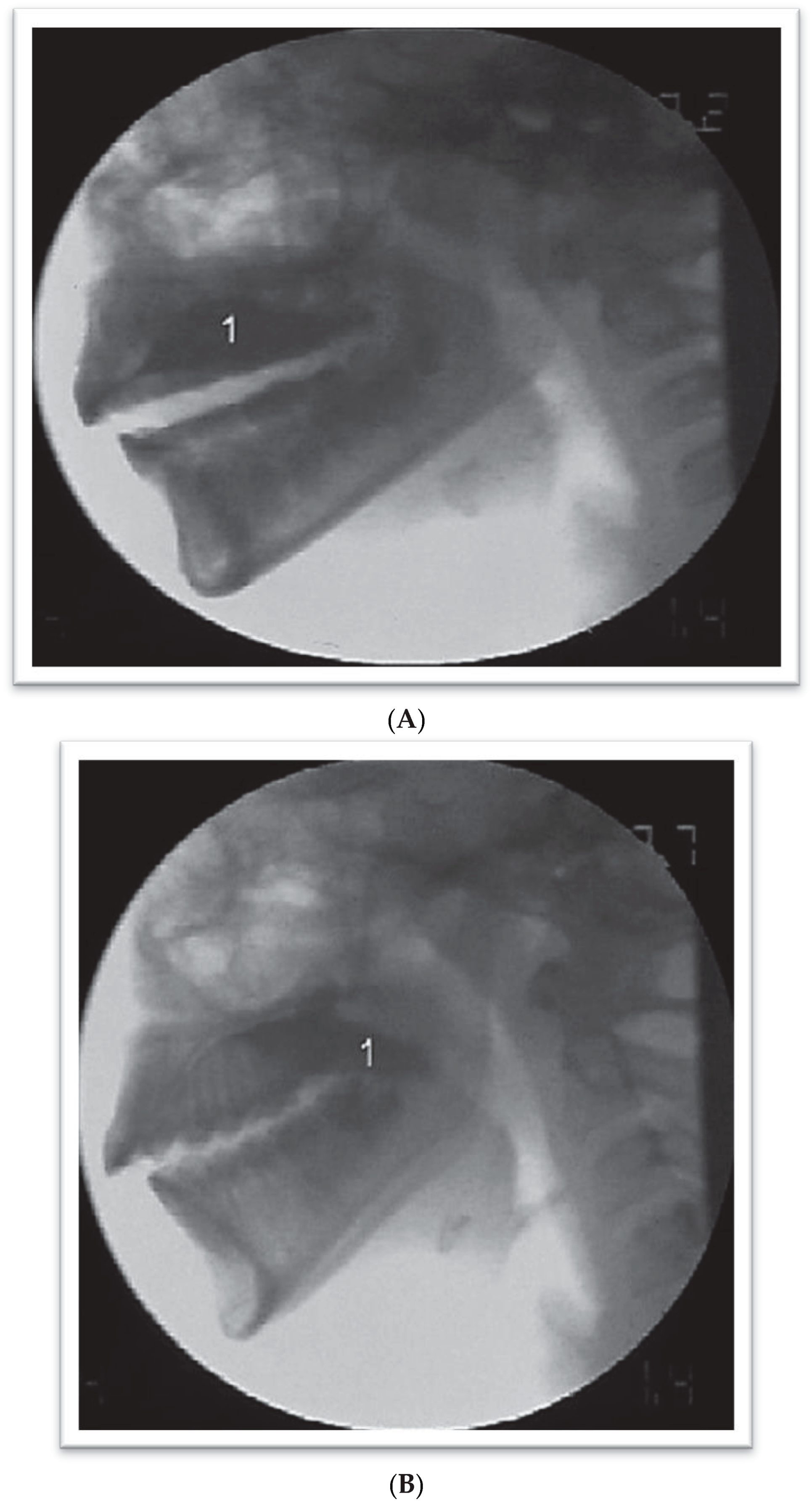
Dodds et al. (1989) found that 72% of the participants with normal oral motor function used the Tipper type of swallow, and 19% used the Dipper type of swallow. In the present study subjects the Tipper (Dodds et al., 1989; Logemann, 1993, 2007; Logemann, Pauloski, Rademaker & Kahrilas, 2002) swallowing type occurred in 94.87% (Figure 1A,B) of the children with normal dental occlusion, while the Dipper (Dodds et al., 1989; Logemann, 1993, 2007; Logemann et al., 2002) swallowing type occurred in 2.56% of the swallows. In 2.56% of the swallows, the observation of the oral phase indicated swallowing with premature loss of the liquid consistency bolus, consistent with the description by Logemann (1993, 2007), Logemann et al. (2002).
Figure 1. Normal swallowing occurred in 94.87% of the gulps in children presenting normal occlusion. (A) Videofluoroscopic exam on individuals with normal dental occlusion during oral organization phase. 1—Contrasted solution organized on the dorsal surface of the tongue. (B) Videofluoroscopic exam on individuals with normal dental occlusion during oral ejection phase. 1—Beginning of the projection of the bolus to the pharynx.
Ijom 39 00054 g001
In order to standardize the oral phase of swallowing for individuals with dental malocclusion, it was necessary for the researchers to create a classification method to describe their observations of the act of swallowing by modifying the Tipper and Dipper types of swallow as provided by Dodds et al. (1989). This classification method was named Adaptation by the researchers. There currently is not a description in the existing literature that defines the changes in the swallowing process that were observed in this study which included variations in the act of swallowing occurring in the presence of a tongue protrusion.
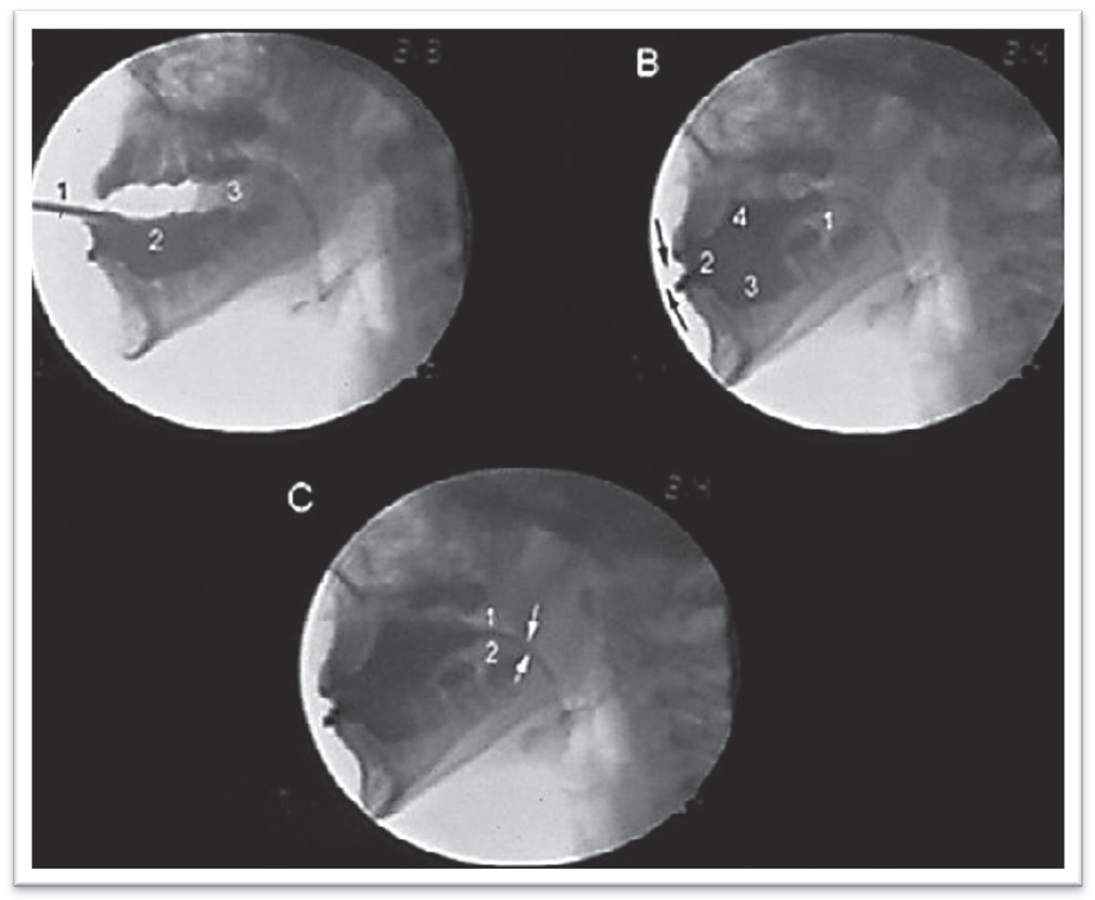
Four different types of consistent adaptation and one of inconsistent adaptation were observed. These observations were classified according to the way the volume was organized and ejected for each individual. The researchers defined a consistent adaptation as one in which the volume of each gulp ingested was organized and ejected as a whole bolus during the swallow. Inconsistent adaptation was defined as one in which the volume of each gulp ingested was organized and ejected in parts during the swallow, rather than as a whole bolus. This type of organization and oral ejection was sub-divided into different gulps for ejection and, therefore, not swallowed as a whole. Characteristics of the observed swallows were able to be organized into the following types:
  • Type I: Consistent Adaptation—Adapted Tipper
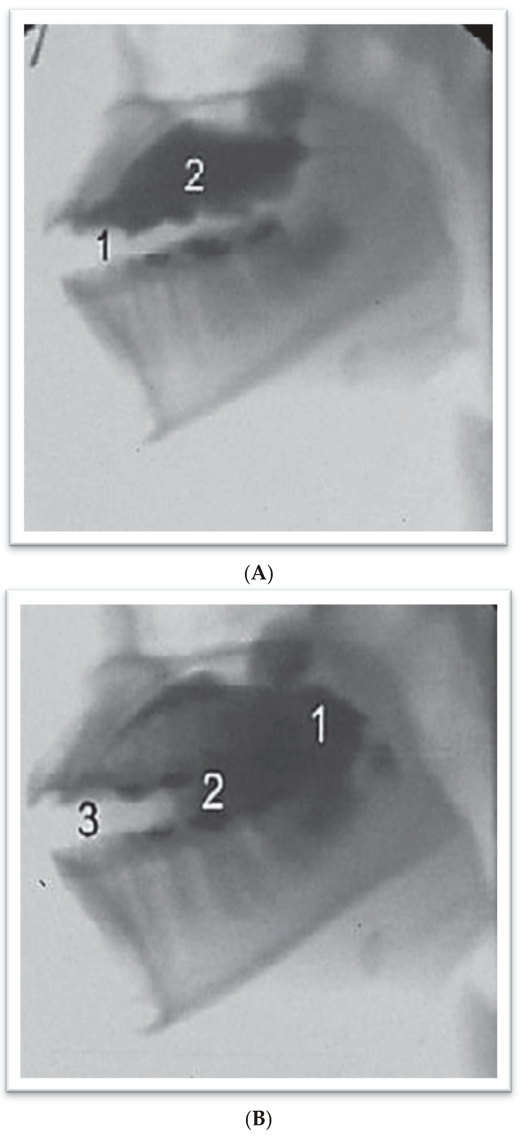
    Individuals in Group B with Class II malocclusion demonstrated the Adapted Tipper Swallow with the bolus going onto the tongue in 51.66% of the gulps observed. Individuals in Group C with Class III malocclusion demonstrated the Adapted Tipper Swallow in 59.52% of the gulps. The bolus was organized on the tongue and, in some cases this was accomplished with difficulty. It is defined by active mobilization of the tongue in maintaining the content. When ejection takes place, the tip of the tongue is placed between the incisors. Ejection is productive with apparent lower effectiveness and the strong influence of gravity.
  • Type 2 Consistent Adaptation—In two stages
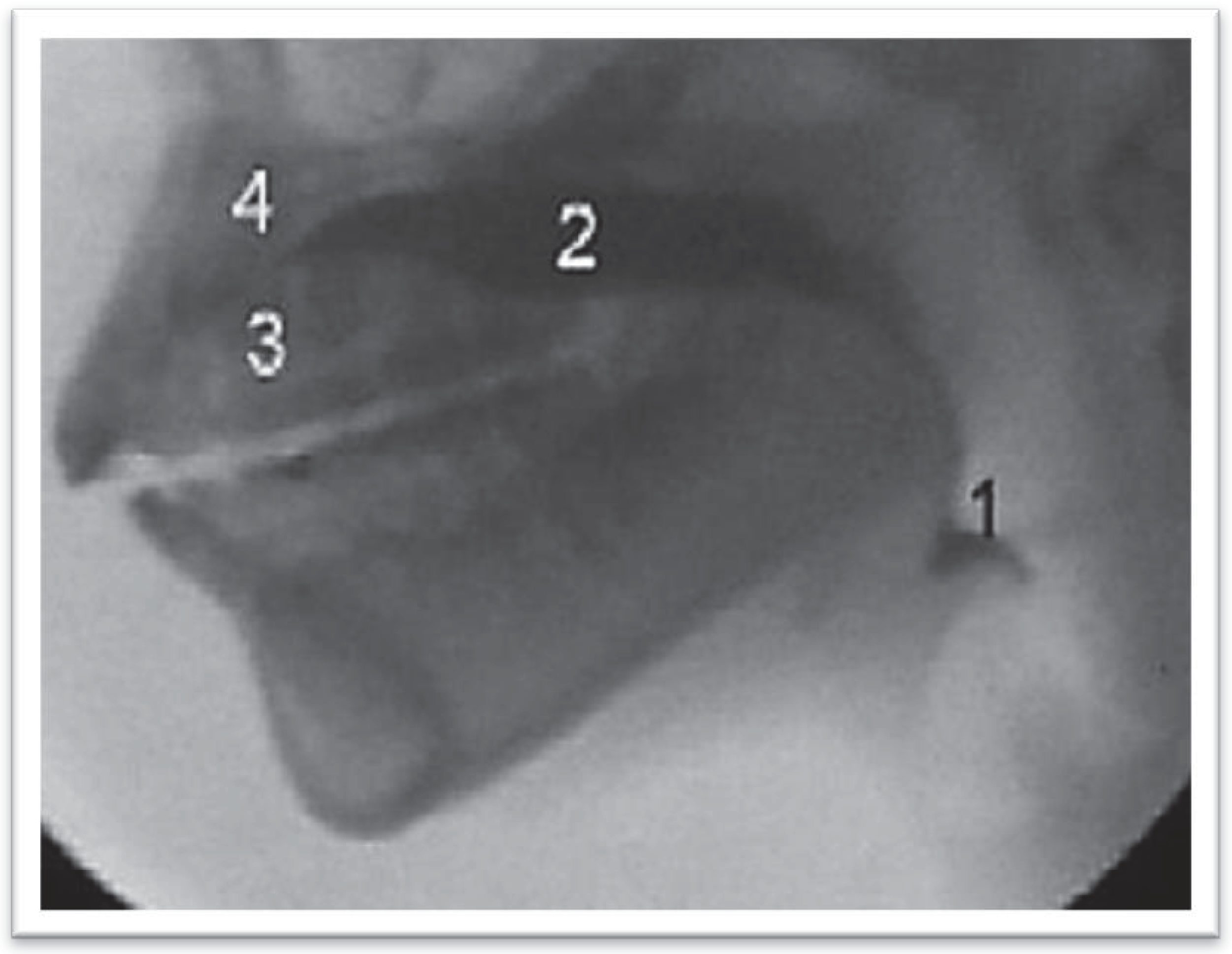
    Individuals in Group B with Class II malocclusion demonstrated the Type 2 Consistent Adaptation—In two stages in 25% of the gulps. Individuals in Group C with Class III malocclusion demonstrated the Type 2 Consistent Adaptation—In two stages in 11.9%. During organization or at the beginning of oral ejection, part of the bolus escapes to the oropharynx and it is only ejected after it reaches the level of the vallecula. The bolus that remains in the oral cavity for this period of time and that is placed on the dorsal surface of the tongue is also propelled to the pharynx by a second ejection. Both ejection processes occur with the tip of the tongue placed between the incisors.
  • Type 3 Consistent Adaptations—Extended Time
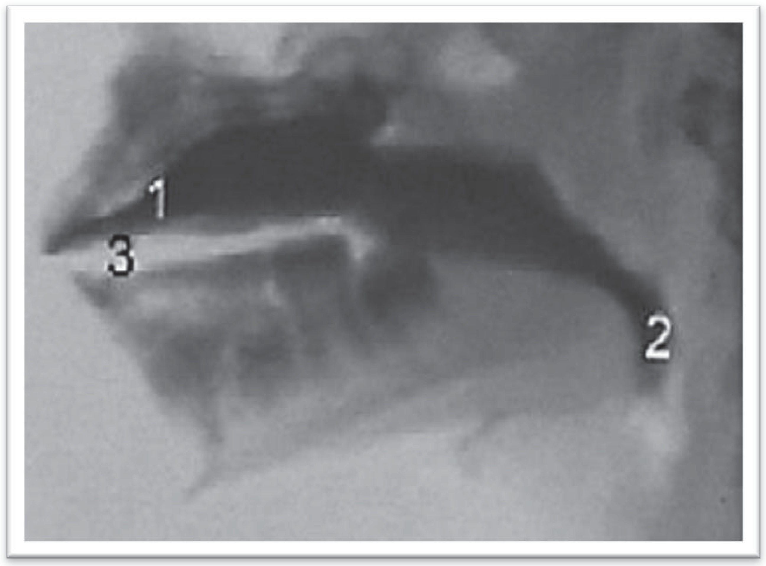
    Individuals in Group B with Class II malocclusion demonstrated the Type 3 Consistent Adaptation—Extended Time in 11.66% of the gulps. Individuals in Group C with Class III malocclusion demonstrated the Type 3 Consistent Adaptation-Extended Time in 7.14% of the gulps. In this Type 3, the bolus was placed on the dorsal surface of the tongue; however the bolus spread over the entire tongue and did not concentrate in the center of the tongue. The extended time for ejection occurred with the bolus being continuously propelled from the oral cavity to the pharynx, with strong help of gravity. During ejection, the tip of the tongue was placed between the incisors.
  • Type 4 Consistent Adaptations—Adapted Dipper
    Individuals in Group B with Class II malocclusion demonstrated the Type 4 Consistent Adaptation—Adapted Dipper in 1.66% of the gulps. Individuals in Group C with Class III malocclusion demonstrated the Type 4 Consistent Adaptation-Adapted Dipper in 14.28% of the gulps. It was observed in Type 4 that most of the bolus, was placed under the anterior part of the tongue to start swallowing. Consequently, the tip of the tongue makes an anterior dip to place the bolus on the tongue before transporting it to the oropharynx. When most of the bolus is placed on the tongue, the tip moves from anterior to posterior to eject the bolus into the pharynx. The bolus that is on the tongue and the bolus under the tongue are simultaneously ejected. First, the bolus on the tongue was propelled to the posterior oral cavity, while the bolus under the tongue moved onto it and they both were finally ejected to the pharynx. During ejection, the tip of the tongue was placed between the incisors.

Irregular Inconsistent Adaptation

Individuals in Group B with Class II malocclusion demonstrated Irregular Inconsistent Adaptation in 10% of the gulps. Individuals in Group C with Class III malocclusion demonstrated the Irregular Inconsistent Adaptation in 7.14% of the gulps.
In Irregular Inconsistent Adaptation, swallowing occurred two or more times for the same bolus that was introduced into the oral cavity. In addition with these cases, consistent adaptation types 1, 2, 3 and 4 occurred irregularly for the same bolus. The organization/ejection of the oral content varied constantly and it was not possible to identify a consistent pattern (Type 2, Type 1; or Type 3, Type 1) for the method of swallow adaptation.
The research results indicated that the most common swallowing type observed both for individuals with Class II malocclusion and for those with Class III malocclusion, was Type 1 Consistent Adaptation—Adapted Tipper. There was more Type 2 Consistent Adaptation—In two stages for the gulps of individuals belonging to Class II, and more Type 4 Consistent Adaptation—Adapted Dipper for the gulps of individuals belonging to Class III.
It was not possible to carry out analytical statistics of all results due to restrictions of Cochran (1954).
The adapted swallowing occurred in 100% of the gulps, both for Class II malocclusion and Angle Class III.
The statistical analysis indicated that there was significant statistical difference between the Group A with normal occlusion and the groups with dental occlusion Class II Group B and Angle Class III Group C occlusion. There was no significant difference between malocclusions in Group B and Group C.
Table 1. Type of dental occlusion versus the swallowing mode.
OcclusionNormalII III Total
SwallowingN%N%N%N
Tipper3794.8700.0000.0037
Adapted Tipper00.003151.672559.5256
In Two Stages00.001525.00511.9020
Extended Time00.00711.6737.1410
Adapted Dipper00.0011.67614.297
Inconsistent Adaptation00.00610.0037.149
Dipper12.5600.0000.001
Premature Loss of Bolus12.5600.0000.001
Total39100.0060100.0042100.00141
Figure 2. Videofluoroscopic exam on patients with dental malocclusion, Angle Class III type with adaptation defined as Type 1 Adapted Tipper (A and B). (A) Oral Organization Phase. 1—Interposing of the tongue between the upper and the lower dental arch; 2—Contrasted solution organized on the dorsal surface of the tongue. (B) Beginning of the oral ejection phase. 1—Beginning of the projection of the bolus to the pharynx; 2—Escape of part of the contrast from under onto the tongue during ejection; 3—Interposing of the tongue between the upper and the lower dental arch.
Ijom 39 00054 g002
Figure 3. Videofluoroscopic exam on patient with dental malocclusion Angle Class II type, with adaptation defined as Type 2 in two stages during Oral Ejection Phase. 1—Part of the contrast that escaped to the vallecula. 2—Remaining contrast to be ejected; 3, 4—Anterior part of the dorsal surface of the tongue, against the hard palate.
Ijom 39 00054 g003
Figure 4. Videofluoroscopic exam on patient with dental malocclusion Angle Class II type, with adaptation defined as Type 3 extended during the beginning of the oral ejection phase. 1—Contrast on the dorsal surface of the tongue, starting from the anterior part of the oral cavity and 2—escaping to the oropharynx, already at the vallecula level; 3—Interposing of the tongue between the upper and lower dental arch.
Ijom 39 00054 g004
Figure 5. Videofluoroscopic exam on patient with dental malocclusion Angle Class III, with adaptation defined as Type 4 Adapted Dipper. (A) 1—Admission of the contrasted solution, 2—taking anterior space, 3—generated by tongue retraction. (B) 1—Affixed lips (arrows), retracted tongue, 2—with contrasted solution taking anterior space. of the oral cavity, 3, 4—and also taking the space under and on the tongue. (C) Escape of contrast between the palate (1) and the tongue (2).
Ijom 39 00054 g005
Table 2. Type of dental occlusion and the normal versus adapted swallowing mode.
SwallowingNormal AdaptedTotal
OcclusionN%N%N
Normal3794.8725.1339
II00.0060100.0060
III00.0042100.0042
Total3726.2410473.76141
❖ X calc = 131.197 *Xcrit = 5.99
❖ II and III × NormalXcalc = 131.197 *
❖ II × IIIXcalc = 0.000

DISCUSSION

The first point to be considered regarding this study’s findings is that swallowing modes, either adapted or different from the normal, were not related to ineffective swallowing, i.e., even for patients who sometimes had difficulties in organizing and ejecting the liquid bolus, the sample was able to be swallowed satisfactorily, demonstrating that there were structures involved in swallowing that had undergone adaptation (Marchesan, 1999, 2000; Junqueira & Marchesan, 2008; Yamada et al., 2004; Junqueira, 2003). In agreement with Tosello, Vitti and Berzin (1998) and Gay, Rendell and Spiro (1994), the results here indicate that individual morphology may play a significant role in the functional control of swallowing. In addition, these results are in agreement with previous research that indicates there is plasticity of the swallowing function which has an ongoing adaptation, and compensation dependent on existing orofacial myofunctional conditions (Marchesan, 1999; Junqueira & Marchesan, 2008; Yamaguchi & Sueishi, 2003).
These findings induce it not be more suitable to correct the malocclusion problem first and re-evaluate the need for therapy to reflect on the best moment to initiate therapy sessions, and whether it is actually necessary to conduct swallowing rehabilitation for those patients who present with dental malocclusion (Junqueira, 1997; Marchesan, 1999, 2000; Maciel & Leite, 2005; Junqueira & Marchesan, 2008). If a patient adapts his swallowing according to his abilities and anatomic conditions, should the speech therapist interfere? If the speech therapist interferes, will they be able to correct the swallowing mode or just practice a new adaptation? Would intervention later on?
The question that begs to be answered is “What should be done with those patients who swallow with tongue protrusion between the incisors teeth during the ejection step of the oral phase of swallowing and who are indicated for treatment by orthodontists? This is frequently one of the main reasons why orthodontists send patients for speech therapy sessions (Marchesan, 2005a). Many orthodontists think that tongue protrusion between the incisors may cause, keep, or make the anterior malocclusion worse (Fraser, 2006). There are professionals who even consider that if the tongue protrusion problem during swallowing is not removed, it may impair the process or even prevent the closing of the anterior malocclusion.
Throughout their clinical practice, the authors observed that some types of malocclusion make a correct swallowing pattern difficult and in some cases unfeasible. The complex and dynamic process of swallowing depends on anatomical structures, which are not always in the condition to positively influence rehabilitation. In these cases, it seems more prudent to wait for anatomical conditions to be resolved. Once resolved, automation of swallowing can be made more effective and consequently there are better results.
The authors do not aim to say that the speech language pathologist should assist patients with malocclusion and myofunctional alterations only at the end of orthodontic treatment. Instead, the practitioner should highlight the importance of the relationship between the function of swallowing and dental occlusion. Furthermore, the partnership between the speech language pathologist and orthodontist is essential. It is important to identify what the limits of rehabilitation and swallowing are, and define the positive and negative factors for rehabilitation.
Thus when a patient is referred to a speech language pathologist, clarity in the differential diagnosis is facilitated, as they know what time to act and why, and can justify their recommendation with objective data. Depending on the patient, the therapist can initially work on strengthening the orofacial muscles, and correcting mouth breathing or oral habits, before commencing the rehabilitation of swallowing (Junqueira, 1997; Marchesan, 1999, 2005a, 2005b, 2005c; Junqueira & Marchesan, 2008). In this way, partnership with the orthodontist is strengthened. The therapist helps the patient to gain myofunctional conditions that will encourage and facilitate orthodontic treatment.
In addition, the therapist will reassess the patient to determine whether his swallowing is adequate or needs to be corrected. In this way, all parties benefit. The patient has shortened therapy time and the automation of new postures is facilitated, as he or she is motivated by the results achieved. The orthodontist and their patient with adequate muscular conditions, nasal breathing and without oral habits, has more effective treatment. Speech therapists are pleased to obtain excellent results in a short time, bringing credibility to their work.
It is necessary to look at the stomatognathic system as a set of interdependent parts, and determine the priorities for the beginning of rehabilitation. Until now, therapy has characteristically focused on swallowing. It is now time to reflect on the issues of orthodontic patients from a broader perspective and consider the interdependence among the orofacial myofunctional implications and the occlusal factors, in order to determine the criteria for, and limits of therapy.
The objective of this study was to learn more about the oral phase of swallowing in patients with dental malocclusion. This is because until now, every type of speech therapy, for swallowing rehabilitation in patients with malocclusion was based on dogmas and pre-established concepts. There is no doubt that controversy in relation to this matter is not going to come to an end after this study, much on the contrary. This piece of work has demonstrated the necessity of conducting more research in this extensive area of study of orofacial myofunctional alterations. Finally, additional research is necessary in order to identify the right moment to initiate speech therapy and avoid prolonged and sometimes even unnecessary periods of therapy.

CONCLUSIONS

1)
The swallowing mode (oral phase) is different between individuals who present with normal occlusion and individuals who present with malocclusion.
2)
There is no consistent pattern of swallowing based on the volume of liquids ingested for individuals with an Angle Class II or Class III malocclusion.
3)
Anterior tongue protrusion occurred in every adapted swallowing mode, no matter the type of malocclusion with an Angle Class II or Class III.

References

  1. Altmann, E. B. C. 1994. Deglutição atípica. In Fisioterapia, fonoaudiologia e terapia ocupacional em pediatria, Edited by A. M. Kudo, E. Marcondes and L. Lins. , 2nd ed. São Paulo: Sarvier: pp. 125–133. [Google Scholar]
  2. American Speech-Language-Hearing Association [ASHA]. 2004. Guidelines for speech-language pathologists performing videofluoroscopic swallowing studies [guidelines]. Available online: www.asha.org/policy.
  3. Ballard, C. F. 1967. The morphological basis of prognosis determination and treatment planning. Dental Practitioner and Dental Record 18, 63: 107–115. [Google Scholar]
  4. Beck, J. T., and B. W. Gayler. 1990. Image quality abd radiation levels in videofluoroscopy for swallowing studies: A review. Dysphagia 5: 118–128. [Google Scholar] [PubMed]
  5. Bianchini, E. M. G. 2001. Avaliação fonoaudiológica da motricidade oral: distúrbios miofuncionais ou situações adaptativas. Revista Dental Press de Ortodontia e Ortopedia Facial 6: 73–83. [Google Scholar]
  6. Bianchini, E. M. G. 2005. Mastigação e ATM: Avaliação e terapia. In Fundamentos em Fonoaudiologia: aspectos clínicos da Motricidade Oral, I. Q. Marchesan (Org.), 2nd ed. Rio de Janeiro: Guanabara Koogan, pp. 45–58. [Google Scholar]
  7. Braga, G. C., and C. P. Machado. 1992. Deglutição atípica. In Ortodontia para Fonoaudiologia. Edited by E. Petrelli. São Paulo: Lovise, pp. 145–162. [Google Scholar]
  8. Cayley, A. S., A. P. Tindall, W. J. Sampson, and A. R. Butcher. 2000. Electropalatographic and cephalometric assessment of tongue function in open bite and non-open bite subjects. European Journal of Orthodontics 22: 463–474. [Google Scholar]
  9. Cleall, J. F. 1965. Deglutition: A study of form and function. American Journal of Orthodontics 51, 8: 566–594. [Google Scholar]
  10. Cochran, W. G. 1954. Some methods for strengthening the common X2: Test. Biometric 10: 417–451. [Google Scholar]
  11. Costa, M. M. B. 1998. Edited by M. M. B. Costa, L. Emo and K. Há. Dinâmica da deglutição: fases oral e faríngea. In Colóquio multidisciplinar deglutição e disfagia 1. Rio de Janeiro: PAEDD. [Google Scholar]
  12. Costa, M. M. B. 1999. Videofluoroscopia da deglutição. In Arquivos de motilidade digestiva e neurogastroenterologia. Edited by V. R. G. Michelsohn. São Paulo: Cop L. Print. [Google Scholar]
  13. Costa, M. M. B. 2010. Videofluoroscopy: The gold standard exam for studying swallowing and its dysfunction. Arquivos de Gastroenterologia 47, 4. [Google Scholar]
  14. Costa, M. M. B., J. L. L. Nova, M. T. Carlos, A. A. Pereira, and H. A. Koch. 1992. Videofluoroscopia: um novo método. Radiologia Brasileira 25: 11–18. [Google Scholar]
  15. Dantas, O. R. 1996. A videofluoroscopia no estudo das fases oral e faringeana da deglutição. Arquivos de Gastroenterologia 33, 3: 122–123. [Google Scholar]
  16. Dodds, W. J., J. A. Taylor, T. E. Stewart, M. K. Kern, A. J. Logemann, and I. J. Cook. 1989. Tipper and dipper types of oral swallows. American Journal of Roentgenology 153: 1197–1199. [Google Scholar]
  17. Felício, C. M. 1999. Fonoaudiologia aplicada a casos odontológicos. São Paulo: Pancast. [Google Scholar]
  18. Fink, F. S. 1986. The tongue, the lingometer and the role of accommodation in oclusion. The Angle Orthodontist 56, 3: 225–243. [Google Scholar]
  19. Fränkel, R. 1980. Lip seal training in the treatment of the skeletal open bite. European Journal of Orthodontics 2: 219–228. [Google Scholar] [CrossRef]
  20. Fraser, C. 2006. Tongue thrust and its influence in orthodontics. International Journal of Orthodontics (Milwaukee, Wis.) 17, 1: 9–18. [Google Scholar]
  21. Fuhrmann, R. A., and P. R. Diedrich. 1994. B-moode ultrasound scanning of the tongue during swallowing. Dentomaxillofacial Radiology 23, 4: 211–215. [Google Scholar] [PubMed]
  22. Gay, T., K. J. Rendell, and J. Spiro. 1994. Oral and laryngeal muscle coordination during swallowing. Laryngoscope 104: 341–349. [Google Scholar] [CrossRef] [PubMed]
  23. Hanson, M. L., and M. S. Cohen. 1973. Effects of form and function on swallowing and the developing dentition. American Journal of Orthodontics and Dentofacial Orthopedics 64, 1: 63–82. [Google Scholar] [CrossRef] [PubMed]
  24. Ichida, T., R. Takakiguchi, and K. Yamada. 1999. Relationship between the lingual-palatal contact duration associated with swallowing and maxillofacial morphology with the use of electropalatography. American Journal of Orthodontics and Dentofacial Orthopedics 116, 2: 146–151. [Google Scholar]
  25. Jann, H. W. 1960. Tongue thrusting as a frequente unrecognized cause of maloclusion and speech defects. The New York State Dental Journal 26: 72. [Google Scholar]
  26. Junqueira, P. 1997. A postura em repouso dos órgãos fonoarticulatórios frente aos limites anatômicos do paciente na terapia miofuncional. Pró-Fono: Revista de Atualização Científica 9, 1. [Google Scholar]
  27. Junqueira, P. 2003. A importância da fase oral na dinâmica da deglutição. In Tópicos em deglutição e disfagia. M. Costa, & L. P. Castro (Orgs.). Rio de Janeiro: Medsi, pp. 20–35. [Google Scholar]
  28. Junqueira, P., and M. M. B. Costa. 2001. Protocolo para avaliação videofluoroscópica da dinâmica da fase oral da deglutição de volume líquido. Pró-Fono: Revista de Atualização Científica 13: 165–168. [Google Scholar]
  29. Junqueira, P., and I. Q. Marchesan. 2008. Atípica ou adaptada: como considerar os problemas da deglutição? In Prática e aspectos atuais. Cap. 1. Rio de Janeiro: Revinter. [Google Scholar]
  30. Kortsch, W. E. 1965. The tongue implicated in classe II maloclusion. Journal of the Wisconsin Dental Association 41: 261–262. [Google Scholar]
  31. Langmore, S. E., K. Schatz, and N. Olson. 1988. Fiberoptic endoscopic evaluation of swallowing safety: A new procedure. Dysphagia 2: 216–219. [Google Scholar] [CrossRef]
  32. Logemann, J. A. 1993. Manual for the videofluorographic study of swallowing. Texas: Austin. [Google Scholar]
  33. Logemann, J. A. 2007. Swallowing disorders. Best Practice & Research Clinical Gastroenterology 21, 4: 563–573. [Google Scholar]
  34. Logemann, J. A., B. R. Pauloski, A. W. Rademaker, and P. J. Kahrilas. 2002. Oropharyngeal swallow in Young and older women: videofluorospic analysis. Journal of Speech, Language, and Hearing Research 45, 3: 434–445. [Google Scholar] [CrossRef] [PubMed]
  35. Maciel, C. T. V., and I. C. G. Leite. 2005. Aspectos etiológicos da mordida aberta anterior e suas implicações nas funções orofaciais. Pró-Fono: Revista de Atualização Científica 17, 3: 293–302. [Google Scholar] [CrossRef] [PubMed]
  36. Marchesan, I. Q. 1996. Deglutição atípica ou adaptada? [Vídeo]. São Paulo: Pró Fono. [Google Scholar]
  37. Marchesan, I. Q. 1998. Deglutição: como e quando tratar. In Temas: em deglutição e disfagia abordagem multidisciplinar. Edited by M. M. B. Costa, E. M. O. Lemme and H. A. Koch. Rio de Janeiro: Supra, pp. 59–63. [Google Scholar]
  38. Marchesan, I. Q. 1999. Adapted or atypical thrusting? The International Journal of Orofacial Myology 25: 15–17. [Google Scholar] [CrossRef]
  39. Marchesan, I. Q. 2000. The speech pathology treatment with alterations of the stomatognathic system. The International Journal of Orofacial Myology 26: 5–12. [Google Scholar] [CrossRef]
  40. Marchesan, I. Q. 2005a. Edited by Q. Marchesan I. Deglutição: diagnóstico e possibilidades terapêuticas. In Fundamentos em fonoaudiologia: aspectos clínicos da motricidade oral, 2nd ed. Rio de Janeiro: Guanabara Koogan: pp. 59–68. [Google Scholar]
  41. Marchesan, I. Q. 2005b. Tratamento da deglutição: a atuação do fonoaudiólogo em diferentes países. São Jose dos Campos: Pulso. [Google Scholar]
  42. Marchesan, I. Q. 2005c. Comparando as diferentes formas de atuar na deglutição. In Tratamento da deglutição: a atuação do fonoaudiólogo em diferentes países. I. Q. Marchesan (Org.). São Jose dos Campos: Pulso, pp. 341–360. [Google Scholar]
  43. Marchesan, I. Q., and A. M. Furkim. 2003. Manobras utilizadas na reabilitação da deglutição. In Tópicos em deglutição e disfagia. Edited by M. Costa and L. P. Castro. Rio de Janeiro: Medsi, pp. 375–384. [Google Scholar]
  44. Marchesan, I. Q., and N. T. Sanseverino. 2004. Fonoaudiologia e ortodontia/ortopedia facial: esclarecendo dúvidas sobre o trabalho conjunto. São Jose dos Campos: Pulso. [Google Scholar]
  45. Mason, R. M. 2005. A retrospective and prospective view of orofacial mycology. The International Journal of Orofacial Myology 31: 5–14. [Google Scholar] [CrossRef]
  46. Mason, R. M. 2008. A retrospective and prospective view of orofacial myology. Reprint of The International Journal of Orofacial Myology 31: 5–14. [Google Scholar] [CrossRef]
  47. Mason, M. R., and W. R. Proffit. 1974. The tongue thrust controversy: Background and recommendations. Journal of Speech and Hearing Disorders 39, 2: 115–132. [Google Scholar] [CrossRef]
  48. Medeiros, A. M. C., and M. Medeiros. 2006. Motricidade orofacial: inter-relação entre fonoaudiologia e odontologia. São Paulo: Lovise. [Google Scholar]
  49. Milne, J. M., and J. F. Cleall. 1970. Cinefluorographic study of functional adaptation of the oropharyngeal structures. The Angle Orthodontist 40: 267–283. [Google Scholar]
  50. Moyers, R. E. 1988. Ortodontia. Rio de Janeiro: Guanabara Koogan. [Google Scholar]
  51. Overstatake, C. P. 1970. An investigation of tongue thrust swallowing and the functional relationship of deyiant swallowing, orthodontic problems, and speech defects. American Speech and Hearing Association Convention. [Google Scholar]
  52. Segovia, M. L. 1977. Interrelaciones entre la Odontoestomatologia y la Fonoaudiologia: la deglución atípica. Buenos Aires: Panamericana. [Google Scholar]
  53. Steps for Radiation Safety in Pediatric Interventional Radiology. n.d.The alliance for radioation safety in pediatric imaging. Available online: www.imagegently.org.
  54. Sonies, C. B., G. Chi-Fishman, and L. J. Miller. 2003. Ultrasound Imaging and swallowing. In Normal and abnormal swallowing. Edited by B. M. D. Jones. New York: Springer Science Business Media. [Google Scholar]
  55. Subtelny, J. D. 1970a. Orofacial muscle activity: To train or not to train? Malocclusions, orthodontic corrections and orofacial muscle adaptation. The Angle Orthodontist 40, 3: 182–185. [Google Scholar]
  56. Subtelny, J. D. 1970b. Malocclusions, orthodontic corrections and orofacial muscle adaptation. The Angle Orthodontist 40, 3: 171–201. [Google Scholar]
  57. Subtelny, J. D., and A. Sakuda. 1964. Open-bite: Diagnosis and treatment. American Journal of Orthodontics 50: 337–358. [Google Scholar] [CrossRef]
  58. Tosello, D. O. O., M. Vitti, and F. Berzin. 1998. EMG activity of the orbicularis oris and mentalis muscles in children with malocclusion, incompetent lips and atypical swallowing—Part I. Journal of Oral Rehabilitation 25: 838–846. [Google Scholar] [CrossRef] [PubMed]
  59. Valentin, J., ed. 2007. Annals of ICRP/International Commission on Radiological Protection Publication. p. 103. The 2007. Recommendations of the International Commission on Radiological Protection 37, 2–4: 1–332. [Google Scholar]
  60. Van der Linden, F. P. G. M. 1990. Crescimento e ortopedia funcional dos maxilares. Rio de Janeiro: Quintessence. [Google Scholar]
  61. Yamada, E. K., K. O. Siqueira, D. Xerez, H. A. Koch, and M. M. B. Costa. 2004. A influência das fases oral e faríngea na dinâmica da deglutição. Arquivos de Gastroenterologia 41, 1: 18–23. [Google Scholar] [CrossRef] [PubMed]
  62. Yamaguchi, H., and M. Sebata. 1995. Changes in oral functions and posture at rest following surgical orthodontic treatment and myofunctional therapy. The International Journal of Orofacial Myology 21: 29–32. [Google Scholar] [CrossRef]
  63. Yamaguchi, H., and K. Sueishi. 2003. Malocclusion associated with abnormal posture. Bulletin of Tokyo Dental College 44: 43–54. [Google Scholar] [CrossRef]