INTRODUCTION
Although swallowing is the first function to be established in the stomatognathic system, it is the last process to mature. While the bone structures are growing and dentition has not erupted, the tongue cannot acquire mature posture and movement. When the child is around two years of age, a pattern of transitory swallowing is expected to occur until the child develops the mature swallow pattern. The mature swallow pattern is represented by the tongue being within the limits of the dental arcade and the lips sealed. An immature type of swallow can persist well after the fourth year of life. However, it is then considered a dysfunction or abnormality because of its association with certain dental malocclusions and facial growing alterations (
Graber et al., 1985;
Peng et al., 2004) Such deglutition is classified as atypical (
Bertolini et al., 2003; Machado Júnior et al., 2010, 2011, 2012;
Ovsenik et al., 2007;
Peng et al., 2004).
A recent study has investigated the swallowing pattern in the child’s development and the authors concluded that atypical swallowing was present in half of the children examined at age three, has changed significantly after age 6, but was still present in 25% of children at age twelve (
Ovsenik et al., 2007). The movements of the tongue during swallowing may be clinically assessed by asking the child to swallow liquids, semi-solids, solids, or even only saliva. The child is observed in an attempt to determine if protrusion of the tongue occurs when the lips are half-open. In some instances the lips are opened by the clinician by placing a finger on the upper lip and the thumb on the lower lip and gently pulling the lips apart to break the lip seal (forced opening method) during the execution of a swallow. By palpating the masseters it is possible to observe the presence or absence of contraction during a swallow. When fingers are gently placed at the hyoid bone, the hyoid excursion upwards may also be observed during a swallow. Additional observations include: the participation of the perioral muscles; if the swallowing is loud; if there is a retraction movement with the head; or the presence of any signal which characterizes the child’s swallowing behaviors (Machado Júnior et al., 2010, 2011, 2012;
Ovsenik et al., 2007;
Peng et al., 2004). For a variety of reasons that so far remain incompletely explained, immature swallowing may continue beyond the replacement of the deciduous teeth. Atypical deglutition has been attributed to sucking without nutritive purposes, use of feeding bottles, oral respiration, abnormalities of the central nervous system, and anatomical abnormalities (
Bertolini et al., 2003;
Cheng et al., 2002). However, there is no consensus regarding the etiology of atypical deglutition (
Bertolini et al., 2003;
Cheng et al., 2002; Machado Júnior et al., 2010, 2011).
Synchronization of sucking and swallowing represents an integrated relationship between the muscles of the orofacial region for generating suction pressure, for opening and closing the mandible, tongue movements for bolus formation, and the peristaltic transport of the bolus to the pharynx (Felício et al., 2010;
Valera et al., 2003). During oral feeding, mechanical respiration involves the proper activation of the diaphragm by the abdominal muscles, intercostal muscles, and the muscles of the upper airways from the nose to the glottis (
Valera et al., 2003). Among the likely anatomical variations in cases of atypical deglutition is the position of the hyoid bone, because the hyoid is the origin or insertion point of several muscles relating to deglutition (Adamidis et al., 1983;
Cheng et al., 1988;
Paskay, 2006).
Recent studies have identified a correlation between craniofacial morphology and the hyoid bone in healthy adults (
Ishida et al., 2002;
Mays et al., 2009;
Palmer et al., 1992). This correlation has not yet been studied in children, especially in children who present with atypical swallowing. Therefore, the purpose of this study was to examine the possible relationship between selected facial measures and the position of the hyoid bone in children with atypical swallowing.
MATERIAL AND METHODS
The research protocol of this study received unrestricted prior approval from the Research Ethics Committee of the Scholl of Medical Sciences, Unicamp (#619/2005). This observational study evaluated lateral teleradiographs from children of both sexes at the phase of mixed dentition. This was a retrospective analysis of lateral teleradiographs which were stored in the archives from patients whose treatment had been completed. The client’s case history information was also available. The study did not involve carrying out experiments on human beings, therefore, it was deemed unnecessary to obtain written informed consent from the patients.
To define the control and experimental groups archival patient records were reviewed. An initial test of type of swallow used by the patient had been conducted by senior orthodontists simultaneously, using forced opening of the lips during a saliva swallow. This assessment had been conducted (Machado Júnior et al., 2010, 2011, 2012;
Ovsenik et al., 2007;
Peng et al., 2004) on the initial visit, and this information was available in each patient’s record. Prior to the study it was determined that a total sample size of 110 was necessary for the study with approximately 55 children in each group. The case history information providing the type of swallow as defined by consensus to which group the teleradiography of the child should belong: to the control group (normal swallowing) or to the experimental group (atypical swallowing). Records were reviewed on children who were in the stage of mixed dentition, and between 7 and 11 years of age. 110 patients’ records were selected with 52 of those being female and 58 being male. 55 patients were assigned to the control group and 55 were assigned to the experimental group based on type of swallow. Corresponding teleradiographs for each patient were reviewed.
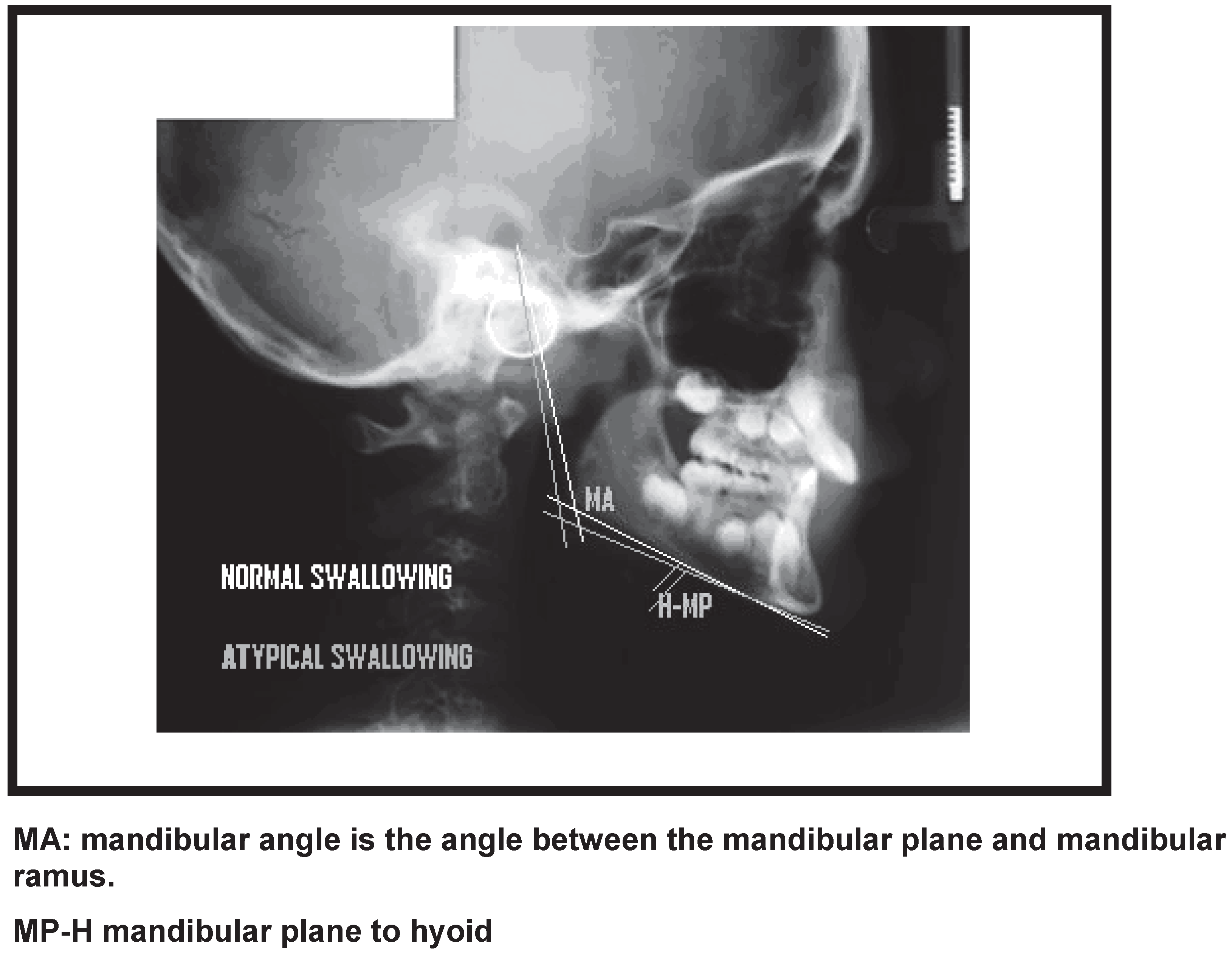
All lateral view teleradiographs selected for the present study were sized 18x24 cm, and were obtained using the same Siemens apparatus in one second at 6 kVp with a focal length of 1.5 meters. The examinations were performed with the patient’s head in a natural position (mirror position), performed by the same examiner. Using the selected lateral teleradiographs, cephalometric examination was performed in a darkened room with a negatoscope. An acetate sheet was laid over the teleradiograph and the following anatomoradiographic landmarks were marked on the sheet (
Figure 1):
MP-H: mandibular plane (line from the midpoint of the mandibular angle to the lowest point on the outline of the mentonian symphysis) to hyoid (most anterosuperior point of the body of the hyoid bone);
MA: mandibular angle (angle between mandibular plane and mandibular ramus).
Figure 1.
Cephalogram Measurements.
Figure 1.
Cephalogram Measurements.
Lateral teleradiographs that did not provide a good view of the anatomical structures used in the cephalometric examination were excluded from the study sample. Patients with dental agenesis, congenital poor orofacial formation, orthodontic and/or functional orthopedic treatment prior to the study, or doubts and imprecision regarding the diagnosis of deglutition were also excluded. A lack of unanimity among the examiners on the clinical diagnosis was also a factor of exclusion of the sample. The skeletal pattern and malocclusion of the patients were not taken into consideration in this study.
The lateral teleradiographs from the experimental group and the control group were randomly put aside and numbered sequentially. The examiner performing the manual measurements was blinded to patient data. The sequentially numbered teleradiographs were handed over to the examiner for the standardized above mentioned measurements to be made, and measurement results were recorded to a data collection instrument. To minimize systematic errors, the same examiner carried out data collection of the whole sample on two occasions separated by a 20-day interval. After the collection of radiographic data, age and sex data were added, along with whether atypical deglutition was present or not. All appropriate measures were taken to ensure confidentiality of the subjects’ personal data.
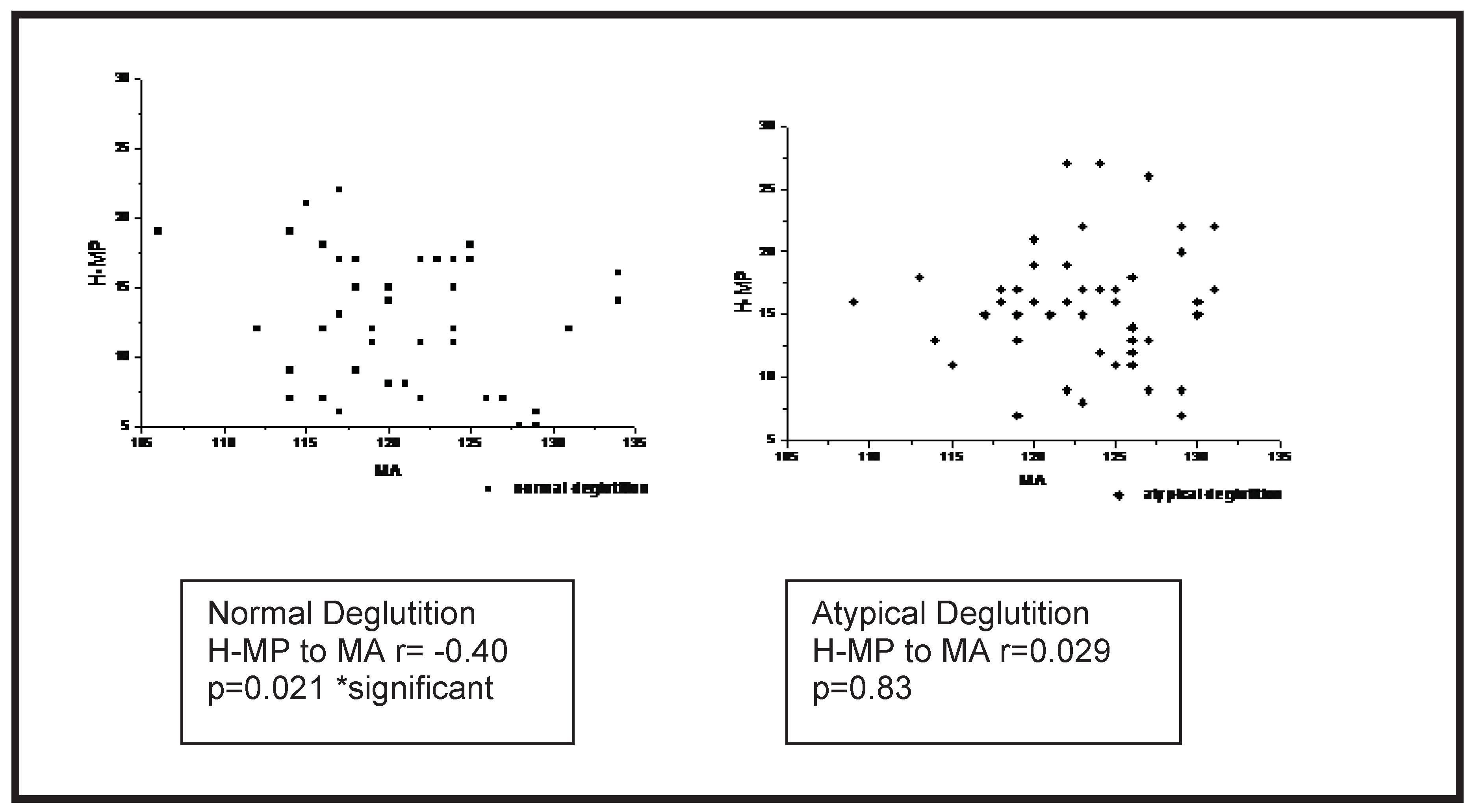
To investigate a possible correlation between variables H-MP and MA, Spearman’s correlation analysis was performed. To investigate the intra-examiner consistency, Wilcoxon test for related samples was used to detect possible differences between measurements obtained in two different occasions. The significance level used in the statistical tests was p=0.05.
Figure 2.
Spearman’s correlation between variables H-MP and MA.
Figure 2.
Spearman’s correlation between variables H-MP and MA.
RESULTS
The study sample consisted of 110 teleradiographs in lateral view, from 52 female and 58 male subjects. The mean age of the control group (normal deglutition) and the experimental group (atypical deglutition) was 9.58 years (+-2.13), and 9.32 years (+-1.83), respectively, without any significant difference between the groups. The age range for each group was from 7 to 11 years of age. To investigate the intra-examiner consistency, the Wilcoxon test for related samples was used to detect possible differences between measurements obtained on two different occasions. However, no significant difference was found between these two measurements.
There was a significant moderate negative correlation between MA (mandibular angle) and hyoid bone (H-MP) in the normal swallow group (R= −0.40, p= 0.021; Table 1). However, there was no significant correlation between the MA and H-MP (R= 0.029, p = 0.83; Table 1) in the atypical deglutition.
DISCUSSION
Interest in the further study of atypical deglutition continues due to many gaps in the literature on this topic. Appealing study topics include the expansion of the classifications of deglutition (
Peng et al., 2004); the causes of atypical deglutition and its consequences (
Cheng et al., 2002); diagnostic methods (Machado Júnior et al., 2011); age at its onset, and treatment methods.
A variety of instrumental techniques are available for diagnosing atypical deglutition. Prominent among the methods that have been used for diagnosis during the oral phase of deglutition is videofluoroscopy. This instrumentation has limited availability in dental practice since the equipment is usually a part of hospital-based radiology departments. The evaluation of the images obtained by videofluoroscopy involves rather subjective assessments (
Peng et al., 2004). However, the multiple views of swallows, involving the frontal, lateral, and coronal dimensions, provide useful descriptions of the mechanical properties of deglutition.
Teleradiographs are standardized extra-oral images that are routinely used as an orthodontic/orthopedic functional diagnostic tool (
Palmer et al., 1992). Teleradiography has been used in a large number of studies on craniofacial growth. Through this method, the spatial relationships between the cranium, vertebrae, mandible, hyoid bone and oropharyngeal space can be easily examined (
Ishida et al., 2002). Data collection was carried out over the entire sample on two occasions, in an attempt to identify any systematic error. An analysis was completed to determine whether the data collected by the same examiner at two different times might vary significantly. However, no significant difference was found between the two measurements, confirming intra-examiner consistency of the method.
This radiographic study evaluated the relationships between known parameters, but these relations were used in a novel manner in that they were correlated with normal and abnormal deglutition. In addition to studying a normal group, this study also evaluated patients with atypical deglutition: a quite prevalent clinical condition that can impact orofacial, nutritional, esthetic, and psychosocial development (
Cheng et al., 1988).
Since deglutition is a highly complex and coordinated function, it requires activation of many anatomical structures related to the tongue. Insufficient functional stimulation of the stomatognathic system, especially the tongue, might be the main factor in the persistence of childlike deglutition (
Peng et al., 2003).
The relationship between craniofacial morphology and extrinsic factors which have an influence on the development of the face has been arousing great interest among researchers. Empirically it is believed that in the cases of atypical swallowing there is a tendency to increase the vertical dimensions of the face, although this possible alteration is not considered a cause or a consequence of atypical swallowing. If this were an absolute truth and if the position of the hyoid bone was dependant exclusively on facial type, the hyoid would be closer to the mandibular plane in atypical swallowing in cases of faces with a tendency to vertical growth, a factor which was not observed in our results. Although we did not plan to correlate data of malocclusion and atypical swallowing, we speculate that this possibility is real, and deserving of further studies (
Graber et al., 1985). Studies have investigated the influence of atypical swallowing on the craniofacial pattern and on mandibular morphology, especially the vertical dimensions of the face, but there is no consensus among researchers (Felício et al., 2010;
Paskay, 2006).
Swallowing is a complex and coordinated function, in which a certain number of muscles is involved, especially the muscles of the tongue, which include the intrinsic muscles and extrinsic muscles (
Peng et al., 2003). The extrinsic muscles of the tongue (genioglossus, styloglossus, palatoglossus, hyoglossus and geniohyoid) may possibly have their tonus altered for individuals with atypical swallowing. This possibility has already been observed in studies with ultrasonography (
Peng et al. 2003,
2004). These studies suggest that in atypical swallowing the activity of the genioglossus muscle is increased, which would explain the lowered resting position of the tongue in atypical swallowing group. This study also mentions that the geniohyoid and mylohyoid were adequate for the distinction of immature swallowing based on the change of muscle tone from the normal swallowing pattern.
The results of this study indicated that there is a negative correlation of the radiographic position of the hyoid bone to the mandibular plane in the group of normal swallowing children. These results are similar to those observed in healthy adults (
Ishida et al., 2002;
Mays et al., 2009;
Palmer et al., 1992). However, there was no significant correlation between the MA and H-MP in the atypical deglutition group. Our results confirm the hypothesis raised by
Mays et al. (
2009) that the mandibular plane is in a position below the gonial angle in relation to a higher FMA (angle between mandibular plane and Frankfurt plane) and the inferior position of the mandibular plane could put the geniohyoid in a position of mechanical disadvantage, reducing its traction on the hyoid bone during swallowing.
Another hypothesis that might be raised is that the differences in the position of the gonial angle and length of the mandible may have additional effects on the control of movement of the hyoid bone. Mays et al., (2009) suggested that the FMA may be a predictor of displacement of the hyoid bone during swallowing; we suggest the use of the mandibular angle to assess the position of the hyoid bone as well.
Finally, our results demonstrate that the smaller the mandibular angle, the greater the distance from the mandibular plane to hyoid bone. The only result this observation could predict is that the hyoid maintains a stable position to the mandibular plane in normal swallowing group. However not observing this correlation in the group of atypical swallowing leads us to believe that the oral functions, specifically deglutition, are able to change the position of the hyoid bone in relation to the mandibular plane.


