Abstract
Publications throughout the world attribute to the artificial teat and the pacifier (dummy) the reason why some mothers, who suspend breast-feeding for a while, are unable to resume it afterwards. The authors wanted to evaluate the specific characteristics of the various commercially made teats and pacifiers. This evaluation examined the physical characteristics of such commercially available teats. It has been possible to affirm that the specific features of the various teats tested are important in the resumption of breast-feeding after such an interruption. It’s easier to resume breast-feeding after interruption if artificial teats are prescribed with an understanding of the muscular movements during swallowing.
INTRODUCTION
The literature is rich with studies supporting the advantages of breast-feeding versus bottle-feeding (Goldfield, Richardson, Lee, Margetts S. (2006) Wojdan-Godek, Mikiel-Kostyra, Mazur, 2005). The passing of antibodies, the suitability of the mothers milk for her child rather than milk from another species, the positive psychological effect of the physical contact and eye contact between mother and child, and the greater intelligence suggested in breast-fed babies (Lehtonen, Kononen, Purhonen, Partanen, Saarikoski, Launiala, 1998; Gomez-Sanciz,
Canete, Rodero, Baeza, Avila, 2003)) are thought to be among the many advantages of breast-feeding. However, sometimes it becomes necessary for the mother to suspend or cease breast-feeding for medical reasons such as illness, exposure to toxins, medications, or for social reasons, such as returning to work. The result is the interruption of breast-feeding (Victora,
Tomasi, Olinto, Barros, 1993). This study investigated the effects produced by the use of artificial teats and dummies (pacifiers) on the swallowing mechanism and the impact of the use of such artificial products on the ability of a mother to resume breast-feeding. Attempts were made to identify: (1) if the rapid transition from mixed feeding to exclusively bottle-feeding is indeed common to every feeding aid used (Gorbe, Kolhalmi,
Gaal, Szantho, Rigo, Harmath, Csabay,
Szabo, 2002; Howard CR, Howard FM,
Lanphear, deBlieck, Eberly, Lawrencw,
1999; Lamounier, 2003); (2) if there are artificial teats that do not modify the swallowing mechanism, and (3) if so, to identify the specific characteristics of those teats.
In order to understand exactly what damage occurs from the use of artificial teats and pacifiers, it is important to highlight the fundamental differences between correct and incorrect swallowing patterns. Swallowing, like breathing, is a basic life function. Development of swallowing precedes breathing as it can be observed by scans showing the first sucking movements in ten-week embryos and rhythmic swallowing three weeks later (Balercia & Balercia, 1993). These early tongue movements are extremely important for shaping the palatal structure and generally for the development of the splancnocranium and the neurocranium (Raymond, & Bacon, 2006). There are many etiologies, such as an anatomical ankylosis (tied tongue or a short lingual frenulum), that may create functional abnormalities in the delicate swallowing mechanism. Although these conditions may be overlooked, they are able to inhibit the correct movements of the tongue. More frequently the etiologies are functional in nature and may be determined by various events.
Labor and birth can lead to an abnormal functioning of the tongue (Frymann, 1966) due to the prolonged compression on the occiput, which is an area particularly vulnerable due to the risk of compressing the hypoglossal nerves. This pair of cranial nerves supplies motor innervation to fifteen of the sixteen tongue muscles (Frymann, 1966; Ferrante, 1997). The umbilical chord twisted round the baby’s neck may create edema in the area under the lower jaw, and difficulty of movement of the hyoid. The hyoid provides a fundamental connection point for the tongue muscles, as well as for the muscles of the upper body, by means of the infra-hyoid muscles. This reduced mobility of the hyoid may cause difficulty of tongue movement after birth with consequent sluggishness in latching on to the breast nipple.
The tongue, however, has within itself a wonderful capacity to regain normal functioning. By means of its connection with the cranial base, via the insertion of the styloglossus muscle, the tongue is capable of resolving most of the problems that may be caused by birth. According to osteopathic principles (Frymann, 1966), the innate reflex that causes the baby’s tongue to grasp the mother’s nipple and squeeze it against the palate, allows a resumption of the cranial breathing rhythm and permits the muscles to perform adequately. This forward to backward movement is the same one the baby has been practicing in the womb for six months already. The styloglossus, inserted on the temporal styloid process and activated during swallowing, produces movement of the cranial base. Any hindrance to a correct tongue movement impedes the memorization of movements of the swallowing mechanism, with consequent dysfunction.
The hallmark of swallowing is a specific timing in muscle activation. After the contraction of the chewing muscles to stabilize the mandible, the tongue squeezes the nipple against the palate. At this stage, the most active muscles are the intrinsic and extrinsic tongue muscles (Ferrante, 1995; Ferrante, 1997; Garliner, 1968; Logeman, 1988). After this tongue movement, comes the peristalsis of the pharyngeal muscles, which is essential for the progression of food without adding air, and for the opening of the Eustachian tubes, which is necessary for aeration of the middle ear and drainage of the mucus (Tamura, Horikawa, & Yoshid, 1996).
When there is an incorrect swallowing pattern the tongue is unable to move as described above and tends to thrust forward, and may thrust between the dental arches. This means that other muscles have to be recruited to allow swallowing to occur (Ferrante, 1995; Ferrante, 1997; Garliner, 1968). The most important of these are the buccinators, which press against the dental arches when contracting. This contraction may cause, over the years, a narrowing of the palate and subsequent breathing problems. Breathing problems may occur as a result of the increased nasal resistance due to the decrease air space available. However, the most important characteristic of an incorrect swallowing pattern is probably the swallowing of air, which is the cause of aerophagia typical of bottle-fed babies.
Therefore, it is important to consider the causes which most easily affect tongue movement: sucking of the thumb or fingers (Straub, 1951; Aarts, Hornell, Kylberg,
Hofvander, Gebre-Medhin, 1999), bottle-feeding, and the use of pacifiers. Much has been said about the emotional and psychological factors involved in thumb sucking (Clauzade & Marty, 2004). The authors, who have carried out research which is about to be published, wish to point out the frequent association of difficulty of tongue movement and the impossibility of the tongue to directly stimulate the exteroceptors present at the end of the nasopalatal nerve during thumb sucking and while using feeding aids (2003). Thumb sucking, using pacifiers and bottle-feeding, are able to block the tongue movement almost 100% of the time (Gorbe et al., 2002; Howard et al., 1999; Lamounier, 2003; Rigard, 1998). A pacifier, a bottle teat, or a thumb takes up a large part of the functional space of the tongue and does not allow full tongue movement. The only movement possible is horizontal, either back to front, or side-to-side. A tongue unable to move correctly does not contribute to the swallow process, as it should. This difficulty forces the buccinators (Ferrante, 2003; Janskey, &
Zelny, 1961) to compensate for the tongue function. The buccinators then become indispensable for swallowing.
Inhibiting proper tongue function influences the complete and correct use of the surrounding muscles, in particular the styloglossus and the palatoglossus. The styloglossus and the palatoglossus become increasingly weaker. This progressive decline in muscle function may be the reason why in a matter of months or weeks the baby becomes dependent on the bottle (Howard
CR, Howard FM, Lanphear, Eberly, deBlieck,
Oakes, Lawrence, 2003). If the mother wishes to speed up the feeding sessions and makes the hole of the artificial teat larger, the tongue thrusts forward even more forcefully, to attempt to control the flow of milk and to allow the baby to breathe. The size of the hole in the bottle teat is very important because the larger the hole, the less the baby has to suck, as the milk comes out by force of gravity. This leads to a weakening of the facial muscles and especially the orbicularis oris, which then may lead to lack of lip seal.
METHOD
The 78 babies included in this research were between two and six months of age, in perfect health, without any restriction of the lingual frenulum to influence tongue movement. The babies were divided into three groups to observe their movements more precisely. Group 1 included 27 babies who were exclusively breast-fed, representing 34.6% of the total population. These 27 exclusively breast-fed babies made up the control group, and demonstrated correct tongue movement and swallowing after every 3-5 suctions. Group 2 included 14 babies on mixed feeding programs, representing 17.9% of the total population. Group 3 included 37 babies exclusively bottle-fed, representing 47.5% of the total population.
Seven different artificial bottle teats were tested: one was the “traditional” type (defined as type F); one that appeared to be anatomical (type G); two defined by the producer as “physiological” (A, B), and lastly three “newly designed” ones (types C, D, E). These last three, although of different sizes had all the same design characteristics. The artificial teats types A, B, C, D, E were advertised as being similar to the mother’s nipple, due to the characteristic of the teats to lengthen during suction.
It was not possible to test all the babies with all the seven artificial teats because some of those teats were refused by some babies. The babies who were exclusively bottle-fed, or on a mixed feeding regime (both breastfed and bottle fed) were tested with the teat they were already using. Other types of teats were then presented. Some babies did not accept teats other than their own.
Research was conducted on three levels
- 1)
- A consideration of the teats’ characteristics was deemed necessary to avoid iatrogenic damage to correct function. The flexibility and expandability of the teats and the compressibility of the pacifiers most used in Italy were tested.
- 2)
- An ultrasound scan filming the sucking and swallowing of the baby was completed. A comparison was made of tongue movement when using teats and while breast-feeding. Raymond & Bacon, 2006; Smith, Erenberg, Novak, 1988; Novak, Smith, Erenberg,1995). Various different types of teats and pacifiers were tested to evaluate any differences of tongue posture and movement during swallowing.
- 3)
- The mother’s opinion was also sought. While a mother’s opinion is not an objective judgment, it was considered important if the results of this study were to be implemented in the community.
Instrumentation
Ultrasound scans were performed using the AU3 Partner (Advanced Ultrasonographie) produced by the Esaote Biomedica connected to a video recorder. The function of the tongue and the swallowing muscles were filmed first during breast-feeding, and then after introducing seven brands of teat. The babies who were exclusively breast-fed made up the control group. This control group was used to evaluate if the changes in tongue function were due to the teat tested or to a natural consequence due to the developmental growth of the baby.
The following tests were carried out with the artificial teats:
- (a)
- Measurement of the force needed to bend the teat 5mm: The Deflection Test.
- (b)
- Measurement of the force needed to extend the teat to a length of 5mm: The Extension Test.
The DeflectionTest
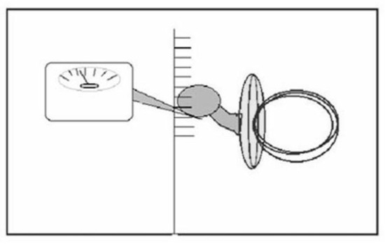
To measure the deflection of the artificial teat (Deflection Test), defined as the force necessary to bend the teat 5mm from its longitudinal axis, the teat was held blocked horizontally and a tangent force was applied to raise the teat 5mm. For this measurement a hand held dynamometer (Haag-Streit A.G. Correx) was used. The measurement of the strength needed to raise the artificial teat 5mm from its horizontal position is thought to be an indication of the physiological movement or force necessary to press the mother’s nipple against the palate and squeeze out the milk. The more force needed to move the artificial teat the more the teat is able to modify tongue function.
Seven types of teats of the four brands most widely available on the Italian market were tested. The measurement of 5mm was chosen because all of the teats (and later, pacifiers) were able to bend this amount, and because it is a measurement similar to that of the space between the teat/pacifier and the palate. The same methods were later used for the Deflection Test of 13 pacifiers from three different brands.

Figure 1.
Deflection Test.
The Extension Test
To measure the extension of the artificial teat, the lengthening or drawing out of the teat, a clip was attached to a tubular dynamometer “Pesola”, corresponding to the equator of the teat, and traction was exerted until the teat was lengthened by 5mm. The force needed to stretch the teat to this length was then recorded. This process was repeated ten times consecutively and averaged to minimize the risk of error. Fluctuations of the individual extension readings were within 5% of the maximum measurement of each teat.
The measurement of the strength needed to obtain a lengthening of the artificial teat to 5 mm informs us of the strength needed to fulfill the motion of squeezing out the milk as done on the mother’s nipple. The physiological movement of squeezing the nipple occurs with the raising of the tongue under the nipple and the dragging it backwards.
The less strength needed to stretch the nipple to 5mm the less the likelihood that tongue function will be altered. This test also makes possible an assessment of the truth of the claims made by the various brands about the similarity of their own teats to that of the mother’s nipple. The same methods were later used to complete the Extension Test on pacifiers.
RESULTS
During testing, differences were observed between the various types of bottle teats. The material composition of the teat made a difference. The mixture of rubber used demonstrated very different levels of elasticity, however, the largest differences noted were related to the shape of the teat. Teats C, D, E all produced by the same brand name required minimum strength to be raised to 5mm, while others required more than double the strength to fulfill the same requirement. Table 1
Greater differences were noted in the deflection tests. The same bottle teats, shown to be resistant during the deflection tests, were found to be even less mobile when it came to the possibility of lengthening the teats. In testing two of the teats, it was determined that it was necessary to apply eight times more strength to attain an extension of 5 mm, when compared to the remaining bottle teats. Table 2
An important finding in regard to the pacifiers was the variation in force necessary to raise them against the palate. In addition to the impact of the rubber used for construction of the pacifiers, the most conclusive finding was that better results were obtained from the anatomically shaped pacifiers. Table 3
Ultrasound Scanning
Seventy-eight scans were recorded with the following results:
The five babies filmed using the “traditional” bottle teat (F) attempted to raise the tongue but also thrust the tongue forward while swallowing (probably due to the non-deformability of the teat).
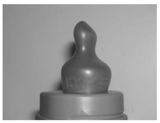
The “anatomical” teats (G) produced mixed results: in 50% of the babies the tongue did indeed produce an upward and backward movement, but in the other 50% the movement was forward.
The control group was evaluated with mother’s nipple and various artificial bottle teats to see if the dysfunction induced by the use of the teats was immediate or not. There were no differences between the babies who were already using a specific teat and those who were trying it for the first.

Figure 2.
The anatomical teat does not have the hole at the end of the teat.
The A and B type teats produced raising of the tongue towards the palate in 50% of the babies. However, there was consistently a space between the teat and the palate suggesting that additional strength would be needed to completely press the teat against the palate.

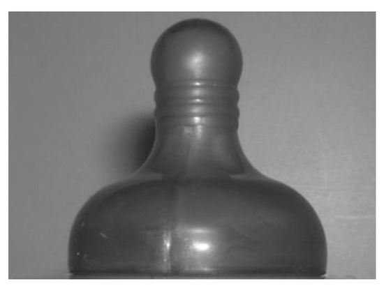
Figure 3.
The “physiological” teat. (The ripples are there to enhance the lengthening of the teat but do not appear to be very effective.).
In 35.9% of cases the teat advertised as “ physiological” induced difficulty of tongue movement, while in 14.1% the tongue maintained a correct functional movement, probably because they were breast-fed babies and had maintained a superior muscular efficiency.

Table 1.
Strength needed to raise the teat 5mm (Deflection).
Table 1.
Strength needed to raise the teat 5mm (Deflection).
| Teat A | 0-4 months | 65 g |
| Teat B | 4 + months | 55 g |
| Teat C | 4 + months, 3 holes | 25 g |
| Teat D | 4 + months, semi solids hole | 25 g |
| Teat E | 0-4 months | 20 g |
| Teat F | Silicone | 90 g |
| Teat G | 3 holes | 85 g |

Table 2.
Strength needed to lengthen the teat 5 mm (Extension).
Table 2.
Strength needed to lengthen the teat 5 mm (Extension).
| Teat A | 0-4 months | 420 g |
| Teat B | 4 + months | 300 g |
| Teat C | 4 + months, 3 holes | 120 g |
| Teat D | 4 + months, semi solids hole | 120 g |
| Teat E | 0-4 months | 100 g |
| Teat F | Silicone | 910 g |
| Teat G | 3 holes | 850 g |

Table 3.
Deflection in pacifiers (dummies).
Table 3.
Deflection in pacifiers (dummies).
| Dummy A (cherry) 0-6 months | 100 g |
| Dummy A (cherry) 6-18 months | 55g |
| Dummy A (anatomical) 0-6 months | 40 g (60 g upside down) |
| Dummy A (anatomical) 6-18 months | 25 g (35 g upside down) |
| Dummy A (all rubber cherry) | 55 g |
| Dummy A (all rubber anatomical) | 20 g (35 g upside down) |
| Dummy B (rubber drop shape) Medium | 70 g |
| Dummy B (all rubber drop shape) Medium | 70 g |
| Dummy B (silicone cherry) | 125 g |
| Dummy C (rubber cherry) diameter 18,6 mm | 125 g |
| Dummy C (silicone cherry) diameter 15 mm | 150 g |
The “newly designed” teats (C, D, E) allowed the tongue to preserve its normal movement during swallowing in over 80% of cases. In the remaining 20% of the babies, the tongue movement was deemed to be within functional limits although not within normal limits. The “physiological” type of teats were the least accepted by the babies, except when they had used these teats from the first day of life. These teats require additional muscular work when compared to traditional teats, which allow a more easily obtained flow of milk. Only one baby accepted a “ physiological” teat even though it was using a “traditional” type for daily feedings.

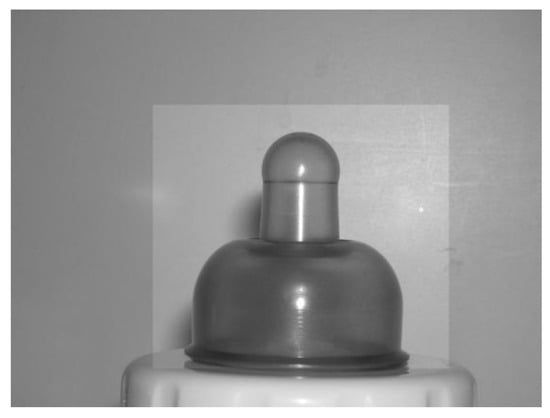
Figure 4.
The Newly Designed Teat is characterized by a blending between the false nipple and the areola. This permits for lengthening in this area.
Scans were also taken of pacifier sucking. None of the pacifiers were found to be without risk but the soft flexible anatomical pacifiers allowed pressing against the palate in 70% of cases, while in 50% the tongue moved in both vertical and horizontal directions. Large pacifiers or pacifiers with non-anatomical shapes (particularly cherry or olive shaped) were observed to condition the tongue negatively, inducing it to thrust forward and impeding it to rise toward the palate.
CONCLUSIONS
Most of the bottle teats and pacifiers tested showed entirely different characteristics from those of the mother’s nipple, and resulted in very obvious impediment to free tongue movement. Free tongue movement is necessary for correct functioning during swallowing. Of a particular importance, all of the pacifiers/dummies were found to have risks associated with their use. Of the pacifiers sampled in this study, some produced much more damage than others as indicated by the data contained in tables previously presented.
Both the Deflection and Extension Tests, and the ultrasound scans provided results, which varied significantly from teat to teat. The strength needed to press the teat against the palate and to lengthen it is the strength necessary to swallow correctly. If the strength required to perform this function is far beyond the baby’s capacity, the teat will force the tongue down from the palate and will promote a permanent incorrect swallowing pattern. The less the hindrance to tongue movements, the more likely the baby will preserve the capacity to return to breastfeeding.
The results indicate that the two most important characteristics the teat should offer are pliability/softness and a shape, which facilitates ease of deflection and extention. These qualities were indeed indicated by the extension tests in the last group of teats C, D, E, and the “newly designed” teat during ultrasound scanning. However, in order to achieve the same results of deflecting the teat 5 mm, the “physiological” teat needed between three and eight times the strength exerted against it when compared to the group of teats C, D, E, and the “newly designed” teat. A baby may not have the necessary strength to raise and extend these kinds of bottle teats, and the muscular pattern of sucking and swallowing from these teats will continue to be functional but not normal. Unfortunately, the worst results were obtained by one of the teats most frequently found in Italian maternity wards.
What emerged from the information given by the mothers, during informal interviews, confirms the findings of the tests conducted. The mother’s judgments, although not scientific, can provide significant information, which can help us to understand the importance of choosing the right teat. However, the anecdotal experience of mothers may be enhanced by the scientific information provided by an informed neonatologist or pediatrician.
The most important point which emerged is that almost all the milk bottle teats sold today, like their predecessors, have been shown to alter correct tongue function and swallowing to the point of making a mixed feeding program very improbable. This is due to the activation of different muscles during breast and bottle-feeding, which makes establishing a correct swallowing pattern almost impossible to achieve. Most of the breast-fed babies who were exposed to bottle-feeding very quickly stopped breast-feeding, favoring the quick and energy-saving bottle teat to the mother’s more tiring and time-consuming nipple. For this reason we were rather surprised to hear that most of the babies who suspended breast-feeding for a while, substituting the “newly designed” teat, did not have difficulty in returning to the breast afterwards, and had no problems with a mixed feeding regime. The “newly designed” teat was shown to be a valid option, as described above, when used exclusively as a first and only feeding aid.
The only limitation noted was that, when it was tested on babies who had already tried other teats, the “newly designed” teat was frequently refused. The babies at this point were already negatively conditioned in their tongue function. A possible explanation is that the baby who used the “newly designed” teat for the first time did not have to change the existing swallowing pattern and continued to activate the same muscles as they did in breast-feeding in the same way, with the same force. The baby already fed with other types of teats had, at this point, already lost a correct swallowing pattern and was unable to recreate a correct activation of the muscles. The baby would not accept a feeding method demanding the same force required for breast-feeding from muscles which had become inactive.
Acknowledgments
The authors wish to extend their sincere appreciation to Licia Coceani Paskay, MS, CCC-SLP, COM, for her assistance in the preparation of this article.
Conflicts of Interest
Until Dec. 2005, Dr A. Ferrante was employed as consultant for a company that produced artificial nipples and pacifiers. During his tenure at that company, he had the opportunity to test some of the products. The specific brand names were omitted from this report. The purpose of this research is to assist mothers in their choice of a more advantageous product, if breast-feeding is interrupted and artificial means of feeding, such as bottle-feeding, becomes necessary.
References
- Aarts, C., A. Hornell, E. Kylberg, Y. Hofvander, and M. Gebre-Medhin. 1999. Breastfeeding patterns in relation to thumb sucking and pacifier use. Pediatrics 104, 4: e50. [Google Scholar]
- Balercia, L., and P. Balercia. 1993. Fisiopatologia della deglutizione. Relazioni tra occlusione e postura. Dentista Moderno 1: 57–83. [Google Scholar]
- Clauzade, M., and J.P. Marty. 2004. Ortoposturodonzia. GLM Ed. Roma (4): 97–98. [Google Scholar]
- Ferrante, A. 1995. La deglutizione atipica. Il dentista moderno 2: 227–239. [Google Scholar]
- Ferrante, A., and S. Benedetto del Tronto. 1997. Terapia Miofunzionale. Futura Ed.
- Ferrante, A. 2003. Considerazioni sui danni al meccanismo deglutitorio causati da succhietti e tettarelle. Estratti I° Congresso Nazionale IMA, 33–36. [Google Scholar]
- Ferrante, A., C. Montinaro, R. Silvestri, and V. Stile. 2003. Studio comparativo clinico e tecnico-strumentale su varie tipologie di succhietti. International Myofunctional Association 1: 15–17. [Google Scholar]
- Frymann, V. M. 1966. Relation of disturbances of craniosacral mechanism to symptomatology of the newborn: Study of 1250 infants. Journal of American. Osteopathy 65: 1059–1075. [Google Scholar]
- Garliner, D. 1968. Effects of unrecognized abnormal swallowing. Journal of Canadian Dental Association 34: 301–304. [Google Scholar]
- Goldfield, E. C., M. J. Richardson, K. G. Lee, and S. Margetts. 2006. Coordination of sucking, swallowing, and breathing and oxygen saturation during early infant breast-feeding and bottle feeding. In Pediatric Research. Epub: vol. 60, 4, pp. 450–455. [Google Scholar]
- Gomez-Sanciz, M., R. Canete, I. Rodero, J. E. Baeza, and O. Avila. 2003. Influence of breastfeeding on mental and psychomotor development. Clinical Pediatrics (Philadelphia) 42: 35–42. [Google Scholar] [CrossRef]
- Gorbe, E., B. Kolhalmi, G. Gaal, A. Szantho, J. Rigo, A. Harmath, L. Csabay, and G. Szabo. 2002. The relationship between pacifier use, bottle feeding and breast feeding. Journal of Maternal, Fetal and Neonatal Medicine 12, 127–131. [Google Scholar]
- Howard, C. R., F. M. Howard, B. Lanphear, E. A. deBlieck, S. Eberly, and R. A. Lawrencw. 1999. The effects of early pacifier use on breastfeeding duration. Pediatrics 103, 3: E33. [Google Scholar] [CrossRef] [PubMed]
- Howard, C. R., F. M. Howard, B. Lanphear, S. Eberly, E. A. deBlieck, D. Oakes, and R. A. Lawrence. 2003. Randomized clinical trial of pacifier use and bottle-feeding or cupfeeding and their effect on breastfeeding. Pediatrics 111, 3: 511–518. [Google Scholar] [CrossRef] [PubMed]
- Janskey, Z., and A. Zelny. 1961. “Investigation of the muscular forces exerted by the human tongue”. Dental Abstracts 6: 267. [Google Scholar]
- Lamounier, J. A. 2003. The influence of nipples and pacifiers on breastfeeding duration. Journal of.Pediatrics. (Rio Journal) 79, 4: 284–286. [Google Scholar] [CrossRef]
- Lehtonen, J., M. Kononen, M. Purhonen, J. Partanen, S. Saarikoski, and K. Launiala. 1998. The effect of nursing on the brain activity of the newborn. Journal of.Pediatrics 132, 4: 646–651. [Google Scholar] [CrossRef]
- Logemann, J. A. 1988. Swallowing physiology and patho-physiology. Otological. Clinic of North America 21: 4. [Google Scholar]
- Novak, A. J., W. L. Smith, and A. Erenberg. 1995. Imaging evaluation of breast-feeding and bottle-feeding systems. Journal of Pediatrics 126, 6: S130–S134. [Google Scholar]
- Raymond, J. L., and W. Bacon. 2006. Influence of feeding method on maxillofacial development”. Orthodontie Francaise 77, 1: 101–3. [Google Scholar] [CrossRef]
- Rigard, L. 1998. Are breastfeeding problems related to incorrect breastfeeding techniques and the use of pacifiers and bottles? Birth 25, 1: 40–44. [Google Scholar] [CrossRef]
- Smith, W. L., A. Erenberg, and A. Novak. 1988. Imaging evaluation of the human nipple during breast feeling. American Journal Disease in Children 142, 1: 76–8. [Google Scholar]
- Straub, W. J. 1951. The etiology of the perverted swallowing habit. American Journal of Orthodontics, 603–612. [Google Scholar]
- Tamura, Y., Y. Horikawa, and S. Yoshida. 1996. Co-ordination of tongue movements and peri-oral muscle activities during nutritive sucking. Developmental Medicine and Children Neurology 38, 6: 503–510. [Google Scholar] [CrossRef] [PubMed]
- Victora, C. G., E. Tomasi, M. T. Olinto, and F. C. Barros. 1993. Use of pacifiers and breastfeeding duration. Lancet 341, 8842: 404–406. [Google Scholar] [CrossRef] [PubMed]
- Wojdan-Godek, E., K. Mikiel-Kostyra, and J. Mazur. 2005. Effect of feeding pattern on the body mass of infants in the first six months of life. Medecine Wieku Rozwoj 4: 611–620. [Google Scholar]
Disclaimer/Publisher’s Note: The statements, opinions and data contained in all publications are solely those of the individual author(s) and contributor(s) and not of MDPI and/or the editor(s). MDPI and/or the editor(s) disclaim responsibility for any injury to people or property resulting from any ideas, methods, instructions or products referred to in the content. |
© 2008 by the authors. 2008 Antonio Ferrante, Raffaele Silvestri, Carlo Montinaro