Article

Antitumor Potential of Bioactive Crude Extracts Derived from Actinomycetes

by
Hassan K. Dhaini
1,
Bahaa Fahed Hassanieh
1,
Rana El Hajj
1,* and
Mahmoud I. Khalil
1,2
1
Department of Biological Sciences, Faculty of Science, Beirut Arab University, Beirut P.O. Box 11-5020, Lebanon
2
Molecular Biology Unit, Department of Zoology, Faculty of Science, Alexandria University, Alexandria 21568, Egypt
*
Author to whom correspondence should be addressed.
Bacteria 2025, 4(4), 51; https://doi.org/10.3390/bacteria4040051
Submission received: 24 June 2025 / Revised: 31 July 2025 / Accepted: 4 September 2025 / Published: 1 October 2025

Abstract

Marine actinomycetes constitute a vigorous source of bioactive compounds with potential anti-tumor activity. This study investigates the antitumor activity and classification of actinomycetes isolated from 32 marine soil samples collected across four seasons from Tyr City Beach, Lebanon. A total of 80 morphologically diverse isolates were recovered and characterized, with dominant genera including Streptomyces, Kocuria, and Micrococcus. Among these, three promising strains—Kocuria rosea, Micrococcus luteus, and Streptomyces longisporoflavus—were selected for further analysis. Crude extracts were tested against human colorectal adenocarcinoma (Caco-2) and human hepatocellular carcinoma (HepG-2) cancer cell lines using MTT and Western blot assays. At the highest concentration (8 µg/µL), the extracts reduced cell viability to 24–37% in Caco-2 and 12–25% in HepG-2. The IC50 values ranged from 1.72 to 3.53 µg/µL, depending on the extract and cell line. Western blot analysis showed dose-dependent increases in the Bax/Bcl-2 ratio, with fold changes reaching 4.35 (Kocuria), 11.39 (Micrococcus), and 14.25 (Streptomyces) in HepG-2 cells. The p53 protein expression also increased significantly, with fold changes up to 7.79 in Caco-2 and 3.0 in HepG-2 cells. These results indicate that marine actinomycetes from the Lebanese coastline hold strong potential as a source of antitumor agents targeting apoptosis pathways.

1. Introduction

Actinomycetes represent a phylogenetically and functionally distinct group of Gram-positive bacteria, bridging characteristics of both prokaryotic bacteria and eukaryotic fungi. These symbiotic microorganisms are isolated from a wide variety of plant species across diverse ecosystems, suggesting their widespread occurrence and ecological importance. Notably, genera such as Streptomyces, Micromonospora, and Nocardiopsis are recognized for producing pharmacologically significant compounds, including antibiotics, antitumor agents, and immunomodulators. This positions endophytic actinomycetes as a promising frontier for drug discovery and sustainable agricultural innovations.
Natural products have long served as a cornerstone in the discovery and development of anticancer therapeutics, offering structurally diverse and biologically potent molecules, underscoring their pivotal role, particularly in oncology drug pipelines [1]. Among these sources, the marine environment has emerged as a particularly rich and underexploited reservoir of bioactive secondary metabolites. To date, more than 22,000 marine-derived natural compounds have been isolated and documented, many of which exhibit unique chemical architectures not found in terrestrial organisms. Several of these marine compounds have demonstrated potent antitumor properties, with some progressing through various phases of clinical evaluation or serving as lead structures for the rational design and synthesis of next-generation anticancer drugs. This highlights the vast and largely untapped therapeutic potential of marine biodiversity to fight cancer [2].
Actinomycetes contribute substantially to bioactive compound production, notably with 7600 of the 23,000 identified secondary metabolites coming from Streptomyces species alone. Their biological roles vary depending on their source of isolation, and they can produce bioactive compounds with antiviral, anti-inflammatory, antimicrobial, and antitumor properties, though data on their antioxidant capabilities remain limited. Additionally, actinomycetes diversity, influenced by isolation methods, is essential for ecological understanding and identifying beneficial strains [3,4,5].
Streptomyces sp. extract from soil sample culture demonstrated an antitumor effect against some colon cancer cell lines by the upregulation of Bax and downregulation of Bcl-2, suggesting the induction of the intrinsic apoptotic pathway. On the other hand, the extract decreased the expression of cyclin D1 and increased the expression of the P21 gene, leading to a cell cycle arrest in the G0/G1 phase [6].
Additionally, Streptomyces isolates contain compounds such as viridenomycin, violapyrone A, elaiomycin L, and feigrisolide B with anticancer and antimicrobial activities through the induction of apoptosis and cell cycle arrest [7].
Moreover, actinomycetes-derived compounds downregulated the expression of key tumor metabolic enzymes, such as hexokinase 2 (HK2), phosphofructokinase (PFKFB3), and pyruvate kinase (PKM2), showing a promising antitumor effect [8].
This study aimed to examine the antitumor activities of active metabolites derived from multiple actinomycetes from marine soil in Tyr City Beach, Lebanon, against colon and liver cancer cells. The novelty of this work lies in the isolation and characterization of marine actinomycetes from an underexplored region of the Eastern Mediterranean—Tyr City Beach, Lebanon—and the demonstration of their antitumor potential for the first time against Caco-2 and HepG-2 cancer cell lines. The study not only identifies three promising strains (Kocuria rosea, Micrococcus luteus, and Streptomyces longisporoflavus) but also links their bioactive metabolites to molecular apoptotic pathways via Bax/Bcl-2 modulation and p53 upregulation, highlighting their mechanistic relevance. Furthermore, preliminary in vitro assays such as the MTT and Western blot are critical first steps in drug discovery. They allow for rapid screening of cytotoxicity, evaluation of dose–response relationships, and identification of mechanistic targets, providing essential data to prioritize candidates for further in vivo validation and potential therapeutic development.

2. Materials and Methods

2.1. Sample Preparation and Identification

Across four seasons, Fall/Winter 2021 and Spring/Summer 2022, 32 marine soil samples were obtained from Tyr City Beach. Samples collected at 15.0 cm depth from the sandy shore were designated as A1, B1, C1, and D1, totaling sixteen. The remaining sixteen samples, labeled A2, B2, C2, and D2, were gathered from 2.0 m beneath the sea surface. Details regarding the sampling methodology are depicted in Figure 1.
After collection, 1 g of each sediment sample was mixed with 9 mL of sterile distilled water and subjected to a series of ten-fold serial dilutions. From each dilution, 100 μL aliquots were plated onto Starch Casein Agar (SCA) supplemented with cycloheximide (100 μg/mL) and nalidixic acid (30 μg/mL). The plates were incubated at 28 °C for a period ranging from 7 to 21 days [9].
Marine actinomycete samples were cultured on Starch Casein Agar and initially identified based on colony morphology, pigmentation, and growth characteristics. Further identification was carried out using morphological traits, Gram staining, and a series of biochemical tests (catalase, oxidase, citrate utilization, urease, and nitrate reduction), followed by genus-level confirmation using the VITEK 2® Gp automated system (manufactured by bioMérieux, Marcy-l’Étoile, France) [10].
These procedures were described in detail in our previously published study [10], which reported the recovery of 80 marine actinomycete isolates from seasonal samples collected along Tyr City Beach. Based on their distinct biochemical profiles and preliminary antimicrobial activity, isolates belonging to the genera Kocuria, Micrococcus, and Streptomyces were selected for further investigation. One representative strain from each genus was chosen for fermentation in Starch Casein Broth, followed by extraction of secondary metabolites using ethyl acetate and concentration by rotary evaporation.
In order to obtain a crude extract from the studied actinomycetes, 1.5 mL of stock suspension of the actinomycetes associated with the highest antimicrobial activities was poured onto 200 mL of SCB and incubated at 30 °C at 200 rpm for 10 days. The supernatant was then collected and supplemented with an equal volume of ethyl acetate. Following centrifugation, the upper layer containing secondary metabolites was collected and evaporated using a rotary evaporator at 40 °C [11,12].

2.2. Cell Culture

Caco-2 and HepG-2 cells were maintained in Dulbecco’s Modified Eagle Medium (DMEM) supplemented with 10% Fetal Bovine Serum (FBS), 100 units/mL penicillin, and 100 μg/mL streptomycin, under conditions of 37 °C and 5% CO2 atmosphere. Cells were subcultured every 2–3 days after reaching 70–80% confluency (according to ATCC protocol).

2.3. Cell Viability Assay

Cells grown to 70–80% confluency in complete DMEM were seeded into 96-well plates at a density of 3 × 10⁴ cells per well and incubated under standard conditions for 24 h. Subsequently, they were treated with various concentrations of bacterial extracts ranging from 0.125 µg/µL to 8 µg/µL in half-fold dilutions. After 24 h, crude extracts were dissolved in DMSO and added to achieve a final concentration of 50 μg/mL. Plates were then incubated for 24 and 48 h. Following incubation, the culture medium was replaced with fresh medium containing MTT (3-(4,5-dimethylthiazol-2-yl)-2,5-diphenyl tetrazolium bromide) at a concentration of 0.5 mg/mL. After a 3 h incubation, the medium was removed, plates were dried, and the resulting formazan crystals were dissolved in 150 μL of DMSO. Absorbance was measured at 570 nm. Doxorubicin and DMSO served as positive and negative controls, respectively. The activity of each extract was expressed as a percentage relative to the positive control, with extracts considered cytotoxic if they inhibited more than 75% of cell growth at the tested concentration.

2.4. Western Blot Assay

Caco-2 and HepG-2 cells were treated in triplicate for 24 h using concentrations corresponding to the IC50 and ½ IC50 values. Following treatment, cells were harvested, lysed with NP-40 whole cell lysis buffer, and the protein was extracted after centrifugation, subjected to electrophoresis, and transferred onto nitrocellulose membrane, which was incubated with Bax rabbit monoclonal antibody (1:1000, Cell Signaling, Danvers, MA, USA, Cat no. 2774S), rabbit anti-Bcl-2 antibody (Cell Signaling, Cat no. D55G8), rabbit anti-p53 antibody (Cell Signaling, Danvers, MA, USA, Cat no. 9282T), and rabbit anti-β-actin antibody (1:1000, Cell Signaling, Cat no. 13E5). Protein was later incubated with HRP-conjugated anti-rabbit IgG secondary antibody (1:1000, Cell Signaling, Cat no. 7074). Detection was performed using a chemiluminescent HRP substrate (Cell Signaling, Danvers, MA, USA, Cat no. 6950S), and band intensities were analyzed using ImageJ software version 1.54p. All experiments were independently repeated at least three times [13].

2.5. Statistical Analysis

Statistical analysis of the data and experimental design was performed using Excel® 2016 MSO (16.0.4266.1001) 32-bit and GraphPad Prism version 9.5.1. Differences between groups were assessed using t-tests and one-way ANOVA, with a p-value of less than 0.05 considered statistically significant.

3. Results

3.1. Effect of Extracts Derived from Kocuria Rosea, Micrococcus luteus, and Streptomyces longisporoflavus on the Proliferation and Viability of Caco-2 and HepG-2 Cells

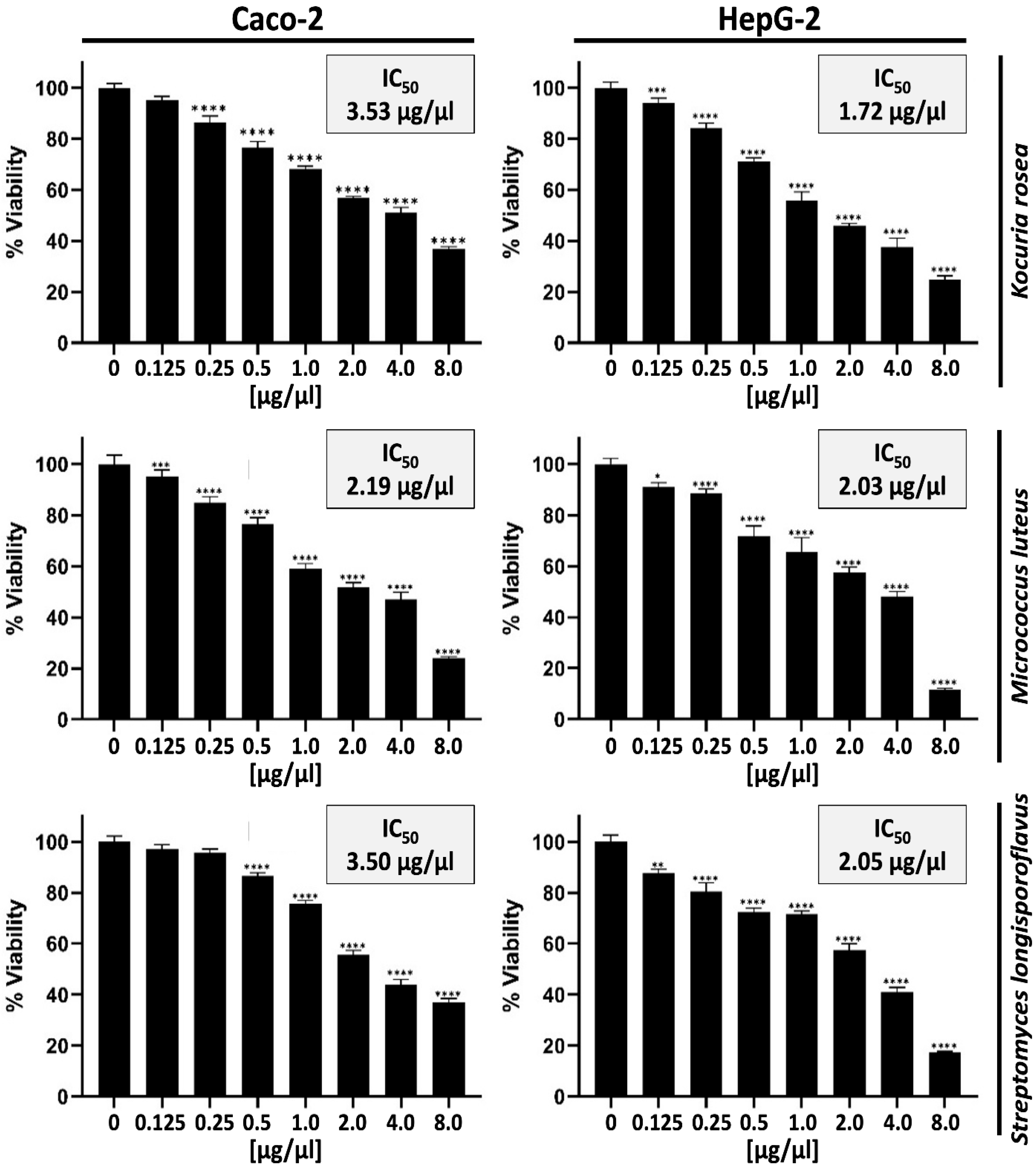
A significant decrease in cell viability was observed in both Caco-2 and HepG-2 cell lines when treated with all three extracts at the highest concentration of 8 µg/µL. Kocuria rosea extracts reduced cell viability to 37 ± 1.1% in Caco-2 and 25 ± 0.8% in HepG-2 cells, Micrococcus luteus extracts reduced it to 24 ± 0.7% and 12 ± 0.4%, respectively, and Streptomyces longisporoflavus extracts to 37 ± 0.9% and 17 ± 0.5%, respectively (Figure 2).
The IC50 of all three extracts was greater in Caco-2 than HepG-2 when treated with Kocuria rosea extracts 3.53 µg/µL and 1.72 µg/µL, respectively, showing a major difference between the two cell lines of 1.81 µg/µL; however, Micrococcus luteus extracts reached 2.19 µg/µL and 2.03 µg/µL, respectively, a 0.16 µg/µL difference, and Streptomyces longisporoflavus extracts reached 3.50 µg/µL and 2.05 µg/µL, respectively, a 1.45 µg/µL difference (Figure 2).

3.2. Effect of Bacterial Extracts on the Induction of Mitochondrial-Dependent Apoptosis in Caco-2 and HepG-2 Cell Lines

The expression of Bax and Bcl-2 increased with respect to control in a dose-dependent manner in all three extracts (Figure 3A); however, a significant change in Kocuria rosea and Micrococcus luteus extracts was observed between the IC50 and the control, where there was a fold change of 3.38 and 64.8, respectively, for both extracts, with no significant increase for Streptomyces longisporoflavus extract (Figure 3B).
On the other hand, the expression of the tumor suppressor protein p53 was increased in all three extracts with respect to control (Figure 3A). A highly significant fold change with respect to control was observed in Kocuria rosea and Micrococcus luteus extracts in the ½ IC50 concentration, reaching 7.38 and 3.16, respectively, and showing a significant increase in the same concentration with Streptomyces longisporoflavus extract, reaching 2.28. The IC50 concentration demonstrated significant changes in all three bacterial extracts, where Kocuria rosea and Micrococcus luteus showed a slightly significant fold change reaching 7.79 and 3.29, respectively; however, Streptomyces longisporoflavus showed a highly significant change of 2.75 with respect to control (Figure 3C).
When tested with HepG-2 cells, similar results were observed increasing the expression of Bax and decreasing the expression of Bcl-2 in all three extracts in a dose-dependent manner (Figure 4A), where Kocuria rosea demonstrated a highly noticeable change in the Bax/Bcl-2 ratio reaching 4.35 in comparison to the control in the IC50 concentration similarly in Micrococcus luteus recorded an 11.39 increase and Streptomyces longisporoflavus showed a 14.25 increase; however, Streptomyces longisporoflavus demonstrated a highly significant increase in the ½ IC50 concentration showing a 10.19 increase (Figure 4B).
The p53 protein also demonstrated an increased expression in HepG-2 cells in all three extracts (Figure 4A), where the most significant increase was in the IC50 in all extracts, with Kocuria rosea, Micrococcus luteus, and Streptomyces longisporoflavus reaching 2.3, 3, and 1.95, respectively; a notable increase was recorded in the ½ IC50 concentration for the Kocuria rosea extract, reaching a 1.93 expression (Figure 4C).

4. Discussion

Marine actinomycetes constitute a source of various bioactive substances with wide medical applications. Due in large part to their significant production of secondary metabolites, which have demonstrated promise in offering cancer treatments with fewer side effects than traditional chemotherapy, Actinomycetes have recently gained attention [14,15].
This study examined the effects of extracts from marine actinomycetes that were isolated from Tyr Beach on Lebanon’s Mediterranean coast on colon cancer cell lines.
A previous study showed that seasonal sampling of marine soils from Tyre City Beach resulted in the isolation of 80 morphologically diverse actinomycete strains. Colonies developed varied textures and pigmentation over time, indicating significant phenotypic diversity. Identified genera included Kocuria, Kytococcus, Dermacoccus, Micrococcus, and Streptomyces. Microscopic analysis confirmed Gram-positive, filamentous hyphae and spore production, consistent with actinomycete characteristics. These findings highlight the rich taxonomic and morphological diversity of actinomycetes in coastal marine environments [10].
The biochemical and physiological analysis of the ten isolated actinomycetes revealed distinct enzymatic activities. The isolates consistently exhibited strong catalase, oxidase, and citrate activities. All tested genera-Kocuria spp., Dermacoccus nishinomiyaensis, Kytococcus sedentarius, Micrococcus spp., and Streptomyces spp., were positive for these three enzymes, indicating their robust oxidative metabolism and ability to utilize citrate as a carbon source. However, variability was observed in other enzymatic activities. Notably, urease production activity was only present in Micrococcus spp. and Streptomyces spp., while Kocuria spp., Dermacoccus nishinomiyaensis, and Kytococcus sedentarius did not exhibit this activity. Similarly, nitrate reductase activity was only present in Kocuria spp., Dermacoccus nishinomiyaensis, and Kytococcus sedentarius, while Micrococcus spp. and Streptomyces spp. did not exhibit this activity. In terms of fermentation characteristics, the methyl red (MR) test was negative for all isolates, highlighting the absence of mixed acid fermentation pathways. The Voges–Proskauer (VP) test, indicating acetoin production, showed positive results for Kocuria spp., Dermacoccus nishinomiyaensis, and Kytococcus sedentarius, whereas Micrococcus spp. and Streptomyces spp. were negative. Additionally, none of the isolates produced hydrogen sulfide (H2S) or exhibited indole production, suggesting a lack of certain sulfur and tryptophan metabolic pathways. Overall, the isolates demonstrated consistent oxidative capabilities with selective fermentative and reductive enzymatic traits, which could reflect their ecological roles and adaptability in their native environments. In addition, in this study, all ten isolated actinomycetes demonstrated different activities against the studied bacterial and fungal species [10].
Marine actinomycetes have been demonstrated in earlier research to reduce breast cancer cell viability [16]. Similar results were found when extracts from Lebanese isolates decreased the viability of the Hep-G2 (human hepatocellular carcinoma) and Caco-2 (human colorectal adenocarcinoma) cell lines. This is consistent with previous results revealing that Caco-2 cells treated with marine extracts exhibit lower viability [17]. Hep-G2 cells treated with deep-sea bacterial extracts also showed comparable results [18].
Interestingly, bacterial extracts contain secondary metabolites that cause apoptosis in resistant cancer cells [19,20]. Our findings demonstrate that marine actinomycete extracts increase Bax/Bcl-2 ratio, a measure of mitochondrial apoptosis, by modulating apoptotic proteins. Similar results were obtained for bacterial extracts, including those from Staphylococcus aureus, which likewise caused cytochrome C release and raised the Bax/Bcl-2 ratio [21]. Furthermore, bacterial extracts increased the tumor suppressor protein p53 in breast cancer cells [13]. Our results imply that colon cancer cells treated with actinomycete extracts exhibit a comparable upregulation in p53 levels.
Moreover, our results are consistent with previous studies demonstrating that mRNA regulates Bcl-2 expression in colon cancer cells. Transcriptional control is essential for the observed variations in Bcl-2 protein levels, as previously shown, for example, that β-catenin/T-cell factor signaling promotes Bcl-2 expression via a transcriptional pathway containing E2F1 and c-Myc. However, our study and others revealed that in colon cancer cell lines, Bcl-2 regulation often occurs mainly at the post-transcriptional level. Western blot examination revealed that treatment with marine actinomycete extracts resulted in considerable changes in Bcl-2 protein levels, consistent with the results achieved by Li, Q. et al., demonstrating similar changes in Bcl-2 levels [22].
This result is consistent with recent results from a qPCR-based study on bacterial endotoxin lipopolysaccharides (LPS), which demonstrated that Bcl-2 mRNA levels in human colon cancer cells remained unchanged, such as those involving the bacterial endotoxin LPS. Because there was little change in Bcl-2 mRNA levels, the authors of the study concluded that Bcl-2 is primarily regulated at the protein level. Several Western blot experiments demonstrated that treatments with natural products such as berberine, curcumin, and red seaweed agarose significantly reduced Bcl-2 protein without inducing similar reductions in mRNA levels, further corroborating this hypothesis [23].
In addition, Streptomyces species are well-documented producers of doxorubicin, actinomycin D, and mitomycin C, which induce apoptosis, inhibit DNA synthesis, and promote cell cycle arrest. Kocuria and Micrococcus species have been reported to produce metabolites such as carotenoids, diketopiperazines, and quinones, which exhibit cytotoxic, antioxidant, and antimicrobial activities. These compounds are known to act through mechanisms involving mitochondrial apoptosis, Bax/Bcl-2 modulation, and p53 upregulation, all of which were observed in our in vitro assays [24].
Altogether, our findings imply that Bcl-2 regulation is mostly post-transcriptional in response to the bacterial extracts Kocuria, Kytococcus, Dermacoccus, Micrococcus, and Streptomyces. Our results’ higher Bax/Bcl-2 protein ratio lends further credence to the idea that apoptosis is caused by post-transcriptional control of apoptotic proteins rather than modifications in mRNA expression.

5. Conclusions

Marine actinomycete extracts demonstrated significant antitumor potential by activating the intrinsic apoptotic pathway, as evidenced by the upregulation of pro-apoptotic proteins such as Bax and the tumor suppressor protein p53, along with the downregulation of anti-apoptotic Bcl-2. These molecular changes resulted in reduced viability of colon (Caco-2) and liver (HepG-2) cancer cells, highlighting their potential as effective anticancer agents.
The chemical diversity of secondary metabolites produced by marine actinomycetes offers a rich reservoir for the discovery of novel bioactive compounds with distinct structures and mechanisms of action. This diversity enables them to interfere with multiple oncogenic signaling pathways, supporting their utility not only as standalone therapeutics but also as synergistic agents in combination therapies to overcome multidrug resistance.
To fully realize their therapeutic potential, future research should focus on the isolation and structural elucidation of the active metabolites, mechanistic pathway analysis, formulation and delivery optimization, and comprehensive pharmacokinetic and toxicological profiling in both in vivo and clinical models. The promising findings of this study reinforce the value of marine actinomycetes from the Lebanese coastline as a powerful source for next-generation anticancer drug development.

Author Contributions

H.K.D., Methodology, Formal analysis, Investigation, Data curation, Writing—original draft. B.F.H., Methodology, Data curation, Formal analysis, Visualization. R.E.H., Conceptualization, Formal analysis, Validation, Writing—review and editing, Supervision. M.I.K., Conceptualization, Formal analysis, Validation, Writing—review and editing, Supervision. All authors have read and agreed to the published version of the manuscript.

Funding

This research received no external funding.

Data Availability Statement

The data generated and analyzed during this study are available from the corresponding author upon reasonable request.

Conflicts of Interest

There are no conflicts of interest to declare.

References

  1. Newman, D.J.; Cragg, G.M. Natural products as sources of new drugs over the 30 years from 1981 to 2010. J. Nat. Prod. 2012, 75, 311–335. [Google Scholar] [CrossRef]
  2. Gerwick, W.H.; Moore, B.S. Lessons from the past and charting the future of marine natural products drug discovery and chemical biology. Chem Biol 2012, 19, 85–98. [Google Scholar] [CrossRef]
  3. Berdy, J. Bioactive microbial metabolites. J. Antibiot. 2005, 58, 1–26. [Google Scholar] [CrossRef]
  4. El-Gendy, M.M.A.; El-Bondkly, A.M.A.; Shaaban, M.T. Streptomyces spp. with antitumor activity: Isolation, identification and biological evaluation. Res. J. Pharm. Biol. Chem. Sci. 2008, 1, 1–12. [Google Scholar]
  5. Genilloud, O. Streptomyces as a source of novel antibiotics. Antibiotics 2017, 6, 21. [Google Scholar]
  6. Kouroshnia, A.; Zeinali, S.; Irani, S.; Sadeghi, A. Induction of apoptosis and cell cycle arrest in colorectal cancer cells by novel anticancer metabolites of Streptomyces sp. 801. Cancer Cell Int. 2022, 22, 235. [Google Scholar] [CrossRef]
  7. Osama, N.; Bakeer, W.; Raslan, M.; Soliman, H.A.; Abdelmohsen, U.R.; Sebak, M. Anti-cancer and antimicrobial potential of five soil Streptomycetes: A metabolomics-based study. R. Soc. Open Sci. 2022, 9, 211509. [Google Scholar] [CrossRef]
  8. Ye, X.; Anjum, K.; Song, T.; Wang, W.; Liang, Y.; Chen, M.; Huang, H.; Lian, Y.; Zhang, Z. Antiproliferative cyclodepsipeptides from the marine actinomycete Streptomyces sp. P11-23B downregulating the tumor metabolic enzymes of glycolysis, glutaminolysis, and lipogenesis. Phytochemistry 2017, 135, 151–159. [Google Scholar] [CrossRef] [PubMed]
  9. Saurav, K.; Krishnan, K. Diversity and Optimization of Process Parameters for the Growth of Streptomyces VITSVK9 spp. İsoled From Bay of Bengal, İndia. J. Nat. Environ. Sci. 2010, 1, 9–18. [Google Scholar]
  10. Dhaini, H.K.; Khalil, M.I.; El Hajj, R. The Antimicrobial Potential of Actinomycetes Isolated from Marine Soils in Tyre City Beach, Lebanon: A Promising Source of Novel Bioactive Metabolites. Appl. Microbiol. 2025, 5, 27. [Google Scholar] [CrossRef]
  11. Selvameenal, L.; Radhakrishnan, M.; Balagurunathan, R. Antibiotic pigment from desert soil actinomycetes; biological activity, purification and chemical screening. Indian J. Pharm. Sci. 2009, 71, 499–504. [Google Scholar]
  12. Janardhan, A.; Kumar, A.P.; Viswanath, B.; Saigopal, D.V.R.; Narasimha, G. Production of bioactive compounds by actinomycetes and their antioxidant properties. Biotechnol. Res. Int. 2014, 20, 217030. [Google Scholar] [CrossRef]
  13. Kuzmich, A.S.; Romanenko, L.A.; Kokoulin, M.S. Cell-cycle arrest and mitochondria-dependent apoptosis induction in T-47D cells by the capsular polysaccharide from the marine bacterium Kangiella japonica KMM 3897. Carbohydr. Polym. 2023, 320, 121237. [Google Scholar] [CrossRef]
  14. Ngamcharungchit, C.; Chaimusik, N.; Panbangred, W.; Euanorasetr, J.; Intra, B. Bioactive Metabolites from Terrestrial and Marine Actinomycetes. Molecules 2023, 28, 5915. [Google Scholar] [CrossRef] [PubMed]
  15. Jagannathan, S.V.; Manemann, E.M.; Rowe, S.E.; Callender, M.C.; Soto, W. Marine Actinomycetes, New Sources of Biotechnological Products. Mar. Drugs 2021, 19, 365. [Google Scholar] [CrossRef] [PubMed]
  16. Rammali, S.; Idir, A.; Aherkou, M.; Ciobică, A.; Kamal, F.Z.; El Aalaoui, M.; Rahim, A.; Khattabi, A.; Abdelmajid, Z.; Aasfar, A.; et al. In vitro and computational investigation of antioxidant and anticancer properties of Streptomyces coeruleofuscus SCJ extract on MDA-MB-468 triple-negative breast cancer cells. Sci. Rep. 2024, 14, 25251. [Google Scholar] [CrossRef]
  17. Kanchanasin, P.; Salahong, T.; Sripreechasak, P.; Suriyachadkun, C.; Harunari, E.; Igarashi, Y.; Tanasupawat, S.; Tawinwung, S.; Vimolmangkang, S.; Chaotham, C.; et al. Discovery of two new actinobacteria, Micromonospora palythoicola sp. nov. and Streptomyces poriticola sp. nov., isolated from marine invertebrates. Sci. Rep. 2024, 14, 22140. [Google Scholar] [CrossRef]
  18. Su, Z.; Li, K.; Luo, X.; Zhu, Y.; Mai, S.-Y.; Zhu, Q.; Yang, B.; Zhou, X.; Tao, H. Aromatic Acids and Leucine Derivatives Produced from the Deep-Sea Actinomycetes Streptomyces chumphonensis SCSIO15079 with Antihyperlipidemic Activities. Mar. Drugs 2022, 20, 259. [Google Scholar] [CrossRef] [PubMed]
  19. Jelali, H.; Mansour, L.; Deniau, E.; Sauthier, M.; Hamdi, N. An Efficient Synthesis of Phthalimides and Their Biological Activities. Polycycl. Aromat. Compd. 2022, 42, 1806–1813. [Google Scholar] [CrossRef]
  20. Liu, M.; Li, W.; Xiang, X.; Xie, J. Mycobacterium tuberculosis effectors interfering host apoptosis signaling. Apoptosis 2015, 20, 883–891. [Google Scholar] [CrossRef]
  21. Wang, X.; Li, H.; Wang, J.; Xu, H.; Xue, K.; Liu, X.; Zhang, Z.; Liu, J.; Liu, Y. Staphylococcus aureus extracellular vesicles induce apoptosis and restrain mitophagy-mediated degradation of damaged mitochondria. Microbiol. Res. 2023, 273, 127421. [Google Scholar] [CrossRef] [PubMed]
  22. Li, Q.; Dashwood, W.M.; Zhong, X.; Nakagama, H.; Dashwood, R.H. Bcl-2 overexpression in PhIP-induced colon tumors: Cloning of the rat Bcl-2 promoter and characterization of a pathway involving β-catenin, c-Myc and E2F1. Oncogene 2007, 26, 6194–6202. [Google Scholar] [CrossRef]
  23. Cao, H.; Sethumadhavan, K. Identification of Bcl2 as a Stably Expressed qPCR Reference Gene for Human Colon Cancer Cells Treated with Cottonseed-Derived Gossypol and Bioactive Extracts and Bacteria-Derived Lipopolysaccharides. Molecules 2022, 27, 7560. [Google Scholar] [CrossRef] [PubMed]
  24. Gao, Q.; Deng, S.; Jiang, T. Recent developments in the identification and biosynthesis of anticancer metabolites from actinomycetes. Front. Microbiol. 2022, 13, 11611020. [Google Scholar]
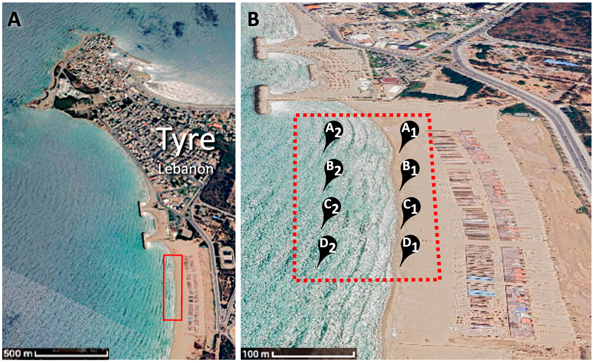
Figure 1. Satellite imagery (Google. 2025. Google Earth. Version 7.3.6.9796.) showing the study sites. (A) Overview of Tyre City, Tyre Beach and the study area (Red-boxed). Scale bar: 500 m. (B) Tyre Beach sampling sites at the study area (Red-dotted box); identified as A1, A2, B1, B2, C1, C2, D1, and D2. Scale bar: 100 m.
Bacteria 04 00051 g001
Figure 2. MTT assay showing the cell viability of Caco-2 and HepG-2 cells after treatment with increasing concentrations of metabolites derived from Kocuria rosea, Micrococcus luteus and Streptomyces longisporoflavus. Significance differences are represented by asterisk (*) as follows: (*) for p < 0.05 *, (**) for p < 0.01 **, (***) for p < 0.001 ***, and (****) for p < 0.0001. The data represent the average of four repeats.
Bacteria 04 00051 g002
Figure 3. Effect of bacterial extracts of Kocuria rosea, Micrococcus luteus and Streptomyces longisporoflavus on the expression of key proteins in Caco-2 cells. (A) Cells were either untreated (Ctrl) or treated with ½ IC50, IC50 of the bacterial extracts. Protein lysates were subjected to immunoblotting analysis for the expression levels of Bax, Bcl-2, and p53. β-actin was used as a loading control. (B) Bax/Bcl-2 ratio was calculated after normalization with β-actin and the untreated control cells. (C) The expression levels of p53 were calculated after normalization with β-actin and the control. Significance differences are represented by asterisk (*) as follows: (*) for p < 0.05 *, (**) for p < 0.01 **, and (***) for p < 0.001 ***.
Bacteria 04 00051 g003
Figure 4. Effect of bacterial extracts of Kocuria rosea, Micrococcus luteus and Streptomyces longisporoflavus on the expression of key proteins in HepG-2 cells. (A) Cells were either untreated (Ctrl) or treated with ½ IC50, IC50 of the bacterial extracts. Protein lysates were subjected to immunoblotting analysis for the expression levels of Bax, Bcl-2, and p53. β-actin was used as a loading control. (B) Bax/Bcl-2 ratio was calculated after normalization with β-actin and the untreated control cells. (C) The expression levels of p53 were calculated after normalization with β-actin and the control. Significance differences are represented by asterisk (*) as follows: (*) for p < 0.05 *, (***) for p < 0.001 ***, and (****) for p < 0.0001.
Bacteria 04 00051 g004
Disclaimer/Publisher’s Note: The statements, opinions and data contained in all publications are solely those of the individual author(s) and contributor(s) and not of MDPI and/or the editor(s). MDPI and/or the editor(s) disclaim responsibility for any injury to people or property resulting from any ideas, methods, instructions or products referred to in the content.